戚益军DNA Methylation Mediated by a MicroRNA Pathway
Sirtuin家族及其生物学特性

Sirtuin家族及其生物学特性戚欣欣;孙莉【摘要】沉默蛋白(sir2-related enzymes,sirtuin)或沉默信息调节因子2(silence information negulator2,Sir2)是一类从古细菌到人类都高度保守的烟酰胺腺嘌呤二核苷酸(nicotinamide adenine dinucleotide,NAD)依赖的组蛋白去乙酰化酶(histone deacetylase,HDAC),哺乳动物有7种sirtuin同源基因SIRT1-SIRT7,具有不同的亚细胞定位和功能.这些蛋白在细胞周期控制、维持线粒体的动态平衡、自噬和细胞生长调节等过程中发挥重要作用.笔者将对sirtuin家族的蛋白结构、酶学功能、家族成员及其生物学功能做一综述.【期刊名称】《华夏医学》【年(卷),期】2016(029)001【总页数】6页(P169-174)【关键词】sirtuin;沉默信息调节因子2;去乙酰化酶【作者】戚欣欣;孙莉【作者单位】桂林医学院基础医学院,广西桂林541000;桂林医学院基础医学院,广西桂林541000【正文语种】中文【中图分类】Q5;R34翻译后修饰在细胞中有重要作用,如DNA识别、蛋白-蛋白相互作用、催化活性和蛋白质稳定性[1]。
蛋白乙酰化/去乙酰化属于组蛋白共价修饰,主要由组蛋白乙酰化酶(histone acetyltransferases,HAT)和组蛋白去乙酰化酶(histone deacetvlase,HDAC)分别催化[2]。
共有Ⅳ类HDAC,sirtuin属于Ⅲ类HDAC,与酵母转录抑制因子Sir2同源[2]。
Sirtuin蛋白家族在不同的细胞过程如细胞凋亡、线粒体生物合成、脂质代谢、脂肪酸氧化、细胞应激反应、胰岛素分泌和衰老都发挥着重要作用。
1.1 蛋白结构X线晶体衍射显示(图1),细菌、酵母和哺乳动物sirtuin具有相似的催化核心区域,即275氨基酸残基构成的一大一小两个基本结构域。
RNA中6_甲基腺嘌呤的研究进展

关键词:
RNA 修饰 ; 6- 甲基腺嘌呤 ; IP-seq
Recent progresses in RNA N6-methyladenosine research
LI Yu-Li1,3, YU Jun1, SONG Shu-Hui2
1. CAS Key Laboratory of Genome Sciences and Information, Beijing Institute of Genomics, Chinese Academy of Sciences, Beijing 100101, China; 2. Core Genomic Facility, Beijing Institute of Genomics, Chinese Academy of Sciences, Beijing 100101, China; 3. University of Chinese Academy of Sciences, Beijing 100049, China
生物 mRNA 内部序列中最常见的一种转录后修饰形式, 由包含 3 个独立组分的复合物 mRNA: m6A 甲基转移酶 催化生成。最新研究发现肥胖相关蛋白 FTO 可以脱掉 m6A 上的甲基 , 表明该甲基化过程是可逆的。抑制或敲 除 m6A 甲基转移酶会引起重要的表型变化, 但是由于过去的检测方法受限 , m6A 确切的作用机制目前为止还不 甚清楚。二代测序技术结合免疫沉淀方法为大规模检测 m6A 修饰并研究其作用机制提供了可能。文章主要综 述了 m6A 的发现史、生成机制、组织和基因组分布、检测方法、生物学功能等及其最新研究进展, 并通过比较 3 种 IP-seq 技术和数据分析的异同及优缺点, 对 m6A 这种 RNA 表观修饰研究中尚未解决的问题进行了讨论。
分子生物学RNA干扰(RNAi)

Craig Mello A professor of Molecular Medicine University of Massachusetts Medical School
In 1998: Fire & Mello in Nature 证实在RNAi 中,真正起作用的是dsRNA
表达与C. elegant worm unc-22基因同源的dsRNA的 细菌喂食线虫,则线虫表现出类似unc-22缺失的表型
•Dicer protein :
Kenneth Kemphues Professor of Genetics
Su Guo Cornell graduate student
Two-cell
Four-cell for distribution of Two-cell to visualize germline-specific mitotic spindles granules.
( The discovery of RNA-mediated interference )
In 1990: Dr. Jorgensen 共抑制现象(cosuppression)
Richard Jorgensen, PhD. Univ. of Arizona RNAi Innovator Awardee
Negative control
Endogenous mex-3 RNA
Injected with mex-3 antisense RNA
Injected with dsRNA corresponding to mex-3
Effects of mex-3 RNA interference on levels of the endogenous mRNA
靶向性DNA甲基化酶在毕赤酵母中的表达和鉴定

维普资讯
76 5
生
物
技
术
通
讯
L E 兀 RS I B OT N I ECHN0L ) Vo. No5 S p ,2 0 (GY 11 . e . 0 7 8
文 章编 号 :09 00 (070 - 7 6 0 10 - 0 22 0 )5 0 5 — 3
[ 键 词 】 D A 甲基 化 酶 ; 向性 ; 赤酵 母 关 N 靶 毕 [ 图分类号】 Q 8 中 7 [ 献标 识 码 】 A 文
一种荧光检测5_羟甲基胞嘧啶的新方法_毛伍祥

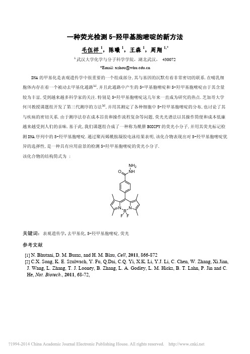
一种荧光检测5-羟甲基胞嘧啶的新方法毛伍祥1,陈曦1,王森1,周翔1,*1武汉大学化学与分子科学学院,湖北武汉, 430072*Email: xzhou@DNA 的甲基化是表观遗传学中很重要的一个组成部分,其与基因的沉默有着非常密切的联系.在哺乳细胞体内存在着一个被动去甲基化通路[1],并且此通路中产生的5-甲基胞嘧啶和5-羟甲基胞嘧啶由于其含量较为丰富,受到越来越多科学家的关注.特别是5-羟甲基胞嘧啶这几年来一直成为研究的热点.芝加哥大学何川教授课题组开发了第三代测序的方法[2],并用其测定了各种细胞中5-羟甲基胞嘧啶的分布,也讨论了其与疾病的密切关系.由于测序法存在成本昂贵和操作流程复杂等问题,荧光光谱法以其操作简便和成本低廉越来越受到人们的亲昧.基于此,我们课题组合成了一种称为酰肼BODIPY 的荧光小分子,并用其荧光标记检测DNA 序列中的5-羟甲基胞嘧啶.通过聚丙烯酰胺凝胶电泳结果表明,该化合物表现出对5-羟甲基胞嘧啶优异的选择性,是一种具有应用前景的检测5-羟甲基胞嘧啶的荧光小分子.该化合物的结构简式为 : N N BO NH NH 2F F关键词:表观遗传学,去甲基化,5-羟甲基胞嘧啶,荧光参考文献[1] N. Bhutani, D. M. Burns, and H. M. Blau , Cell , 2011, 866-872[2] C.X. Song, K. E. Szulwach, Y. Fu, Q.Dai, C.Q. Yi, X.K. Li, Y.J. Li, C. Chen, W. Zhang, Xi.Jian, J. Wang, L. Zhang, T. J. Looney, B. Zhang, L. A. Godley, L. M. Hicks, B. T. Lahn, P. Jin and C. He, Nat. Biotech ., 2011, 68-72,A fluoresent method for detecting the 5-hydromethylcytosine in DNAsequencesWuxiang Mao 1, Xi Chen 1, Sen Wang 1 , Xiang Zhou1,*1College of Chemistry and Molecular Sciences, Wuhan University, Hubei, Wuhan, 430072, P. R. of ChinaThe DNA methylation is closely connection to the cancer, which leads to more and more scientists take part in the research of DNA methylation. In mammalia cell, a demethylation pass was investigated. At first, the C5 positon of cytosine was methylated by DNA methyltransferase, and then oxidized to5-hydromethylcytosine by TET family enzyme. The 5-hydromethylcytosine in many kinds of cells have been investigated by DNA sequencing. Due to the Operational Complexity and expensive cost of DNA sequencing, we synthesized a fluorescent small molecular, which can react with 5-hydromethylcytosine specifically. So we can afford a convenient method for detecting the 5-hydromethylcytosine in DNA sequences.Keywords: demethylation, fluorescence, 5-hydromethylcytosine。
创伤弧菌溶血素基因vvhA质粒定性标准样品的研制及其在水产品检测中的应用

㊀山东农业科学㊀2024ꎬ56(1):147~155ShandongAgriculturalSciences㊀DOI:10.14083/j.issn.1001-4942.2024.01.020收稿日期:2023-04-07基金项目:国家重点研发计划 食品安全关键技术研发 重点专项(2017YFC1601400)ꎻ山东省重点研发计划项目(2022TZXD0022)ꎻ泰山学者工程专项经费资助(tsqn201909168)ꎻ山东省自然科学基金青年基金项目(ZR2020QC226)ꎻ济南市 新高校20条 项目(202228062)ꎻ山东省农业科学院国际科技合作专项(CXGC2022F09)作者简介:袁玮(1997 )ꎬ女ꎬ硕士研究生ꎬ研究方向为食品微生物检测技术研究ꎮE-mail:794393617@qq.com通信作者:陈相艳(1973 )ꎬ女ꎬ研究员ꎬ研究方向为食源性病原微生物检测及标准物质研究ꎮE-mail:315478845@qq.com创伤弧菌溶血素基因vvhA质粒定性标准样品的研制及其在水产品检测中的应用袁玮1ꎬ2ꎬ陈蕾蕾1ꎬ2ꎬ杨金玉1ꎬ周庆新1ꎬ2ꎬ裘纪莹1ꎬ赵双枝1ꎬ付恩君1ꎬ赵国琰2ꎬ陈相艳1(1.山东省农业科学院农产品加工与营养研究所/山东省农产品精深加工技术重点实验室/农业农村部新食品资源加工重点实验室ꎬ山东济南㊀250100ꎻ2.山东师范大学生命科学学院ꎬ山东济南㊀250014)㊀㊀摘要:针对我国缺乏适用于创伤弧菌检测的质粒标准样品的现状ꎬ本研究开展了创伤弧菌鉴定即用型定性质粒标准样品的研制和应用工作ꎮ首先构建了创伤弧菌毒力基因vvhA的重组质粒ꎬ经测序验证后制备成质粒标准样品冻干粉ꎬ然后对其进行PCR定性检测及紫外分光光度计法定量分析ꎮ均匀性检验结果表明ꎬ样品间无显著差异ꎬ均匀性良好ꎬ符合预期目标ꎻ短期稳定性检验结果表明ꎬ样品能在4ħ㊁37ħ条件下稳定保存14天ꎻ长期稳定性检验结果表明ꎬ样品能在-20ħ条件下稳定保存至少12个月ꎮ研究结果表明ꎬ创伤弧菌质粒定性标准样品的均匀性和稳定性均符合国家定性标准样品的要求ꎬ为创伤弧菌的快速㊁高通量的定性鉴定分析提供了可靠的参考物质ꎮ将标准样品应用于鱼类㊁贝类等10份海鲜类食品样品的检测中ꎬ经传统培养法验证ꎬ检测结果准确无误ꎮ本研究所研制的创伤弧菌质粒定性标准样品具有较好的商业应用潜力ꎬ为其在食品检测领域的推广应用奠定了重要基础ꎮ关键词:创伤弧菌ꎻ质粒定性标准样品ꎻ水产品ꎻ检测中图分类号:S852.61㊀㊀文献标识号:A㊀㊀文章编号:1001-4942(2024)01-0147-09DevelopmentofCertifiedPlasmidReferenceMaterialforHemolysinGenevvhAofVibriovulnificusandIt sApplicationinAquaticProductsDetectionYuanWei1ꎬ2ꎬChenLeilei1ꎬ2ꎬYangJinyu1ꎬZhouQingxin1ꎬ2ꎬQiuJiying1ꎬZhaoShuangzhi1ꎬFuEnjun1ꎬZhaoGuoyan2ꎬChenXiangyan1(1.InstituteofFood&NutritionScienceandTechnologyꎬShandongAcademyofAgriculturalSciences/KeyLaboratoryofAgro ̄productsProcessingTechnologyofShandongProvince/KeyLaboratoryofNovelFoodResourcesProcessingꎬMinistryofAgricultureandRuralAffairsꎬJinan250100ꎬChinaꎻ2.CollegeofLifeSciencesꎬShandongNormalUniversityꎬJinan250014ꎬChina)Abstract㊀DuetolackofplasmidstandardsamplesuitablefordetectionofVibriovulnificusinChinaꎬaready ̄to ̄usecertifiedplasmidreferencematerialwasdevelopedherein.TherecombinantplasmidsofvvhAgeneofV.vulnificuswasconstructedfirstlyandthentheirfreeze ̄driedplasmidpowderswerepreparedaftersequen ̄cingverificationandbeingdetectedbyPCRandUVspectrophotometry.Theuniformitytestshowednosignifi ̄cantdifferencebetweensamplesꎬandtheuniformitywasasexpected.Thestabilitytestshowedthatthepre ̄paredplasmidsamplescouldbestablypreservedfor14daysat4ħor37ħꎬandforatleast12monthsat-20ħ.TheoverallresultsindicatedthattheuniformityandstabilityofthevvhAgenerecombinantplasmidqualitativestandardsamplescouldmeettherequirementsofnationalqualitativestandardsamplesꎬwhichpro ̄videdreliablereferencematerialforrapidandhigh ̄throughputqualitativeidentificationofV.vulnificus.Thesampleswereusedaspositivecontrolinthedetectionof10seafoodsamplessuchasfishandshellfish.andtheresultswereconfirmedtobeaccuratebytraditionalculturemethod.AboveallꎬthequalitativestandardsamplesofvvhAgenerecombinantplasmiddevelopedinthisstudyhadgreatpotentialincommercialapplicationꎬlayinganimportantfoundationforitsapplicationinfooddetection.Keywords㊀VibriovulnificusꎻCertifiedplasmidreferencematerialꎻAquaticproductsꎻDetection㊀㊀创伤弧菌(Vibriovulnificus)是一种革兰氏阴性菌ꎬ主要特性为嗜盐㊁喜温ꎬ自然分布于世界各地沿海和河口水域[1-2]ꎬ是一种人畜共患病原菌ꎬ容易感染鱼㊁虾㊁牡蛎㊁蛤㊁螃蟹等海产品ꎬ人类通过生食或食用未完全煮熟的海产品㊁破损皮肤直接接触被其污染的海水或海产品而患病[3]ꎮ创伤弧菌与霍乱弧菌㊁副溶血弧菌并称为人类三大致病弧菌[4]ꎬ弧菌感染病例具有明显的季节性ꎬ大多数发生在夏季和初秋气温较高的时期[5]ꎮ随着全球气候变暖㊁海洋温度升高等自然条件的变化ꎬ创伤弧菌的感染率也逐年增加ꎮ在全世界范围内创伤弧菌感染的死亡率高达60%ꎬ在美国约为33%ꎬ使其成为严重的公共卫生和食品安全问题[6-7]ꎮ创伤弧菌感染的主要症状包括肠胃炎㊁原发创伤性感染㊁败血症和坏死性筋膜炎等ꎬ免疫力低下或患有糖尿病㊁肝脏疾病等慢性基础病的患者属于易感人群[8]ꎮ创伤弧菌伤口感染通常以肿胀㊁红斑和剧烈疼痛为特征ꎬ潜伏期短ꎬ发病迅速ꎬ病变经常演变为可坏死的囊泡或充满液体的大泡ꎬ最终引起多脏器衰竭[9]ꎮ创伤弧菌菌体产生的创伤弧菌外毒素通过特定的毒力机制引发疾病ꎮ创伤弧菌毒力因子主要包括溶细胞素㊁铁载体㊁金属蛋白酶㊁荚膜多糖等[10]ꎬ由vvhA基因编码的创伤弧菌溶细胞素是唯一分泌到细胞外的外毒素ꎬ具有创伤弧菌种属特异性ꎬ可作为鉴定创伤弧菌的指标[11]ꎮ当前ꎬ对创伤弧菌进行定量检测主要通过平板计数㊁MPN法等传统培养法ꎬ这些方法需要进行过夜培养㊁选择性平板分离㊁生化鉴定㊁血清学检测等繁琐的试验步骤ꎬ不仅耗时费力ꎬ同时样本中杂菌的过量繁殖也会对鉴别结果产生影响ꎮ另外ꎬ由于水产品中的创伤弧菌通常处于 活的且不可培养 的状态[12-13]ꎬ传统的培养方法很难对该部分创伤弧菌进行有效鉴定ꎬ严重降低了检测结果的准确度ꎮ作为最危险的食源性细菌之一ꎬ创伤弧菌造成了95%的海鲜相关死亡ꎬ已成为一个主要的食品安全问题[14]ꎮ随着食品供应链的全球化ꎬ创伤弧菌的定期监测变得更加重要ꎮ为了满足当下快速㊁高通量检测的需求ꎬ分子生物学方法在食品微生物的检测中得到了越来越广泛的应用ꎬ但相关的参考物质相对匮乏ꎮ食源性微生物检测即用型标准样品的研制ꎬ有助于解决目前国内食品微生物检测中存在的参考物质不足的难题ꎬ使具有自主知识产权的标准样品在相关领域得到更好的推广和应用ꎬ从而摆脱对国外标准样品的依赖ꎮ因此ꎬ建立创伤弧菌鉴定检测即用型质粒定性标准样品用于其快速㊁高通量鉴定ꎬ对提高水产品的质量安全㊁保证人类健康具有重要意义ꎮ1㊀材料与方法1.1㊀试验材料1.1.1㊀菌株来源㊀创伤弧菌(CICC21615)来源于中国工业微生物保藏中心ꎮ1.1.2㊀主要试剂㊀琼脂糖购自上海贝晶生物技术有限公司ꎻPNCC增菌液基础培养基㊁PNCC添加剂㊁mCPC琼脂基础培养基㊁多粘菌素E㊁多粘菌素B购自北京陆桥技术股份有限公司ꎻ核酸染料GelStain㊁Trans15000marker㊁Trans2000mark ̄er㊁质粒大提试剂盒购自北京全式金生物技术股份有限公司ꎻ50ˑTAE缓冲溶液购自生工生物工程(上海)股份有限公司ꎻ2ˑTaqPCRMix㊁琼脂糖841山东农业科学㊀㊀㊀㊀㊀㊀㊀㊀㊀㊀㊀㊀㊀㊀第56卷㊀凝胶DNA回收试剂盒㊁pLB零背景快速克隆试剂盒购自天根生化科技(北京)有限公司ꎮ1.1.3㊀仪器和设备㊀ZHWY-200H恒温培养振荡器购自上海智城分析仪器制造有限公司ꎻSW-CJ-2D双人净化工作台购自苏州净化设备有限公司ꎻC1000TouchPCR仪㊁NanoDropTM2000超微量分光光度计购自赛默飞世尔科技公司ꎻJY600C水平电泳仪㊁JY04S-3C凝胶成像系统购于北京君意东方电泳设备有限公司ꎮ1.2㊀试验方法1.2.1㊀重组质粒的获取与验证㊀创伤弧菌vvhA基因片段由生工生物工程(上海)股份有限公司合成ꎬ获得重组质粒PUC-SP-vvhAꎬ并保存于大肠埃希氏菌Top10菌株中ꎮ依据GB4789.44 2020«食品安全国家标准食品微生物学检验创伤弧菌检验»[15]中创伤弧菌PCR检测的引物序列ꎬ由生工生物工程(上海)股份有限公司合成引物(表1)ꎮ以重组质粒为模板ꎬ利用创伤弧菌鉴定引物ꎬ对目的基因进行PCR扩增ꎬ扩增产物进行琼脂糖凝胶电泳分析ꎬ使用琼脂糖凝胶DNA回收试剂盒对PCR产物进行胶回收ꎬ使用pLB零背景快速克隆试剂盒将PCR纯化产物连接至pLB-simpleVector上ꎬ并转化大肠杆菌DH5α感受态细胞ꎬ置于37ħ培养箱培养12hꎮ从平板上挑取单菌落ꎬ经PCR反应鉴定为阳性的克隆送生工生物工程(上海)股份有限公司进行测序鉴定ꎬ测序结果与预期一致的ꎬ即为验证正确的重组质粒ꎮ将携带正确重组质粒的大肠埃希氏菌于-80ħ超低温冰箱中甘油管保存ꎮ㊀㊀表1㊀创伤弧菌vvhA基因的引物序列引物名称引物序列(5ᶄң3ᶄ)片段大小/bpvvhA-FCCGCGGTACAGGTTGGCGCAvvhA-RCGCCACCCACTTTCGGGCC5191.2.2㊀重组质粒的提取㊀将携带重组质粒的大肠埃希氏菌甘油管解冻ꎬ接入4mLLB液体培养基中ꎬ放入200r/min的振荡摇床中37ħ培养12hꎬ取2mL种子液转接于200mL的LB液体培养基中扩大培养ꎬ获取大量携带重组质粒大肠埃希氏菌的培养液ꎬ利用细菌质粒大提试剂盒提取质粒ꎮ琼脂糖凝胶电泳分析质粒完整性ꎬPCR验证目的基因ꎬ紫外分光光度计检测质粒的浓度和纯度ꎮ1.2.3㊀重组质粒的含量检测㊀取重组质粒样品1μLꎬ于NanoDropTM2000超微量分光光度计中检测浓度ꎬ每管检测两次ꎮ1.2.4㊀重组质粒的分装㊀将大提的质粒样品混合至1管中ꎬ采用紫外分光光度计测定浓度ꎬ然后用无菌去离子水稀释至20ng/μLꎬ每管100μL分装至螺口冻存管中ꎬ贴上标签ꎮ1.2.5㊀重组质粒的冷冻干燥㊀由于质粒样品较为稳定ꎬ分装后可直接冻干ꎮ在-25ħ㊁真空度为50Pa条件下冻干20hꎮ1.2.6㊀质粒定性标准样品的均匀性分析㊀按照随机抽号系统抽取的号码ꎬ抽取质粒定性标准样品12管ꎬ用100μL无菌水溶解质粒冻干粉ꎮ取0.5μL溶解的质粒样品作为PCR模板ꎬ按照1.2.1方法进行基因定性检测ꎻ取质粒样品2μLꎬ按照1.2.1方法琼脂糖凝胶电泳分析质粒样品的完整性ꎻ取质粒样品1μLꎬ采用紫外分光光度计法进行复溶质粒样品的定量试验ꎬ检测样品的浓度ꎬ每管测两次ꎬ核酸含量=核酸浓度ˑ水化体积ꎬ核酸含量结果统计分析采用方差分析法ꎮ1.2.7㊀质粒定性标准样品的稳定性分析㊀稳定性检验方法同均匀性检验ꎬ核酸含量结果统计分析采用单因素方差分析法ꎮ短期稳定性检验:采取两种短期稳定性试验ꎮ第一种模拟冰袋运输:在4ħ条件下的短期储存稳定性试验ꎬ随机取样21管置于4ħ保温箱ꎬ分别在第1㊁3㊁5㊁7㊁9㊁11㊁14天每天检测3管ꎬ每管2个重复ꎻ第二种模拟高温运输:在37ħ条件下稳定性试验ꎬ随机取样21管置于37ħ保温箱ꎬ分别在第1㊁3㊁5㊁7㊁9㊁11㊁14天时每天检测3管ꎬ每管2个重复ꎮ长期稳定性检验:质粒样品经冷冻干燥后ꎬ需要在-20ħ条件下长期冷冻保存ꎮ为了测定质粒样品长期保存时间及其稳定性ꎬ每次检测时ꎬ从冷冻样品中随机取出3管样品ꎬ每管重复2次ꎮ抽样时间点遵循先密后疏的原则ꎬ分别在第1㊁2㊁4㊁6㊁8㊁10㊁12个月共7个时间点抽样检测ꎮ1.2.8㊀创伤弧菌质粒定性标准样品在食品检测中的应用㊀食品样本的前处理:将购买的食品样本按照GB4789.44 2020要求处理ꎬ在无菌条件941㊀第1期㊀㊀㊀㊀袁玮ꎬ等:创伤弧菌溶血素基因vvhA质粒定性标准样品的研制及其在水产品检测中的应用下ꎬ称取鲫鱼㊁偏口鱼㊁银鲳鱼㊁鲤鱼㊁鲅鱼㊁黄花鱼6种鱼类样品的表面组织㊁肠和腮各25gꎬ花蛤㊁海蛎子和生蚝3种贝类样品内容物各25gꎬ明虾的头足部组织25gꎬ分别放入含225mLPNCC增菌液的无菌均质袋ꎬ用拍打式无菌均质器拍打2min制成样品匀液ꎻ将均质袋放入培养箱37ħ培养18h获得增菌液ꎬ取距液面1cm深处菌液1mL放入离心管中ꎬ9000r/min离心3minꎬ去上清ꎻ用1mLPBS磷酸盐缓冲液悬浮清洗后9000r/min离心3minꎬ去上清ꎬ重复2次ꎻ加入1mL无菌去离子水ꎬ煮沸10minꎬ12000r/min离心5minꎬ吸取上清液ꎮPCR分析:将上清液用作PCR反应的DNA模板ꎻ随机抽取1管创伤弧菌重组质粒定性标准样品ꎬ加入100μL无菌去离子水溶解ꎬ作为阳性对照ꎻ将大肠埃希氏菌CICC10003基因组DNA冻干粉用300μL无菌水溶解(终浓度为20ng/μL)后作为阴性对照ꎻ无菌去离子水为空白对照ꎬ按照1.2.1的方法进行PCR分析ꎮ创伤弧菌的分离验证:取检测为阳性的食品样本的增菌液ꎬ用接种环将其划线接种于CC平板和mCPC平板ꎬ37ħ培养18hꎬ验证菌落形态是否符合创伤弧菌菌落形态特征ꎬ即圆形㊁扁平ꎬ光照下透明但中心不透明的黄色或橘黄色菌落ꎬ直径1~2mmꎮ创伤弧菌的生化鉴定:按照GB4789.442020进行创伤弧菌的培养和生化特性鉴定ꎮ1.3㊀数据统计与分析使用SPSS26.0软件的ANOVA法进行单因素方差分析ꎬ统计各处理组之间的差异性ꎬ数据用平均值ʃ标准误 表示ꎬP<0.05表示差异显著ꎮ2㊀结果与分析2.1㊀创伤弧菌重组质粒的验证以创伤弧菌重组质粒为模板ꎬ对其进行目的基因PCR扩增验证ꎬ以大肠埃希氏菌CICC10003为阴性对照ꎬ无菌去离子水为空白对照ꎮ琼脂糖凝胶电泳分析PCR扩增结果(图1A)显示ꎬ创伤弧菌重组质粒vvhA基因为阳性ꎬ条带清晰ꎬ片段大小为519bpꎬ符合目的条带大小ꎬ说明成功构建了创伤弧菌溶血素基因vvhA重组质粒ꎬ质粒图谱如图1B所示ꎮM:Trans2000markerꎻ1:vvhA质粒ꎻ2:阴性对照ꎻ3:空白对照ꎮ图1㊀创伤弧菌重组质粒PCR扩增(A)及PUC-SP-vvhA重组质粒图谱(B)2.2㊀创伤弧菌重组质粒的浓度及纯度分析取重组质粒1μLꎬ利用NanoDropTM2000测定其浓度和纯度ꎬ结果如表2所示ꎬA260/280㊁A260/230均超过1.8ꎬ说明所提取的质粒纯度高ꎬ无蛋白质和有机物污染ꎻ对其携带的基因进行PCR扩增ꎬ琼脂糖凝胶电泳分析结果(图2A)显示ꎬ目的基因vvhA条带单一且片段大小符合预期ꎬ质粒完整性分析(图2B)显示质粒条带完整ꎬ说明所制备的质粒符合预期ꎮ㊀㊀表2㊀创伤弧菌重组质粒的浓度与纯度样品管号浓度/(ng/μL)A260/A280A260/A230VA1167.01.862.21VA2189.51.882.29VA3173.61.882.26VA482.11.892.53051山东农业科学㊀㊀㊀㊀㊀㊀㊀㊀㊀㊀㊀㊀㊀㊀第56卷㊀M:Trans15000markerꎻ1~8:vvhA质粒样品ꎮ图2㊀创伤弧菌重组质粒vvhA的PCR扩增(A)及质粒完整性(B)分析㊀㊀将4管大提的创伤弧菌重组质粒样品混合至1管中ꎬ采用紫外分光光度计测定浓度ꎬ然后用无菌去离子水稀释至20ng/μLꎬ取100μL分装至2mL螺口冻存管中ꎬ所有质粒溶液分装450管后ꎬ置于真空冷冻干燥机中按照冷冻程序真空冷冻干燥ꎬ获得白色粉末状的冻干质粒样品ꎬ设计创伤弧菌重组质粒定性标准样品标签纸ꎬ打印并贴在管外(图3)ꎮ质粒定性标准样品置于-20ħ冰箱冻存ꎮ2.3㊀创伤弧菌溶血素基因vvhA质粒定性标准样品的均匀性分析从450管质粒标准样品中随机抽取12管进行PCR定性试验ꎬ结果如图4A所示ꎬ各管PCR扩增目的基因均为阳性ꎬ条带单一且清晰ꎬ图4B显示质粒完整无降解ꎮ使用NanoDropTM2000超微量分光光度计进行质粒标准样品的浓度㊁纯度分析ꎬ对所测数据进行单因素方差分析ꎬ结果如表3所示ꎬ在95%的置信概率下ꎬF值小于F临界值ꎬ各管间质粒含量无显著性差异ꎬ表明质粒定性标准样品均匀性良好ꎮ图3㊀创伤弧菌溶血素基因vvhA质粒定性标准样品M:DNAmarkerꎻ1~12:vvhA质粒标准样品ꎮ图4㊀创伤弧菌溶血素基因vvhA质粒定性标准样品PCR扩增(A)及均匀性(B)分析㊀㊀表3㊀创伤弧菌溶血素基因vvhA质粒定性标准样品均匀性试验方差分析平方和SS自由度均方MSF值F临界值置信概率P值组间0.021110.022.7002.720.950.051组内0.008120.012.4㊀创伤弧菌溶血素基因vvhA质粒定性标准样品的稳定性分析温度是运输过程中影响质粒定性标准样品质量的主要因素ꎬ因此设计不同温度模拟质粒样品运输条件ꎬ以质粒标准样品目的基因阳性检出㊁质粒完整性及核酸含量变化确定其短期稳定性(运输稳定性)ꎮ创伤弧菌溶血素基因vvhA质粒定性标准样品分别在4ħ和37ħ条件下保存14dꎬ琼脂糖凝胶电泳分析结果(图5㊁图6)显示第1天和第14天目的基因均为阳性ꎬ电泳条带单一ꎬ符合预期条带大小ꎬ质粒无降解ꎻ质粒含量统计学分析结果如图7所示ꎬ各时间点间无显著性差异ꎬ表明质粒含量无明显变化ꎬ说明质粒定性标准样品在上述条件下是稳定的ꎮ因此创伤弧菌溶血素基因vvhA质粒定性标准样品可与冰袋(4ħ)一起运输ꎬ也可在温度较高(37ħ)且无降温装置条件下运输ꎮ151㊀第1期㊀㊀㊀㊀袁玮ꎬ等:创伤弧菌溶血素基因vvhA质粒定性标准样品的研制及其在水产品检测中的应用M:DNAmarkerꎻ1~6:vvhA质粒标准样品ꎻ7:阴性对照ꎻ8:空白对照ꎬ下同ꎮ图5㊀4ħ条件下保存1天创伤弧菌溶血素基因vvhA质粒定性标准样品PCR扩增(A㊁B)及完整性(C㊁D)分析图6㊀37ħ条件下保存14天创伤弧菌溶血素基因vvhA质粒定性标准样品PCR扩增(A㊁B)及完整性(C㊁D)分析图7㊀创伤弧菌溶血素基因vvhA质粒定性标准㊀㊀样品短期稳定性定量分析为了分析创伤弧菌溶血素基因vvhA质粒定性标准样品的长期稳定性ꎬ在第1㊁2㊁4㊁6㊁8㊁10㊁12个月随机抽取3管样品进行定性和定量分析ꎮ创伤弧菌溶血素基因vvhA质粒定性标准样品在-20ħ条件下保存12个月ꎬ琼脂糖凝胶电泳分析结果显示第1个月和第12个月目的基因均为阳性ꎬ电泳条带单一ꎬ符合预期条带大小(图8A㊁B)ꎬ质粒无降解(图8C㊁D)ꎮ质粒含量统计学分析结果如图9所示ꎬ各时251山东农业科学㊀㊀㊀㊀㊀㊀㊀㊀㊀㊀㊀㊀㊀㊀第56卷㊀间点间无显著性差异ꎬ表明质粒定性标准样品在-20ħ条件下稳定ꎬ说明创伤弧菌溶血素基因vvhA质粒定性标准样品能在-20ħ条件下稳定保存至少12个月ꎮ图8-20ħ条件下保存1㊁12个月创伤弧菌溶血素基因vvhA质粒定性标准样品PCR扩增(A㊁B)及完整性(C㊁D)分析图9-20ħ条件下创伤弧菌溶血素基因vvhA质粒㊀㊀定性标准样品长期稳定性定量分析2.5㊀创伤弧菌溶血素基因vvhA质粒定性标准样品在水产品检测中的应用为了验证创伤弧菌溶血素基因vvhA质粒定性标准样品在水产品检测中的应用效果ꎬ将其作为阳性对照ꎬ参照GB4789.44 2020的方法ꎬ利用PCR对10种水产品中的创伤弧菌基因vvhA进行检测ꎬ每种样品取样7次ꎬ每个样品7个重复ꎮ结果如表4所示ꎬ在10种水产品样本中检测出1例vvhA基因阳性ꎮ将阳性样本的增菌液划线至创伤弧菌鉴定平板CC平板和mCPC平板中ꎬ每种平板重复划线3次ꎬ37ħ培养18hꎬ平板菌落为黄色㊁圆形且扁平(图10)ꎮ挑取菌落按照GB4789.44 2020要求进行生化鉴定(图11㊁表5)ꎬ验证此阳性样本感染创伤弧菌ꎮ㊀㊀表4㊀海鲜样品中创伤弧菌溶血素基因vvhA检出结果海鲜类型检测份数检出阳性次数鱼类60贝类31虾类10总计101㊀㊀表5㊀创伤弧菌溶血素基因vvhA阳性样本生化鉴定结果项目结果赖氨酸紫色氨基酸对照黄色无盐胰胨水微弱生长6%氯化钠胰胨水生长旺盛8%氯化钠胰胨水不生长10%氯化钠胰胨水不生长V-P半固体穿刺周围扩散增长351㊀第1期㊀㊀㊀㊀袁玮ꎬ等:创伤弧菌溶血素基因vvhA质粒定性标准样品的研制及其在水产品检测中的应用图10㊀CC平板和mCPC平板创伤弧菌菌落特征图11㊀创伤弧菌溶血素基因vvhA阳性样本菌体的生化鉴定结果3㊀讨论与结论本研究构建了创伤弧菌溶血素基因vvhA的重组质粒PUC-SP-vvhAꎬ经测序验证后ꎬ将质粒样品真空冷冻干燥制成创伤弧菌溶血素基因vvhA质粒定性标准样品冻干粉ꎮ对其进行均匀性和稳定性分析ꎬ检测结果均为阳性且符合目的基因片段的大小ꎬ质粒样品无降解ꎻ均匀性分析定量检测结果表明ꎬ质粒定性标准样品无管间差异ꎬ均匀性良好ꎻ稳定性分析定量检测结果表明ꎬ质粒定性标准样品在低温(4ħ)或高温(37ħ)下可稳定保存14天ꎻ在-20ħ下长期存储12个月ꎬ亦未观测到不稳定性ꎮ将所研制的创伤弧菌溶血素基因vvhA质粒定性标准样品应用于10种海鲜产品的检测ꎬ检出1份阳性样本ꎬ经传统培养法和生化鉴定验证ꎬ证实阳性样本确为创伤弧菌污染ꎬ表明质粒定性标准样品能够满足水产品中创伤弧菌溶血素基因vvhA的快速㊁精确检测ꎮ该研究填补了我国创伤弧菌鉴定即用型质粒定性标准样品的空白ꎬ为创伤弧菌质粒定性标准样品在食品检测领域的应用研究打下了良好的基础ꎮ参㊀考㊀文㊀献:[1]㊀YunNRꎬKimDM.Vibriovulnificusinfection:apersistentthreattopublichealth[J].KoreanJournalofInternalMedi ̄cineꎬ2018ꎬ33(6):1070-1078.[2]㊀Hernánde ̄CabanyeroCꎬAmaroC.PhylogenyandlifecycleofthezoonoticpathogenVibriovulnificus[J].EnvironmentalMicrobiologyꎬ2020ꎬ22(10):4133-4148.[3]㊀彭钟琴ꎬ黄璐.水产品中创伤弧菌检测方法的研究进展[J].食品安全导刊ꎬ2021(36):190-192.[4]㊀WangMYꎬHuCJ.PathogenicityandvirulencefactorsofVib ̄riovulnificus:researchadvances[J].ChineseJournalofMicro ̄ecologyꎬ2017ꎬ29(12):1470-1473.[5]㊀IwamotoMꎬAyersTꎬMahonBEꎬetal.Epidemiologyofsea ̄food ̄associatedinfectionsintheUnitedStates[J].ClinicalMi ̄crobiologyReviewsꎬ2010ꎬ23(2):399-411.[6]㊀JonesMKꎬOliverJD.Vibriovulnificus:diseaseandpathogen ̄esis[J].InfectionandImmunityꎬ2009ꎬ77(5):1723-1733.[7]㊀HengSPꎬLetchumananVꎬDengCYꎬetal.Vibriovulnificus:anenvironmentalandclinicalburden[J].FrontiersinMicrobi ̄ologyꎬ2017ꎬ8:997.[8]㊀HorsemanMAꎬSuraniS.AcomprehensivereviewofVibriovulnificus:animportantcauseofseveresepsisandskinandsoft ̄tissueinfection[J].InternationalJournalofInfectiousDis ̄easesꎬ2011ꎬ15(3):e157-e166.451山东农业科学㊀㊀㊀㊀㊀㊀㊀㊀㊀㊀㊀㊀㊀㊀第56卷㊀[9]㊀Baker ̄AustinCꎬTrinanesJꎬGonzalez ̄EscalonaNꎬetal.Non ̄choleravibrios:themicrobialbarometerofclimatechange[J].TrendsinMicrobiologyꎬ2017ꎬ25(1):76-84.[10]LiGꎬWangMY.TheroleofVibriovulnificusvirulencefactorsandregulatorsinitsinfection ̄inducedsepsis[J].FoliaMicro ̄biologicaꎬ2020ꎬ65(2):265-274.[11]GavinHEꎬBeubierNTꎬSatchellKJ.Theeffectordomainre ̄gionoftheVibriovulnificusMARTXtoxinconfersbiphasicepi ̄thelialbarrierdisruptionandisessentialforsystemicspreadfromtheintestine[J].PLoSPathogensꎬ2017ꎬ13(1):e1006119.[12]RaoNVꎬShashidharRꎬBandekarJR.Inductionꎬresuscita ̄tionandquantitativereal ̄timepolymerasechainreactionanaly ̄sesofviablebutnonculturableVibriovulnificusinartificialseawater[J].WorldJournalofMicrobiologyandBiotechnologyꎬ2014ꎬ30(8):2205-2212.[13]包秋华ꎬ刘倩宇.基于WebofScience细菌活的非可培养状态研究文献的可视化分析[J].食品科学ꎬ2023ꎬ44(5):248-256.[14]ZhangXꎬGuoBꎬYangLHꎬetal.CRISPR/Cas12acombinedwithrecombinasepolymeraseamplificationforrapidandsensi ̄tivedetectionofVibriovulnificusinonetube[J].ActaBio ̄chimicaetBiophysicaSinicaꎬ2023ꎬ55(2):322. [15]中华人民共和国国家卫生和计划生育委员会.食品安全国家标准:食品微生物学检验创伤弧菌检验:GB4789.442020[S].北京:中国标准出版社ꎬ2020.551㊀第1期㊀㊀㊀㊀袁玮ꎬ等:创伤弧菌溶血素基因vvhA质粒定性标准样品的研制及其在水产品检测中的应用。
O-GlcNAc糖基化修饰和Akt1对胃癌细胞体外增殖及侵袭力的影响
O-GlcNAc糖基化修饰和Akt1对胃癌细胞体外增殖及侵袭力的影响章诺贝;陈新【摘要】目的探讨O-连接N-乙酰氨基葡萄糖(O-GlcNAc)糖基化修饰对胃癌细胞体外增殖及侵袭力的影响,并评价Akt1在O-GlcNAc糖基化促进胃癌细胞体外增殖及侵袭过程中的作用.方法通过使O-GlcNAc转移酶(OGT)过表达(过表达OGT组)或沉默表达(沉默OGT组)及使用O-GlcNAc水解酶(OGA)特异性抑制剂Thiamet-G(抑制剂组)下调O-GlcNAc水解酶活性(抑制剂组)等构建O-GlcNAc糖基化水平升高或降低的细胞模型;采用MTT法检测各组胃癌细胞增殖活力;软琼脂集落形成实验观察各组胃癌细胞集落形成;Transwell细胞迁移实验各组胃癌细胞体外迁移和侵袭能力;蛋白免疫印迹法(Western blot)检测各组胃癌细胞Akt1活性;Thiamet-G处理Akt1表达沉默(沉默Akt1组)的胃癌细胞以评价Akt1在O-GlcNAc糖基化促进胃癌细胞侵袭中的作用;利用Thiamet-G处理Akt1过表达(过表达Akt1组)的胃癌细胞以进一步验证Akt1在O-GlcNAc糖基化调控胃癌细胞侵袭性过程中的作用.结果 O-GlcNAc糖基化水平升高可促进胃癌细胞增殖并显著提高细胞集落形成、体外迁移和侵袭的能力;Akt1活性被由O-GlcNAc糖基化水平升高所介导的Ser473磷酸化上调而激活;Thiamet-G诱导的细胞侵袭性被Akt1 shRNA所抑制;Akt1高表达可进一步促进由Thiamet-G诱导的细胞侵袭性增强.结论 O-GlcNAc糖基化可部分通过Akt1途径增强胃癌细胞的体外增殖及侵袭力.%Objective To study the influence of O-GlcNAcylation on on proliferation and invasion of gastric cancer cells and evaluate the role of Aktl on O-GlcNAcylation promotting cells proliferation and invasion in gastric cancer.Methods Build the cell model:O-GlcNAc glycosylation levelsrise or fall.The cell viability was determine by MTT.To investigate whether O-GlcNAcylation affected colony formation ability of gastric cancer cells,soft agar colony assays were carried out.Cell migration or invasion was using transwell chambers.The expression of Akt1 was detected through Western blot.Thiamet-G was used to eualuate the role of Akt1 on O-Gcnac cylation regulating invasion in gastric Cancei.Results O-GlcNAcylation was increased the gastric cancer cells proliferation ability,colony formation ability,migration and invasion ability in vitro.Akt1 was activated by Ser473 phosphorylation upregulation though O-GlcNAcylation.Akt1 shRNA was inhibition the cell invasive which induced by Thiamet-G.Akt1 overexpression was promoted by Thiamet-G-induced cell invasion.Conclusion O-GlcNAcylation enhanced oncogenic phenotypes possibly partially involving Akt1.【期刊名称】《重庆医学》【年(卷),期】2017(046)008【总页数】6页(P1027-1031,1035)【关键词】胃肿瘤;肿瘤侵润;细胞增殖;O-GlcNAc糖基化;Akt1【作者】章诺贝;陈新【作者单位】南昌大学第二附属医院消化科 330006;南昌大学第二附属医院核医学科 330006【正文语种】中文【中图分类】R735.7O-连接N-乙酰氨基葡萄糖(O-Linked N-acetylglucosamine,O-GlcNAc)糖基化是细胞核与细胞质蛋白的丝氨酸和苏氨酸残基以O-GlcNAc修饰的一种高丰度可逆性翻译后修饰方式,它被认为能够调节细胞内蛋白质的功能及活性[1]。
小麦穗发芽抗性相关Vp1基因启动子的分离及功能验证
本研究由国家重点基础研究发展计划 (973 计划 )项目 (2009CB118305)资助。
*
通讯作者 (Corresponding author): 夏兰琴 , E-mail: xialq@, Tel: 010-82105804
第一作者联系方式 : E-mail: suenyw@, Tel: 010-82105921 Received(收稿日期 ): 2011-05-09; Accepted(接受日期 ): 2011-06-25; Published online(网络出版日期 ): 2011-07-28. URL: /kcms/detail/11.1809.S.20110728.1000.004.html
1744
作 物 学 报
第 37 卷
分析 Vp1 的转录本结构 , 发现每个同源基因都会产 生一套大小不同的转录本, 原因是转录后前体 mRNA 发 生 了 选 择 性 的 剪 接 , 导 致 无 法 编 码 全 长 根据 Vp1 基因 Vp1 蛋白 , 从而表现对穗发芽敏感 [4]。 序列中跨越 5 个内含子的 B3 区段设计特异性引物 , 对 2 个 CIMMYT 人工合成六倍体小麦进行 RT-PCR, 发现这 2 个合成小麦之间存在不同的剪切模式, 进一 步证明 Vp1 在转录时不同的剪切方式是导致品种间 穗发芽抗性差异的重要因素之一 [5]。然而 , Nakamura 等 [4] 研 究 了 强 休 眠 品 种 Minamino 和 非 休 眠 品 种 Tozan 成熟胚中 Vp1 基因的表达 , 发现在 Minamino 中 Vp1 的表达量大于在 Tozan 中。燕麦 Vp1 只形成 一种转录本 , 把燕麦 Vp1 cDNA 转入小麦时 , 转基 因小麦穗发芽程度明显降低 [6]。小麦的 3 个 Vp1 同 源基因中 , 位于 3B 染色体上的 Vp1B 对穗发芽抗性 起关键作用, 并且存在广泛的多态性[7-11]。Yang 等[7-8] 在小麦 3B 染色体上发现了 2 个与穗发芽抗性相关的 Vp1B 的新型等位变异 Vp1Bb 和 Vp1Bc, 由 Vp1B 编 码区第 3 内含子中反转座子的插入和转座子的缺失 引起 , 尽管存在错误剪切现象 , 但 Vp1B 各等位基 因 正 常 转 录 本 的 表 达 量 与 种 子 的 ABA 敏 感 性 和 穗发芽抗性呈正相关。对中国和欧洲小麦品种 Vp1 变异类型检测 , 共发现 5 种基因型 , 分别是 Vp1Ba、 Vp1Bb、 Vp1Bc、 Vp1Bd 和 Vp1Be[9-10]。 Utsugi 等 [11] 同样发现具有较高休眠性的品种对 ABA 敏感性要 高于非休眠性品种 , 3B 染色体上 Vp1B 表达起主导 作用 , 并与种子休眠性呈正相关 ; ABA 处理后胚中 TaVp1B mRNA 含量明显提高 , 其编码蛋白 Vp1B 可 以激活 Em 基因表达和抑制 α-淀粉酶的活性 , 从而 影响种子休眠性。但小麦 Vp1 启动子在穗发芽抗性 中的作用和其所介导的 ABA 应答机制目前尚未见 报道 。 玉米 Vp1 启动子和拟南芥的同源基因 ABI3 启动 子均为组织特异性启动子 , 主要在胚中表达。但当 受到 ABA 和干旱、高盐等逆境诱导时 , 其表达程度 均有提高 , 并在茎等组 织中表现出 不同量的表 达 , 呈现出非组织特异性特征 [12-13]。深入研究小麦 Vp1 启动子 , 对了解不同逆境条件下 Vp1 的表达调控及 其在小麦穗发芽抗性中的作用具有重要的意义 , 同 时也可为进一步利用此启动子进行抗逆基因工程操 作提供依据。本研究克隆了小麦 B 基因组 Vp1 启动 子 , 并构建了系列缺失载体。通过瞬时表达和在转 基因小麦中的稳定表达 , 明确了启动子及所含元件
枯草芽孢杆菌Y_6产抗菌肽的体外抗氧化效果研究
malondialdehyde ( MDA) formation inhibiting effects of mice liver homogenate. The results showed that antimicrobial
peptide has certain activities on scavenging free radicals and inhibiting lipid oxidation,and the effects were better when
核 农 学 报 2011 ,25 (3) :0518 ~ 0522 Journal of Nuclear Agricultural Sciences
枯草芽孢杆菌 Y-6 产抗菌肽的体外抗氧化效果研究
张 捷 丁韩英 戚向阳 袁勇军
( 浙江万里学院生物与环境学院,浙江 宁波 315100 )
摘 要:为研究枯草芽孢杆菌 Y-6 产抗菌 肽 的 抗 氧 化 活 性,通 过 化 学 发 光 法 测 定 抗 菌 肽 清 除 H2 O2 、·OH 和 O2-·3种氧自由基的性能,再通过分光光度法测定抗菌肽的还原能力、对 DPPH·的清除率以及对肝匀 浆丙二醛( MDA) 生成的抑制率。结果表明,抗菌肽有一定清除自由基、抑制脂质过氧化的作用,试验范 围内抗菌肽浓度越高,抗氧化效果越好。抗菌肽具有一定的还原能力,在试验范围内 10mg / ml 时,还原 力达到最大值,为 0. 88;对 DPPH·的 IC50 为 1. 71mg / ml,对 MDA 生 成 的 抑 制 率 随 抗 菌 肽 浓 度 增 加 而 增 加,抗菌肽浓度为 6mg / ml,无诱导组的抑制率达到 76. 3% ,比 H2 O2 和 FeSO4 诱导组分别高出 52. 1% 和 45. 2% 。枯草芽孢杆菌 Y-6 产抗菌肽是一种高效的天然抗氧化剂。 关键词:抗菌肽;自由基;抗氧化
线粒体转录因子A的调节和功能
线粒体转录因子A的调节和功能宋银娟;廖轶;赵德明;周向梅【摘要】线粒体转录因子A(mitochondrial transcription factor A,TFAM)是一种由核基因编码的高迁移率族蛋白,在细胞质内合成后与HSP60-70复合体结合转运至线粒体内发挥作用.TFAM可调控线粒体DNA(mitochondrial DNA,mtDNA)的复制和转录,具有结合和缠绕mtDNA及解螺旋能力,与mtDNA拟核结构的形成、mtDNA的修复和维持mtDNA的稳定性密切相关.论文就TFAM的调控、TFAM与mtD-NA相互作用、TFAM定向疗法以及对线粒体的保护作用等方面的相关研究进行了概述.【期刊名称】《动物医学进展》【年(卷),期】2017(038)011【总页数】5页(P112-116)【关键词】线粒体转录因子A;线粒体DNA;转录调控;线粒体功能;定向疗法【作者】宋银娟;廖轶;赵德明;周向梅【作者单位】中国农业大学动物医学院,北京 100193;中国农业大学动物医学院,北京 100193;中国农业大学动物医学院,北京 100193;中国农业大学动物医学院,北京100193【正文语种】中文【中图分类】S852.2线粒体是一种广泛存在于各类真核细胞中的细胞器,是细胞能量产生的核心。
线粒体还参与钙离子稳态的调节、血红素和类固醇激素的生物合成以及细胞凋亡的发生。
线粒体具有特殊的双膜结构,且内外膜所含脂质和蛋白质的比例不同。
线粒体内膜表面积大,含有电子传递链(electron transport chain,ETC)复合物,可通过氧化磷酸化(oxidative phosphorylation,OXPHOS)过程产生ATP。
据研究估计,线粒体正常结构和功能的维持需要1 500种蛋白质,而线粒体DNA(mitochondrial DNA,mtDNA)仅能编码13种基本的氧化磷酸化亚基以及2个rRNA和22个tRNAs,其余超过99%的线粒体蛋白质都是由细胞核基因所编码的[1]。
- 1、下载文档前请自行甄别文档内容的完整性,平台不提供额外的编辑、内容补充、找答案等附加服务。
- 2、"仅部分预览"的文档,不可在线预览部分如存在完整性等问题,可反馈申请退款(可完整预览的文档不适用该条件!)。
- 3、如文档侵犯您的权益,请联系客服反馈,我们会尽快为您处理(人工客服工作时间:9:00-18:30)。
Molecular CellArticleDNA Methylation Mediated by a MicroRNA PathwayLiang Wu,1,2,3Huanyu Zhou,1,3Qingqing Zhang,1Jianguang Zhang,1Fangrui Ni,1Chang Liu,1and Yijun Qi1,*1National Institute of Biological Sciences,Zhongguancun Life Science Park,Beijing102206,China2College of Life Sciences,Beijing Normal University,Beijing100875,China3These authors contributed equally to this work*Correspondence:qiyijun@DOI10.1016/j.molcel.2010.03.008SUMMARYIn plants,the known microRNAs(miRNAs)are pro-duced as 21nucleotide(nt)duplexes from their precursors by Dicer-like1(DCL1).They are incorpo-rated into Argonaute1(AGO1)protein to regulate target gene expression primarily through mRNA cleavage.We report here the discovery of a class of miRNAs in the model monocot rice(Oryza sativa). These are24nt in length and require another mem-ber of the Dicer family,DCL3,for their biogenesis. The24nt long miRNAs(lmiRNAs)are loaded into AGO4clade proteins according to hierarchical rules, depending on the upstream biogenesis machinery and the50-terminal nucleotide.We demonstrated that lmiRNAs direct DNA methylation at loci from which they are produced as well as in trans at their target genes and play roles in gene regulation.Con-sidered together,ourfindings define a miRNA path-way that mediates DNA methylation. INTRODUCTIONSmall silencing RNAs(sRNAs)have emerged as key compo-nents in the gene regulatory networks of eukaryotes.Based upon their origins,structures,associated effector proteins,and biological functions,sRNAs are classified into three major cate-gories:microRNAs(miRNAs),small interfering RNAs(siRNAs), and piwi-interacting RNAs(piRNAs)(Carthew and Sontheimer, 2009).miRNAs are transcribed by RNA polymerase II as long pri-mary transcripts(termed pri-miRNAs)that contain an miRNA-embedded hairpin.In animals,the maturation of an miRNA from its pri-miRNA involves two sequential processing steps: (1)the pri-miRNA is processed into an miRNA precursor(pre-miRNA)by RNase III family enzyme Drosha in the nucleus,and (2)the pre-miRNA is exported into the cytoplasm,where it is further processed by another RNase III enzyme Dicer to release an 21nucleotide(nt)duplex formed by the miRNA and miRNA star(the miRNA*is the miRNA partner strand that arises from the opposite arm in the precursor)(Carthew and Sontheimer, 2009;Kim,2005).Plants lack Drosha homologs,and both pro-cessing steps are catalyzed by a Dicer-like enzyme(DCL1)in the nucleus(Kurihara et al.,2006;Kurihara and Watanabe, 2004;Park et al.,2002;Reinhart et al.,2002).miRNAs are loaded into an Argonaute(AGO)protein to form the RNA-induced silencing complexes(RISCs).In animals,the loading of miRNAs appears to be independent of their biogenesis,and the speci-ficity of loading is primarily provided by the structure of the miRNA/miRNA*duplex(Forstemann et al.,2007;Steiner et al., 2007;Tomari et al.,2007),whereas in plants,the prevailing 50-terminal U of miRNAs contributes to their specific loading into AGO1,which has the strongest binding affinity to sRNAs that initiate with U(Mi et al.,2008;Montgomery et al.,2008; Takeda et al.,2008).In animals,miRNAs modulate target gene expression through both translational repression and mRNA destabilization(Carthew and Sontheimer,2009).However,plant miRNAs repress their target gene expression primarily through mRNA cleavage(Llave et al.,2002;Voinnet,2009),with a subset of them also repressing translation(Aukerman and Sakai,2003; Brodersen et al.,2008;Chen,2004;Dugas and Bartel,2008; Gandikota et al.,2007;Lanet et al.,2009).In plants,the most abundant class of sRNAs are heterochromatic siRNAs that orig-inate from transposon loci and DNA repeats.These siRNAs direct de novo cytosine DNA methylation in all sequence contexts(CG,CHG,and CHH where H is A,C,or T)at the loci from which they are produced,a process termed RNA-directed DNA methylation(RdDM)(Chan et al.,2005;Matzke et al.,2009; Wassenegger et al.,1994).In the RdDM pathway,transcripts from transposons and other repetitive elements are produced presumably by Pol IV,a plant-specific DNA-dependent RNA polymerase(Herr et al.,2005;Kanno et al.,2005;Onodera et al.,2005).These transcripts serve as templates for an RNA-dependent RNA polymerase,RDR2,to generate double-stranded RNAs(dsRNAs)(Xie et al.,2004).dsRNAs are then pro-cessed by DCL3into24nt siRNAs(Xie et al.,2004).The siRNAs are incorporated into proteins in the AGO4clade(Li et al.,2006; Qi et al.,2006;Zheng et al.,2007;Zilberman et al.,2003)to guide DNA methylation that is catalyzed by the de novo DNA methyl-transferase DRM2(Cao and Jacobsen,2002;Matzke et al., 2009),often resulting in transcriptional silencing of transposons and some genes that are adjacent to repeats.Rice(Oryza sativa)is the staple food for more than half of the world’s population and has served as a model system for the study of monocots.Exhaustive bioinformatic and sRNA sequencing efforts have identified many conserved and noncon-served 21nt miRNAs in this organism(Heisel et al.,2008; Liu et al.,2005;Lu et al.,2008;Morin et al.,2008;Sunkar et al.,2005,2008;Wang et al.,2004;Wu et al.,2009;Zhu et al., 2008).These 21nt miRNAs are produced by DCL1 (Liu et al.,2005)and associated with AGO1clade proteins (Wu et al.,2009).Recently,a high-throughput mRNA degradome Molecular Cell38,465–475,May14,2010ª2010Elsevier Inc.465sequencing approach has identified many targets of miRNA-mediated cleavage in rice.The validated targets include tran-scription factors that play key roles in development and genes involved in a variety of other physiological processes,indicating a broad regulatory role for miRNAs in rice(Wu et al.,2009).In addition to the 21nt miRNAs(for convenience,they are referred to as canonical miRNAs,cmiRNAs,hereafter), DCL3-dependent24nt variants of many cmiRNAs have been detected at lower accumulation level in Arabidopsis thaliana (Dunoyer et al.,2004;Vazquez et al.,2008).In rice,others and we have previously identified dozens of miRNA loci that pro-duce only24nt miRNAs(referred to as long miRNAs,lmiRNAs, hereafter)in rice or both cmiRNAs and lmiRNAs(Wu et al., 2009;Zhu et al.,2008).A catalog of annotated lmiRNAs is included in Table S1,available online.However,the biogenesis, associated effector protein,and function of these lmiRNAs remain unknown.Here we demonstrate that rice lmiRNAs are generated by DCL3and that they are specifically sorted into AGO4clade proteins by a hierarchy of rules.More importantly, we show that some lmiRNAs can direct cytosine DNA methyl-ation at their own loci in cis and at their target genes in trans, sometimes resulting in transcriptional gene silencing.Our results reveal a role for plant miRNAs in chromatin modification in addition to their well-known roles in posttranscriptional gene regulation.RESULTSA Class of DCL3-Dependent24nt Long miRNAs in Rice Rice encodesfive DCL homologs,DCL1,DCL2,DCL3a,DCL3b, and DCL4(Kapoor et al.,2008;Liu et al.,2005).It has been shown that DCL1is required for the production of cmiRNAs (Liu et al.,2005),whereas DCL4is involved in the biogenesis of trans-acting siRNAs(Liu et al.,2007).The functions of rice DCL2,DCL3a,and DCL3b remain unknown.Genetic and biochemical studies have demonstrated that Arabidopsis DCL3 catalyzes the production of24nt siRNAs and miRNA variants (Qi et al.,2005;Vazquez et al.,2008;Xie et al.,2004).The gener-ation of siRNAs of a specific size is presumably attributed to the intrinsic structural characteristics of a DCL protein(Qi et al., 2005;Qi and Hannon,2005).Thus,we sought to examine whether rice DCL3proteins are responsible for the biogenesis of24nt lmiRNAs.We used an RNAi approach to knock down DCL3a in rice,as DCL3a is expressed at a much higher level than DCL3b(Kapoor et al.,2008).We obtained two strong DCL3a RNAi lines,dcl3a-17and dcl3a-21,in which the accumu-lation of DCL3a transcript was decreased to 13%–14%of that in wild-type(WT),whereas the accumulation of other DCLs remained essentially unchanged(Figure S1A).We also gener-ated RNAi lines(rdr2-2and rdr2-6)targeting the rice RDR2,in order to confirm the RDR2independence of lmiRNA biogenesis (Figure S1B).We employed Illumina deep sequencing technology to profile sRNA populations in WT,dcl3a-17,rdr2-2,and a published DCL1RNAi line,DCL1IR-2(Liu et al.,2005).In total,5,063,556, 4,576,301,4,912,205and4,517,177genome-matching sRNA reads were obtained for WT,DCL1IR-2,dcl3a-17,and rdr2-2, respectively.In WT,24nt sRNAs were the predominant size class,with sRNAs of21nt forming a weaker peak(Figure1A). In DCL1IR-2,21nt sRNAs were underrepresented;whereas in dcl3a-17and rdr2-2,the21nt size class was overrepresented and the24nt size class was underrepresented compared to those in WT(Figure1A).The abundance of miRNAs was calcu-lated as reads per million(RPMs).Wefirst examined the overall abundance of cmiRNAs and lmiRNAs,respectively.Consistent with the role for DCL1in the biogenesis of cmiRNA(Liu et al., 2005),the abundance of cmiRNAs was reduced dramatically in DCL1IR-2,while that of lmiRNAs was only mildly affected (Figure1B).In contrast,in dcl3a-17line there was an85%reduc-tion in the abundance of lmiRNAs and a marginal increase in cmiRNA production(Figure1B).This indicates the involvement of DCL3in lmiRNA production.In rdr2-2,the abundance of clustered24nt siRNAs from two intergenic regions(named OsCluster1and OsCluster2)was dramatically reduced in rdr2-2 (Table S2),but the RPMs of cmiRNAs and lmiRNAs were not decreased and instead increased to different extents(Fig-ure1B),indicating that the biogenesis of both types of miRNAs does not require RDR2.The increased RPMs of miRNAs in rdr2-2could be interpreted as their overrepresentation in the sequenced population when the RDR2-dependent24nt siRNAs were lost(Figure1A).Similar observations have previously been made in the Arabidopsis rdr2mutant(Kasschau et al., 2007).We also calculated RPMs for individual miRNAs and obtained similar results(Table S2).To verify the results obtained from the deep sequencing,we used northern blots to examine the expression of miRNAs in WT as well as the RNAi lines.The amounts of three examined cmiRNAs(miR160,miR169,and miR528)were significantly reduced in the two DCL1IR lines but unaffected in the dcl3a and rdr2RNAi lines(Figure1C).By contrast,the accumulation of all examined lmiRNAs remained unchanged or was mildly increased in the DCL1IR lines but was dramatically reduced in the dcl3a lines(Figure1C).In the rdr2lines,the accumulation of both size classes of miRNAs was unaffected,but the expres-sion of two intergenic siRNA clusters was reduced to barely detectable levels(Figure1C).Taken together,these data demonstrate that DCL3a is responsible for the biogenesis of lmiRNAs.DCL1and DCL3Act Cooperatively to Produce Both CmiRNAs and LmiRNAs from Dual-Coding Precursors Most rice miRNA precursors give rise to a single cmiRNA or lmiRNA.Intriguingly,we found that some precursors can pro-duce both cmiRNAs and lmiRNAs,indicating that they are recog-nized and processed by both DCL1and DCL3(Figure2and Figure S2;Table S2).Pri-miR1850encodes two miRNA species,miR1850.1(21nt) and miR1850.2(24nt).In the precursor,miR1850.1and miR1850.2are arrayed in tandem,with miR1850.1being closer to the base of the stem-loop structure(Figure2A and Fig-ure S2).Thus,we reasoned that miR1850.1and miR1850.2 would be sequentially released from the precursor by the coordi-nated actions of DCL1and DCL3.DCL1would catalyze the first two cleavages to produce miR1850.1and the precursor of miR1850.2,which is further processed by DCL3to generate miR1850.2(Figure2A).Supporting this model,results fromMolecular CellMicroRNA Mediates DNA Methylation466Molecular Cell38,465–475,May14,2010ª2010Elsevier Inc.deep sequencing (Table S2)and northern blot analyses (Figure 2A)demonstrated that the accumulation of miR1850.1was dramatically reduced in DCL1IR lines,whereas that of miR1850.2was strongly affected in both DCL1IR and dcl3a lines.We also found that cmiRNA and lmiRNA can be generated from the same region within their precursor (Figures 2B and 2C).Pri-miR168a encodes three miRNA species,miR168a-5p (21nt),miR168a-3p.1(21nt),and miR168-3p.2(24nt)(Fig-ure 2B and Figure S2).miR168a-5p and miR168a-3p.1form a perfect duplex and thus must be produced by the same pared to miR168-3p.1,miR168-3p.2contains three additional nucleotides on its 50end.Deep sequencing (Table S2)and northern blot analysis (Figure 2B)showed that the biogenesis of miR168a-5p and miR168a-3p.1was depen-dent on DCL1,but not DCL3,whereas miR168a-3p.2needed both for its production,indicating that the first cut was carried out by DCL1.Similar results were also obtained for pri-miR396e and pri-miR396f (Table S2).Pri-miR820a/b/c give rise to two miRNA variants,miR820.1(21nt)and miR820.2(24nt)(Figure 2C and Figure S2).Figure 1.A Class of DCL3-Dependent LmiRNAs(A)Size distribution of sequenced sRNAs isolated from wild-type rice (WT),DCL1IR-2,dcl3a-17,and rdr2-2lines.(B)Relative abundances of canonical miRNAs (cmiRNAs)and long miRNAs (lmiRNAs)in wild-type rice (WT),DCL1IR-2,dcl3a-17,and rdr2-2lines.The relative abundance of each class of miRNAs is represented by its normalized RPMs.(C)Detection of miRNAs and two siRNA clusters in wild-type rice (WT),DCL1IR ,dcl3a ,and rdr2RNAi lines.The length and the 50-terminal nucleotide of the sRNAs are shown in the parentheses.The northern blots were stripped and reprobed multiple times.U6was probed and used as load-ing control.The positions of RNA size markers,electrophoresed in parallel,are shown to the right of the blots.See Figure S1for the characterization of dcl3a and rdr2RNAi lines,Table S1for the list of lmiRNAs,and Table S2for the relative abundances of individual miRNAs in the RNAi lines determined by Illumina deep sequencing.miR820.2shares the 5021nucleotides with miR820.1and has three additional nucleotides in the 30end.As shown in Figure 2C and Table S2,the production of miR820.2was abolished in dcl3a RNAi lines but remained unaffected in DCL1IR lines,whereas that of miR820.1was significantly reduced in both.This indicates that DCL3catalyzes the first cleavage to generate the pre-miRNA,which is further diced by DCL1and DCL3to release miR820.1and miR820.2,respectively.These results together indicate that the dual-coding pri-miRNAs are processed into cmiRNAs and lmiRNAs through well-coordinated actions of DCL1and DCL3.CmiRNAs and LmiRNAs Are Sorted into AGO1and AGO4Clade Proteins,RespectivelySmall RNAs must associate with AGO proteins to exert their func-tions.Next we investigated with which AGO proteins lmiRNAs interact.Rice contains 19AGO family members that can be phylo-genetically divided into MEL1,AGO1,AGO4,and AGO7clades (Kapoor et al.,2008;Wu et al.,2009).We have previously purified three rice AGO1clade proteins (AGO1a,AGO1b,and AGO1c)and profiled their associated sRNA populations (Wu et al.,2009).We found that AGO1s predominantly recruit cmiRNAs,but not lmiRNAs (Wu et al.,2009),suggesting the interaction of lmiRNAs with alternative AGOs.In Arabidopsis ,DCL3-dependent hetero-chromatic siRNAs are recruited by proteins in the AGO4clade (Li et al.,2006;Qi et al.,2006;Zheng et al.,2007;Zilberman et al.,2003).This led us to examine whether the rice DCL3-depen-dent lmiRNAs are incorporated into AGO4clade proteins.Molecular CellMicroRNA Mediates DNA MethylationMolecular Cell 38,465–475,May 14,2010ª2010Elsevier Inc.467There are four members in the rice AGO4clade,namely AGO4a,AGO4b,AGO15,and AGO16(Kapoor et al.,2008;Wu et al.,2009).We immunopurified AGO4a,AGO4b,and AGO16complexes using antibodies that specifically recognize the N-terminal sequence of each AGO but failed to purify AGO15.As shown on silver-stained gels (Figure 3A,upper panels),AGO proteins of the expected sizes were recovered,and their identities were further verified by mass spectrometry (Fig-ure S3).No other AGO family proteins were detected by mass spectrometry in the immunoprecipitates (data not shown).We found that AGO4a,AGO4b,and AGO16complexes all con-tained sRNAs of 24nt in length (Figure 3A,lower panels).After conversion into cDNAs,the identities of these sRNAs were determined by deep sequencing.In total,2,723,872,6,079,715,and 4,276,937genome-matched sRNA reads were obtained for AGO4a,AGO4b,and AGO16complexes,respectively.ForFigure 2.Processing of Dual-Coding miRNA Precursors by Coordinated Activi-ties of DCL1and DCL3Models for processing of representative dual-coding miRNA precursors are shown (panels on the left).Northern blot analyses (panels on the right)were performed to examine the production of the cmiRNAs and lmiRNAs in wild-type rice (WT)and different RNAi lines as indicated.The blots were stripped and reprobed multiple times.U6was probed and used as loading control.The positions of RNA size markers,electrophor-esed in parallel,are shown to the right of the blots.See Figure S2for the sequences and secondary structures of the miRNA precursors.comparison,sRNA data sets for AGO1complexes were also included in all following analyses.Consistent with the results from SYBR Gold-stained gels,sRNAs of 24nt were the predominant size class in AGO4a,AGO4b,and AGO16complexes (Figure S4).This is in sharp contrast to AGO1clade proteins that predominantly bind to sRNAs of 21nt (Wu et al.,2009).Rice AGO1s have a strong preference for sRNAs with a 50-terminal U (Wu et al.,2009),whereas AGO4a and AGO16were predominantly bound to sRNAs that initiate with an A (Figure 3B).Intriguingly,AGO4b did not appear to have a strong specificity for recruiting sRNAs with a particular 50-terminal nucleotide (Figure 3B).We next examined the distribution of cmiRNAs and lmiRNAs in the AGO com-plexes.The cmiRNAs were primarily associated with AGO1s,but not with AGO4clade proteins.In contrast,lmiR-NAs were present in AGO4clade proteins and excluded from AGO1s (Figure 3C;Table S3).Interestingly,we observedthat lmiRNAs were not evenly distributed among AGO4clade proteins.The majority of the lmiRNAs that initiate with an A appeared to be preferentially loaded into AGO4a,and less into AGO4b and AGO16,whereas the lmiRNAs that begin with other nucleotides (U,G,or C)were predominantly recruited by AGO4b (Table S3).To confirm the results from the deep sequencing analysis,we performed northern blots with sRNAs prepared from the purified AGO complexes.In agreement with the sequencing results,cmiRNAs were detected in AGO1a and AGO1b but barely detected in AGO4a and AGO4b,whereas lmiRNAs were seen in AGO4a and/or AGO4b but not in AGO1a and AGO1b (Fig-ure 3D).It is noteworthy that lmiRNAs with 50-terminal U or G had much stronger signal in AGO4b than in AGO4a (Figure 3D).The preferential recruitment of different subsets of lmiRNAs by AGO4a and AGO4b prompted us to test whether they haveMolecular CellMicroRNA Mediates DNA Methylation468Molecular Cell 38,465–475,May 14,2010ª2010Elsevier Inc.Figure 3.CmiRNAs and LmiRNAs Are Sorted into AGO1and AGO4Clade Proteins,Respectively(A)The AGO4clade complexes (AGO4a,AGO4b,and AGO16)were immunopurified by using peptide-specific antibodies and were separated on 10%SDS-PAGE (upper panels).Preimmune antisera were used for the control purifications.The proteins were visualized by silver staining.The positions of protein size markers,electrophoresed in parallel,are shown to the left of each gel.The AGO protein bands are indicated by solid arrowheads.sRNAs were extracted from each AGO complex,analyzed by denaturing polyacrylamide gels,and visualized by SYBR Gold staining (lower panels).The positions of RNA size markers are shown to the left.(B)The relative frequency of each 50-terminal nucleotide of sRNAs in total RNA (Total)and those bound by AGO1and AGO4clade proteins.(C)Relative abundances of cmiRNAs and lmiRNAs in AGO1and AGO4clade proteins.Relative abundances of cmiRNAs and lmiRNAs in AGO1and AGO4clade proteins were assessed by calculating enrichment or depletion in the AGO1and AGO4immunoprecipitates relative to the total extract.(D)Detection of miRNAs and two siRNA clusters in total extract (Total)and AGO1and AGO4clade proteins as indicated.The length and the 50-terminal nucleotide of the sRNAs are shown in the parentheses.The northern blots were stripped and reprobed multiple times.The positions of RNA size markers,electrophoresed in parallel,are shown to the right of the blots.(E)Immunopurified AGO4a and AGO4b were incubated with single-stranded 32P-labeled 21and 24nt siRNAs bearing the indicated 50-terminal nucleotides.Mixtures were irradiated with UV and resolved by 10%SDS-PAGE.Gels with shorter exposure (middle panels)are shown to indicate that comparable amounts of siRNAs were added into each reaction.Silver-stained gels (bottom panels)are shown as controls for the proteins used in the crosslinking reactions.See Figure S3for the confirmation of purified AGO4clade proteins by mass spectrometry and Table S3for relative abundances of individual cmiRNAs and lmiRNAs in AGO1and AGO4clade proteins.Molecular CellMicroRNA Mediates DNA MethylationMolecular Cell 38,465–475,May 14,2010ª2010Elsevier Inc.469different intrinsic binding affinities for sRNAs with different 50-terminal nucleotides.Indeed,UV crosslinking experiments demonstrated that AGO4a had much higher binding affinity for sRNA beginning with an A than for sRNAs initiating with other nucleotides,whereas AGO4b had comparable binding affinities for sRNAs with any 50-terminal nucleotides (Figure 3E).The Mechanisms of Sorting CmiRNAs and LmiRNAs into AGO1and AGO4Clade ProteinsOthers and we have previously shown that Arabidopsis AGO proteins have differential binding affinities for sRNAs having different 50-terminal nucleotides,and sorting of sRNAs into a specific AGO complex can be directed by the 50-terminal nucleotide (Mi et al.,2008;Montgomery et al.,2008;Takeda et al.,2008).The sorting of lmiRNAs among rice AGO4clade proteins fits this sorting mechanism well (Figure 3D;Table S3).However,the allocation of rice cmiRNAs and lmiRNAs between AGO1and AGO4clade proteins did not appear to observe this rule.Regardless of the 50-terminal nucleotide,cmiRNAs were predominantly incorporated into AGO1proteins,whereas lmiRNAs were exclusively recruited by AGO4clade proteins (Figures 3D and 3E;Table S3).This was more evident for those cmiRNAs and lmiRNAs that initiate with 50-terminal U.Although AGO1s and AGO4b both have binding affinity for sRNAs with a 50-terminal U,cmiRNAs with 50U were loaded into AGO1s,not into AGO4b.Conversely,lmiRNAs with 50U were recruited by AGO4b,not by AGO1s (Figures 3D and 3E;Table S3).These observations imply that another mechanism for sorting must exist.CmiRNAs and lmiRNAs differ in both their size and biogenesis.It was possible that the distinct sizes of cmiRNAs and lmiRNAs led to their respective association with AGO1and AGO4clade proteins.To test this possibility,we performed UV crosslinking assays to determine the binding affinities of AGO1a,AGO1b,AGO4a,and AGO4b for 21and 24nt sRNAs.As shown in Figure 4A,each AGO displayed similar binding affinities for sRNAs of 21nt or 24nt.This suggests that the size of an sRNA is not a determinant for its sorting into a particular AGO.This premise holds true in Arabidopsis ,where we found that a DCL1-dependent 24nt miRNA,miR163,was predominantly loaded into AGO1,and much less into AGO4,as shown by deep sequencing (Figure 4B)and northern blots (Figure 4C).This was in contrast to a cluster of DCL3-dependent 24nt siRNAs (AtREP2),which were primarily loaded into AGO4,not into AGO1(Figure 4C).Taken together,our data indicate that the 50-terminal nucleo-tide of sRNAs is important for their sorting into individual AGOs within the same clade but does not play a primary role in the sorting between AGOs in different clades.We propose that the specificity of sRNA sorting between different AGO clades is provided by a signal emanating from the sRNA biogenesis machinery.An LmiRNA Directs Cytosine DNA Methylation at Its Own LocusThe association of lmiRNAs with AGO4clade proteins prompted us to investigate whether lmiRNAs can direct DNA methylation at loci from which they are produced.We used bisulfite sequencing to examine the status of DNA methylation at sev-eral lmiRNA-producing loci.We detected cytosine methylation in all sequence contexts at the miR1873locus (Figure 5A).Strik-ingly,the methylated sites were centered on the miRNA or miRNA*regions,and few methylated sites were detected at the sequences outside of the stem-loop coding region.This indi-cates that the methylation is highly sequence specific.As expected,in dcl3a-17both CHG and CHH methylation were significantly reduced (Figure 5B).To rule out the possibility that the methylation at miR1873was mediated by other undetected siRNAs,we examined methylation at this locus in the rdr2-2line.We found that the methylation at this locus remained essen-tially unchanged in rdr2-2(Figure 5B).This was in contrast to the two siRNA-producing loci,OsCluster1and OsCluster2,whose CHG and CHH methylation were almost completely erased in both dcl3a-17and rdr2-2(Figure 5C).These results demon-strated that the methylation at miR1873locus was mediated by miR1873and not by the known siRNApathway.Figure 4.Size of a Small RNA Is Not a Deter-minant for AGO Loading(A)Immunopurified AGO1and AGO4clade proteins were incubated with single-stranded 32P-labeled 21and 24nt siRNAs bearing the indi-cated 50-terminal nucleotides.Mixtures were irra-diated with UV and resolved by 10%SDS-PAGE.The crosslinked products are indicated by solid arrowheads.Gels with shorter exposure (middle panels)are shown to indicate that comparable amounts of siRNAs were added into each reac-tion.Silver-stained gels (bottom panels)are shown as controls for the proteins used in the crosslinking reactions.(B)Relative abundances of Arabidopsis miR163in total extract (Total),AGO1,and AGO4complexes.The relative abundance of miR163is represented by its normalized RPMs.(C)Detection of Arabidopsis miR163and a clusterof heterochromatic siRNAs (AtREP2)in total RNAs isolated from WT plants (La-er and Col-0),dcl1-7and dcl3-1mutants,and in AGO1and AGO4immunopre-cipitates.The northern blots were stripped and reprobed.The positions of RNA size markers,electrophoresed in parallel,are shown to the right of the blots.Molecular CellMicroRNA Mediates DNA Methylation470Molecular Cell 38,465–475,May 14,2010ª2010Elsevier Inc.miR1873has two variants,miR1873.1and miR1873.2.miR1873.1was predominantly associated with AGO4b,whereas miR1873.2was loaded into both AGO4a and AGO4b,and much less into AGO16(Table S3).Thus we predicted that AGO4aand AGO4b would play redundant roles in methylation at the miR1873locus.To test this,we constructed rice RNAi lines (ago4a-1,ago4b-1,and ago4ab-1)in which AGO4a or AGO4b or both were knocked down (Figure S1).Confirming our pre-diction,bisulfite sequencing results showed that CHG and CHH methylation at the miR1873locus were decreased in ago4ab-1but not significantly changed in ago4a-1and ago4b-1(Figure 5B).These data indicate that an lmiRNA can act in cis to direct DNA methylation at the locus from which it is produced.LmiRNAs Direct Cytosine DNA Methylation at Target Genes and Regulate Their ExpressionWe next asked whether lmiRNAs could also act on their target genes.We predicted targets of the lmiRNAs (Table S1)using a published method (Allen et al.,2005).We first searched for cleavage products of the predicted targets in a rice mRNA degradome library where mRNAs cleaved by cmiRNAs were easily identified (Wu et al.,2009).No target cleavage was detected (data not shown).This suggested that lmiRNAs are unlikely to function like cmiRNAs to direct target mRNAs for cleavage.We then investigated whether lmiRNAs could direct DNA methylation at their target loci.We used bisulfite sequencing to examine the status of DNA methylation at some of the predicted targets.We detected cyto-sine methylation in all sequence contexts at several targets (Figure 6and Figure S5).Strikingly,the methylation was con-fined within an 80nt region around the target sites,indicat-ing that the methylation is highly sequence specific.These targets include Os06g38480(a target of miR1863,Figure 6),Os03g02010(a target of miR820.2,Figure S5A),Os05g01790(a target of miR1873.1,Figure S5B),Os07g41090,and Os02g05890(targets of miR1876,Figures S5C and S5D).Meth-ylation levels at all of these loci were dramatically reduced in dcl3a-17but mostly remained unchanged in rdr2-2(Figure 6B and Figure S5),indicating that the methylation was directed by the lmiRNAs,rather than by any other RDR2-dependent siRNAs.To experimentally test whether association of lmiRNAs with AGO4clade proteins is critical for the observed DNA methylation patterns,we conducted further analyses with two lmiRNAs.miR1863is preferentially loaded into AGO4a,and much less into AGO4b (Figure 3D and Table S3).Thus,we predicted that AGO4a would play a major role in miR1863function.Confirming our prediction,bisulfite sequencing results showed that methyl-ation at Os06g38480(a target of miR1863)was dramatically reduced in ago4ab-1and ago4a-1but essentially not changed in ago4b-1(Figure 6B).By contrast,at Os03g02010(a target of miR820.2),the methylation was significantly decreased in ago4ab-1and ago4b-1but remained at a comparable level in ago4a-1(Figure S5A),suggesting a predominant role for AGO4b in the methylation at this locus.This is consistent with the fact that miR820.2has a 50-terminal U and is recruited predominantly by AGO4b (Figure 3D and Table S3).Collectively,these experimental results demonstrate that lmiRNAs,in association with AGO4clade proteins,can indeed direct DNA methylation at their target genes in trans .We next asked whether lmiRNA-directed DNA methylation has an effect on the expression of target genes.Quantitative RT-PCR was employed to determine target gene expression in WTandFigure 5.miR1873Directs DNA Methylation at Its Own Locus(A)Analysis of DNA methylation at miR1873locus in WT rice by bisulfite sequencing.Sequencing data were analyzed by using Kismeth software (/homepage/kismeth/revpage.pl ).The colored lines above the x axis show the percent methylation at individual cytosine sites.The colored lines below the x axis indicate the positions of cytosine sites.The regions corresponding to mature miR1873and miR1873*are indicated by red and blue horizontal bars,respectively.(B)DNA methylation status at the miR1873locus in the indicated RNAi lines.The 50-terminal nucleotide of miR1873or miR1873*is set as position 1.Percent methylations of individual cytosine sites located in ±25nt of miR1873or miR1873*are shown.Cytosine sites within miR1873and miR1873*regions are highlighted.(C)DNA methylation status at two siRNA-producing loci,OsCluster1and OsCluster 2,in WT rice,dcl3a-17,and rdr2-2lines.Presented is overall percent methylation of cytosine sites in different sequence contexts.Molecular CellMicroRNA Mediates DNA MethylationMolecular Cell 38,465–475,May 14,2010ª2010Elsevier Inc.471。
