超微型显微成像系统(中英文版)
ZEISS Axioscope 5智能生物学研究镜说明书

ZEISS Axioscope 5Your Smart Microscope forBiomedical Routine and Research2ZEISS Axioscope 5Your Smart Microscope forBiomedical Routine and ResearchThis smart microscope system teams Axioscope 5 with a high-performance LED light source and a sensitive, standalone microscope camera to document your samples in transmitted light or fluorescence – all in one go. In particular, it’s never been easier to acquire fluorescent images consisting of up to four different channels. Now, instead of going through that tedious process of at least 15 steps with a manual microscope, all you have to do is push a button.Axioscope 5’s high quality optics deliver brilliant images of your sample. In addition, this smart, encoded microscope facilitates digital documentation enormously, andSnap images without taking your hands off the microscopeMink Uterus Endometrium Epithelial Cells, vimentin – red, F-actin – green, nucleus – blue; acquired with ZEISS Axioscope 5, Colibri 3 and Axiocam 202 mono in stand-alone mode, objective: Plan-Apochromat 40× /0.95without getting in the way of your established workflow in the lab. You’ll work more efficiently, save time and produce high contrast images with the very best resolution.Enjoy Fast Digital Documentation with Smart MicroscopyAxioscope 5 makes documenting your specimens very efficient. The color impression shows up in the camera image exactly the same as it appears through the eyepieces. The smart Axioscope 5 system makes automatic adjustments for illumination and white balance to keep digital documentation easy. All you have to do is focus on your sample, press the ergonomic Snap button on the microscope, and that’s it. Acquiring high quality images with high color fidelity has never been easier – and quicker.Visualize Your Sample Clearlyand EffortlesslyAxioscope 5 uses its transmittedwhite light LED to provide powerful illumination with high color reproductivity. You will clearly seethe subtle differences in your sample. And experience all the advantagesof LED illumination properties suchas stable color temperature, low energy consumption and long lifetime. Axioscope 5 comes with a light intensity manager that produces uniform brightness at all magnifications so adjusting lamp brightness whenyou change magnification is a thingof the past. That saves you time and reduces eye fatigue, too.Histological specimen (human). CDx immuno-histological stain; Red: immunoreactive antigens in cytoplasm; Blue: nuclear counterstainingZ iehl-Neelsen-Färbung,objective: EC Plan-Neo fl uar 63× / 0.95 Korr.Chromosome specimen (human). Giemsa stain,objective: Plan-Apochromat 63× / 1.4Renal tissue (human). Trichrome stain,objective: Plan-Apochromat 40× / 0.95Capture Multichannel FluorescenceImages with Just One ClickAcquiring fluorescent images has neverbeen so easy. Using Axioscope 5 inconjunction with its LED light sourceColibri 3, you have the perfect setupfor easy multichannel fluorescencedocumentation. Switch effortlesslybetween the channels for UV, blue,green and red excitation. Just selectthe relevant channels and press Snap.The system then takes over andautomatically adjusts the exposuretime, acquires the image, switchesthe channel and starts again. So thereyou have it in one go: your overlayedmultichannel fluorescence image,complete with a scale bar – evenwithout a PC.3N o t a l l p r o d u c t s a r e a v a i l a b l e i n e v e r y c o u n t r y . U s e o f p r o d u c t s f o r m e d i c a l d i a g n o s t i c , t h e r a p e u t i c o r t r e a t m e n t p u r p o s e s m a y b e l i m i t e d b y l o c a l r e g u l a t i o n s . C o n t a c t y o u r l o c a l Z E I S S r e p r e s e n t a t i v e f o r m o r e i n f o r m a t i o n .E N _41_012_193 | C Z 04-2019 | D e s i g n , s c o p e o f d e l i v e r y , a n d t e c h n i c a l p r o g r e s s s u b j e c t t o c h a n g e w i t h o u t n o t i c e . | © C a r l Z e i s s M i c r o s c o p y G m b HYour Flexible Choice of ComponentsCarl Zeiss Microscopy GmbH 07745 Jena, Germany ******************** /axioscopeThis is Smart Microscopy –Digital Documentation Made Easy Used in combination with themicroscope cameras Axiocam 202mono or Axiocam 208 color, you have the full advantage of a smart standalone m icroscope solution.Camera settings such as white balance, contrast and exposure time are done automatically.W ithout needingadditional imaging software or even acomputer, you can:• Snap images and record videosdirectly from your stand• Use mouse (and optionally keyboard) to control your c amera via OSD (on screen display)• Save settings• Store images with all metadata of them icroscope and camera as well as scaling i nformation• Predefine the name or rename your imageStand-alone for Basic Routine Imaging ZEISS Axioscope 5 operatesi ndependently of a computer system.VZEISS Labscope for Advanced Routine ImagingOperating ZEISS Axioscope 5 with ZEISS Labscope imaging software is ideal forc onnected microscopy and standard multichannel fluorescence imaging.VVZEISS ZEN for Research ApplicationsUse ZEN imaging software to perform advanced imaging tasks with ZEISSA xioscope 5.VVBoost your Efficiency –with Smart MicroscopyEfficiency and quality are key in your lab, but it can take a lot of time to acquire detail-rich, true-color images. You know the drill: place the sample, focus your region of interest, switch to the computer, a djust settings such as white balance, exposure time and gain, then acquire an image, insert a scale bar, switch back to the microscope … and so on. That’s what a typical documentation workflow looks like. Now, with the Axioscope 5 system, you can stay focused on your sample at all times, thanks to smart microscopy. Digital documentation is inherent in the system design. Just press the e rgonomic Snap button on the microscope and you’re done. The procedure integrates perfectly with your established microscopy workflow and boosts your efficiency tremendously.。
单分子定位显微成像技术介绍
单分子定位显微技术(Single Molecule Localization Microscopy,简称SMLM)是一种基于荧光显微技术的高分辨率成像方法,它的原理是通过利用单个荧光分子的闪烁信号来定位样品中的单个分子,从而实现高分辨率成像。
SMLM技术可以实现分子级别的可视化,其分辨率可以达到10纳米以下,远高于传统显微技术。
由于其高分辨率和高灵敏度,SMLM 技术已经成为生物学、化学、材料科学等多个领域中重要的研究工具。
一、原理SMLM的原理基于单个荧光标记的瞬时发光特性。
当激光或其他光源激发荧光标记时,标记会发射荧光光子。
这些光子在相机或光探测器上被捕获,并转化为电子信号。
在SMLM 中,通过分析这些光子的时间和空间分布,可以确定每个荧光标记的位置。
SMLM的关键在于利用光学显微镜的成像系统将荧光标记限制在光学焦点内,使得每次只有单个标记被激发发光。
因此,可以记录每个标记的光子发射时间和位置信息,并将这些信息用于精确定位标记的位置。
SMLM技术包括多种方法,其中最常用的是单分子激发的STORM(stochastic optical reconstruction microscopy)和PALM(photoactivated localization microscopy)。
在SMLM过程中,需要解决许多技术挑战,包括背景噪声的影响、荧光标记的失活和漂移等问题。
为了解决这些问题,研究者们开发了各种算法和图像处理技术,如滤波、校正和重建方法等。
这些技术的应用可以提高SMLM的空间分辨率和信噪比,并为更深入的细胞生物学研究提供了新的工具。
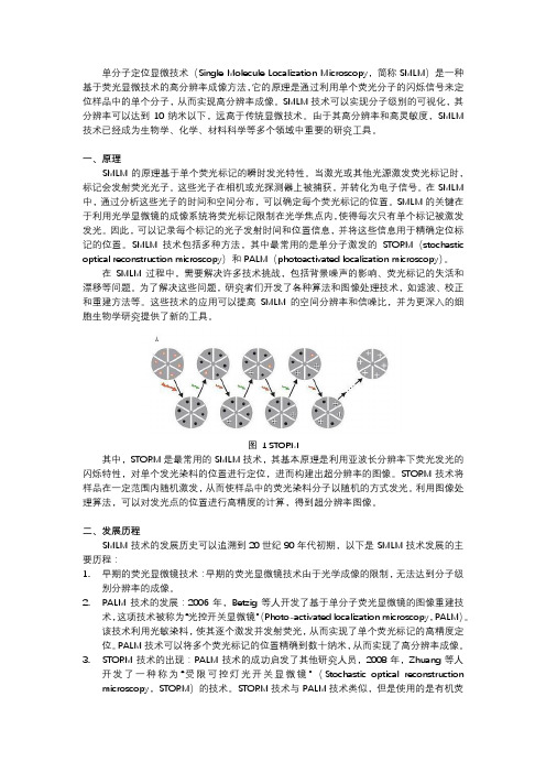
图 1 STORM其中,STORM是最常用的SMLM技术,其基本原理是利用亚波长分辨率下荧光发光的闪烁特性,对单个发光染料的位置进行定位,进而构建出超分辨率的图像。
STORM技术将样品在一定范围内随机激发,从而使样品中的荧光染料分子以随机的方式发光。
利用图像处理算法,可以对发光点的位置进行高精度的计算,得到超分辨率图像。
光纤非线性光学显微成像
中国科学 G 辑: 物理学 力学 天文学 2007年 第37卷 增刊: 138~145收稿日期: 2007-05-20; 接受日期: 2007-09-03Australia Research Council 和国家自然科学基金(批准号: 60410131, 90508003)资助项目* E-mail:《中国科学》杂志社SCIENCE IN CHINA PRESS 光纤非线性光学显微成像付 玲①②* M. Gu ①②(① 华中科技大学武汉光电国家实验室(等), 武汉 430074; ② Centre for Micro-Photonics, Faculty of En-gineering and Industrial Sciences, Swinburne University of Technology, Hawthorn, Victoria 3122, Australia)摘要 在光学显微成像领域, 基于光纤的小型非线性光学显微镜和内窥镜作为传统显微镜和其他光学成像方法的一种重要补充形式, 近几年来受到人们的关注. 该文介绍和总结了光纤非线性光学显微成像技术及其在生物医学中的应用.首先介绍了结合非线性光学显微技术和单模光纤耦合器获得小型非线性光学显微镜的方法; 特别对基于双包层光子晶体光纤和微电机系统扫描镜的光纤非线性内窥成像系统进行了分析; 最后通过消化器官的组织成像实验说明了光纤非线性光学显微镜的重要应用. 研究证明了基于光纤和微电机系统MEMS 扫描镜的非线性内窥镜的新概念, 并用于生物组织的成像.关键词 非线性光学显微成像 光纤耦合器 双包层光子晶体光纤 微电机系统扫描镜 组织成像显微镜改变了人类认识世界的方法, 是人类历史上最重要的发明之一. 经过300多年的发展, 光学显微成像已成为自然科学研究领域中的重要分支, 为生命和信息等科学领域不断提供新方法和新概念[1]. 特别是上世纪末期出现的非线性光学显微镜具有光学层析能力并能在厚组织(毫米量级)内实现高空间分辨率(亚微米)成像, 是现代光学显微成像技术的革新, 为人们研究厚组织中的分子和细胞功能提供了有力的工具[2,3]. 非线性光学成像技术主要基于多光子吸收(multiphoton absorption)[4]、高次谐波(higher harmonic)[5]和相干反斯托克斯拉曼散射(coherent anti-Stokes Raman scattering, CARS)[6]等光学非线性效应, 其中以双光子荧光成像的应用最为广泛. 自非线性光学显微镜诞生以来[4], 科学家们一直致力于将其用于活体(in vivo )成像. 然而, 复杂的光学系统和沉重的光学器件使非线性光学显微成像的研究局限于庞大的显微镜系统, 无法应用到活体的内部器官(internal organs)和整体动物(intact animals)成像.近年来, 随着新型光纤和微制造技术的迅猛发展, 光纤非线性显微镜和内窥镜(fiber-optic nonlinear optical microscopy/endoscopy)的研究正在扭转这一局面[7]. 光纤非线性显微镜和内窥镜是小型的非线性光学显微成像系统, 由光纤传输激发光或者非线性光信号, 并利用微型扫增刊付玲等: 光纤非线性光学显微成像139描机制构建光学图像. 光纤的超小尺寸(一般只有数百个微米的直径)与良好的机械和光学性能增加了成像系统的灵活性并减小了系统的尺寸, 使非线性光学成像技术在内部器官和活体动物中的研究成为可能. 自Denk研究小组和Gu研究小组先后研制出基于单模光纤的双光子荧光显微镜之后[8,9], 光纤非线性显微镜和内窥镜的研究在短短6年之内进展迅速[10~19]. 光纤非线性显微镜和内窥镜研究中的关键问题是: (ⅰ) 近红外波段超短脉冲激光的传输和非线性光信号的收集. 光纤的色散和自相位调制等非线性效应会导致脉冲宽度和光谱的展宽, 降低激发效率[20]; 光纤的低数值孔径和小芯径限制了可见光信号的高效收集、系统的信噪比和成像深度. (ⅱ) 扫描机制. 扫描器件应该具有毫米量级的尺寸, 并能实现快速扫描和高图像分辨率来实时监测生物过程. (ⅲ) 系统设计. 系统设计是非线性内窥镜研究的关键, 直接决定了系统的灵活性, 功能和应用范围. 该文研究了基于单模光纤耦合器(single-mode fibre coupler)和双包层光子晶体光纤(double-clad photonic crystal fibre, DCPCF)的非线性光学显微成像系统, 首次实现了基于光纤耦合器的双光子荧光和二次谐波显微成像技术, 并证明了基于DCPCF和微电机系统(microelectromechanical system, MEMS)扫描镜的非线性内窥镜的新概念, 使光纤成像系统的信噪比提高了两阶.1基于单模光纤耦合器的非线性光学显微镜Bird和Gu[10,11]首次将单模光纤耦合器引入双光子荧光显微成像, 证明了三端口的单模光纤耦合器能传输近红外波段的超短脉冲激发光并收集荧光. 与基于单模光纤的非线性光学显微镜相比[9], 使用单模光纤耦合器能将非线性显微镜的纵向分辨率提高30%, 且使系统更紧凑并具有自准直的特性. 然而, 二次谐波比双光子荧光更远离单模光纤耦合器的工作波长; 二次谐波源于相干过程, 其偏振各向异性的测量(polarisation anisotropy measurement)要求光纤耦合器能同时传输偏振的激发光和谐波信号[15]. 因此, 我们首先研究了单模光纤耦合器在不同波长的传输特性, 结果如图1(a). 实验中端口3单模光纤耦合器(Newport, F-CPL-S12785)的工作波长为780 nm, 分束比(Splitting ratio)是50:50, 纤芯与包层的直径分别为5和125 μm. 当波长范围在770~870 nm时, 耦合器中端口3~1的耦合效率为20%~41%; 端口2~1在435~532 nm 波长范围内的耦合效率为29%~41%. 当长度为2 m的光纤耦合器传输80~150 mW的激发光至样品端时, 尽管光纤中的群速度色散和自相位调制使脉冲展宽到数皮秒, 系统仍有足够的激发光功率用于非线性成像. 实验结果还表明, 单模光纤耦合器在可见光波段的功能类似于普通光学显微镜中的二向色镜(Dichroic mirror). 当435 nm的激光耦合入端口1, 端口2和端口3的分束比为99.6:0.4, 而且两个端口的输出模式分别表现出单模和多模的特征(图1(b), (c)). 532 nm的激光在单模光纤耦合器中传播时, 测量得到的分束比和输出模式也具有相同特征(图1(d)). 因此, 为了优化在基于单模光纤耦合器的非线性显微镜系统中激发光与非线性信号的传输, 超短脉冲激光通过端口3(激发臂)传递到端口1(样品臂), 后向散射的双光子荧光和二次谐波则由端口1通过端口2(信号臂)被探测器收集(图1). 光纤纤芯能消除焦点以外的杂散光, 在成像系统中具有共聚焦针孔(Pinhole)的功能.除了单模光纤耦合器传输特性的研究, 二次谐波光谱的研究也是验证光纤成像系统的关键. 在图1的实验系统中, 我们以AF-50为样品, 用光谱仪(Acton Research Corporation)测量了经过单模光纤耦合器的双光子荧光和二次谐波的光谱. 当激发波长是800 nm时, 通过单模光纤耦合器的辐射谱包含400 nm的二次谐波谱线和430~600 nm的双光子荧光(图2). 二次谐波140中国科学G辑物理学力学天文学第37卷的光谱宽度约为9 nm, 约为激发光带宽的. 当激发光在780~870 nm范围内调谐时, 二次谐波光谱的中心波长则随激发光波长改变, 始终展示出倍频的特性.图1 基于单模光纤耦合器的双光子荧光和二次谐波成像系统(a) 单模光纤耦合器在可见光和近红外波段的耦合效率. (b)~(d) 可见光在激发臂和信号臂的输出模式; O1和O2为 0.25 NA10×显微物镜, O3为0.85 NA 40×显微物镜双光子荧光和二次谐波显微镜中的光学层析能力源于信号光强度与激发光强度的非线性关系, 纵向分辨率是非线性显微成像系统的重要参数. 通过在纵向位置扫描薄层AF-50, 我们得到了基于单模光纤耦合器的显微镜系统对双光子荧光和二次谐波的响应曲线(图2中的插图). 此结果表明, 在激发波长为800 nm时, 光纤显微镜对二次谐波和双光子荧光成像的纵向分辨率分别为1.8和2.1 μm. 可见二次谐波较短的信号波长使其成像的纵向分辨率比双光子荧光成像的纵向分辨率提高了14%. 当激发光波长由800 nm调谐到860 nm时, 二次谐波成像的纵向分辨率增加到 1.9 μm. 通过三维相干传递函数的理论计算, 我们发现与光纤参数和光学系统参数相关的归一化光纤尺寸对基于单模光纤的二次谐波成像系统的纵向分辨率起决定性的作用, 光纤中二次谐波波长的增加使归一化光纤尺寸增大, 纵向分辨率降低[21].图2 基于单模光纤耦合器的非线性显微镜的双光子荧光(TPEF)和二次谐波(SHG)光谱与纵向响应曲线增刊付玲等: 光纤非线性光学显微成像141用偏振的二次谐波信号来反应结构蛋白的形态是二次谐波显微镜的特性之一. 为了证明基于单模光纤耦合器的成像系统具有探测二次谐波偏振各向异性的能力, 我们研究了单模光纤耦合器对偏振光传输和成像的影响. 测量结果表明, 在整个近红外和可见光波段, 单模光纤耦合器中的材料双折射特性使其能传播特定偏振方向的线性偏振光[22]. 因此单模光纤耦合器能传递线性偏振的激发光与高度有序的蛋白纤维(例如鱼鳞, black tetra fish scale)相互作用, 二次谐波经过光纤耦合器后的偏振状态可通过转动探测器前的偏振片并测量收集到的二次谐波强度来确定. 图3(a)和(b)是在探测器前偏振片(Newport, 10GT04)转角正交情况下光纤系统收集到的鱼鳞的二次谐波图像, 激发波长为800 nm, 探测器前的滤光片为400/9 nm. 当偏振片转角改变时, 收集到的二次谐波强度与转角θ 满足cos2θ 的关系(图3(c)). 此结果证明, 基于单模光纤耦合器的显微镜系统能保持二次谐波信号的线性偏振态, 并对结构蛋白成像.图3 二次谐波偏振各向异性的测量(a)和(b)偏振方向正交的二次谐波成像, 箭头方向为探测器前偏振片的转角方向. 标尺为20 μm. (c) 二次谐波的强度与探测器前偏振片转角的cos2θ 关系2基于双包层光子晶体光纤的非线性光学内窥镜由于单模光纤能传输高质量的激光束并改善成像系统的层析能力, 因而在光纤成像系统中被普遍采用. 但是单模光纤的低数值孔径(约0.1~0.2)和较小的纤芯尺寸极大地限制了对生物样品中微弱光信号的收集和探测, 使非线性光学显微镜系统的灵敏度较低. 作为光纤领域的革新产物, 光子晶体光纤能通过设计光纤中二维光子晶体的结构实现普通光纤不能具备的功能, 彻底地改变了光纤传输光束的方式. 我们首次改造了有源的DCPCF, 并将其用于光学显微成像来提高系统的灵敏度. 由于MEMS器件具有小尺寸、低功耗、利于建造“芯片显微镜”等优点, 显微内窥成像系统中的扫描机制则由MEMS扫描镜来实现. 基于DCPCF和MEMS 扫描镜的非线性内窥成像系统如图4. 飞秒脉冲光经过光栅对(Newport, 1200 grooves/mm)和一个显微物镜(Melles Griot, 4×/0.12NA)后被耦合到DCPCF. 光纤中出射的光经过MEMS扫描镜后被渐变折射率(Gradient index, GRIN)透镜聚焦到样品上. 非线性光信号则通过MEMS扫描镜后被DCPCF收集到探测器.142 中国科学 G 辑 物理学 力学 天文学 第37卷图4 基于DCPCF 和MEMS 扫描镜的非线性光学内窥镜系统右上角是双包层光子晶体光纤的扫描电子显微镜图像与800 nm 激发光输出模式的叠加, 右下角是能实现两维扫描的MEMS 扫描镜非线性光学内窥成像系统中的DCPCF 是由用于光纤激光器的有源DCPCF 演化而来. 我们除去了有源光纤纤芯中的掺杂元素, 其目的是使近红外波段的激发光能在纤芯进行单模传输, 而位于可见光波段的非线性光信号则能被具有高数值孔径的内包层收集[16]. 成像系统中DCPCF(Crystal Fiber A/S)的纤芯和内包层直径分别为20和165 μm(图4). 纤芯被孔间距比(Hole to hole pitch ratio)为0.26的空气小孔包围. DCPCF 的内包层和外包层则被由一圈空气小孔分开, 使内包层拥有高数值孔径(~ 0.6). 需要指出的是, 这种大面积和高数值孔径的光纤只能利用光子晶体的结构实现, 而非普通光纤的制造技术. 通过对DCPCF 光传输特性的研究, 我们发现如果选择与纤芯数值孔径接近的耦合物镜, 改造过后的DCPCF 在410~870 nm 的耦合效率可达到80%~90%(图5). 而使用高数值孔径(0.65)的耦合物镜则会导致光泄露到内包层, 使耦合效率降低20%. 因此, 低数值孔径的耦合物镜能使近红外光耦合到纤芯中的效率为28%, 可见光耦合到内包层的效率为90%, 此数值是单模光纤对可见光耦合效率的两倍.GRIN 透镜是一种利用特定折射率分布使光线聚焦的小型透镜. 当光纤和GRIN 透镜结合用于非线性光学成像系统, GRIN 透镜的有效数值孔径依赖于光纤和GRIN 透镜之间的距离, 因而直接影响到光纤成像系统的效果[23]. 例如,光束的耦合效率与有效数值孔径有关; 成像的分辨率与有效数值孔径的平方成正比; 双光子荧光的强度与有效数值孔径的4次方成正比. 因此, 研究光纤系统的纵向分辨率和信号水平随光纤和GRIN 透镜距离的变化是优化系统设计的重要途径. 实验采用了直径为0.5 mm, 节距(pitch)为0.2, 数值孔径为0.5的GRIN 透镜(GRINTECH)和AF-50为样品. 实验结果表明, 当DCPCF 与GRIN 透镜之间的距离增大时, 双光子荧光成像的纵向分辨率逐渐得到改善, 而且探测到的荧光强度逐渐增大(图6(a)).如果光纤与透镜之间图5 双包层光子晶体光纤在可见至近红外波段的耦合效率增刊付玲等: 光纤非线性光学显微成像143的距离为5 mm, 激发光束充满GRIN透镜的后孔径, 系统能得到最佳的纵向分辨率, 即双光子荧光和二次谐波的纵向分辨率分别为6和5.4 μm (图6(b)). 与基于单模光纤耦合器和GRIN透镜的非线性显微镜系统相比, 由于DCPCF具有高数值孔径和大截面面积, 使用DCPCF取代单模光纤能实现纵向分辨率和非线性光信号的同时最优化, 而使用单模光纤的成像系统在探测到的荧光信号最强时却不具备最高的纵向分辨率[17,23].图6(a) 双光子荧光强度和纵向分辨率随GRIN透镜和光纤间距的变化, 荧光强度和纵向分辨率能在间距较大时被同时优化;(b) 双光子荧光与二次谐波的纵向响应曲线MEMS扫描镜是成像系统中实现二维光束扫描的器件. 我们使用的MEMS扫描镜是基于电热驱动(Electrothermal actuation)的原理, 在驱动电压少于12 V的情况下其光学扫描角度可达到30°[24]. 镜面的尺寸为0.5 mm, 表面铝膜对800 nm激发光的发射率约为80%(图4). 通过在MEMS扫描镜的快、慢轴应用特殊设计的波形和2.5~7.5 V的电压, 激光束的反射角被改变从而实现二维的线扫描(raster scanning), 其扫描速率为7线/s. 图7(a)是由基于DCPCF、MEMS 扫描镜、和GRIN透镜的显微镜系统获得的荧光微球的双光子荧光图像. 此结果证明了MEMS 扫描镜不仅能传递近红外超短脉冲用于非线性效应的激发, 而且能收集位于可见波段的荧光信号; 飞秒脉冲经过MEMS镜面之后, 脉冲大约展宽了25%, 故可通过镀膜技术来改善这一现象. 更重要的是, 以MEMS扫描镜作为扫描机制的非线性光学成像系统所产生的图像与通过普通扫描台获得的图像高度一致(图7(b)).图7 基于DCPCF, MEMS扫描镜、GRIN透镜(a)和基于单模光纤耦合器、扫描台、GRIN透镜(b)的显微镜系统对微球的双光子荧光成像标尺为10 μm144中国科学G辑物理学力学天文学第37卷DCPCF的独特光学性质使非线性显微镜成像系统的灵敏度得到极大改善. 通过比较由DCPCF和单模光纤耦合器得到的双光子荧光图像(图7(a)和(b)), 我们发现DCPCF的应用使成像系统的信号水平提高了两个量级. 如果使用光栅对补偿脉冲光在DCPCF中经历的群速度色散, 成像系统的信号水平将会在此基础上再提高一个量级. 成像系统灵敏度的改善主要是因为DCPCF比单模光纤具有更高的数值孔径和更大截面面积, 减少了飞秒脉冲在光纤中传输的自相位调制效应, 使飞秒脉冲具有最小的畸变, 而且对GRIN透镜的色差有更强的兼容性.3基于光纤非线性光学显微镜的组织成像利用基于DCPCF, MEMS扫描镜和GRIN透镜的非线性光学内窥镜系统(图4), 我们对大鼠的大肠和胃部组织进行了成像, 结果如图8. 为了增加荧光成像的对比度, 大肠和胃部组织从大鼠(Sprague-Dawley)中剥离后, 用浓度为1%的Acridine Orange(Sigma)对其内表面上皮组织染色, 成像实验在大鼠牺牲后两小时之内完成. 大肠组织双光子荧光成像的穿透深度约为100 μm, 图8(a)是双光子荧光成像的截面之一. 光纤显微镜系统能清晰地显示表面上皮细胞和肠道孔穴(intestinal crypts, 箭头)的形态信息. 从大鼠胃部组织获取的双光子荧光图像(图8(b))展示出与大肠截然不同的形态结构, 显微系统的分辨率亦能将胃部的坑穴(gastric pits, 箭头)从上皮细胞中区分出来.图8 使用基于DCPCF, MEMS扫描镜和GRIN透镜的光纤内窥镜系统对大鼠大肠组织(a)和大鼠胃部组织(b)的双光子荧光成像标尺为20 μm4总结和展望本文介绍和总结了基于单模光纤耦合器和DCPCF的非线性显微镜系统及其生物医学应用. 单模光纤耦合器能传递近红外波段的激发光和收集可见光波段的双光子荧光和二次谐波, 并在非线性显微镜系统中展示出二向色镜的特性. 基于单模光纤耦合器的成像系统能保持激发光和二次谐波的线性偏振态并用于研究结构蛋白的形态. 通过引入新型的DCPCF, 光纤非线性光学显微镜的灵敏度得到极大改善. MEMS扫描镜和GRIN透镜的结合使基于DCPCF的成像系统更加小型化, 大鼠大肠和胃部的组织成像也证明了非线性光学内窥镜系统的可行性. 在将来的研究中, DCPCF耦合器[25]与内窥镜系统将会实现整个成像系统的全光纤化, 这对光纤非线性光学显微镜的发展有着重要意义. 另外, 此光纤成像技术也可与显微成像领域中新的扫描技术[26,27]结合, 拓展多光子荧光成像的应用. 随着光纤器件和微制造技术的不断发展, 光纤非线性光学显微镜系统将日趋小型化, 为传统的光学显微成像技术提供重要的补充并在生物医学领域发挥作用.增刊付玲等: 光纤非线性光学显微成像145参考文献1 Yuste R. Fluorescence microscopy today. Nat Methods, 2005, 2: 902—904[DOI]2 Zipfel W R, Williams R M, Webb W W. Nonlinear magic: multiphoton microscopy in the biosciences. Nat Biotech, 2003, 21:1369—1377[DOI]3 Helmchen F, Denk W. Deep tissue two-photon microscopy. Nat Methods, 2005, 2: 932—940[DOI]4 Denk W, Strickler J H, Webb W W. Two-photon laser scanning fluorescence microscopy. Science, 1990, 248: 73—75[DOI]5 Campagnola P J, Loew L M. Second harmonic imaging microscopy for visualizing biomolecular arrays in cells, tissues andorganisms. Nat Biotech, 2003, 21: 1356—1360[DOI]6 Cheng J X, Xie X S. Coherent anti-Stokes Raman scattering microscopy: instrumentation, theory, and applications. J PhysChem B, 2004, 108: 827—840[DOI]7 Flusberg B A, Cocker E D, Piyawattanametha W, et al. Fiber-optic fluorescence imaging. Nat Methods, 2005, 2: 941—950[DOI]8 Helmchen F, Fee M S, Tank D W, et al. A miniature head-mounted two-photon microscope: high-resolution brain imaging infreely moving animals. Neuron, 2001, 31: 903—912[DOI]9 Bird D, Gu M. Resolution improvement in two-photon fluorescence microscopy with a single-mode fiber. Appl Opt, 2002,41: 1852—1857[DOI]10 Bird D, Gu M. Compact two-photon fluorescence microscope based on a single-mode fiber coupler. Opt Lett, 2002, 27:1031—1033[DOI]11 Bird D, Gu M. Two-photon fluorescence endoscopy with a micro-optic scanning head. Opt Lett, 2003, 28: 1552—1554[DOI]12 Jung J C, Schnitzer M J. Multiphoton endoscopy. Opt Lett, 2003, 28: 902—904[DOI]13 GÖbel W, Kerr J N D, Nimmerjahn A, et al. Miniaturized two-photon microscope based on a flexible coherent fiber bundleand a gradient-index lens objective. Opt Lett, 2004, 29: 2521—2523[DOI]14 Levene M J, Dombeck D A, Kasischke K A, et al. In vivo multiphoton microscopy of deep brain tissue. J Neurophysiol,2004, 91: 1908—1912[DOI]15 Fu L, Gan X, Gu M. Use of a single-mode fiber coupler for second-harmonic-generation microscopy. Opt Lett, 2005, 30:385—387[DOI]16 Fu L, Gan X, Gu M. Nonlinear optical microscopy based on double-clad photonic crystal fibers. Opt Express, 2005, 13:5528—5534[DOI]17 Fu L, Jain A, Xie H, et al. Nonlinear optical endoscopy based on a double-clad photonic crystal fiber and a MEMS mirror.Opt Express, 2006, 14: 1027—1032[DOI]18 Myaing M T, MacDonald D J, Li X. Fiber-optic scanning two-photon fluorescence endoscope. Opt Lett, 2006, 31: 1076—1078[DOI]19 Fu L, Gu M. Fibre-optic nonlinear optical endoscopy. J Microscopy, 2007, 226: 195—206[DOI]20 Agrawal G P. Nonlinear Fiber Optics. San Diego: Academic. 198921 Gu M, Fu L. Three-dimensional image formation in fiber-optical second harmonic generation microscopy. Opt Express,2006, 14: 1175—1181[DOI]22 Fu L, Gan X, Bird D, et al. Polarisation characteristics of a 1×2 fiber coupler under femtosecond pulsed and continuouswave illumination. Opt Laser Technol, 2005, 37: 494—497[DOI]23 Fu L, Gan X, Gu M. Characterization of the GRIN lens-fiber spacing toward applications in two-photon fluorescence endo-scopy. Appl Opt, 2005, 44: 7270—7274[DOI]24 Jain A, Xie H. An electrothermal SCS micromirror for large bi-directional 2D scanning. 13th International conference onSolid-State Sensor, Actuators and Microsystems. Seoul: IEEE Computer Society Press, 2005. 988—99125 Fu L, Gu M. A double-clad photonic crystal fiber coupler for compact nonlinear optical microscopy imaging. Opt Lett, 2006,31: 1471—1473[DOI]26 Zeng S, Lv X, Zhan C, et al. Simultaneous compensation for spatial and temporal dispersion of acousto-optical deflectorsfor two-dimensional scanning with a single prism. Opt Lett, 2006, 31: 1091—1093[DOI]27 Li D, Zeng S, Lv X, et al. Dispersion characteristics of acousto-optic deflector for scanning Gaussian laser beam of femto-second pulses. Opt express, 2007, 15: 4726—4734[DOI]。
庄小威超分辨率显微成像PPT
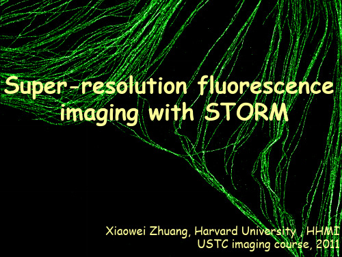
Huang et al, Science (2008)
EM
STORM
STORM
200 nm
Clathrin FBP17
Dynamin FBP17
Wu et al, Nat Cell Biol (2010)
Huang, Jones et al, Nat. Meth. (2008)
Endoplasmic Reticulum
Red light excitation Cy5
Near IR light excitation Cy7
Dempsey, Vaughan, Chen et al, Nat Methods (2011)
Mitochondria Tubulin Acetylated Tubulin Endoplasmic reticulum
Rust et al, Nat Methods (2006) -- STORM Betzig et al, Science (2006) -- PALM Hess et al, Biophys. J (2006) -- FPALM
Single-molecule localization
σ ≈ σPSF / N1/2
1 nm
Diffraction-limited resolution
Diffraction-limited resolution
d 200 250 350 450 550 d= = 650 750 nm nm
Resolution of light microscopy
Abbe resolution (diffraction) Limit: A few hundred nanometers ~Wavelength/2NA
ZEISS Axio Imager 2研究显微镜说明书
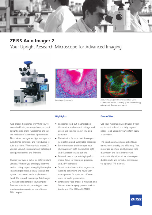
Pilidium larvae of the Nemertean ribbon worm,Cerebratulus lacteus. Courtesy of the Marine BiologyLaboratory & Development journalEsophagus (guinea pig)ZEISS Axio Imager 2Your Upright Research Microscope for Advanced ImagingAxio Imager 2 combines everything you’veever asked for in your research environment:brilliant optics, bright fl uorescence and vari-ous methods of transmitted light contrast.The contrast manager and light manager en-sure defi ned conditions and reproducible re-sults at all times. With your Axio Imager.Z2you can use ACR to automatically detect andconfi gure objectives and fi lter sets.Choose your system out of six different standversions. Whether you are simply observingand recording, or performing highly compleximaging experiments, it’s easy to adapt thesystem components to the application athand. The research microscope Axio Imager2 resolves fi nest details of your samples -from tissue sections in pathology to brainspecimens in neuroscience to multi-colorFISH samples.Highlights• Encoding: read-out magnifi cation,illumination and contrast settings, andautomatic transfer to ZEN Imagingsoftware• Motorization for reproducible compo-nent settings and automated processes• Excellent optics and homogeneousillumination in both transmitted lightand fl uorescence applications• Research microscope with high perfor-mance focus for maximum precisionand 24/7 operation• Smart control concept for ergonomicworking conditions and multi-usermanagement for up to ten differentusers or working scenarios.• Extend your Axio Imager 2 with high endfl uorescence imaging systems, such asApotome.2, LSM 800 and LSM 880Ease of UseUse your motorized Axio Imager 2 withequipment tailored precisely to yourneeds - and upgrade your system easilyat any time.The smart automated contrast settingslet you work quickly and effi ciently. Themotorized aperture and luminous fi elddiaphragm and light intensity areautomatically adjusted. Achieve repro-ducible results and control all componentsvia optional TFT monitor.***************/axioimagerN o t a l l p r o d u c t s a r e a v a i l a b l e i n e v e r y c o u n t r y . U s e o f p r o d u c t s f o r m e d i c a l d i a g n o s t i c , t h e r a p e u t i c o r t r e a t m e n t p u r p o s e s m a y b e l i m i t e d b y l o c a l r e g u l a t i o n s . C o n t a c t y o u r l o c a l Z E I S S r e p r e s e n t a t i v e f o r m o r e i n f o r m a t i o n .E N _41_012_113 | C Z 08-2015 | D e s i g n , s c o p e o f d e l i v e r y a n d t e c h n i c a l p r o g r e s s s u b j e c t t o c h a n g e w i t h o u t n o t i c e . | © C a r l Z e i s s M i c r o s c o p y G m bHPerformance:The motorized reflector turret accommo-dates either six or ten Push & Click filtermodules. Auto-configure all motorized components with Smart Setup of the ZEN imaging software.Acquire fluorescence images with an excellent signal-to-noise ratio. The fluorescence beam path and high efficiency fluorescence filter sets of this research microscope deliver exposure times that are up to 50 percent shorter.Stand Versions:• Axio Imager.A2 LED • Axio Imager.A2• Axio Imager.D2• Axio Imager.M2p • Axio Imager.M2• Axio Imager.Z2 Suitable Applications:• Cell biology • Neuroscience • Molecular genetics • PathologyNote: This product is primarily for research use. Only Axio Imager.M2p is for use in diagnostic procedures or patient management.ZEISS Axio Imager 2Your Upright Research Microscope for Advanced Imaging。
超声波扫描显微镜sonscan

SKLMS 机械制造系统工程国家重点实验室
Advantage of Acoustic Micro Imaging (超声波微成像的优点)
超声波显微成像(AMI)利用高频率的超声波(一般在5MHz以上)探 测物体内的结构、缺陷、以及对材料做定性分析。
其优点如下: • Non Destructive (无损检测,非破坏性) • Sensitive inspection technique for bond evaluation (对粘结层面非常敏感) • Penetrates most materials (能穿透大多数的材料) • Subsurface structures (浅表层结构的分析) • Mechanical properties (材料力学性能的检测-非线性超声测试)
• Lower attenuation materials such as Si can be inspected using higher frequencies. (低衰减的材料:如硅材等能使用较高频率来扫描检测)
STATE KEY LABORATORY FOR MANUFACTURING SYSTEMS ENGINEERING
Greater Penetration
5 MHz 15 MHz 30 MHz 50 MHz 75 MHz
Higher Resolution
100 MHz 150 MHz
230 MHz 300 MHz
Sample Surface
Sample Back Surface
STATE KEY LABORATORY FOR MANUFACTURING SYSTEMS ENGINEERING
• The pressures generated at the focal spot are in the micro Joule range and are not harmful to even moderately delicate samples. • The pulse in the acoustic microscope has very low energy in comparison.
ZEISS OPMI pico 诊断微镜说明书

/OPMI-picoZEISS OPMI picoSeeing without compromise.S 100 / O P M I p i c o23ZEISS OPMI picoSimple, compact, provenOPMI ® pico from ZEISS, the most frequently sold ZEISS surgical microscope worldwide, is the trusted choice among doctors. It makes details and fine structures clearly visible. The system enables you to better visualize regions of interest and consistently provide your patients with high-quality examinations and treatments.ZEISS OPMI pico’s cost-effective LED illumination meets your standards and needs. The complete integration of the light source makes it easier to clean the system.Restorative dentistry Quickly detect enamel and dentine fractures as well as approximal caries. High-precision views enable accurate assessment of crown edges, preparation levels, and veneers.EndodonticsVisualize fine anatomical structures and details of root canals and isthmuses for a clear view right down to the apex.Implantology Conduct high-precision examinations and implant treatments quickly and confidently. Reliably detect important anatomical structures.Periodontics Benefit of support for soft-tissue evaluation and management to assist healing, low scarring, and improved cosmetic outcomes.The compact and easy-to-use ZEISS OPMI pico offers solid support for even the most demanding applications – whether in restorative dentistry, endodontics, implantology, or periodontics:Ergonomic seating enables users to conveniently maintain an upright working posture.ZEISS OPMI pico also features camera options to facilitate patient consultation and documentation, depending on thespecific needs in your practice.4Better viewLong-lasting LEDFor color rendition and light that strongly resembles natural daylight. It is a very cost-effective and virtually maintenance-free light source. The low heat output requires minimal ventilation and generates little noise.Full overview of fine detailsZEISS OPMI pico delivers high-quality images at every magnification level.ZEISS OPMI pico enables you to visualize high-contrast, true-color images – the key to improving the quality of examination and treatment. With ZEISS OPMI pico, optics and illumination go hand in hand.View the whole mouth The Varioskop ® 100 objective lens allows you to adjust the focal length over a range of 200 mm to 300 mm to focus on the whole oral cavity in the vertical axis – without moving themicroscope.5While treating your patients you can sit comfortably in an upright, ergonomically correct position for a more relaxed working day.Comfortable working conditions and improved visionOver 75% of dentists found that using a dental microscope had a positive effect on their neck and back pain.5 Working in a comfortable posture allows you to fully focus on your patient and their procedure, and increases your productivity.6A dental microscope can offer other ergonomic benefits, such as improved vision and reduced eye fatigue, common in dental professionals and corresponding to increasing age.7–9Better ergonomicsAdjust the ZEISS OPMI pico to your demands and not vice versa Reduce or increase the distance for the required treatment field as needed with the patented multi-link design of Foldable Tube f170 / f 260. With its long reach, this highly flexible system easily accommodates the needs of the user and different positions of the patient.6Better integrationBenefits of an integrated full HD camera • Complete function integration • Co-observation and documentation • Simplified cleaning of the housing • Images and videos recorded onto a shared network drive or a USB device• Capture of full HD images during recording or from a recorded videoBenefits of live viewing and streamings • Video livestream into the network• Smart Recording: retroactively record video that occurred in the previous 30 secondsComplete integration of technology and design in the suspension arm for a well-balanced architectureFunctional elements such as the video control console, HD video camera, cables, light sources, and light guides are completely integrated into the stand to avoid workspace clutter.Better educationReal-life pictures are very convincing. ZEISS OPMI pico can accommodate a full HD video camera with recording and streaming, allowing you to present patients with high-definition material to explain procedures.A highly compact instrument with a small footprint, ZEISS OPMI pico fits seamlessly into virtually any practice workflow environment. Simply plug in the cable, switch on the power, and the ZEISS OPMI pico isready for use.7Basic configurationPackage optionsAdd-onsSuspension system optionsFloor standCeiling mountWall mount (wallplate)Workplace integrationFurther space-saving suspension system options are available for the Centro carrier system and specified treatment units.Carl Zeiss Meditec AG Goeschwitzer Strasse 51–52 07745 JenaGermany/opmi-pico /med/contacts en-INT_3_1_12IPrintedinGermany.CZ-II/223Internationaledition:Onlyforsaleinselectedcountries.Thecontentsofthebrochuremaydifferfromthecurrentstatusofapprovaloftheproductorserviceofferinginyourcountry.Pleasecontactourregionalrepresentativesformoreinformation.Subjecttochangesindesignandscopeofdeliveryandduetoongoingtechnicaldevelopment.OPMI,VarioskopandVisionGuardareeithertrademarksorregisteredtrademarksofCarlZeissMeditecAGorothercompaniesoftheZEISSGroupinGermanyand/orothercountries.©CarlZeissMeditecAG,223.Allrightsreserved.Application images courtesy of:1 Dr. Claudia Cia Worschech, Sao Paulo, Brazil2 Dr. José Aranguren Cangas, Madrid, Spain3 Dr. Behnam Shakibaie, Tehran, Iran4 Dr. Rino Burkhardt, Zurich, Switzerland5Z augg B et al. Influence of magnification tools on the recognition of artificaila preparation and restoration defects (in German).Schweiz Monatsschr Zahnmed 2004;114:890-896. [Abstract]6L inger W. Advantages for patients under the dental microscope.Available from: https:///blog/dental-microscope7P errin P, et al. Visual acuities of dentists in their respective clinical conditions. Clin Oral Investig 2014;19:2055-2058.8E ichenberger M, et al. Visual acuity of dentists under simulated clinical conditions. Clin Oral Investig 2013;17:725-729.9Y adav VS, et al. Periodontal microsurgery: Reaching new heights of precision.J Indian Soc Periodontol 2018;22(1):5-11.。
蔡司 Axiovert 5 智能倒置细胞培养显微镜说明书

质臻至简蔡司Axiovert 5用于细胞培养和研究的智能显微镜/axiovert20 µm HeLa Kyoto细胞,物镜:LD Plan-Neofluar 63×。
双通道荧光成像:细胞核为蓝色,微管蛋白为红色。
正在为您的实验室寻找一款功能强大的显微镜?想要一款成像时间短、图像质量优的显微镜?这很有必要!拥有一款高质易用的显微镜,对于需要在实验室进行长时间工作的您来说显得十分重要。
智能的倒置细胞培养显微镜蔡司Axiovert 5是您明智的选择:您仅需专注于样品和工作流,按下拍照按钮,即可获得用于数据记录的清晰图像。
该设备将透射光配备的各种观察方式与多通道荧光相结合,用于研究您的细胞或组织培养。
不仅如此,当实验室空间紧张时,您甚至可以将该智能显微镜作为单机使用,将图像保存在U盘上,而无需使用额外的计算机或软件。
用于细胞培养和研究的智能显微镜› 简介› 优势› 应用› 系统› 技术参数›售后服务更简单、更智能、更高度集成使用智能显微技术,让工作更智能蔡司Axiovert 5显微镜十分智能,且成像快速、结果出众。
您只需专注于样品,按下按钮,即可保存细胞或组织培养的清晰图像。
这款智能显微镜还会自动为您调整透射光以及多通道荧光图像的设置及参数。
自动叠加的多通道荧光图像包含标尺信息,该信息将自动保存在图像文件的元数据中。
轻松自如,享受您的日常工作Axiovert 5让您不用再时时刻刻焦急地等待实验结果。
其设计符合人体工程学,功能巧妙,可为您全天候的工作提供支持。
您只需专注于样品本身,使用单手便能完成包括拍照、移动载物台、调焦和控制亮度在内的各种主要操作。
光强管理功能可在所有放大倍率下提供统一的亮度,让您无需在更换物镜时手动调节灯泡亮度。
为了进一步提高细胞分析流程的速度和数据可靠性,您可以选择使用Labscope 中的AI 细胞融合度和AI 细胞计数分析功能,实时获得可重复的结果。
放眼未来,选择一款立足前沿的活细胞 显微镜从常规细胞培养到研究,Axiovert 5可无缝融入您的实验室和工作流。
- 1、下载文档前请自行甄别文档内容的完整性,平台不提供额外的编辑、内容补充、找答案等附加服务。
- 2、"仅部分预览"的文档,不可在线预览部分如存在完整性等问题,可反馈申请退款(可完整预览的文档不适用该条件!)。
- 3、如文档侵犯您的权益,请联系客服反馈,我们会尽快为您处理(人工客服工作时间:9:00-18:30)。
一、超微型显微成像系统产品介绍如下所示:
1.功能和用途
1.1功能
1.1.1系统组件包括显微镜镜体、固定板、GRIN透镜、CMOS、图像采集卡及采集软件等。
1.1.2在单细胞分辨水平,记录一群神经元的钙信号。
1.1.3适用于自由活动动物的在体实验。
1.1.4通过植入GRIN透镜,可以实现深脑成像。
1.1.5系统体积小、重量轻,不影响小鼠自由运动和行为实验。
2.1用途:
2.1.1用于行为动物在体钙成像的超微型显微成像系统。
2.1.2检测新型可遗传编码的乙酰胆碱和多巴胺等探针的荧光变化,即可实时监测乙酰胆碱、多巴胺等浓度的动态变化情况。
二、产品彩图:
Miniature Fluorescent Microscope
1.1 function
1.1.1 System Components include Miniscope body、Base Plate、GRIN Lens、CMOS、DAQ card and software;
1.1.2 Record the calcium signal of a group of neurons at the single cell resolution level;
1.1.3 experiments for freely moving animals;
1.1.4 Deep brain imaging can be achieved by implanting a GRIN lens;
1.1.5 The system is small in size and light in weight, and does not affect the free movement and behavioral experiments of mice.
2.1 Uses:
2.1.1 Ultra-microscopic microscopic imaging system for in vivo calcium imaging of behavioral animals.
2.1.2 To detect the changes in the fluorescence of new genetically-encoded probes such as acetylcholine and dopamine, the dynamic changes of concentrations of acetylcholine and dopamine can be monitored in real time.。