虹膜识别算法的纹理分析
生物识别技术的虹膜识别教程(Ⅲ)

生物识别技术的虹膜识别教程生物识别技术作为一种高效、安全的身份认证方式,近年来得到了广泛的应用。
其中,虹膜识别作为一种高端的生物识别技术,具有独特的优势。
本文将就虹膜识别技术进行介绍,包括其原理、应用场景、使用方法等方面进行详细的探讨。
一、虹膜识别技术的原理虹膜识别技术是利用虹膜独特的纹理和颜色进行身份识别的一种技术。
虹膜是人眼的一部分,其纹理和颜色是每个人独一无二的,与指纹、声纹等生物特征相比,虹膜更加稳定和安全。
虹膜识别技术通过摄像头等设备获取虹膜图像,然后通过算法进行处理和比对,最终确认个体身份。
虹膜识别技术的原理可以简单概括为:采集虹膜图像、提取特征、进行比对。
在图像采集过程中,需要充分保证光线充足、清晰度高,以确保后续的特征提取和比对的准确性。
特征提取是通过图像处理算法将虹膜图像中的纹理和颜色等信息提取出来,形成特征向量。
最后,比对阶段将采集到的特征向量与已有的数据进行比对,确认身份信息。
二、虹膜识别技术的应用场景虹膜识别技术在各个领域都有着广泛的应用。
首先,虹膜识别技术在安防领域得到了广泛的应用,例如用于监控系统、门禁系统等。
由于虹膜识别技术的高准确性和安全性,可以有效防止非法闯入和身份冒用等问题。
此外,虹膜识别技术还在金融领域、医疗领域、政府机构等各个领域得到了应用,为各行各业提供了更加便捷和安全的身份认证手段。
三、虹膜识别设备的使用方法虹膜识别设备通常包括虹膜摄像头、图像处理器、比对算法等组成部分。
在使用虹膜识别设备进行身份认证时,需要注意以下几点。
首先,要保持眼睛距离摄像头适当的距离,通常为15~30厘米。
然后,需要保持眼睛处于摄像头的视野范围内,并尽量避免眨眼或者移动眼睛。
在采集虹膜图像时,要保持光线充足,避免强光直射眼睛,以免影响图像质量。
最后,在使用虹膜识别设备时,要注意保持设备的清洁和稳定,避免影响识别效果。
四、虹膜识别技术的优势与不足虹膜识别技术作为一种高端的生物识别技术,具有诸多优势。
生物识别技术的虹膜识别教程(Ⅰ)

生物识别技术的虹膜识别教程在当今数字化社会中,生物识别技术得到了广泛的应用,其中虹膜识别作为一种高精度、高安全性的生物识别技术,被广泛应用于身份验证、门禁系统等领域。
虹膜识别技术的原理是利用虹膜的独特纹理特征来进行身份识别,虹膜识别技术的高安全性和准确性使其成为当前生物识别技术中的佼佼者。
本文将介绍虹膜识别技术的原理和应用,并简要介绍虹膜识别设备的选购和使用方法。
虹膜识别技术的原理虹膜识别技术是利用虹膜的独特纹理特征进行身份验证的一种生物识别技术。
虹膜是人眼的一部分,它在光线的照射下会呈现出丰富多彩的纹理特征,这些纹理特征是每个人独一无二的,就如同指纹一样。
虹膜识别技术通过扫描和识别虹膜的纹理特征,来验证用户的身份。
虹膜识别技术具有高安全性和准确性,因为虹膜的纹理特征不会受到年龄、外界环境等因素的影响,而且每个人的虹膜纹理都是独一无二的,因此虹膜识别技术具有很高的辨识度。
虹膜识别技术的应用虹膜识别技术的高安全性和准确性使其被广泛应用于各个领域。
在企业和政府机构中,虹膜识别技术被应用于门禁系统、考勤系统等,可以有效保障机构内部安全,并且可以有效防止考勤打卡等作弊行为。
在金融领域,虹膜识别技术被应用于身份验证、ATM取款等,可以有效防止用户身份被盗用。
在医疗领域,虹膜识别技术被应用于病人身份识别、医疗记录管理等方面,可以有效保障医疗信息的安全性。
虹膜识别技术还被应用于手机解锁、电脑登录等个人设备的安全性保障。
虹膜识别设备的选购和使用方法在选择虹膜识别设备时,首先需要考虑设备的识别准确性和响应速度,好的虹膜识别设备应该具有很高的识别准确性和快速的响应速度,以提高用户体验。
其次需要考虑设备的安全性和稳定性,好的虹膜识别设备应该具有很高的安全性和稳定性,以保障用户数据的安全。
最后需要考虑设备的适用场景和成本,不同的场景对虹膜识别设备的要求不同,而且不同的虹膜识别设备价格不同,需要根据实际需求进行选择。
在使用虹膜识别设备时,首先需要保持眼睛的清洁,因为眼睛的清洁程度会影响虹膜识别的准确性。
基于纹理方向能量特征的虹膜识别算法

1引言随着社会的发展和科技的进步,生物特征识别技术已经脱离了电影屏幕,在生活中的应用越来越平常。
生物特征识别技术应用面很广,包括身份鉴别、公司考勤、社会公共安全、金融等领域。
主要的生物特征技术包含指纹识别、手静脉识别、掌纹识别、虹膜识别、声音识别和步态识别。
虹膜识别因虹膜具有特征稳定、不易盗取、非侵犯采集的特点,已成为安全保密性最好的生物特征识别手段之一。
近年来关于虹膜识别的算法得到了学术界的广泛关注。
国外在虹膜识别领域的研究比较早,也获得了很多开创性的成果。
如英国J.Daugman博士[1-3]发明Gabor虹膜识别算法。
Bobles[4]发明了基于小波过零点特征检测的虹膜识别算法。
这些算法对于后面虹膜识别的研究者有很大的借鉴作用。
国内最早研究虹膜识别的是中国科学院自动化研究所谭铁牛院士团队,该团队于1999年首次发明了一种结合多尺度多方向的Gabor分解和Daubechies小波分解的虹膜识别算法,并获得了“基于基于纹理方向能量特征的虹膜识别算法邓玉波,苏娟DENG Yubo,SU Juan湖南大学电气与信息工程学院,长沙410082College of Electrical and Information Engineering,Hunan University,Changsha410082,ChinaDENG Yubo,SU Juan.Iris recognition algorithm based on texture energy puter Engineering and Appli-cations,2017,53(15):196-199.Abstract:In order to solve the problem that the traditional iris recognition system’s performance isn’t good enough under non-ideal image conditions,a novel algorithm based on texture energy features is proposed.Firstly,a set of horizontal and vertical direction filter is designed to extract iris texture edge.The characteristic figures exhibiting the differences of direc-tion energy are generated by comparing the energy intensity of iris texture edge in the two directions,horizontal and vertical. Secondly,the figures are divided into different parts,and the maximal point of every energy difference is selected as an effective point to obtain eigenvector.Besides,swelling the noise template and ruling out the influence of noise point and convolution noise point on the surrounding effective figures.Finally,the hamming distance matching vector is obtained to implement iris recognition.The method is tested by CASIA3.0iris database provided by Chinese Academy of Sciences gaining a good recognition rate.Key words:iris recognition;feature extraction;directional energy characteristics;rotational and translational invariance摘要:为了解决传统虹膜识别系统在非理想虹膜图像下识别性能不够好的问题,提出了基于纹理方向能量特征的虹膜识别方法。
基于局部纹理分析的虹膜识别算法

基于局部纹理分析的虹膜识别算法
卢光明;杨文;廖庆敏
【期刊名称】《计算机应用》
【年(卷),期】2007(27)6
【摘要】提出了一种新的基于局部纹理分析的虹膜识别算法.基于多通道Gabor滤波器,滤波图像的系数定量地描述了虹膜纹理局部子块和给定滤波器之间的相似度,系数越大,说明该子块在空间形态上和滤波器越相似.采用局部滤波图像系数的加权平均作为特征编码,那些具有较大系数的点,即和滤波器最相似的点对特征点的位置贡献较大,更能真实的反应虹膜纹理结构.基于欧氏距离的特征匹配试验表明了该方法的有效性.
【总页数】3页(P1490-1492)
【作者】卢光明;杨文;廖庆敏
【作者单位】清华大学,深圳研究生院,广东,深圳,518055;哈尔滨工业大学,计算机科学与技术学院,黑龙江,哈尔滨,150001;哈尔滨工业大学,计算机科学与技术学院,黑龙江,哈尔滨,150001;清华大学,深圳研究生院,广东,深圳,518055
【正文语种】中文
【中图分类】TP391.4
【相关文献】
1.基于局部频率特征和局部方向特征的虹膜识别算法 [J], 姚鹏;叶学义;庄镇泉;吴亮;李斌
2.基于局部方向特征的虹膜识别算法 [J], 姚鹏;叶学义;张文聪;庄镇泉;李斌
3.基于局部小波变换与奇异值分解的虹膜识别算法 [J], 甘俊英;梁宇
4.基于局部奇异值检测的虹膜识别算法 [J], 印勇;庞尚珍
5.基于Gabor滤波的改进虹膜识别算法 [J], 马晓峰;高玮玮
因版权原因,仅展示原文概要,查看原文内容请购买。
提取虹膜信息的原理

提取虹膜信息的原理虹膜识别技术是一种生物识别技术,它通过获取和分析人眼虹膜图像来辨认个体身份。
虹膜是人眼内部与瞳孔相接触的部分,它具有独特而稳定的纹理结构,每个人的虹膜纹理都是独一无二的,就像指纹一样。
该技术已经被广泛应用于安全领域,如进出口管理、金融交易、边境安检等。
虹膜识别的原理是基于人眼虹膜的纹理特征。
下面将详细介绍虹膜识别的工作原理。
首先,虹膜识别系统首先通过一个摄像机来获取人眼的虹膜图像。
这通常涉及到近红外摄像机,因为近红外光对人眼没有伤害,而且可以在光线不足的情况下获取清晰的图像。
随后,虹膜图像被数字化并转换为灰度图像。
这一过程包括去除噪声、图像增强和边缘检测等步骤,以提高图像的质量。
接下来,系统通过分析虹膜图像中的纹理特征来提取虹膜信息。
虹膜纹理是指虹膜中的纹理结构,它包括细小的纹理纹线、结构、颜色等特征。
常用的虹膜纹理特征提取方法包括小波变换、主成分分析、Gabor滤波器等。
虹膜纹理特征提取后,系统将特征进行标准化处理,以便实现不同虹膜之间的比较。
通常,虹膜特征向量是由虹膜纹理特征的数值表示,如向量的长度和方向。
在识别阶段,系统将输入的虹膜图像与已知的虹膜数据库中的虹膜特征进行比对。
通常采用的方法是计算输入虹膜特征与数据库中所有虹膜特征的相似度得分,并将其与预定的阈值进行比较。
如果输入虹膜图像与数据库中的某个虹膜特征的相似度得分超过了阈值,则识别成功,系统将返回该虹膜对应的身份信息。
否则,识别失败,系统将拒绝访问。
虹膜识别技术相对其他生物识别技术具有很多优势。
首先,虹膜是人眼内部的一部分,不易受到环境因素的影响,稳定性较高。
其次,虹膜纹理的复杂性和独特性使得虹膜识别具有极高的准确性和低的错误率。
此外,虹膜识别无需直接接触,对用户来说更为舒适和方便。
虹膜识别技术在实际应用中也需要面临一些挑战。
其中最主要的挑战是虹膜图像的采集。
由于虹膜实际上是人眼内部的一部分,因此需要用户准确地对准摄像机来获取清晰的虹膜图像。
基于局部纹理分析的虹膜识别算法
特征 匹配试验表 明 了该 方 法的有效 性 。 关键 词 : 生物 特征识 别 ; 虹膜 识 别 ; ao 滤 波 G br
中 图分类 号 : 9 . 14 文献标 识 码 : A
I i e o n to b s d o h nay i fl c lr go rs r c g iin a e n t e a lss o o a e i ns
像 的 系数 定量 地描 述 了虹 膜纹 理局部 子块 和给 定 滤波 器之 间的相 似度 , 系数 越 大 , 明该 子块在 空间 说 形态上和滤波器越相似。采用局部滤波图像 系数的加权平均作为特征编码 , 那些具有较大系数的点 ,
即和 滤 波 器最相似 的点对 特征 点的位 置贡 献较 大 , 能 真 实的反 应 虹 膜 纹理 结构 。基 于 欧 氏距 离的 更
uigsm ao l r h r n gs e a s r e t sme l rdi ms nw i ece ii tC ecie s o eG br t s eoi a i e r t nf m di o o t e n i f e ,t i g l ma w e r o n i fe ma ,i h ht ofc ns a dsr c h e n b
Ke r s B o t c i s rc g i o ; G b rf e y wo d : ime r : r e o n t n i i i a o k r i
0 引言
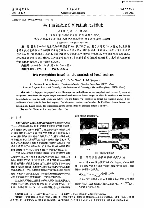
虹膜识别技术是 目前生物特征识别技术领域 的研究热点 之一 。与其他生物特征相 比, 虹膜有着更加丰富 的纹理信息 , 具有更高 的稳 定性 和可 靠性 …。虹 膜识别 技术 的研 究 已有 2 0多年 的 历史 , 中最 具 代 表 性 的虹 膜 识 别 算 法有 基 于 其 Gbr ao 滤波器 的虹膜纹理相位编码算 法 J基于一 维小波 过 , 零 检测的虹膜识 别方法 L , 通道 空 间滤波器 的方 法等 。 4多 J 这 些方法从 不同的层面和角度对虹 膜纹 理特 征的刻 画做 了有 益 的尝试 , 取得 了良好 的效果 。但 由于虹膜纹 理 的高 度复杂
虹膜识别技术介绍
虹膜识别技术介绍虹膜识别技术的原理是利用红外光或可见光对眼睛进行扫描,然后通过图像处理和模式识别算法提取出虹膜纹理特征,最后与预先存储的虹膜特征进行比对来进行身份认证。
虹膜的纹理特征包括纹路、纹孔和纹脊等,这些特征在每个人的双眼中不同,具有极高的独特性和稳定性。
虹膜识别技术具有许多优点。
首先,虹膜对光照、年龄和表情等因素的影响相对较小,因此具有更高的准确度和可靠性。
其次,虹膜是固有的,不易被伪造或复制,具有较高的防伪能力。
此外,虹膜识别技术对被识别者没有侵犯隐私的行为,可以实现非接触式的身份认证。
虹膜识别技术目前已经广泛应用于多个领域。
在安全领域,虹膜识别技术可以应用于边境安检、机场安全、银行保险、政府机关等场所的身份认证和门禁控制。
在企业管理领域,虹膜识别技术可以应用于考勤打卡、会议签到、机房管理等场景,提高工作效率和管理效果。
在医疗领域,虹膜识别技术可以应用于医院的身份验证和病历管理,提高医疗服务的质量和效率。
此外,虹膜识别技术还有一些挑战和限制。
首先,由于虹膜识别需要高分辨率的图像和专门的设备支持,因此成本较高,不适用于一些资源有限的场所。
其次,在使用虹膜识别技术时,需要被识别者主动合作,有时可能会出现用户不适应或者不配合的情况。
最后,一些疾病、手术或眼部损伤等因素可能会影响虹膜辨识的准确性。
虹膜识别技术的发展前景广阔。
随着技术的进步,虹膜识别设备将变得更加小型化、高效化和便携化。
同时,虹膜识别技术也可以与其他技术结合,如人脸识别、声纹识别等,进一步提高识别准确度和可靠性。
虹膜识别技术将在更多领域得到应用,推动社会的数字化、智能化和安全化进程。
总之,虹膜识别技术是一种高安全性、高准确性的生物特征识别技术,具有广泛的应用前景。
随着技术的进步和成本的降低,虹膜识别技术将在更多领域发挥重要作用,为社会带来便利和安全。
浅谈虹膜识别的原理与应用
浅谈虹膜识别的原理与应用虹膜识别是一种生物识别技术,通过分析虹膜的特征来进行个体的身份识别。
虹膜是人眼中的一部分,位于瞳孔和巩膜之间,具有独一无二的纹理和颜色。
虹膜识别利用计算机图像分析和模式识别算法,对虹膜图像进行处理和比对,确定一个人的身份。
虹膜识别的原理基于虹膜的两个基本特征:纹路和颜色。
虹膜的纹路是由一系列的纵向和横向的纹线组成的,个体间的纹路差异非常明显。
虹膜的颜色则由血管和色素质的分布决定,不同的人虹膜颜色不同。
虹膜识别的过程一般包括图像采集、特征提取和匹配三个步骤。
首先,使用虹膜摄像机采集被识别者的虹膜图像。
在采集过程中,要求被识别者与摄像机保持一定的距离和角度,以确保图像质量。
然后,对采集到的虹膜图像进行预处理和特征提取。
预处理包括图像增强、边缘检测等操作,以去除图像中的噪声和干扰。
特征提取则是将虹膜的纹路和颜色信息转换成数字特征。
最后,将提取到的特征与注册在数据库中的特征进行比对,确定一个人的身份。
虹膜识别技术具有许多优势,使其在多个行业和领域得到广泛应用。
首先,虹膜识别凭借其高精度和安全性,可以用于身份认证和门禁控制。
例如,可以应用于企事业单位、政府机构等需要高安全性的场所。
其次,虹膜识别不受个体年龄、表情、情绪等因素的影响,具有很高的稳定性和稳定性。
这使得它可以应用于金融、医疗等领域的用户身份验证,确保信息的安全性和准确性。
此外,虹膜识别技术还可以应用于公共交通、边境入境等领域,提高安全性和效率。
然而,虹膜识别技术也存在一些局限性和挑战。
首先,虹膜识别需要较高的设备成本和复杂的设备安装。
其次,虹膜图像的采集需要被识别者与设备保持一定的距离和角度,不便于大规模采集和使用。
此外,虹膜识别技术也面临着误识别和攻击的风险,例如伪造虹膜图像、存储和传输中的安全问题。
综上所述,虹膜识别作为一种生物识别技术,具有独特的优势和应用前景。
随着技术的不断发展和改进,虹膜识别技术有望在各行各业得到广泛应用,为社会生活和信息安全提供更多的保障。
解析虹膜识别技术
解析虹膜识别技术1.虹膜的生物特性图1:人眼结构人眼眼球结构如图1所示,其生物特征最明显、最丰富的地方是虹膜区域,虹膜位于角膜与晶状体之间,在红外光下,虹膜表面呈现出形态各异的皱纹、条纹、斑点、凹点、射线、水晶体、细丝等大量可供识别的生物信息,如图2所示。
每个人的虹膜纹理特征差异显著,易于区分,且不同种人的虹膜颜色有差异。
不论虹膜颜色还是各种生物特征,都是由遗传基因决定的,不会随外界条件的改变而改变。
虹膜是人体唯一一个能够从外界直接看到并被清晰采集图像的内部组织,虹膜纹理特有的生理特征为虹膜识别技术的唯一性、高防伪性、不可侵犯性奠定了基础。
图2:虹膜结构不同人种的虹膜纹理与颜色也大致不同,西方人的虹膜纹理分布在整个虹膜范围,而东方人的虹膜纹理则集中在虹膜边缘附近,如图3所示为不同人种的虹膜图像。
并且这些特性都是由基因决定的,不受外界条件的左右。
图3:不同人种的虹膜2.人眼虹膜的特点人眼虹膜具有如下特点:(1)高防伪性。
作为身份识别的依据,防伪性是首当考虑因素。
角膜是虹膜的保护伞,固有与外界环境的隔离性,若想要改变虹膜纹理特征,则需冒失明的危险;另外,瞳孔可以自由缩放,使虹膜纹理具有活体特征,无疑为虹膜识别的防伪性再加保障。
(2)高稳定性。
据医学研究表明,人眼虹膜特征在出生三个月后就基本不会再变,也不易病变,一般疾病不会对虹膜组织造成损伤。
生物学家通过大量观察发现人体的虹膜特征一旦发育完全,则在一生之中基本保持不变。
(3)唯一性。
虹膜组织携载大量可视的生物信息,纹理结构的形成与胚胎发育环境息息相关,随机性较大,即便双胞胎,其虹膜特征也是不同的;同一人的左右眼,虹膜细节特征也不同。
(4)非侵犯性。
无需接触,虹膜图像在一定范围内即可采集,身份识别只需用虹膜识别装置扫描用户的眼睛即可,既不会弄脏采集设备,影响拍摄的质量而降低识别准确率,又不会对用户的眼睛造成伤害。
综上所述,虹膜丰富的纹理结构使其具有唯一性;遗传因素决定的虹膜特征,具有终身不变性;不易被手术模仿和改变,使其具有非侵犯性,高稳定性,高防伪性。
生物识别技术的虹膜识别教程(Ⅱ)
生物识别技术的虹膜识别教程虹膜识别技术是一种利用虹膜图像进行身份验证的生物识别技术。
它是一种高精度、高安全性的身份识别技术,已经被广泛应用于各种领域。
本文将介绍虹膜识别技术的基本原理、使用方法以及注意事项,希望能够帮助读者更好地了解和使用这一先进的技术。
虹膜识别技术的基本原理虹膜是人眼的一部分,位于眼睛的睫状体之后。
每个人的虹膜纹路都是独一无二的,类似于指纹,具有高度的个体特征性。
虹膜识别技术利用摄像机拍摄人眼的虹膜图像,然后使用图像处理算法提取虹膜纹路的特征点,最终通过比对输入的虹膜图像与已注册的虹膜特征库进行身份验证。
使用虹膜识别技术的步骤首先,用户需要在虹膜识别系统中注册自己的虹膜信息。
注册时,系统会要求用户注视摄像头,然后通过摄像头拍摄用户的虹膜图像。
注册完成后,用户的虹膜信息将被存储在系统的虹膜特征库中。
在进行身份验证时,用户只需再次注视摄像头,系统会自动拍摄用户的虹膜图像,然后进行比对验证。
如果虹膜图像与已注册的虹膜信息匹配成功,系统将通过验证,并允许用户进行相应的操作。
虹膜识别技术的注意事项虹膜识别技术在使用过程中需要注意以下几点:首先,保持眼部卫生。
在进行虹膜图像采集时,用户需要保持眼部清洁,避免有异物或眼屎影响图像的清晰度。
其次,注视摄像头时需要保持眼睛稳定。
在进行虹膜识别时,用户需要注视摄像头,保持眼睛的位置和姿态稳定,以确保摄像头能够准确拍摄虹膜图像。
另外,避免光线干扰。
强光或弱光都会影响虹膜图像的质量,因此在使用虹膜识别技术时需要避免光线干扰,保持适当的光线条件。
最后,保护个人隐私。
在使用虹膜识别技术时,用户的虹膜信息可能会被存储在系统的数据库中,因此需要确保系统具有足够的安全性,以防止虹膜信息泄露。
虹膜识别技术的应用虹膜识别技术已经被广泛应用于各种领域,包括安防监控、金融支付、边境口岸等。
在安防监控领域,虹膜识别技术可以用于门禁系统、考勤打卡等场景,提高安全性和便利性。
- 1、下载文档前请自行甄别文档内容的完整性,平台不提供额外的编辑、内容补充、找答案等附加服务。
- 2、"仅部分预览"的文档,不可在线预览部分如存在完整性等问题,可反馈申请退款(可完整预览的文档不适用该条件!)。
- 3、如文档侵犯您的权益,请联系客服反馈,我们会尽快为您处理(人工客服工作时间:9:00-18:30)。
Iris Recognition Algorithms Based on Texture AnalysisRichard Yew Fatt NgComputer Vision and Intelligent Systems (CVIS)Group Universiti Tunku AbdulRahman, Malaysia richng01@Yong Haur TayComputer Vision andIntelligent Systems (CVIS)GroupUniversiti Tunku AbdulRahman, Malaysiatayyh@.myKai Ming MokComputer Vision andIntelligent Systems (CVIS)GroupUniversiti Tunku AbdulRahman, Malaysiamokkm@.myAbstractIris recognition has become a popular research in recent years due to its reliability and nearly perfect recognition rates. Iris recognition system has three main stages: image preprocessing, feature extraction and template matching. An innovative method is proposed to extract iris features based on texture analysis. Iris textures are analyzed to capture the discriminating frequency information. Specific filters with different center frequency are applied to three different zones to extract the texture of the iris. Different weightings are given to each zone depending on its contribution to the recognition. The encoded binary templates are compact in size and can avoid the visibility of the individual iris images. The templates are suitable for implementing iris recognition devices using DSP (Digital Signal Processor). The proposed method was evaluated using CASIA iris image database version 1.0 [1]. Experimental results show that the proposed approach has achieved high accuracy of 98.62%.1. IntroductionBiometric identification is an emerging technology which gains more attention in recent years. Iris has distinct phase information which spans about 249 degrees of freedom [2,3]. This advantage let iris recognition be the most accurate and reliable biometric identification.The three main stages of an iris recognition system are image preprocessing, feature extraction and template matching.The iris image needs to be preprocessed to obtain useful iris region. Image preprocessing is divided into three steps: iris localization, iris normalization and image enhancement. Iris localization detects the inner and outer boundaries of iris. Eyelids and eyelashes that may cover the iris region are detected and removed. Iris normalization converts iris image from Cartesian coordinates to Polar coordinates. The iris image has low contrast and non-uniform illumination caused by the position of the light source. All these factors can be compensated by applying local histogram equalization.The paper proposed an innovative method for feature extraction and template matching stages. The iris region is divided into three zones according to the characteristic of the iris texture. Texture of the iris in each zone is analyzed in terms of the discriminating frequency information. Log Gabor filters with different center frequency are chosen accordingly to extract iris most significant texture features. Different weightings are selected for each zone based on its contribution to the recognition. The encoded binary templates are compact in size and can prevent the visibility of individual iris images. The templates can be stored and processed effectively using DSP technology.Iris segmentation method is presented in next section. Section 3 discusses iris normalization and image enhancement algorithms. Section 4 and Section 5 describe details of the proposed feature extraction and template matching stages. Experimental results and discussions are illustrated in Section 6 while the conclusion is drawn in the last section.2. Iris segmentationThis section discusses the iris segmentation method. It includes iris inner and outer boundaries localization, upper and lower eyelids detection andeyelashes, reflection and pupil noise removal algorithms.2.1. Iris inner boundary localizationAs pupil is a black circular region, it is easy to detect the pupil inside an eye image. Firstly, pupil is detected using thresholding operation. An appropriate threshold is selected to generate the binary image which contains pupil only. Morphological operator is applied to the binary image to remove the reflection inside the pupil region and other dark spots caused by eyelashes.Since the inner boundary of an iris can be approximately modeled as circles, circular Hough transform is used to localize the iris [4]. Firstly, edge detector is applied to binary image to generate the edge map. Gaussian filter is applied to smooth the image to select the proper scale of edge analysis. The center coordinate and radius of the circle with maximum number of edge points are defined as the pupil center and iris inner boundary respectively.2.2. Iris outer boundary localizationIn order to locate the iris outer boundary, the proposed method selects two search regions including the outer iris boundary. The pupil center is referred as origin. The search region is a sector with radius from pupil boundary to a maximum radius.The intensities of each radius in the two search regions are added up. The right and left iris boundary is the maximum difference between the sum of intensities of two outer radius and two inner radius.(a) (b)Figure 1.(a) Right and left search regions ofthe iris image. (b) Iris inner and outerboundaries localization.2.3. Upper and lower eyelids detectionTwo search regions are selected to detect upperand lower eyelids. The pupil center, iris inner and outerboundaries are used as reference to select the twosearch regions.(a) (b)Figure 2. (a) Upper and lower search regionsof the iris image. (b) Upper and lower eyelidsdetection.Sobel edge detection is applied to the searchregions to detect the eyelids. In order to reduce thefalse edges detection caused by eyelashes, Sobel kernelis tuned to the horizontal direction.After edge detection step, the edge image isgenerated. The eyelids are detected using linear HoughTransform method.2.4. Eyelashes, reflection and pupil noiseremovalThe eyelashes and pupil noises are observed tohave lower intensity values. A simple thresholdingtechnique is applied to segment eyelashes and pupilnoises accurately.Reflection regions are characterized by highintensity values close to 255. A high threshold valuecan be used to separate the reflection noise.Figure 3. Normalized iris image with pupil,eyelashes and reflection noises.3. Normalization and enhancementIris may be captured in different size with varyingimaging distance. Due to illumination variations, theradial size of the pupil may change accordingly.Therefore, the iris region needs to be normalized tocompensate for these variations. Figure 3 shows the irisimage after normalization.Normalization remaps each pixel in the localizediris region from the Cartesian coordinates to polarcoordinates. The non-concentric polar representation isnormalized to a fixed size rectangular block.Figure 4. Normalization process.)](,[θn p R R r ∈ ]2,0[πθ∈)cos(θ×+=r x x c i (1) )sin(θ×+=r y y c i (2)where (x i ,y i ) denotes the polar coordinates of a point inside iris region. (x c ,y c ) and R p are the center coordinates and radius of the pupil respectively. R n (θ) is the distance from pupil center to the iris outer boundary which is in the function of θ. It can be calculated using Equation (3) and (4).Figure 5. Geometry representation for irisnormalization.(x d ,y d ) and R s denote center coordinates and radius of the iris respectively.22)()(c d c d y y x x d −+−= (3) θθθcos )sin ()(22×+×−=d d R R s n (4)The normalized iris image has low contrast and non-uniform illumination caused by the light source position. The image needs to be enhanced to compensate for these factors.Local histogram equalization is applied to the normalized iris image to reduce the effect of non-uniform illumination and obtain well-distributed texture image.Figure 6. Enhanced iris image.Figure 7. (a) Three zones of the iris image. (b) Pupillary zone, collarette boundary andciliary zone of the iris.The iris region is divided into three zones according to the characteristic of the iris texture. Zone Z1, Z2 and Z3 are illustrated in Figure 6 and 7. Iris is divided into two major regions: pupillary zone and ciliary zone. Zone Z1 is the pupillary zone which contains abundant texture information. Collarette boundary is the part of iris separating pupillary zone and ciliary zone. It often appears in the middle zone Z2. Zone 3 corresponds to the ciliary zone with flat textures.4. Feature extractionIris has abundant of unique texture features, especially inside the pupillary zone. Based on texture analysis, local spatial pattern of the iris consists of orientation and frequency information. Frequency information is discriminating in recognizing irises from different people [7].1D Log Gabor filter is used to extract the frequency information which represents the iris textures. A Log Gabor filter is a Gaussian transfer function on a logarithmic scale [5]. It has strictly band pass filter to remove the DC components caused by background brightness. The 1D Log Gabor filter on the linear frequency scale has a transfer function as in Equation (5).)))/(log(2/))/log(exp(()(2020w k w w w G −= (5)where w 0 denotes the filter’s centre frequency and k denotes the bandwidth of the filter. The ratio k/w 0 is set to be constant to generate consistent filter shape [6].Firstly, 2D normalized iris image is decomposed into a few 1D intensity signals. The 1D intensity signals are convolved with the 1D Log Gabor filter in Equation (5). 1D intensity signals are used because the information density is the highest in the angular direction, which corresponds to the horizontal row in the normalized iris images [7].The iris feature is extracted based on texture analysis. It is observed that the inner zone Z1 contains finest iris texture. The variations of the fine textureindicate that it contains high frequency components. The high frequency information can be extracted using Log Gabor filter with high center frequency, w 0. The middle zone Z2 has larger block of texture due to the presence of collarette boundary. It is processed using filter with a lower center frequency. The flattest texture appears in the outer zone Z3. The texture has low frequency components and therefore a coarsest filter with lowest center frequency is used to capture the local details of the outer zone Z3.After convolution, a series of real and imaginary numbers is generated. The phase information is quantized into four quadrants in the complex plane. Each quadrant is represented with two bits phase information. Therefore, each pixel in the normalized image is demodulated into two bits code in the template.The phase information is extracted because it provides the discriminating information for recognizing irises from different people. It does not depend on extraneous factors, such as illumination and imaging contrast.5. Template matchingHamming distance is defined as the measure of dissimilarity between two binary templates [2,3]. A value of zero would represent a perfect match. The two templates that are completely independent would give a Hamming distance near to 0.5. A threshold is set to decide the two templates are from the same iris or different irises.The fractional hamming distance is sum of the exclusive-OR between two templates over the totalnumber of bits. Masking template is used in the calculation to exclude the noise regions. Only those bits in the templates that correspond to ‘1’ bit in themasking template will be used in the calculation.maskBmaskA maskBmaskA templateB templateA HD I I I )(⊗=(6)Total hamming distance is summation of Hamming distance from three different zones with different weightings.321HD HD HD THD γβα++= (7)where HD i , i=1,2,3 denotes the Hamming distance between two templates computed from three different zones, Z1, Z2 and Z3. α, β and γ represent the weightings of the Hamming distance for zone Z1, Z2,and Z3 respectively. The weightings must satisfy the condition defined in Equation (8).1=++γβα (8)α, β and γ have decreasing weightings because inner zone provides more texture information than the outer zone. Zone Z1 contains most significant texture features that contribute to the recognition. Outer zone has less discriminating information and is often occluded by eyelids and eyelashes.In order to account for rotational variance, one of the two templates is shifted right and left bit-wise during matching. Each bit shifting in the template corresponds to rotation of the iris by an angle depends on the angular resolution. A few Hamming distances are calculated from successive shifts of the template. The lowest Hamming distance is chosen as the best match between the two templates. Finally, a threshold is set to distinguish whether the templates are from same iris or different irises.6. Experimental results and discussions6.1. Experimental resultsThe proposed algorithm was evaluated on CASIA iris image database version 1.0 [1]. There are 756 iris images from 108 different irises. For each eye, 7 images are captured in two sessions. The resolution of the iris image is 320×280 pixels. Figure 8. ROC curve for iris recognitionresults.ROC curve is plotted to measure the recognition accuracy. From the experimental results, the algorithm shows an overall accuracy of 98.62%. It is noted that the result is not perfect due to low quality of the iris images. The iris region is heavily occluded by eyelidsand eyelashes or distorted much due to pupil dilation and constriction. Some of the iris images are in defocused or motion blurred condition as shown in Figure 9. Image quality assessment is needed to select clear images with high quality.(a) (b) (c)Figure 9. (a) An occluded eye. (b) A defocusedeye. (c) A motion blurred eye.6.2. DiscussionsThe binary templates are encoded by quantizingthe phase information. Phase information isdiscriminating for recognizing irises from differentpeople. The templates contain no amplitudeinformation of the irises. Actual iris images cannot bereconstructed from the templates. Therefore, the use ofbinary templates could avoid the visibility of individualiris images.Furthermore, the compact templates are encoded inbinary format. The binary templates can be stored andprocessed effectively using DSP technology. Thousandof comparisons between different templates can becomputed within one second. Because the matching iscomputationally efficient, it is suitable for comparisonsof million of templates in large databases. Thisadvantage lets binary templates suitable forimplementing iris recognition devices based on DSPtechnology.7. ConclusionsAn innovative iris recognition algorithm based ontexture analysis is presented in this paper. The irisregion is divided into three zones according to thecharacteristic of the iris texture. Specific filters withdifferent center frequency are defined to extract thefrequency information from each zone. Differentweightings are given to each zone depending on itscontribution to the recognition. The encoded binarytemplates are small in size and able to avoid thevisibility of actual iris images. The binary templates aresuitable for implementing iris recognition devices usingDSP technology. The experimental results show thatthe proposed iris recognition algorithm is effective.The approach has achieved a high recognition rate upto 98.62%.8. AcknowledgementsThe author would like to acknowledge Institute ofAutomation, Chinese Academy of Science forproviding CASIA iris image database [1]. Thisresearch is partially funded by Malaysia MOSTIScienceFund 01-02-11-SF0019.9. References[1] “CASIA Iris Image Database,”/, 2007.[2] J. Daugman, “High Confidence Visual Recognition ofPersons by a Test of Statistical Independence”, IEEE Tans.Pattern Analysis and Machine Intelligence, vol.15, pp.1148-1161, 1993.[3] J. Daugman, “How Iris Recognition Works”, IEEE Trans.CSVT, vol. 14, no. 1, pp. 21 – 30, 2004.[4] R.P. Wildes, “Iris Recognition: An Emerging BiometricTechnology”, Proceedings of the IEEE, vol.85, pp.1348-1363, 1997.[5] D. Field, “Relations between the statistics of naturalimages and the response properties of cortical cells”, Journalof the Optical Society of America, 1987.[6] X. Yuan and P. Shi, “Efficient iris recognition systembased on iris anatomical structure”, IEICE ElectronicExpress, vol. 4, no. 17, pp.555-560, 2007.[7] L. Ma, T. Tan, Y. Wang, and D. Zhang, “Personalidentification based on iris texture analysis”, IEEE Trans. OnPattern Analysis and Machine Intelligence, vol 25, no.12, pp.1519-1533, 2003.。