rapid and scalable preparation of bacterial lysates for cell-free gene expression[2017][acs synth bi
毕赤酵母表达知识归纳

毕赤酵母表达知识归纳1a.配制500×BIOTIN stock solution(0.02%)有这么3种方案:1、懒人是将Biotin直接溶在去离子水中,放过夜,基本就能溶;2、急性子是将溶液配成0.02N的NaOH,就很容易溶解了;3、水浴加热,温度不能高于50度。
D-生物素是具有生物活性的生物素,也就是vitaminH。
在毕赤酵母代谢过程中,作为多种酶的辅基起作用。
天然培养基中一般可以不单独添加,因为YNB中、酵母粉、蛋白胨中均含有一定量的生物素,但是做高密度发酵还是必须要添加的。
b.有几个比较迷惑的问题请教大家:(很典型的小问题)1、制感受态细胞,OD多少比较好?pyrimidine 战友的方法:取1mlGS115过夜培养物(OD约6-10) 分装到1.5ml EP管中。
说明书还有一些文献是说在1.3左右效率高,再高了效率会很低2、关于高效转化法,文献说用(LiAc),而invitrogen的说明书说转化毕赤酵母用(LiAc)没用,要用LiCl。
Lithium acetate does not work with Pichia pastoris. Use only lithium chloride.3、YNB到底能高温灭么?有的说能有的说不能。
过滤灭菌的怎么操作?我是把滤器装好膜绑到瓶口用纱布盖上,报纸包上,瓶盖放烧杯里单灭。
然后把配好的溶液用注射器一点点推进去。
4、葡萄糖为什么在YPD里一起灭颜色很深,单灭则不会。
该115度还是121度灭?网上搜了下,都有人用!5、电转化参数用400欧还是200欧?有的用400,有的还专门说不是用400。
都是从园里看到的!电击参数:1.5KV,25uF,200欧姆(不是400)6、电转后,在MD平板上长的应该就是整合了目的基因的重组子了吧?如果不想筛高拷贝的,是否PCR验证一下即可?网友的回答:ynb最好不灭菌,我是0.22um过滤处理的。
invitrogen手册上可以灭菌的。
TransEasy TM 超高效感受态细胞制备试剂 说明书
使用说明书TransEasy TM超高效感受态细胞制备试剂盒产品套装编号:U0201A / U0201B / U0201C产品成分包装规格储存条件Solution A1×1ml-20ºC保存。
1×10ml1×50mlSolution B1×1ml-20ºC保存。
1×10ml1×50mlpUC19(10 pg/μl) 1×50μl-20ºC保存。
产品概述本试剂盒是在制备高效感受态细胞的标准方法上结合了一步法制备感受态细胞的方法,既可以一步制备超高效的感受态细胞,又可把常态细胞(包括新鲜培养的菌液,新鲜的或4ºC放置数月的培养基菌落,甘油菌种保存液等)快速制备成感受态细胞。
产品特点一步超高效感受态细胞制备方法快速感受态细胞制备方法(细胞转化液)1. 转化率高:所制备的感受态细胞转化率可达109cfu/µgpUC19 DNA(转化效率根据大肠杆菌细胞系及转化用DNA不同稍有差异)。
2. 操作简单:只需离心一次即可完成感受态细胞的制备。
3. 产率高:制备的感受态细胞数量比普通方法多一倍。
4. 稳定性好:所制细胞在-80ºC保存一年以上,其转化率几乎不会降低。
1. 转化率适中:使用10ng质粒转化一般能得到上万个转化子,效率在105到107cfu/μg质粒之间。
2. 操作简单快捷:不需专门制备感受态细胞,可直接使用常态细胞进行质粒转化,随用随配,整个操作过程在一个1.5ml离心管中即可完成。
3. 设备要求低:无需冷冻离心机和恒温摇床,菌种无需过夜培养,最短只需1小时培养时间。
用户需准备的试剂(相关配置见附录)S.O.B培养基(含有20mM Mg2+)液氮或者乙醇干冰S.O.C培养基LB平板(含有抗生素)用户需准备的设备温控摇床离心机(大量制备需冷冻离心机)超净工作台水浴锅分光光度计(可见光)感受态细胞制备操作流程方法一:一步超高效感受态制备方法1.接种从-80ºC冰箱中取出冻存的甘油菌,直接挑取部分菌液(无需解冻)接种于含100ml S.O.B培养基(含相应抗生素)的500ml三角瓶中,也可挑取已经划线纯化的新鲜单菌落接种于S.O.B培养基中,或者将已隔夜摇好的母液按照1:100比例接种于S.O.B培养基中,以上三种接种方法可以根据需要自主选择(直接从冻存的细菌原种接种培养的细菌,所得到的转化效率高于使用连续传代、4ºC或室温贮存的培养物)。
分子诊断1

名词解释分子诊断学(molecular diagnostics):分子诊断学是以分子生物学理论为基础,利用分子生物学的技术和方法来研究人体内源性或外源性生物大分子和大分子体系的存在、结构或表达调控的变化,为疾病的预防、诊断、治疗和转归提供信息和依据的一门学科。
基因组(Genome):指生物体全套的遗传信息。
基因组学是研究生物基因组的组成、组内各基因的结构、功能及表达调控的科学,包括基因组核苷酸序列测定、基因组作图、基因定位及基因功能分析等。
基因组学(genomics):是研究生物基因组的组成,组内各基因的结构、功能及表达调控的科学,包括基因组核苷酸序列测定、基因组作图、基因定位及基因功能分析等。
包括结构基因组学和功能基因组学。
人类基因组多样性(human genome diversity)同一种或不同种基因组均存在或多或少的差异,这种差异称人类基因组多样性。
微卫星DNA多态性:微卫星DNA的排列方式有的三种:完全重复(无间隔),不完全重复(有非重复序列的间隔)和混合重复(2个或多个重复序列彼此毗连连续出现)。
完全出现最多见的方式。
转座因子:能在基因组中从一个位点移至另一位点的DNA序列,称为转座因子或转座元件。
黏性末端:对于双链DNA病毒而言,基因组双链两端具有能够互补的单链DNA部分,称为黏性末端。
重叠基因(overlapping gene):是指两个或两个以上基因的ORF共有一段DNA序列,即某段DNA序列成两个或两个以上基因的共有的组成部位。
分段基因(segnented genome):是指病毒基因组中由几条不同的核酸分子组成,另见于tRNA病毒、RNA病毒及双链RNA病毒。
人类基因组计划(HGP):是旨在测出人类基因组30亿碱基对的核苷酸序列,发现所有人类基因并确定他们在染色体上的位置,破译人类全部遗传信息,发展基因组学新技术,探讨人类基因组研究的社会、法律和伦理等问题的一个国际性研究项目。
诺禾致源-ChIP-seq 流程
ChIP-seq染色体免疫共沉淀(ChIP)是一种用于研究蛋白质与DNA的体内相互作用的经典实验技术。
采用特异性抗体将目的蛋白进行免疫沉淀,由此可以把目的蛋白所结合的基因组DNA片段也富集下来。
通过与高通量测序技术的结合,对ChIP后的DNA产物进行测序分析,从全基因组范围内寻找目的蛋白的DNA结合位点,以高效率的测序手段得到高通量的数据结果。
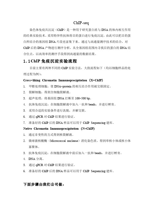
1.1C hIP免疫沉淀实验流程目前主要有两种不同的ChIP实验方法,大致流程如下(均以细胞样品的处理过程为例):Cross-liking Chromatin Immunoprecipitation (X-ChIP)1.甲醛处理细胞,使DNA-protein的相互结合作用被交联固定。
2.裂解细胞,得到全细胞裂解液。
3.超声处理,将基因组DNA打断至100-500 bp。
4.抗体免疫沉淀:在细胞裂解液中加入一抗和beads,并进行孵育。
5.采用合适的实验条件进行洗脱,并解交联。
6.通过qPCR对ChIP结果进行验证。
7.准备好的ChIP后的DNA样品可以用于ChIP Sequencing建库。
Native Chromatin Immunoprecipitation (N-ChIP)1.通过非变性的方式得到核裂解液。
2.微球菌核酸酶(Micrococcal nuclease)消化染色质,得到单核小体或核小体寡聚体。
3.抗体免疫沉淀:在细胞裂解液中前后加入一抗和beads,并进行孵育。
4.DNA分离。
5.通过qPCR对ChIP结果进行验证。
6.准备好的ChIP后的DNA样品可以用于ChIP Sequencing建库。
下面步骤由我们公司做:1.2C hIP Sequencing文库构建流程1.DNA片段末端修复,3’端加A碱基,连接测序接头(详细步骤请参考Illumina 公司Paired-End DNA Sample Prep kit)。
2.PCR扩增及DNA产物的片段大小选择(一般为100-300 bp,包括接头序列在内)合格的文库用于上机测序。
串联亲和层析 protocol
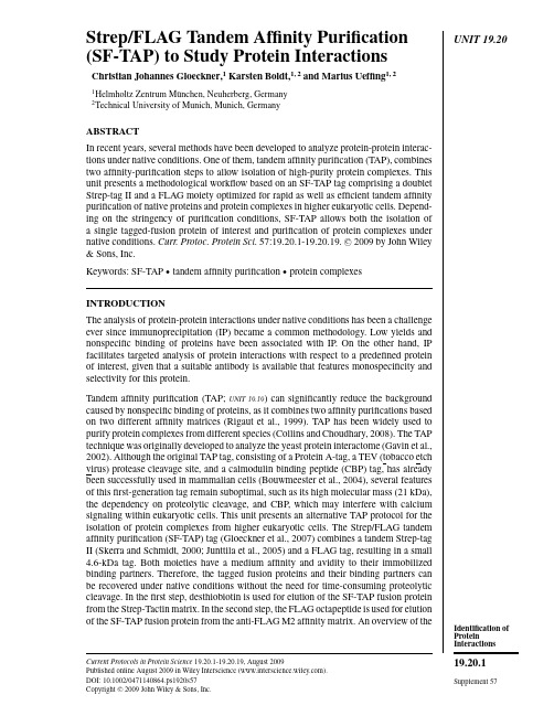
UNIT19.20 Strep/FLAG Tandem Affinity Purification(SF-TAP)to Study Protein InteractionsChristian Johannes Gloeckner,1Karsten Boldt,1,2and Marius Ueffing1,21Helmholtz Zentrum M¨u nchen,Neuherberg,Germany2Technical University of Munich,Munich,GermanyABSTRACTIn recent years,several methods have been developed to analyze protein-protein interac-tions under native conditions.One of them,tandem affinity purification(TAP),combinestwo affinity-purification steps to allow isolation of high-purity protein complexes.Thisunit presents a methodological workflow based on an SF-TAP tag comprising a doubletStrep-tag II and a FLAG moiety optimized for rapid as well as efficient tandem affinitypurification of native proteins and protein complexes in higher eukaryotic cells.Depend-ing on the stringency of purification conditions,SF-TAP allows both the isolation ofa single tagged-fusion protein of interest and purification of protein complexes undernative conditions.Curr.Protoc.Protein Sci.57:19.20.1-19.20.19.C 2009by John Wiley&Sons,Inc.Keywords:SF-TAP r tandem affinity purification r protein complexesINTRODUCTIONThe analysis of protein-protein interactions under native conditions has been a challengeever since immunoprecipitation(IP)became a common methodology.Low yields andnonspecific binding of proteins have been associated with IP.On the other hand,IPfacilitates targeted analysis of protein interactions with respect to a predefined proteinof interest,given that a suitable antibody is available that features monospecificity andselectivity for this protein.Tandem affinity purification(TAP;UNIT19.19)can significantly reduce the backgroundcaused by nonspecific binding of proteins,as it combines two affinity purifications basedon two different affinity matrices(Rigaut et al.,1999).TAP has been widely used topurify protein complexes from different species(Collins and Choudhary,2008).The TAPtechnique was originally developed to analyze the yeast protein interactome(Gavin et al.,2002).Although the original TAP tag,consisting of a Protein A-tag,a TEV(tobacco etchvirus)protease cleavage site,and a calmodulin binding peptide(CBP)tag,has alreadybeen successfully used in mammalian cells(Bouwmeester et al.,2004),several featuresof thisfirst-generation tag remain suboptimal,such as its high molecular mass(21kDa),the dependency on proteolytic cleavage,and CBP,which may interfere with calciumsignaling within eukaryotic cells.This unit presents an alternative TAP protocol for theisolation of protein complexes from higher eukaryotic cells.The Strep/FLAG tandemaffinity purification(SF-TAP)tag(Gloeckner et al.,2007)combines a tandem Strep-tagII(Skerra and Schmidt,2000;Junttila et al.,2005)and a FLAG tag,resulting in a small4.6-kDa tag.Both moieties have a medium affinity and avidity to their immobilizedbinding partners.Therefore,the tagged fusion proteins and their binding partners canbe recovered under native conditions without the need for time-consuming proteolyticcleavage.In thefirst step,desthiobiotin is used for elution of the SF-TAP fusion proteinfrom the Strep-Tactin matrix.In the second step,the FLAG octapeptide is used for elutionof the SF-TAP fusion protein from the anti-FLAG M2affinity matrix.An overview of the Current Protocols in Protein Science19.20.1-19.20.19,August2009Published online August2009in Wiley Interscience().DOI:10.1002/0471140864.ps1920s57Copyright C 2009John Wiley&Sons,Inc.Identification of Protein Interactions19.20.1 Supplement57Strep/FLAGTandem AffinityPurification (SF-TAP)19.20.2Supplement 57Current Protocols in Protein Science A B 1. purification 2. purification binding to Strep-Tactin binding to FLAG matrix elution with desthiobiotin elution with FLAG peptide Key:SF-TAP desthiobiotin FLAG peptide Figure 19.20.1The S trep/FLAG ta n dem affin ity p u rificatio n .(A )N-a n d C-termi n al S F-T AP ta gs (POI,protei n of i n tere s t).(B )Overview of both p u rificatio n s tep s .(1)P u rificatio n by the ta n dem S trep-ta g II moiety:bi n di ng to S trep-T acti n matrix followed by el u tio n with de s thiobioti n .(2)P u rificatio n by the FLAG-ta g moiety:bi n di ng to a n ti-FLAG M2affin ity matrix followed by el u -tio n with FLAG peptide.Abbreviatio ns :s p.,s pecific i n teractor s (s how n a s g ray circle s );n .s p.,n o ns pecific protei ns (co n tami n a n t s ;s how n a s white circle s ).SF-TAP technique and the tag sequence is shown in Figure 19.20.1.The SF-TAP protocol represents an efficient,fast and straightforward purification of protein complexes from mammalian cells within 2hr.This unit describes the full workflow,starting with the cell culture work needed for recombinant expression of the SF-TAP fusion proteins,followed by the SF-TAP protocol (see Basic Protocol 1)and ending with mass spectrometric analysis of the samples (see Basic Protocol 4).Special focus is given to the crucial step of sample preparation for mass spectrometry.For the identification of associated proteins following SF-TAP,the volume of the SF-TAP eluates is reduced by ultrafiltration using centrifugal units with a low molecular weight cut-off or by chloroform/methanol precipitation (see Support Protocol 2).The samples are then directly subjected to proteolytic digestion (see Basic Protocol 2)for analysis on a nano liquid chromatography (LC)–coupled electron sprayIdentification of Protein Interactions 19.20.3Current Protocols in Protein Science Supplement 57Figure 19.20.2Flow chart of a S F-T AP approach i n cl u di ng M S ide n tificatio n of cop u rified pro-tei ns .Thi s figu re co nn ect s all protocol s pre s e n ted i n thi s un it.tandem mass spectrometer.For complex samples,which contain many proteins,an alternative procedure for SDS-PAGE pre-fractionation is provided,including a method for sensitive MS-compatible Coomassie protein staining (see Support Protocol 3)followed by in-gel proteolytic digestion (see Basic Protocol 3).By reducing sample complexity,pre-fractionation helps to increase the number of protein identifications on state-of-the-art LC-coupled tandem mass spectrometers.Representative MS-analysis protocols are provided for an Orbitrap mass spectrometer (Thermo Fisher Scientific),a fast and sensitive system allowing high identification rates from SF-TAP purifications even with low amounts of protein in the sample (see Basic Protocol 4).Finally,a strategy for meta analysis of mass spectrometric data sets using the Scaffold software is provided (see Support Protocol 4).It can generally be used for the analysis of large MS/MS data sets.Figure 19.20.2provides a flowchart of the entire analytical process.Strep/FLAGTandem AffinityPurification (SF-TAP)19.20.4Supplement 57Current Protocols in Protein ScienceBASICPROTOCOL 1STREP/FLAG TANDEM AFFINITY PURIFICATION (SF-TAP)OF PROTEIN COMPLEXES FROM HEK293CELLS A flowchart of the SF-TAP procedure is shown in Figure 19.20.3.Materials HEK293cells (ATCC no.CRL-1573)Complete DMEM containing 10%FBS (APPENDIX 3C )SF-TAP vectors with appropriate insert,and empty control plasmid (see Critical Parameters)Negative control (see annotation to step 3,below)Transfection reagent of choice (see UNIT 5.10)Phosphate-buffered saline (PBS;APPENDIX 2E ),prewarmed Lysis buffer (see recipe)Strep-Tactin Superflow resin (IBA GmbH,cat.no.2-1206-10)Tris-buffered saline (TBS;see recipe)Wash buffer (see recipe)Desthiobiotin elution buffer:dilute 10×buffer E (IBA GmbH,cat.no.2-1000-025)1:10in H 2O (final concentration,2mM desthiobiotin)Anti–FLAG M2agarose (Sigma-Aldrich)FLAG elution buffer (see recipe)14-cm tissue culture plates Cell scraper Millex GP 0.22-μm syringe-driven filter units (Millipore)End-over-end rotator Microspin columns (GE Healthcare,cat.no.27-3565-01)End-over-end rotator Microcon YM-3centrifugal filter devices (Millipore)Additional reagents and equipment for transfection of mammalian cells (UNIT 5.10)Transfect HEK293cells 1.Seed HEK293cells on 14-cm plates at ∼1–2×10⁷cells per dish in complete DMEM medium containing 10%FBS.The amount of cells used for SF-TAP purification can be varied depending on the ex-pression levels of the bait ually,four 14-cm dishes,corresponding to a final amount of ∼4×10⁸HEK293cells,is a good starting point.Strong overexpression of the bait protein usually increases copurification of heat-shock proteins such as HSP70.For in-depth analysis,it is therefore recommended to generate cell lines stably expressing the bait protein.See Support Protocol 1for a stable transfection method.2.Grow cells overnight.3.Transfect cells with the SF-TAP plasmids using a transfection reagent of choice (according to manufacturer’s protocols).HEK293cells can be easily transfected with lipophilic transfection reagents.The trans-fection efficiency is usually >80%.For a typical SF-TAP experiment,1to 4μg plasmid per 14-cm dish is used.Depending on the cell type other transfection reagents may be favorable (also see UNIT 5.10).Although SF-TAP purifications typically exhibit low background caused by nonspecific binding of proteins to the affinity matrix,a suitable negative control should be used in every experiment.Cells transfected with the empty expression vectors may be used in the same amount as for the SF-TAP-tagged bait protein.However,the tag is quite small and expressed at low levels if not fused to a protein.Thus,the untransfected cell line is an acceptable,simple,and inexpensive alternative for a negative control.Identification of Protein Interactions 19.20.5Current Protocols in Protein Science Supplement 571-4 × 10⁸ HEK293 cell s(1-4 co n fl u e n t 14-cm plate s )expre ss i ng S F-TAP f us io n protei nly s i s(15 mi n 4C)vol u mered u ctio nce n trif ug atio n (10 mi n 10,000 × g )a n aly s i sretai n su per n ata n t fi n alel u atei n c u batio n with50 μl/plate S trep-Tacti n matrix (1 hr)el u tio n with200 μl FLAGel u tio n b u ffer(10 mi n )wa s h 3 time s with 500 μl wa s h b u ffer (s pi n 5 s ec, 100 × g )wa s h 3 time s with500 μl wa s h b u ffer(s pi n 5 s ec, 100 × g )el u tio n with 500 μl de s thiobioti n el u tio n b u ffer (10 mi n )i n c u batio n with25 μl/platea n ti-FLAG M2a g aro s e(1 hr)Figure 19.20.3Flow chart for the S F-T AP proced u re.4.Let cells grow for 48hr.If necessary,cells can be starved in DMEM without FBS for 12hr prior to harvesting.Starving might be desirable if cell signaling is to be analyzed,especially prior to differ-ential treatment with growth factors,to eliminate effects of serum growth factors.Lyse cells5.Remove medium from the plates.6.Optional:Rinse cells in warm PBS.Strep/FLAGTandem AffinityPurification (SF-TAP)19.20.6Supplement 57Current Protocols in Protein Science7.Scrape off cells in 1ml lysis buffer per 14-cm plate on ice using a cell scraper,and combine lysates from each experimental condition in a 1.5-ml microcentrifuge tube.8.Lyse cells by incubating 15min on ice with mixing by hand from time to time.9.Pellet cell debris,including nuclei,by centrifuging 10min at 10,000×g ,4◦C.10.Clear lysate supernatant by filtration through a 0.22-μm syringe filter.Perform SF-TAP 11.Wash Strep-Tactin Superflow resin twice,each time with 4resin volumes TBS and once with 4resin volumes lysis buffer.12.Incubate lysates with 50μl per 14-cm plate of settled Strep-Tactin Superflow resin for 1hr at 4◦C (use an end-over-end rotator to keep the resin evenly distributed).Note that a maximum of 200μl settled resin per spin column should not be exceeded.If more than four 14-cm plates (∼4×10⁸HEK293cells)are used,reduce the volume per plate or use additional spin columns in step 13.13.Centrifuge for 30sec at 7000×g ,4◦C,remove the supernatant until 500μl remains,and transfer resin to a microspin column.Snap off bottom closure of the spin column prior to use.The maximum volume of the spin columns is 650μl.Alternatively,centrifugations for wash and elution steps can be performed at room temperature if no cooled centrifuge is available.14.Remove remaining supernatant by centrifugation in the spin column for 5sec at 100×g ,then wash resin three times,each time with 500μl wash buffer (centrifuge 5sec at 100×g each time to remove the supernatant)at 4◦C.Replug spin columns with inverted bottom closure prior to adding the elution buffer in step 15.IMPORTANT NOTE:Do not allow the resin to run dry.Depending on the bait protein,this markedly reduces the yield.15.Add 500μl desthiobiotin elution buffer and gently mix the resin by hand for 10min on ice.16.Remove the plug of the spin column,transfer the column to a new collection tube,and collect the eluate by centrifuging 10sec at 2000×g ,4◦C.If spin columns were closed by the top screw cap during incubation with elution buffer,the cap needs to be removed prior to centrifugation,to allow the pressure to balance out.17.Wash anti–FLAG M2agarose resin three times,each time with 4resin volumes TBS.Suspend resin in TBS and transfer it to microspin columns,then remove the buffer by centrifuging 5sec at 100×g .25μl settled resin per 14-cm plate will be needed.18.Transfer eluate from step 16corresponding to each 14-cm plate to a microspin column containing 25μl settled anti-FLAG M2agarose prepared as in step 17.19.Plug columns,close columns with top screw caps,and incubate for 1hr at 4◦C (on an end-over-end rotator).20.Wash once with 500μl wash buffer,and then twice,each time with 500μl TBS (centrifuge 5sec at 100×g each time to remove the supernatant)at 4◦C.21.For elution,incubate with 4bead volumes (at least 200μl)FLAG elution buffer for 10min,keeping the columns plugged and gently mixing the resin several times.22.After incubation,remove the plugs and top screws of the spin columns,transfer to new collection tubes,and collect the eluate(s)by centrifugation (10sec at 2000×g ).Identification of Protein Interactions 19.20.7Current Protocols in Protein Science Supplement 5723.Depending on downstream method to be used,either precipitate protein (see SupportProtocol 2)or concentrate the eluate by Microcon YM-3centrifugal filter units according to manufacturer’s protocols.SUPPORT PROTOCOL 1GENERATION OF HEK293CLONES STABLY EXPRESSINGSF-TAP-TAGGED PROTEINSIn Basic Protocol 1,SF-TAP-tagged proteins are transiently expressed.However,strong overexpression of the bait protein usually increases copurification of heat-shock proteins such as HSP70.For in-depth analysis,it is therefore recommended to generate cell lines stably expressing the bait protein.This protocol presents a quick method for generating stable HEK293lines.MaterialsHEK293cells (ATCC no.CRL-1573)Complete DMEM containing 10%FBS (APPENDIX 3C )SF-TAP vectors with appropriate insert,and empty control plasmid (see Critical Parameters)Transfection reagent of choice (see UNIT 5.10)Phosphate-buffered saline (PBS;APPENDIX 2E )Complete DMEM medium (APPENDIX 3C )G418(PAA Laboratories, )Freezing solution:90%fetal bovine serum (FBS;Invitrogen)/10%dimethylsulfoxide (DMSO;AR grade)Lysis buffer (see recipe)Blocking reagent:5%(w/v)nonfat dry milk in TBS (see recipe for TBS)containing 0.1%(v/v)Tween 20Anti-FLAG M2antibody (Sigma-Aldrich)10-cm tissue culture dishes12-well and 6-welll tissue culture platesCentrifuge2-ml cryovials (Nunc)Additional reagents and equipment for transfection of mammalian cells (UNIT 5.10),trypsinization and counting of cells (UNIT 5.10),and immunoblotting (UNIT 10.10)Grow and transfect cells1.Grow cells in complete DMEM containing 10%FBS.2.Transfect cells with expression plasmid using a transfection reagent of choice ac-cording to the manufacturer’s protocols.3.Change medium after 6hr.Select cells4.After 48hr,trypsinize and count cells (APPENDIX 3C )and seed them at low density (1×10⁶cells per 10-cm dish)to allow formation of single colonies upon selection.5.Add G418(500to 1000μg/ml)for selection of the SF-TAP expression vectors,which are based on pcDNA3.0and contain a neomycin-resistance gene.6.Grow the cells under G-418selection for 2to 4weeks,changing the medium every second day.7.Collect single colonies with a 200-μl pipet into 12-well plates.8.Keep colonies under G418selection until the cell density is sufficient for expanding them to 6-well dishes (two wells per clone).Strep/FLAGTandem AffinityPurification (SF-TAP)19.20.8Supplement 57Current Protocols in Protein ScienceCryopreserve cells 9.Grow cells to >90%confluency and trypsinize (APPENDIX 3C )one well of each clone for generation of cryostocks.10.Generate cryostocks:a.Wash cells from one well once by adding 3ml PBS,centrifuging 5min at 800×g ,room temperature,and resuspending the pellet in 500μl freezing buffer.b.Transfer resuspended cells to 2-ml cryovials.c.Freeze cells slowly:keep cells for 1hr at −20◦C,then overnight at −80◦C,followed by storage in a liquid nitrogen tank.For cultivation and expansion of confirmed clones,thaw the cryostock at 37◦C,wash cells once with medium,and plate cells onto 10-cm culture dishes.Test for expression of bait protein 11.Lyse one well of each clone in 300μl lysis buffer and test for expression of the bait protein by immunoblotting (UNIT 10.10).SF-TAP proteins can be detected using the anti-FLAG M2antibody (Sigma-Aldrich)at a dilution of 1:1000to 1:5000in blocking reagent.SUPPORTPROTOCOL 2CHLOROFORM/METHANOL PRECIPITATION OF PROTEINS The chloroform/methanol precipitation method described by Wessel and Fl¨u gge (1984)precipitates proteins with high efficiency and yields samples containing low levels of salt contamination.Materials SF-TAP eluate (from Basic Protocol 1)Methanol (AR grade)Chloroform (AR grade)2-ml polypropylene sample tubes 1.Transfer 200μl SF-TAP eluate to a 2-ml sample tube.All steps are performed at ambient temperature.2.Add 0.8ml of methanol,vortex,and centrifuge for 20sec at 9000×g ,room temperature.3.Add 0.2ml chloroform,vortex,and centrifuge for 20sec at 9000×g ,room temperature.4.Add 0.6ml of deionized water,vortex for 5sec,and centrifuge for 1min at 9000×g ,room temperature.5.Carefully remove and discard the upper layer (aqueous phase).The protein precipitate (visible as white flocks)is in the interphase.6.Add 0.6ml of methanol,vortex,and centrifuge for 2min at 16,000×g ,room temperature.7.Carefully remove the supernatant and air dry the pellet.The pellet can be stored for several months at –80◦C.Identification of Protein Interactions 19.20.9Current Protocols in Protein Science Supplement 57BASIC PROTOCOL 2IN-SOLUTION DIGEST OF PROTEINS FOR MASS SPECTROMETRIC ANALYSISThe in-solution digest described here is a quick and efficient method to digest the SF-TAP eluate after protein precipitation (Support Protocol 2).The use of an MS-compatible surfactant helps to solubilize the precipitated proteins.In order to allow the identification of cysteine-containing peptides,random oxidation is prevented,rather than reverted,by applying a DTT/iodoacetamide treatment prior to digestion,leading to a defined-mass adduct.The digested protein sample can then be directly subjected to analysis on an LC-coupled tandem mass spectrometer.MaterialsPrecipitated protein (see Support Protocol 2)50mM ammonium bicarbonate (freshly prepared)RapiGest SF (Waters):prepare 2%(10×)stock solution in deionized water 100mM DTT (prepare from 500mM stock solution;store stock up to 6months at −20◦C)300mM iodoacetamide (prepare fresh)50×(0.5μg/μl)trypsin stock solution (Promega;store at −20◦C)Concentrated (37%)HCl60◦C incubatorPolypropylene inserts (Supelco,cat.no.24722)1to 200μl gel-loader pipet tips (Sorenson Bioscience,/contact.cfm )1.Dissolve the protein pellet in 30μl of 50mM ammonium bicarbonate by extensive vortexing.2.Add 3μl of 10×(2%)RapiGest stock solution (final concentration,0.2%).RapiGest (sodium 3-[(2-methyl-2-undecyl-1,3-dioxolan-4-yl)methoxyl]-1-propanesulfo-nate)is an acid-labile surfactant that helps to solubilize and denature proteins to make them accessible to proteolytic digestion (Yu et al.,2003).3.Add 1μl of 100mM DTT and vortex.4.Incubate 10min at 60◦C.5.Cool the samples to room temperature.6.Add 1μl of 300mM iodoacetamide and vortex.7.Incubate for 30min at room temperature.Samples should be protected from light,since iodoacetamide is light-sensitive.8.Add 2μl trypsin stock solution and vortex.9.Incubate at 37◦C overnight.10.Add 2μl of concentrated (37%)HCl to hydrolyze the RapiGest.For hydrolysis of the RapiGest reagent,the pH must be <2.11.Transfer samples to polypropylene inserts (remove spring).12.Incubate for 30min at room temperature.13.Place inserts in 1.5-ml microcentrifuge tubes and microcentrifuge 10min at 13,000×g ,room temperature.One hydrolysis product of the RapiGest reagent is water-immiscible and can be removed by centrifugation.After centrifugation,it is visible as faint film (oleic phase)on top of theStrep/FLAGTandem Affinity Purification (SF-TAP)19.20.10Supplement 57Current Protocols in Protein Science aqueous sample phase.The other hydrolysis product is an ionic water-soluble component which does not interfere with reversed phase LC or MS analysis.A white pellet might appear.14.Carefully recover the solution between the upper oleic phase and the pellet using gel-loader tips.The sample can now be directly subjected to C18HPLC separation prior to MS/MS-analysis (LC-MS/MS;Basic Protocol 4).Pre-fractionation (Basic Protocol 3)is optional.BASIC PROTOCOL 3PRE-FRACTIONATION VIA SDS-PAGE AND IN-GEL DIGESTION PRIOR TO LC-MS/MS ANALYSIS Pre-fractionation prior to MS analysis increases the number of peptides which can be an-alyzed,and therefore the peptide coverage of identified proteins.This benefit is achieved by overcoming the undersampling problem mainly caused by the limited capacity of the trapping columns used in nano–LC chromatography,or that occurs with high complexity.For these samples,SDS-PAGE pre-fractionation can be used to reduce the complexity.For less complex samples or samples with low protein content,the in-solution digest (Basic Protocol 2)is preferred.Materials Protein sample (e.g.,from Basic Protocol 1or Support Protocol 2)10%NuPAGE gels (Invitrogen)MOPS running buffer (Invitrogen)40%and 100%acetonitrile (AR grade;prepare fresh)5mM DTT (prepare from 500mM stock;store stock up to 6months at −20◦C)25mM iodoacetamide (prepare fresh)Digestion solution:dilute 50×trypsin stock solution (0.5μg/μl,Promega)1:50in 50mM ammonium bicarbonate (freshly prepared)1%and 0.5%(v/v)trifluoroacetic acid (TFA;prepare fresh from 10%v/v stock)50%(v/v)acetonitrile/0.5%(v/v)TFA (prepare fresh)99.5%(v/v)acetonitrile/0.5%(v/v)TFA (prepare fresh)2%(v/v)acetonitrile/0.5%(v/v)TFA Concentration units (e.g.,Microcon from Millipore)Scalpel Polypropylene 96-well microtiter plate:polystyrene material should be avoided since,depending on the product,polymers can be extracted from plastics which produce strong background signals in mass spectrometry 60◦C incubator or heating block Polypropylene 0.5-ml reaction tubes Microtiter plate shaker (e.g.,V ortex mixer equipped with microtiter-plate adaptor)HPLC sample tubes Additional reagents and equipment for SDS-PAGE (UNIT 10.1)and colloidal Coomassie blue staining of gels (Support Protocol 3)Prepare samples 1.Concentrate samples using concentration units (e.g.,Microcon).2.Supplement samples with Laemmli loading buffer (SDS-PAGE loading buffer;UNIT 10.1).A detailed description of the SDS gel electrophoresis and standard buffers can be found in UNIT 10.1or in the protocols supplied with the NuPAGE system.Identification of ProteinInteractions19.20.11Perform electrophoresis and stain gels3.Separate samples on 10%NuPAGE gels according to the manufacturer’s protocols,using MOPS running buffer.4.Stop electrophoresis after the gel front has travelled 1to 2cm.5.Stain gels with colloidal Coomassie blue (see Support Protocol 3).Avoid strong staining of the bands since it increases the time necessary for destaining.6.Excise desired gel pieces with a clean scalpel (three to ten slices,depending on the complexity of the sample).Destain and process gel slices7.Transfer gel pieces into individual wells of a 96-well plate.8.Wash by adding 100μl water to each well and incubating for 30min.9.For destaining:a.Wash twice,each time by incubating the gel slices for 10min in 100μl/well of 40%acetonitrile.b.Wash for 5min in 100μl/well of 100%acetonitrile (if gels are still blue,repeat de-staining).10.Add 100μl of 5mM DTT,then incubate 15min at 60◦C in an incubator or heatingblock.11.Remove DTT solution and cool the plate to room temperature.12.Add 100μl per well of freshly prepared 25mM iodoacetamide,then incubate 30minin the dark.13.Wash twice,each time for 10min with 100μl/well of 40%acetonitrile.14.Wash 5min with 100μl/well of 100%acetonitrile.15.Discard supernatant and air dry (or SpeedVac)the gel pieces to complete dryness.Digest and extract gel slices16.Add 20to 30μl per well of freshly prepared digestion solution (depending on the sizeof the gel plugs).Wrap plates in Parafilm to reduce evaporation during the overnight incubation (or use a humidified incubator in step 17).17.Digest overnight at 37◦C.18.For extraction of the peptides from the gel piece,add 10μl 1%TFA,then shake15min on a V ortex mixer with a microtiter plate adapter.The peptides are extracted in three steps with increasing acetonitrile concentrations (steps 18to 23).19.Transfer liquid (extract 1)to a 0.5-ml polypropylene tube.20.Add 50μl 50%acetonitrile/0.5%TFA to the gel piece and shake 15min on a V ortexmixer with a microtiter plate adapter.21.Remove the liquid (extract 2)and pool extracts 1and 2.22.Add 50μl 99.5%acetonitrile/0.5%TFA to the gel piece,then shake 15min on aV ortex mixer with a microtiter plate adapter.23.Remove the liquid (extract 3)and pool extract 3with 1and 2.Strep/FLAG Tandem AffinityPurification(SF-TAP)19.20.1224.Dry samples to complete dryness in a SpeedVac evaporator.25.Redissolve samples in50μl of2%acetonitrile/0.5%TFA by shaking(e.g.,on aV ortex mixer)for10to15min,then transfer the sample into HPLC sample tubes for LC-MS/MS analysis.SUPPORT PROTOCOL3QUICK MS-COMPATIBLE COLLOIDAL COOMASSIE STAIN OF PROTEINS AFTER SDS-PAGE SEPARATIONThe colloidal Coomassie stain(Kang et al.,2002)represents a fast and sensitive MS-compatible protein staining method.In contrast to the classical staining protocol,no intense and time-consuming destaining is needed to visualize protein bands.Therefore, this method is ideal for a quick staining of the protein bands and provides good orientation on how the gel can be fractionated without splitting predominant bands(see Basic Protocol3).MaterialsElectrophoresed SDS gel containing protein samples of interest(e.g.,from Basic Protocol3)Colloidal Coomassie staining solution(see recipe)Destaining solution:10%(v/v)ethanol/2%(v/v)orthophosphoric acidGel staining trays of appropriate size1.Wash gels twice,each time for10min in deionized water in a staining tray.The SDS must be removed before staining to reduce background signals.2.Incubate gels for10min in colloidal Coomassie staining solution.The incubation steps are kept short for the staining of gels used for pre-fractionation.The staining can be prolonged up to overnight.The maximum staining will be reached after ∼3hr incubation in the staining solution.3.Incubate gels for10min in destaining solution.4.Wash gels twice,each time for10min in deionized water.BASIC PROTOCOL4LC-MS/MS ANALYSIS OF DIGESTED SF-TAP SAMPLESThe following protocol describes MS analysis of digested protein samples on an LC-coupled ESI tandem mass spectrometer.The representative MS-analysis protocol is provided for an Orbitrap mass spectrometer(Thermo Fisher Scientific).The Orbitrap system combines fast data acquisition with high mass accuracy and is therefore ideal for the analysis of SF-TAP samples.Background information on mass spectrometric analysis can be found in UNIT16.11.MaterialsDigested protein sample,either from in-solution digest(Basic Protocol2)or in-gel digest(Basic Protocol3)Nano HPLC loading buffer:0.1%formic acid in HPLC-grade waterNano HPLC buffer A:2%acetonitrile/0.1%formic acid in HPLC-grade waterNano HPLC buffer B:80%acetonitrile/0.1%formic acid in HPLC-grade water HPLC vials(Dionex)Nano HPLC system(UltiMate3000,Dionex)equipped with a trap column (100μm i.d.×2cm,packed with Acclaim PepMap100C18resin,5μm,100◦A;Dionex)and an analytical column(75μm i.d.×15cm,packed with AcclaimPepMap100C18resin,3μm,100◦A;Dionex)Mass spectrometer:Oritrap XL with a nanospray ion source(ThermoFisher Scientific;also see UNIT16.11)。
THP_1巨噬细胞源性泡沫细胞模型的建立及鉴定
稳定可靠的泡沫细胞模型,对研究 =A 及筛选抗 =A 药物有重要的意义 B ! C 。目前实验研究中通常用氧化型 低 密 度 脂 蛋 白 ’ 8DEFE ) 或 乙 酰 化 低 密 度 脂 蛋 白 ’ ,7EFE ) 孵育单核 % 巨噬细胞或平滑肌细胞来复制泡 沫 细 胞 模 型 , 可 用 的 细 胞 有 9GH % #、 I(:&、 J=K!5<6 &、鼠腹腔巨噬细胞、动物主动脉平滑肌细 胞 , 其 中 较 多 文 献 报 道 运 用 9GH % # 细 胞 B : % < C 。 9GH % # 为人类单核细胞株,需经佛波酯 ’ H?8@L8. %
UCV
"N X 丙二醇冲洗 N 7’*,再用 6FI 洗三次,置显微镜 下观察并摄像。 !" ( *+,- . /0 测定胆固醇的条件 ’1 ) !" (" ! 色谱条件 色谱柱: \8^.$/’+ ,B" 柱 R !> B 77 ] BN: 77, @ !7 S ; 流 动 相 : 甲 醇 M 水 R ;: _ B: S ; 流 速::> N 7K Z 7’*;柱温:室温;进样量:B: !K。 !" (" # 质 谱 条 件 离 子 源 为 大 气 压 化 学 电 离 源 R O6,A S ;放电电流 N !O;气化温度 @N: W ;离子传输 毛细管温度 !N: W ;鞘气 R ?! S 流速 @: %$‘’($%$8 1*’(/; 辅助气 R ?! S 流速 N %$‘’($%$8 1*’(/;正离子方式检测;扫 描方式为选择性离子检测 R IA< S ;检测离子:胆固醇, U < a \ V a ,7 Z b @C;,豆甾醇, U < a \ V a ,7 Z b @;N。 !" 2 标准曲线的制备 取胆固醇对照品适量,精密称 定,加无水乙醇制成每毫升含 @:: !2 的储备溶液。 分别精密量取 N,B:,!:,@:,D:,N:,B:: !K 胆固 醇储备液至 B: 7K 量瓶中,各加 B:: !K 浓度为 !:: !2 Z 7K 的豆甾醇内标储备液,用甲醇稀释至刻度, 摇 匀 , 得 质 量 浓 度 为 :> B@"N, :> !H;, :> NN", :> "@H,B> BBC, B> @;N, !> H;: !2 Z 7K 的胆固醇对照 品系列溶液,将上述对照品系列溶液分别注入液质联 用仪,进样量为 B: !K,按前述色谱、质谱条件记录 图谱。以胆固醇的质量浓度为横坐标 R ! S ,其峰面积 与内标峰面积的比值为纵坐标 R " S ,绘制标准曲线。 !" 1 细胞内胆固醇的提取 ’1 ) 细胞接种于 C 孔培养板 内,培养结束后,吸去上清液,冰冻 6FI 洗三次, 用细胞刮刀刮取细胞,加 B 7K 冰冻 :> ; X 生理盐水 重悬细胞,反复冻融三次,冰浴中超声破碎 N 7’*, 得细胞裂解液,分装,每份 !:: !K, M D: W 贮存, 用于测定总胆固醇和游离胆固醇。 总胆固醇的提取:取 BC: !K 上述细胞裂解液,加 @:: !K BN X cG\ 乙醇溶液,N: W 水解 ! -,加正己烷 M 异丙醇 R D _ B S N:: !K 涡漩 @: /, D W 下 @C:: $ Z 7’* 离心 N 7’*,取上层。继用正己烷 M 异丙醇 R D _ B S 如 上法抽提两次,合并三次抽提的有机相,减压真空干 燥,加 !: !K 内标储备液,用甲醇并定容至 ! 7K, 摇匀,作为总胆固醇供试品溶液。 游离胆固醇的提取:取 BC: !K 上述细胞裂解液, 加 @:: !K 乙醇,加正己烷 M 异丙醇 R D _ B S N:: !K 涡漩 @: /,D W 下 @C:: $ Z 7’* 离心 N 7’*,取上层。继用正 己烷 M 异丙醇 R D _ B S 如上法抽提两次,合并三次抽提 的有机相,减压真空干燥,加 !: !K 内标储备溶解并 定容至 ! 7K,摇匀,作为游离胆固醇供试品溶液。 !" 3 *+,- . /0 分析细胞内胆固醇与胆固醇酯的含 量 将正常组和模型组细胞分别用上述方法制得样品
从293中纯化蛋白流程
从293中纯化蛋白流程英文回答:Purification of Protein from 293 Cells.The purification of proteins from 293 cells involves several steps to isolate and concentrate the desired protein from the cellular lysate. Here is a general overview of the purification process:1. Cell Lysis: The first step is to lyse the 293 cells to release the cellular contents. This can be achieved using mechanical methods (e.g., sonication, homogenization) or chemical methods (e.g., detergents, enzymes).2. Clarification: The cell lysate is then subjected to centrifugation to remove cell debris and other insoluble material. The supernatant, containing the soluble proteins, is collected for further processing.3. Initial Purification: The soluble proteins in the supernatant can be further purified using various methods, such as:Affinity Chromatography: This technique utilizes an affinity ligand immobilized on a solid support to selectively bind the target protein. The protein ofinterest binds to the ligand, while other proteins pass through the column. The bound protein is then eluted from the column.Ion Exchange Chromatography: This technique separates proteins based on their net charge. The proteins are loaded onto an ion exchange column, and the elution buffer is adjusted to alter the pH or salt concentration to selectively elute the target protein.Size Exclusion Chromatography: This technique separates proteins based on their molecular size. The proteins are loaded onto a gel filtration column, and the larger proteins elute earlier than the smaller proteins.4. Polishing: The partially purified protein can be further purified using additional techniques to remove any remaining impurities. These techniques may include:Dialysis: This process removes salts and other small molecules from the protein solution by diffusion across a semipermeable membrane.Ultrafiltration: This technique uses a membrane filter to concentrate the protein solution and remove impurities.Electrophoresis: This technique separates proteins based on their charge and molecular size. The proteins are loaded onto a gel and an electric current is applied, causing the proteins to migrate through the gel.5. Characterization: The purified protein is then characterized to confirm its identity and purity. This may involve techniques such as:SDS-PAGE: This technique separates proteins basedon their molecular weight and is used to verify the molecular weight of the purified protein.Western Blotting: This technique uses antibodies to detect specific proteins and is used to confirm theidentity of the purified protein.Mass Spectrometry: This technique can provide detailed information about the protein's amino acid sequence and molecular weight.中文回答:从293细胞中纯化蛋白。
pET表达系统说明书(Novagen公司)
17
17 18 19 20 20 21
III. Cloning Inserts in pET Vectors
A. Ligation B. Transformation Handling Tips Procedure Plating Technique C. Analysis of pET Recombinants ® Transcription/Translation Analysis with EcoPro™ or STP3 Systems Plasmid Templates PCR Templates Ligation PCR for Transcription/Translation Analysis Colony PCR for Transcription/Translation Analysis Colony Screening Plasmid Miniprep Procedure Sequencing
Novagen
1
pET System Manual
Use of Ampicillin Precautions to Maximize Expression Rationale for Plasmid Stability Test F. Difficult Target Proteins Other Factors Influencing Expression Level 33 33 34 34 36
United States & Canada 800-207-0144 Germany 0800 6931 000 United Kingdom 0800 622935 Or your local sales office
30
30 30 30 30 31 31 31 32 32
细菌基因组的抽提
细菌基因组DNA的提取方法综述,提供了5种方法。
1 快速微量提取法A.取1.5ml菌体培养物于一灭菌Ep管中,12000rpm离心1min, 丢去上清夜,收集菌体。
B.加入400ul裂解液(40mMTris-醋酸,20mM醋酸钠,1mMEDTA,1%SDS,pH7.8)混匀,置于37o C水浴1hr。
C.然后加入200ul5mol/L的氯化钠溶液,混匀后于13000rpm离心15min。
D.取上清液,用苯酚抽提2次,氯仿抽提1次。
E.加两倍体积无水乙醇,1/10体积醋酸钾(3M ,pH8.0),-20度保存1小时后,13000rpm离心15min,弃上清液,沉淀用70%乙醇洗2次;置于室温干燥后,溶于50ulTE溶液中,置4℃保存备用。
2 蛋白酶/SDS法制备先用10ml含适当抗生素的GBM过夜培养Delftia sp.,第二天4000rpm离心10min收集菌体,用Washing TE (50mmol/LTris-HCl pH8.0,10mmol/LEDTA pH8.0)洗菌体2次,之后将菌体充分悬浮在5ml 1×TE缓冲液中,先后加入0.5ml 5mg/L的蛋白酶、0.5ml 10% SDS,轻轻混匀后50℃放置3h~5h,接着用等体积的Tris 饱和苯酚抽提2次,苯酚/氯仿/异戊醇抽提一次,氯仿抽提一次后,乙醇沉淀DNA,用自动移液器吸管头将絮状DNA沉淀块吸附到Ep管中,70%乙醇洗2次,干燥后溶于适当1×TE或ddH2O中。
31) 细菌培养:细菌接种于5ml液体培养基中,37℃摇床(300rpm)培养过液。
2) 细菌收集:取1ml培养物于1.5ml EP管中,室温8000rpm离心5min,弃上清,沉淀重新悬浮于1ml TE (pH8.0)中(用ddH2O也行)。
3) 菌体裂解:加入6μl 50mg/ml的溶菌酶,37℃作用2h。
再加2mol/LNaCl50μl,10%SDS 110μl,20mg/ml 的蛋白酶K 3μl,50℃作用3h或37℃过夜。
细菌总RNA提取方法的比较
细菌总RNA提取方法的比较邹晓蕾,刘礼崔,罗立新(华南理工大学生物科学与工程学院,广东广州 510006)摘要:采用改良前后的Trizol法,RNAiso plus法、Qiagen试剂盒法以及Triton X-100法,从大肠杆菌(Escherichiacoli),枯草芽孢杆菌(Bacillus subtilis),乳酸乳球菌(Lactococcuslactis)3株细菌中提取RNA。
用琼脂糖凝胶电泳和核酸浓度测定仪检测总RNA 的提取质量,并用R T-PCR(Reverse Transcription Polymerase Chain Reaction)对RNA的质量进行验证。
结果表明:RNAiso plus法对3株菌的总RNA提取均有很好的效果。
Trizol法和Qiagen试剂盒法对于E. coli总RNA的提取效果不错,但对其他2株菌的提取效果均不明显,并且都伴有DNA污染。
上述几种方法提取出的RNA样品中均含有23S和16S rRNA。
ctis更适合用Triton X-100法提取总RNA。
改良后的Trizol法和RNAiso plus法与Triton X-100法的原理基本相同,都是采用加热的方法,更彻底地裂解细菌细胞,而且得到的RNA中不含有rRNA和tRNA,只保留了mRNA,能更好地用于后续的实验研究。
关键词:细菌;总RNA提取;反转录聚合酶链式反应文章篇号:1673-9078(2013)8-1948-1954Comparison of Different Methods for T otal Bacteria RNA extractionZOU Xiao-lei, LIU Li-cui, LUO Li-xin(College of Bioscience &Bioengineering, South China University of Technology, Guangzhou 510006) Abstract: In this research, RNA was isolated from three bacterial strains, (Escherichia coli, Bacillus subtilis and Lactococcuslactis) by using modified and un-modified Trizol and RNAiso plus methods, Triton X-100 method and Qiagen method. The quality and integrity of the extracted RNA samples were determined with agarose gel electrophoresis, nucleic acid detector and R T-PCR detection. The results demonstrate that un-modified RNAiso plus method can effectively collect high-quality RNA from the three bacterial strains. Qiagen method and un-modified Trizol method showed good efficiency when isolating total RNA from E.coli but other strains. In addition, the RNA samples also contain DNA contamination. L. lactis was more suitable for using Triton X-100 method. With the same principle, modified Trizol method, modified RNAiso plus method and Triton X-100 method reported here can be employed for extraction of RNA that are free from 16S and 23S rRNA and provide simple, rapid and effective tools for the isolation of high-quality RNA appropriate for downstream molecular experiments Key words : bacteria; tatal RNA extraction; RT-PCRRNA是生物体重要的遗传物质,RNA的提取是分子生物学的基础实验,对于cDNA文库构建、荧光定量PCR、R T-PCR、Northern杂交等下游分子生物学实验来说,都需要质量好的,完整性高的RNA[1]。