大鼠系统解剖简述
大鼠解剖图

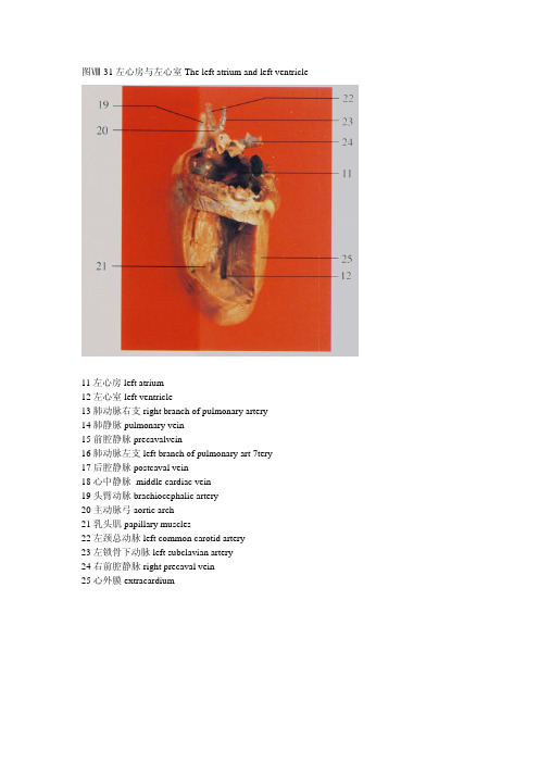
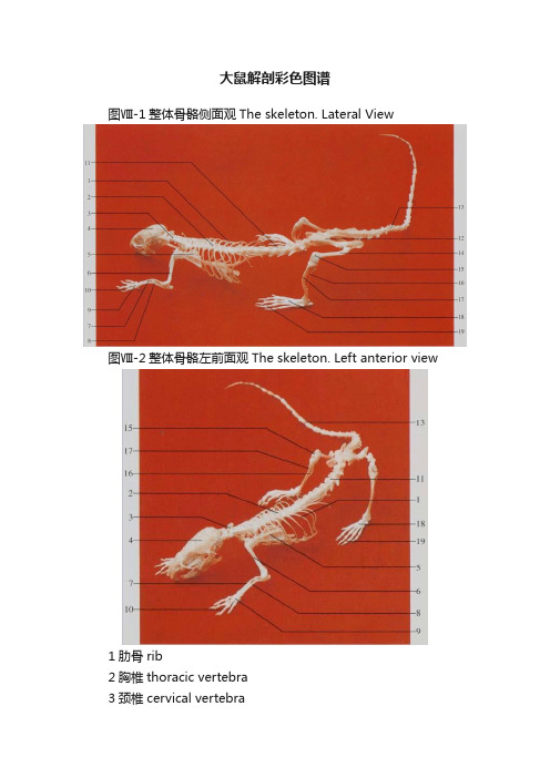
图Ⅷ-31左心房与左心室The left atrium and left ventricle11左心房left atrium12左心室left ventricle13肺动脉右支right branch of pulmonary artery14肺静脉pulmonary vein15前腔静脉precavalvein16肺动脉左支left branch of pulmonary art 7tery17后腔静脉postcaval vein18心中静脉 middle cardiac vein19头臂动脉brachiocephalic artery20主动脉弓aortic arch21乳头肌papillary muscles22左颈总动脉left common carotid artery23左锁骨下动脉left subclavian artery24右前腔静脉right precaval vein25心外膜extracardium图Ⅷ-56前肢内侧面The anterior limb. Medial aspect图Ⅷ-57后肢内侧面(1)The posterior limb. Medial aspect(1)1腋神经axillary nerve 2桡神经radial nerve3头静脉cephalic vein 4正中神经median nerve5正中动脉median artery 6肌支muscular branch7尺神经ulnar nerve 8臂动脉brachial artery9胸长神经long thoracic nerve 10胸外侧动脉external thoracic arteryI1颈总动脉common carotid artery 12主动脉弓aorta arch13左心耳left auricle 14右心耳right auricle15食管esophagus 16胸主动脉thoracic aorta17腹壁浅动脉superficial epigastric artery 18腹壁浅静脉superficial epigastric vein 19隐动脉saphenous artery 20隐大静脉great saphenous vein图Ⅷ-52生殖器官(♂)(5)The genital organs(♂)(5)图Ⅷ-53腹主动脉及其分支The abdominal aorta and branches1 直肠 rectum2精囊seminal vesicle3 膀胱urinary bladder4前列腺prostate5精索spermatic cord6阴茎penis7 附睾epididymis8睾丸testis9膈diaphragm10后腔静脉postcaval vein11腹腔动脉celiac artery12肾动脉renal artery13右肾 right kidkey14前肠系膜动脉anterior mesenteric artery15精索内动脉internal spermatic artery16输尿管ureter17髂腰动、静脉iliolumbar artery and vein18髂总动脉common iliac artery19贮精囊vesicula seminalis20隐动脉saphenous artery21食管esophagus22膈下动脉inferior phrenic artery23肾上腺adrenal glands24肾上腺上动脉superior adrenal artery25左肾left kidney26后肠系膜动脉posterior mesenteric artery27髂外动脉external iliac artery28股动脉femoral artery29腹壁阴部动脉干arterial epigastro-pudendal trunk 30输精管ductus deferens1直肠rectum 2膀胱urinary bladder3肾kidney 4脂肪fat5精囊seminal vesicle 6前列腺prostate7阴茎penis 8附睾 epididymis9精索spermatic cord图Ⅷ-46生殖器官(♀)(1)The genital organs(♀)(1)图Ⅷ-47生殖器官(♀)(2)The genital organs(♀)(2)1小肠small intestine 2输卵管uterine tube 3脂肪fat 4胃stomach5子宫角horn of uterus 6直肠rectum7膀胱urinary bladder 8脾spleen9子宫体body of uterus图Ⅷ-44空肠、回肠与盲肠腹面观The jejunum, ileum and cecum. Ventral view图Ⅷ-45空肠、回肠与盲肠背面观The jejunum, ileum and cecum. Dorsal view1空肠jejunum2横结肠transverse colon3升结肠ascending colon4盲肠cecum5腹壁浅动脉superficial epigastric artery6隐动脉saphenous artery7隐大静脉great saphenous vein8肝liver9胃stomach10脾spleen11降结肠descending colon12 膀胱 urinary bladder13尾中动脉middle coccygeal artery14回肠ileum15盲肠尖apex of cecum16盲肠体body of cecum17盲肠基部base of cecum图Ⅷ-41腹腔器官(6)The organs in the abdominal cavity (6)图Ⅷ-42胃The stomach 图Ⅷ-43胃壁内面The wall of stomach. Inner surface1膈diaphragm2前腔静脉precaval vein3胸腺thymus4肺lung5心heart6肾上腺adrenal gland7肾kidney8肾门静脉renal portal vein9精索内静脉internal spermatic vein10髂腰静脉iliolumbar vein11膀胱urinary bladder12十二指肠duodenum13幽门pylorus14胃大弯greater curvature of stomach15食管esophagus16胃底fundus of stomach17胃小弯lesser curvature of stomach18胃体body of stomach19贲门pylorus20胃粘膜皮区cutaneous part of gastric mucous21胃粘膜腺区glandular part of gastric mucous1肝右中叶right middle lobe of l iver 2肝右外叶right lateral lobe of liver 3小肠small intestine 4大网膜greater omentum5结肠colon 6胸腺thymus7肺lung 8心heart9 膈 diaphragm l0肝左中叶left middle lobe of liver 11肝左外叶left lateral lobe of liver 12胃stomach13脾spleen 14附睾epididymis15睾丸testis 16阴茎penis17股动、静脉femoral artery and vein1肝右中叶right middle lobe of liver 2肝右外叶right lateral lobe of liver 3横结肠transverse colon 4大网膜greater omentnm5空肠jejunum 6剑突xiphoid process7肝左中叶left middle lobe of liver 8肝左外叶left lateral lobe of liver 9胃stomach 10脾spleen1l脂肪fat 12肝liver13胰pancreas 14结肠colon图Ⅷ-35胸主动脉The thoracic aorta1甲状腺thyroid gland 2迷走交感神经链vagosympathetic chain 3颈总动脉common carotid artery 4锁骨下动脉subclavian artery5无名动脉innominate artery 6前腔静脉precaval vein7后腔静脉postcaval vein 8 膈 diaphragm9颈动脉干carotid trunk 10臂神经丛brachial plexus11腋动脉axillary artery 12胸外侧动脉lateral thoracic artery13椎动脉vertebral artery 14肋颈动脉干costocervical trunk15主动脉弓aorta arch 16肺动脉pulmonary artery17胸主动脉thoracic aorta 18肋间动脉intercostal artery19食管esophagus图Ⅷ-33颈总动脉The common carotid artery图Ⅷ-34主动脉弓The aorta arch1舌动脉lingual artery2甲状腺thyroid gland3迷走交感神经链vagosympathetic chain4颈外静脉external jugular vein5颈总动脉common carotid artery6锁骨下动脉subclavian artery7无名动脉anonyma artery8主动脉弓aorta arch9颌外动脉external maxillary artery10耳前动脉anterior auricular artery11甲状腺上动脉superior thyroid artery 12颈动脉干carotiq trunk13腋动脉axillary artery14左心耳left auricle15前腔静脉precaval vein16面后静脉posterior facial vein17面前静脉anterior facial vein18臂神经丛brachial plexus19胸外侧动脉external thoracicartery 20椎动脉vertebral artery21肋颈动脉干costocervicaltrunk图Ⅷ-32右心房和右心室The right atrium and right ventricle4右心房right atrium5冠状沟coronary sulcus6右心室right ventricle7心尖cardiac apex8左颈内动脉left internal cartoid artery9左颈外动脉left external cartoid artery10主动脉弓aortic arch11左心房left atrium12左心室left ventricle13肺动脉右支right branch of pulmonary artery14肺静脉pulmonary vein15前腔静脉precavalvein16肺动脉左支left branch of pulmonary art 7tery17后腔静脉postcaval vein18心中静脉 middle cardiac vein19头臂动脉brachiocephalic artery20主动脉弓aortic arch21乳头肌papillary muscles22左颈总动脉left common carotid artery23左锁骨下动脉left subclavian artery24右前腔静脉right precaval vein25心外膜extracardium26肺动脉pulmonary artery 27心内膜endocardiurn28心肌层myocardium3右锁骨下动脉 right subclavian artery4右心房right atrium5冠状沟coronary sulcus6右心室right ventricle7心尖cardiac apex8左颈内动脉left internal cartoid artery9左颈外动脉left external cartoid artery10主动脉弓aortic arch11左心房left atrium12左心室left ventricle13肺动脉右支right branch of pulmonary artery 14肺静脉pulmonary vein15前腔静脉precavalvein16肺动脉左支left branch of pulmonary art 7tery 17后腔静脉postcaval vein1右颈内动脉right internal carotid artery 2右颈外动脉right external carotid artery 3右锁骨下动脉 right subclavian artery 4右心房right atrium5冠状沟coronary sulcus6右心室right ventricle7心尖cardiac apex8左颈内动脉left internal cartoid artery 9左颈外动脉left external cartoid artery 10主动脉弓aortic arch11左心房left atrium12左心室left ventricle图Ⅷ-27腹肌The abdominal muscle图Ⅷ-28胸、腹腔器官The organs in the thoracic and abdominal cavities1腹直肌rectus abdominis muscle2 股动、静脉femoral artery. femoral vein3阴茎penis4阴囊scrotum5胸浅肌pectoral suferficialis muscle6腹外斜肌external oblique muscle of abdomen7股直肌rectus femoris muscle8股内侧肌vastus medialis muscle9耻骨肌pectineus muscle10长收肌adductor longus muscle11股薄肌gracilis muscle12半腱肌semitendinous muscle13咬肌masseter muscle14胸骨舌骨肌sternohyoid muscle15胸乳突肌sternomastoideus muscle16肩胛横肌transverses scapulae muscle17胸腺thymus18心heart19肺lung20 膈diaphragm21十二指肠duodenum22空肠jejunmn23 膀胱 urinary bladder24二腹肌后腹posterior belly of digastric muscle 25脾spleen26结肠colon27回肠ileum28股静脉 femoral vein29前列腺prostate1二腹肌前腹anterior belly of digastrc muscle 2胸骨舌骨肌sternohyoid muscle3胸浅肌pectoral suferficialis muscle 4二腹肌后腹posterior belly of digastric muscle 5咬肌masseter muscle 6胸乳突肌stemomastoideus muscle7淋巴结lymph nodes 8肱二头肌biceps brachii muscle9桡神经radial nerve 10正中神经median nerve11背阔肌latissimus dorsi muscle 12肩胛舌骨肌omohyoid muscle13腋神经axillary nerve 14胸深肌pectoral profundus muscle图Ⅷ-3头骨背面The skull.Dorsal aspect图Ⅷ-4头骨侧面The skull. Lateral aspect图Ⅷ-5头骨腹面The skull. Ventral aspect图Ⅷ-6下颌骨侧面The mandible. Lateral aspect1枕骨occipital bone2顶间骨interparietal bone3矢状缝sagittal suture4颧骨malar bone5上颌骨maxillary bone6前颌骨premaxillary bone7枕外嵴external occipital creat8顶骨parietal bone9额骨frontal bone10鼻骨nasal bone11鼻间缝internasal suture12前筛孔preethmoid pore13蝶腭孔sphenopalatine foramen 14门齿 incisor tooth15下颌骨mandible16视神经孔optic foramen17枕骨occipital bone18茎突styloid process19外耳道external acoustic meatus 20颞骨temporal bone21 腭裂patoschisis22臼齿molar tooth23腭骨palatine bone24翼孔pterygoid apertures25破裂孔foramen lacerum26枕大孔foramen magnum27腭后孔posterior palatine foramen 28鼻后孔posterior nasal apertures 29卵圆孔foramen ovale30鼓骨tympanic bone31舌下神经孔hypoglossal foramen 32下颌联合mandibular symphysis 33颏孔mental foramen34冠状突coronoid process35下颌支ramus of mandible36角状突process of horn37下颌孔mandibular foramen38翼肌窝pterygoid fossa39髁突condylar process图Ⅷ-1整体骨骼侧面观The skeleton. Lateral View图Ⅷ-2整体骨骼左前面观The skeleton. Left anterior view1肋骨rib2胸椎thoracic vertebra3颈椎cervical vertebra4颅骨cranium5肩胛骨scapula6肱骨hiimeius7桡骨radius8尺骨ulna9掌骨metacarpal bone10指骨digital bone11腰椎lumbar vertebra12髂骨ilium13尾骨coccyx14股骨femur15髌骨patella16腓骨fubula17胫骨tibia18跖骨metatarsal bone19趾骨digital bone。
大鼠和小鼠解剖图谱(照片版)
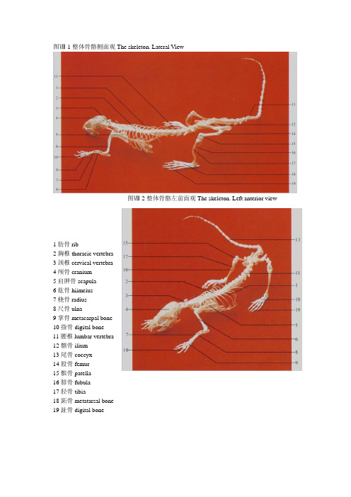
大鼠和小鼠解剖图谱生物秀—专心做生物!www.bbioo.com易生物-领先的生物医药商务平台www.ebioe.com生物秀论坛-学术交流,资源共享,互助社区www.bbioo.com/bbs/图Ⅷ-1整体骨骼侧面观The skeleton. Lateral View图Ⅷ-2整体骨骼左前面观The skeleton. Left anterior view1肋骨rib2胸椎thoracic vertebra3颈椎cervical vertebra4颅骨cranium5肩胛骨scapula6肱骨hiimeius7桡骨radius8尺骨ulna9掌骨metacarpal bone10指骨digital bone11腰椎lumbar vertebra12髂骨ilium13尾骨coccyx14股骨femur15髌骨patella16腓骨fubula17胫骨tibia18跖骨metatarsal bone19趾骨digital bone图Ⅷ-4头骨侧面The skull. Lateral aspect图Ⅷ-6下颌骨侧面The mandible. Lateral aspect1枕骨occipital bone2顶间骨interparietal bone3矢状缝sagittal suture4颧骨malar bone5上颌骨maxillary bone6前颌骨premaxillary bone7枕外嵴external occipital creat8顶骨parietal bone9额骨frontal bone10鼻骨nasal bone11鼻间缝internasal suture12前筛孔preethmoid pore13蝶腭孔sphenopalatine foramen 14门齿 incisor tooth15下颌骨mandible16视神经孔optic foramen17枕骨occipital bone18茎突styloid process19外耳道external acoustic meatus 20颞骨temporal bone21 腭裂patoschisis22臼齿molar tooth23腭骨palatine bone24翼孔pterygoid apertures25破裂孔foramen lacerum26枕大孔foramen magnum27腭后孔posterior palatine foramen 28鼻后孔posterior nasal apertures 29卵圆孔foramen ovale30鼓骨tympanic bone31舌下神经孔hypoglossal foramen 32下颌联合mandibular symphysis 33颏孔mental foramen34冠状突coronoid process35下颌支ramus of mandible36角状突process of horn37下颌孔mandibular foramen38翼肌窝pterygoid fossa39髁突condylar process图Ⅷ-7颈椎与胸椎背面The cervical 图Ⅷ-9前肢骨背面The bone of vertebra and thoracic vertebra.Dorsal aspect anterior 1imb.Dorsal aspect图Ⅷ-8前肢骨外侧面The bone of anteriorlimb. Lateral aspect1枢椎axis 2第4颈椎4th cervical vertebra3第6颈椎6th cervical vertebra 4第1胸椎1st thoracic vertebra5第4胸椎4th thoracic vertebra 6寰椎axis7第3颈椎3rd cervical vertebra 8第5颈椎5th cervical vertebra9第7颈椎7th cervical vertebra 10第3胸椎3rd thoracic vertebral1肱骨体shaft of humcrus 12桡骨radius13尺骨ulna 14尺骨茎突styloid process of ulna15掌骨metacarpal bone 16肱骨头head of hurnerusl7肘突cubital process 18腕骨carpal bone19指骨digital bone图Ⅷ-10股骨前面The femur. 图Ⅷ-11胫骨与腓骨前面The tibia Anterior aspect and fibula. Anterior aspect1大转子greater trochanter2股骨颈neck of femur3第三转子third trochanter4髌骨patella5股骨头femoral headG小转子lesser trochanter7股骨体shaft of femur8股骨内侧髁medial condyle of femur9外侧髁lateral condyle10腓骨fibula11外踝 lateral malleolus12跗骨tarsal bone13胫骨内侧髁medial condyle of tibia14径骨粗隆tibial tuberosity15胫骨tibiaI6内踝medial mallcolus17跖骨metatarsal bone18斜方肌trapezius muscle19臀浅肌glutoeus super(icialis muscle20股直肌rectus femoris muscle21腹外斜肌external oblique muscle of abdomen 22背阔肌latissimus dorsi muscle23肩斜方肌shoulder-trapezius muscle24大圆肌teres major muscle25肩三角肌shoulder-deltoid muscle26前锯肌serratus anterior muscle1锁骨提肌levator clavicle muscle 2肩三角肌shoulder-deltoid muscle 3肱三头肌triceps brachii muscle4臀浅肌gluteus suferficidlis muscle 5股直肌rectus femoris muscle6腓肠肌gastrocnemius muscle7夹肌splenius muscle8颈菱形肌rhomboideus cervicis muscle9胸菱形肌rhomboideus pectoralis muscle10背阔肌latissimus dorsi muscle11腹外斜肌external oblique muscle of abdomen 12股二头肌biceps femoris muscle13半腱肌semitendinosus muscle14二腹肌前腹anterior belly of digastric muscle 15二腹肌后腹posterior belly of digastric muscle 16胸骨舌骨肌sternohyoid muscle17肱二头肌biceps brachii muscle18腹直肌rectus abdominis muscle19股动、静脉femoral artery and femoral vein 20阴囊scrotum21咬肌masseter muscle22胸乳突肌sternomastoideus muscle23肩三角肌shoulder-deltoid muscle24桡侧腕伸肌extensor carpi radialis muscle25指总伸肌extensor digitorum communis muscle 26尺侧腕伸肌extensor carpi ulnaris muscle27胸浅肌pectoral superficialis muscle28前锯肌serratus anterior muscle29股内侧肌vastus medialis muscle30耻骨肌pectineus muscle31长收肌adductor longus muscle32股薄肌gracilis muscle图Ⅷ-15头、颈部侧面The head and neck. Lateral aspect上静脉supraorbitalveine 7颈外静脉external jugular vein 1颞浅动、静脉superficial temporal artery and vein 2眶3面横动、静脉transverse facial artery and vein 4下颌缘神经marginalmandibular nerv 5面后静脉posterior facial vein 6面前静脉anterior facial vein1颞浅动、静脉superficial temporal artery and vein 2眶上静脉supraorbitalvein3面横动、静脉transverse facial artery and vein 4下颌缘神经marginalmandibular nerve 5面后静脉posterior facial vein 6面前静脉anterior facial vein7颈外静脉external jugular vein 8硬腭褶fold of hard palate9臼齿molar tooth 10门齿incisor tooth11软腭soft palate 12舌根root of tongue13舌体body of tongue 14舌尖apex of tongue图Ⅷ-18脑与脊髓背面The brain and 图Ⅷ-19脑与脊髓腹面The brain and spinal cord.Dorsal aspect spinal cordVentral aspect1 喉口aperture of larynx 2舌根root of tongue 3舌尖apex of tongue 4硬腭褶fold of hard palate 5软腭soft palate 6会厌epiglottis 7臼齿molar tooth 8舌体body of tongue 9嗅球olfactory bulb 10中央纵裂central longitudinal fissue 1l 绒球flocculus 12颈膨大cervical enlargement 13腰膨大lumbar enlargement 14外侧纵沟lateral longitudinal sulcus15大脑cerebrum 16小脑(中央部)cerebellum (central part ) 17脊髓圆锥courts medullaris 18 脑垂体 pituitary gland 19脑桥pons 20延髓 myelencephalon 21嗅束olfactory tract 22视神经optic nerve 23三叉神经trigeminal nerve图Ⅷ-20磁共振冠状面定位像(1)The scout view of coronal images(1)图Ⅷ-21磁共振冠状面定位像(2)The scout view of coronal images(2)1眼球eyeball2嗅脑rhinencephalon3大脑皮层cerebral cortex4大脑镰cerebral faix5鼻旁窦paranasal sinus6纵裂longitudinal fissure冠状面T1加权像,颞颌关节线圈,SE序列,层厚3.0mm,无间隔,TR=500rns,TE=20ms。
大鼠解剖-17
பைடு நூலகம்
11 左心房 left atrium 12 左心室 left ventricle 13 肺动脉右支 right branch of pulmonary artery 14 肺静脉 pulmonary vein 15 前腔静脉 precavalvein 16 肺动脉左支 left branch of pulmonary art 7tery 17 后腔静脉 postcaval vein 18 心中静脉 middle cardiac vein 19 头臂动脉 brachiocephalic artery 20 主动脉弓 aortic arch 21 乳头肌 papillary muscles 22 左颈总动脉 left common carotid artery 23 左锁骨下动脉 left subclavian artery 24 右前腔静脉 right precaval vein 25 心外膜 extracardium
图31左心房与左心室theleftatriumleftventricle11左心房leftatrium12左心室leftventricle13肺动脉右支rightbranchpulmonaryartery14肺静脉pulmonaryvein15前腔静脉precavalvein16肺动脉左支leftbranchpulmonaryart7tery17后腔静脉postcavalvein18心中静脉middlecardiacvein19头臂动脉brachiocephalicartery20主动脉弓aorticarch21乳头肌papillarymuscles22左颈总动脉leftcommoncarotidartery23左锁骨下动脉leftsubclavianartery24右前腔静脉rightprecavalvein25心外膜extracardium
大老鼠解剖实验报告
大老鼠解剖实验报告引言解剖实验是生物学实验中的重要一环,通过解剖动物可以深入了解其内部器官的组织结构和功能。
本次实验旨在通过解剖大老鼠,观察和探索其特征解剖结构,进一步了解大老鼠的生物学特性。
实验材料与方法材料- 大老鼠- 刀具- 实验台方法1. 将大老鼠放置在实验台上,保持其身体放松。
2. 用刀具小心地剪开大老鼠的皮肤,从胸部开始向腹部剖开。
3. 慢慢解剖并移除腹肌,保留器官的完整性。
4. 依次观察并记录大老鼠的器官,包括心脏、肺、肝脏、胃、肾脏等。
实验结果1. 心脏:大老鼠的心脏位于胸腔的前部,包裹在围绕心脏的透明薄膜中,被肺脏所遮盖。
心脏分为心房和心室,具有明显的血管与动脉。
2. 肺:大老鼠的肺部位于心脏的两侧,呈粉红色。
通过观察可以看到肺组织细密且有弹性,能够进行有效的气体交换。
3. 肝脏:大老鼠的肝脏位于腹腔的上部,呈褐色。
肝脏是人和动物体内最重要的器官之一,具有解毒、代谢和贮存营养物质等多种功能。
4. 胃:大老鼠的胃位于肝脏下方,通过食道与口腔相连。
胃可以分为贲门、体部和幽门三个区域,用于食物的储存和初步消化。
5. 肾脏:大老鼠的肾脏位于胸腔和腹腔的后部,颜色深红。
肾脏是身体的排泄器官,通过滤波、重吸收和分泌等过程维持体内水和电解质的平衡。
结论通过本次解剖实验,我们对大老鼠的内部器官有了更加直观的了解。
大老鼠的心脏、肺、肝脏、胃以及肾脏等器官在结构和功能上有着与人类相似的特点,但也存在一些差异。
通过观察和学习大老鼠的解剖结构,可以为进一步研究大老鼠的生态学、行为学和疾病模型提供基础。
参考文献(此处列出使用到的参考文献)致谢(此处致谢实验中的指导教师和实验室成员等)。
大鼠解剖图7
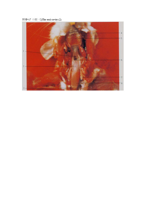
图17口腔2theoralcavity2图18脑与脊髓背面thebrainand图19脑与脊髓腹面thebrainandspinalcorddorsalaspectspinalcordventralaspect1喉口apertureoflarynx2舌根rootoftongue3舌尖apexoftongue4硬腭褶foldofhardpalate5软腭softpalate6会厌epiglottis7臼齿molartooth8舌体bodyoftongue9嗅球olfactorybulb10中央纵裂centrallongitudinalfissue1l绒球flocculus12颈膨大cervicalenlargement13腰膨大lumbarenlargement14外侧纵沟laterallongitudinalsulcus15大脑cerebrum16小脑中央部cerebellumcentralpart17脊髓圆锥courtsmedullaris18脑垂体pituitarygland19脑桥pons20延髓myelencephalon21嗅束olfactorytract22视神经opticnerve23三叉神经trigeminalnerve
4 硬腭褶 fold of hard palate 6 会厌 epiglottis 8 舌体 body of tongue 10 中央纵裂 central longitudinal fissue 12 颈膨大 cervical enlargement 14 外侧纵沟 lateral longitudinal sulcus 16 小脑(中央部)cerebellum (central part) 18 脑垂体 pituitary gland 20 延髓 myelencephalon 22 视神经 optic nerve
大鼠解剖彩色图谱
大鼠解剖彩色图谱图Ⅷ-1整体骨骼侧面观The skeleton. Lateral View图Ⅷ-2整体骨骼左前面观The skeleton. Left anterior view1肋骨rib2胸椎thoracic vertebra3颈椎cervical vertebra4颅骨cranium5肩胛骨scapula6肱骨hiimeius7桡骨radius8尺骨ulna9掌骨metacarpal bone 10指骨digital bone11腰椎lumbar vertebra 12髂骨ilium13尾骨coccyx14股骨femur15髌骨patella16腓骨fubula17胫骨tibia18跖骨metatarsal bone 19趾骨digital bone图Ⅷ-4头骨侧面The skull. Lateral aspect图Ⅷ-6下颌骨侧面The mandible. Lateral aspect1枕骨occipital bone2顶间骨interparietal bone3矢状缝sagittal suture4颧骨malar bone5上颌骨maxillary bone6前颌骨premaxillary bone7枕外嵴external occipital creat8顶骨parietal bone9额骨frontal bone10鼻骨nasal bone11鼻间缝internasal suture12前筛孔preethmoid pore13蝶腭孔sphenopalatine foramen 14门齿 incisor tooth15下颌骨mandible16视神经孔optic foramen17枕骨occipital bone18茎突styloid process19外耳道external acoustic meatus 20颞骨temporal bone21 腭裂patoschisis22臼齿molar tooth23腭骨palatine bone24翼孔pterygoid apertures25破裂孔foramen lacerum26枕大孔foramen magnum27腭后孔posterior palatine foramen 28鼻后孔posterior nasal apertures 29卵圆孔foramen ovale30鼓骨tympanic bone31舌下神经孔hypoglossal foramen 32下颌联合mandibular symphysis 33颏孔mental foramen34冠状突coronoid process35下颌支ramus of mandible36角状突process of horn37下颌孔mandibular foramen38翼肌窝pterygoid fossa39髁突condylar process图Ⅷ-7颈椎与胸椎背面The cervical 图Ⅷ-9前肢骨背面The bone of vertebra and thoracic vertebra.Dorsal aspect anterior 1imb.Dorsal aspect图Ⅷ-8前肢骨外侧面The bone of anteriorlimb. Lateral aspect1枢椎axis 2第4颈椎4th cervical vertebra3第6颈椎6th cervical vertebra 4第1胸椎1st thoracic vertebra5第4胸椎4th thoracic vertebra 6寰椎axis7第3颈椎3rd cervical vertebra 8第5颈椎5th cervical vertebra9第7颈椎7th cervical vertebra 10第3胸椎3rd thoracicvertebral1肱骨体shaft of humcrus 12桡骨radius13尺骨ulna 14尺骨茎突styloid process of ulna15掌骨metacarpal bone 16肱骨头head of hurnerusl7肘突cubital process 18腕骨carpal bone19指骨digital bone图Ⅷ-10股骨前面The femur. 图Ⅷ-11胫骨与腓骨前面The tibia Anterior aspect and fibula. Anterior aspect1大转子greater trochanter2股骨颈neck of femur3第三转子third trochanter4髌骨patella5股骨头femoral headG小转子lesser trochanter7股骨体shaft of femur8股骨内侧髁medial condyle of femur9外侧髁lateral condyle10腓骨fibula11外踝 lateral malleolus12跗骨tarsal bone13胫骨内侧髁medial condyle of tibia14径骨粗隆tibial tuberosity15胫骨tibiaI6内踝medial mallcolus17跖骨metatarsal bone18斜方肌trapezius muscle19臀浅肌glutoeus super(icialis muscle20股直肌rectus femoris muscle21腹外斜肌external oblique muscle of abdomen 22背阔肌latissimus dorsi muscle23肩斜方肌shoulder-trapezius muscle24大圆肌teres major muscle25肩三角肌shoulder-deltoid muscle26前锯肌serratus anterior muscle1锁骨提肌levator clavicle muscle 2肩三角肌shoulder-deltoid muscle 3肱三头肌triceps brachii muscle4臀浅肌gluteus suferficidlis muscle 5股直肌rectus femoris muscle6腓肠肌gastrocnemius muscle7夹肌splenius muscle8颈菱形肌rhomboideus cervicis muscle9胸菱形肌rhomboideus pectoralis muscle10背阔肌latissimus dorsi muscle11腹外斜肌external oblique muscle of abdomen 12股二头肌biceps femoris muscle13半腱肌semitendinosus muscle14二腹肌前腹anterior belly of digastric muscle 15二腹肌后腹posterior belly of digastric muscle 16胸骨舌骨肌sternohyoid muscle17肱二头肌biceps brachii muscle18腹直肌rectus abdominis muscle19股动、静脉femoral artery and femoral vein 20阴囊scrotum21咬肌masseter muscle22胸乳突肌sternomastoideus muscle23肩三角肌shoulder-deltoid muscle24桡侧腕伸肌extensor carpi radialis muscle25指总伸肌extensor digitorum communis muscle 26尺侧腕伸肌extensor carpi ulnaris muscle27胸浅肌pectoral superficialis muscle28前锯肌serratus anterior muscle29股内侧肌vastus medialis muscle30耻骨肌pectineus muscle31长收肌adductor longus muscle32股薄肌gracilis muscle图Ⅷ-15头、颈部侧面The head and neck. Lateral aspect上静脉supraorbitalveine 7颈外静脉external jugular vein 1颞浅动、静脉superficial temporal artery and vein 2眶3面横动、静脉transverse facial artery and vein 4下颌缘神经marginalmandibular nerv 5面后静脉posterior facial vein 6面前静脉anterior facial vein1颞浅动、静脉superficial temporal artery and vein 2眶上静脉supraorbitalvein3面横动、静脉transverse facial artery and vein 4下颌缘神经marginalmandibular nerve 5面后静脉posterior facial vein 6面前静脉anterior facial vein7颈外静脉external jugular vein 8硬腭褶fold of hard palate 9臼齿molar tooth 10门齿incisor tooth11软腭soft palate 12舌根root of tongue13舌体body of tongue 14舌尖apex of tongue图Ⅷ-18脑与脊髓背面The brain and 图Ⅷ-19脑与脊髓腹面The brain and spinal cord.Dorsal aspect spinal cordVentral aspect1 喉口aperture of larynx 2舌根root of tongue 3舌尖apex of tongue 4硬腭褶fold of hard palate 5软腭soft palate 6会厌epiglottis 7臼齿molar tooth 8舌体body of tongue 9嗅球olfactory bulb 10中央纵裂central longitudinal fissue 1l 绒球flocculus 12颈膨大cervical enlargement 13腰膨大lumbar enlargement 14外侧纵沟lateral longitudinal sulcus15大脑cerebrum 16小脑(中央部)cerebellum (central part )17脊髓圆锥courts medullaris 18 脑垂体pituitary gland 19脑桥pons 20延髓myelencephalon 21嗅束olfactory tract 22视神经optic nerve 23三叉神经trigeminal nerve图Ⅷ-20磁共振冠状面定位像(1)The scout view of coronal images(1)图Ⅷ-21磁共振冠状面定位像(2)The scout view of coronal images(2)1眼球eyeball2嗅脑rhinencephalon3大脑皮层cerebral cortex4大脑镰cerebral faix5鼻旁窦paranasal sinus6纵裂longitudinal fissure冠状面T1加权像,颞颌关节线圈,SE序列,层厚3.0mm,无间隔,TR=500rns,TE=20ms。
大鼠解剖图2(PDF)
图Ⅷ-3 头骨背面 The skull.Dorsal aspect 图Ⅷ-4 头骨侧面 The skull. Lateral aspect
图Ⅷ-5 头骨腹面 The skull. Ventral aspect 图Ⅷ-6 下颌骨侧面 The mandible. Lateral aspect
1 枕骨 occipital bone 2 顶间骨 interparietalr bone 5 上颌骨 maxillary bone 6 前颌骨 premaxillary bone 7 枕外嵴 external occipital creat 8 顶骨 parietal bone 9 额骨 frontal bone 10 鼻骨 nasal bone 11 鼻间缝 internasal suture 12 前筛孔 preethmoid pore 13 蝶腭孔 sphenopalatine foramen 14 门齿 incisor tooth 15 下颌骨 mandible 16 视神经孔 optic foramen 17 枕骨 occipital bone 18 茎突 styloid process 19 外耳道 external acoustic meatus 20 颞骨 temporal bone 21 腭裂 patoschisis 22 臼齿 molar tooth 23 腭骨 palatine bone 24 翼孔 pterygoid apertures 25 破裂孔 foramen lacerum 26 枕大孔 foramen magnum 27 腭后孔 posterior palatine foramen 28 鼻后孔 posterior nasal apertures 29 卵圆孔 foramen ovale 30 鼓骨 tympanic bone 31 舌下神经孔 hypoglossal foramen 32 下颌联合 mandibular symphysis 33 颏孔 mental foramen 34 冠状突 coronoid process 35 下颌支 ramus of mandible 36 角状突 process of horn 37 下颌孔 mandibular foramen 38 翼肌窝 pterygoid fossa 39 髁突 condylar process
大鼠全身解剖实验报告(3篇)
第1篇一、实验目的1. 了解大鼠的解剖结构,掌握大鼠全身主要器官的形态、位置和功能。
2. 熟悉大鼠解剖操作步骤,提高实验技能。
3. 深入理解大鼠生理学知识,为后续研究打下基础。
二、实验材料与仪器1. 实验动物:成年大鼠1只2. 实验器材:解剖刀、解剖剪、解剖镊、解剖针、解剖板、生理盐水、棉签、剪刀、止血钳、手术剪、解剖针、注射器、剪刀、解剖板、解剖剪、解剖镊、解剖针、生理盐水、棉签、剪刀、止血钳、手术剪、解剖针、注射器等。
三、实验步骤1. 准备阶段- 将大鼠用乙醚麻醉,待麻醉成功后,将大鼠固定在解剖板上。
- 在大鼠腹侧做一纵切口,从颌下至肛门,注意保护内脏器官。
2. 皮肤解剖- 将皮肤从切口处翻开,暴露出肌肉层。
- 观察肌肉层,注意区分浅层和深层肌肉。
- 观察皮肤下的血管和神经。
3. 肌肉解剖- 将肌肉层从切口处切开,暴露出骨骼。
- 观察骨骼结构,包括头骨、椎骨、胸骨、肋骨、四肢骨等。
- 观察肌肉与骨骼的连接方式。
4. 内脏解剖- 沿着腹中线切开腹腔,暴露出内脏器官。
- 观察并分离内脏器官,包括心脏、肺、肝脏、胃、肠、肾脏、膀胱、生殖器官等。
- 观察内脏器官的形态、位置和功能。
5. 血管解剖- 观察心脏和血管,包括主动脉、肺动脉、肺静脉、肝脏静脉、胃静脉、肠静脉、肾脏静脉、生殖器官静脉等。
- 观察血管的分支和走向。
6. 神经系统解剖- 沿着脊柱切开背部肌肉,暴露出脊髓和神经。
- 观察脊髓和神经的走向,了解神经系统在大鼠体内的分布。
7. 复原图- 根据解剖观察结果,绘制大鼠全身解剖复原图。
四、实验结果1. 大鼠全身骨骼系统由头骨、椎骨、胸骨、肋骨和前后肢骨组成。
2. 大鼠内脏器官包括心脏、肺、肝脏、胃、肠、肾脏、膀胱、生殖器官等。
3. 大鼠血管系统包括主动脉、肺动脉、肺静脉、肝脏静脉、胃静脉、肠静脉、肾脏静脉、生殖器官静脉等。
4. 大鼠神经系统由脊髓和神经组成,负责调节大鼠的生理活动。
五、实验讨论1. 通过本次实验,我们对大鼠的解剖结构有了更深入的了解,为后续研究奠定了基础。
大鼠的解剖图谱
图Ⅷ-2 整体骨骼左前面观 The skeleton. Left anterior view 1 肋骨 rib 2 胸椎 thoracic vertebra 3 颈椎 cervical vertebra 4 颅骨 cranium 5 肩胛骨 scapula 6 肱骨 hiimeius 7 桡骨 radius 8 尺骨 ulna 9 掌骨 metacarpal bone 10 指骨 digital bone 11 腰椎 lumbar vertebra 12 髂骨 ilium 13 尾骨 coccyx 14 股骨 femur 15 髌骨 patella 16 腓骨 fubula 17 胫骨 tibia 18 跖骨 metatarsal bone 19 趾骨 digital bone
图Ⅷ-7 颈椎与胸椎背面 The cervical vertebra and thoracic vertebra.Dorsal aspect
图Ⅷ-9 前肢骨背面 The bone of anterior 1imb.Dorsal aspect
图Ⅷ-8 前肢骨外侧面 The bone of anterior
1 喉口 aperture of larynx 2 舌根 root of tongue 3 舌尖 apex of tongue 5 软腭 soft palate 7 臼齿 molar tooth
4 硬腭褶 fold of hard palate 6 会厌 epiglottis 8 舌体 body of tongue
图Ⅷ-12 整体肌侧面观 The muscles. Lateral view
18 斜方肌 trapezius muscle 19 臀浅肌 glutoeus super(icialis muscle 20 股直肌 rectus femoris muscle 21 腹外斜肌 external oblique muscle of abdomen 22 背阔肌 latissimus dorsi muscle 23 肩斜方肌 shoulder-trapezius muscle 24 大圆肌 teres major muscle 25 肩三角肌 shoulder-deltoid muscle 26 前锯肌 serratus anterior muscle
大鼠解剖-27
图Ⅷ-53 腹主动脉及其分支 The abdominal aorta and branches
1 直肠 rectum 2 精囊 seminal vesicle 3 膀胱 urinary bladder 4 前列腺 prostate 5 精索 spermatic cord 6 阴茎 penis 7 附睾 epididymis 8 睾丸 testis 9 膈 diaphragm 10 后腔静脉 postcaval vein 11 腹腔动脉 celiac artery
12 肾动脉 renal artery 13 右肾 right kidkey 14 前肠系膜动脉 anterior mesenteric artery 15 精索内动脉 internal spermatic artery 16 输尿管 ureter 17 髂腰动、静脉 iliolumbar artery and vein 18 髂总动脉 common iliac artery 19 贮精囊 vesicula seminalis 20 隐动脉 saphenous artery 21 食管 esophagus 22 膈下动脉 inferior phrenic artery 23 肾上腺 adrenal glands 24 肾上腺上动脉 superior adrenal artery 25 左肾 left kidney 26 后肠系膜动脉 posterior mesenteric artery 27 髂外动脉 external iliac artery 28 股动脉 femoral artery 29 腹壁阴部动脉干 arterial epigastro-pudendal trunk 30 输精管
- 1、下载文档前请自行甄别文档内容的完整性,平台不提供额外的编辑、内容补充、找答案等附加服务。
- 2、"仅部分预览"的文档,不可在线预览部分如存在完整性等问题,可反馈申请退款(可完整预览的文档不适用该条件!)。
- 3、如文档侵犯您的权益,请联系客服反馈,我们会尽快为您处理(人工客服工作时间:9:00-18:30)。
. . 第一章 皮肤 一、皮肤由表皮、真皮和皮下组织构成。 二、皮肤腺有皮脂腺、汗腺和乳腺。 1、皮脂腺分布在毛囊周围。口角部、肛门、包皮和乳头周围有特化的皮脂腺。 2、汗腺,大鼠的汗腺只局限于足垫的皮肤。 3、乳腺 数量 大鼠的乳腺共有6对,胸部3对,腹部1对,鼠蹊部2对。个别的大鼠有5对或者7对。 大小和形态 随大鼠的年龄和性周期有明显的变化。 部位 包埋在皮下组织中,由结缔组织隔与胸壁和腹壁松松地相连。 第二章 骨 一.脊柱 大鼠的脊柱由57-61块脊椎骨组成,包括颈椎7、胸椎13、腰椎6、荐锥4、尾锥27-31块。锥式C7T13L6S4Cy27-31。骨性标志为第二胸椎,其棘突最高,超过其它脊椎骨。 1、颈椎 和其它哺乳动物一样,恒为7块,全无肋骨相连,横突上具有横突孔,供锥动脉通过。 2、胸椎 13块,椎骨的长度由前向后逐渐增加(由2毫米增加到4毫米),锥管的直径平均为3.3毫米,较颈部的锥管为狭窄。 3、腰椎 6块,每块锥体的长度比较一致,约6-7毫米。锥管直径由前面的4毫米向后逐渐缩小至2毫米。 二、胸骨 共分为6节,最前一节为胸骨柄,第二到第五节称为胸骨体,最后一节为剑 . . 突,棒状的剑突后面接一盘状的剑状软骨。胸骨柄长约10毫米。 第三章 肌肉系统(省略) 第四章 消化系统 消化系统包括消化管及附属消化器官。消化管可分为口腔、食管、胃、小肠和大肠等部。附属消化器官有齿、舌、唾液腺、肝和胰。 第一节 消化管 一、口腔 1、 舌 全长约30毫米,不具有正中系带,但是有两条侧系带。 一、食管 分为颈、胸、腹三部。成年大鼠食管的颈-胸段长度约75毫米,腹段通过膈的食管裂孔在膈后的长度约15毫米,食管外径约2毫米。 位置:食管主要是沿气管背侧走行,仅在颈部稍偏左侧。 一、胃 位置: 横位于腹腔的左前部,其壁面几乎完全为肝的左叶所覆盖。 大小:胃重为体重的0.5%,属单室胃。 形态:胃小弯朝向背前方,食管在其中部入胃。胃大弯朝向腹后方,其边缘有双层的口袋状大网膜。贲门部外观呈半透明状,壁有粘液腺。 三、小肠 1、 十二指肠 长度:长约100毫米。 位置:从幽门发出向右后行,再折向前终于右侧。可分为降支(向右后行)、横支(水平部)、升支(向前行),它们构成一个不完全的环,包围着部分胰腺。 . . 颜色:淡红色。 2、 空肠 长度: 为小肠的最长部分,大约有700-1000毫米。 位置形态:盘旋在腹腔的右方腹侧部。 3、 回肠 长度: 较短,约有40毫米。 位置形态:以三角形的系膜回盲褶与盲肠的末端相连,向盲肠的开口与结肠的起始部紧密相接。 二、大肠 1、 盲肠 长度:是介于小肠与盲肠之间的一个大的盲囊。长约60毫米,直径约10毫米。位置:通常位于腹腔的左后部。盲肠呈锥体形,分为基部、体部和尖端。 2、 结肠 长度:长约100毫米。 位置形态:升结肠从盲肠发出呈粗管,先弯向后方,然后再度弯向背前方。 横结肠沿十二指肠由右向左横过腹腔,再腹腔左侧突然转折向后为降结肠。 3、 直肠 长度: 长约80毫米。 位置形态:是一直行的沿中线走行的管道,穿过骨盆,在尾根的下方终于肛门。 第二节 消化腺 一、唾液腺 1、 大唾液腺很发达,包括腮腺、颌下腺、大舌下腺。 . . a、 腮腺(耳下腺) 形态位置:呈扁平形,包含3-4个界限清楚的分叶。向上达耳根后方,后缘止于肩部并覆盖锁骨的外测二分之一,前界紧接眶外泪腺,下端达颈部腹面并为其它腺体所覆盖。 颜色:暗肉色。 b、 颌下腺 位置:是颈部腹面最显眼的腺体,其前缘在舌骨水平处与颌淋巴结相连,后界可达胸骨柄,左右两个腺体沿腹中线相接触。 大小:长约16毫米,宽约10-15毫米,厚5毫米。 颜色:红色。 c、 大舌下腺 位置形态:位于紧靠颌下腺的前外侧面,形似眼球的晶状体。 大小:宽约4毫米,厚约1-2毫米。 颜色:淡黄色。 二、肝 1、肝约占体重的4.2%。 位置:位于腹腔的前部,肝的大部分紧贴膈,其壁面向膈面凸出,形状与膈穹隆一致。其腹面沿腹壁延伸并有一小部分越过肋骨,其脏面与腹腔脏(胃、降十二指肠、横结肠、空肠和脾脏)相贴。 2、颜色形态:其色呈暗褐色,每叶都是中心厚,边缘薄。 3、分叶:左外叶、左中叶、中叶、右叶、尾状叶和两个盘状的乳头状突。 A、左外叶,位于左侧,其脏面与胃相连,其前面似较小的左中叶,它与中 . . 叶以一深的裂相隔。 B、中叶稍向前和向右延伸,其脏面与右叶相重叠。 C、右叶位于右侧,部分和右肾相连。 三、胰 颜色形态:为灰粉色且分叶甚多的器官,与周围的脂肪组织相较,可见胰腺的色泽较暗,质地稍坚实。重量从550mg到1000mg。 位置:胰体和右叶包埋在中十二指肠和中空肠的开始处。其扁平的左叶沿胃的背面走行,埋在大网膜的背部,并沿脾动脉到脾的小肠面。 第五章 呼吸系统 呼吸系统包括――鼻腔、咽、喉、气管、和肺两部分。 第一节 呼吸道 一、鼻孔和鼻前庭(省略) 二、鼻腔 可分为前庭部,呼吸部和嗅部。 三、犁鼻器(省略) 四、喉 位置:位于咽的后面,气管的前端。 形态:喉部软骨作为三角形喉腔的支架。具不成对的甲状软骨,会厌软骨,环状软骨和成对的杓状软骨。 五、气管 位置:气管位于食道的腹侧。 形态大小:一般由24个背面不相衔接的U型软骨环构成。气管横肌连接起软骨环的缺口。气管横切面呈扁平椭圆状,水平径约3.5毫米,垂直径约为 . . 2毫米,壁厚约0.5-1.0毫米。从第一气管环到气管的分叉处的距离约33毫米。 气管分支处或支气管与血管之间由大量的淋巴组织。 第二节 肺 位置:位于胸腔。 分叶:分左、右两肺,左肺仅分一叶,右肺分为4叶,即:右上叶、右中叶、右下叶和心脏背侧的右后叶。 第六章 泌尿系统 大鼠的泌尿系统由1对肾脏、1对输尿管、单一的膀胱和单一的尿道组成。 第一节 肾 一、肾 颜色形态:为一对致密的实质性器官,新鲜时红褐色、质地柔软,表面平滑,背腹略扁呈蚕豆形。 位置:左右两肾位于腹膜以外紧贴腰部脊柱两侧,右肾位置靠前,其前缘位于第一腰椎水平处,后端位于第三腰椎水平处。左肾位置稍后,比右肾约后其长度的1/2--1/4。 大小:大鼠肾的长度约为15-25毫米,宽度约10-15毫米,厚度约10毫米。 其它:肾门-位于肾的侧凹陷处,为神经、血管、淋巴管、和输尿管通过的地方。营养好的大鼠,在肾的侧和外测常常围绕着大量脂肪。肾表面有致密的结缔组织构成的纤维膜,沿肾门处纵切剖开肾脏,可见色深红的皮质和色较浅的髓质。 . . 第二节 输尿管 大小长度:输尿管全长约40-55毫米,因左右两肾的位置差异,故右输尿管要比左输尿管约长5毫米,它们的外径约为0.3毫米。 位置:输尿管上端起于肾盂,沿腰肌腹面下降进入盆腔,在膀胱的背外侧注入膀胱。 第三节 膀胱 位置:膀胱位于耻骨前缘,为一肌性的袋囊。膀胱腹侧借膀胱脐韧带与腹腔底壁相连;两侧借助膀胱脐侧褶固定在骨盆的侧壁。 大小形态:其大小和形状均随尿在其中的充盈程度而改变。排空时,呈梨形,长约10毫米,最宽部约5毫米;充盈时,几乎呈圆形,长约25毫米,宽约15毫米或更大。 第七章 生殖系统 第一节 雄性生殖器官 雄性生殖器官的组成由:睾丸、附睾、输精管、阴茎、附性腺及阴囊。 一、阴囊 大鼠的阴囊是由肛门腹侧会阴部皮肤下垂所形成的囊袋。阴囊隔将其分为左右两半。 二、睾丸 位置:位于阴囊。 数量:左右各一。 形态及颜色:睾丸外观呈椭圆形,淡红色。表面包有一层透明的致密结缔组织,即睾丸白膜。 . . 大小:成年的大鼠睾丸长约20毫米,直径14毫米,重约2.0-3.5克。 三、附睾 分为附睾头、附睾体、附睾尾三部。 位置及形态:附睾头呈半月状覆盖在睾丸头端;附睾体狭细,位于睾丸侧;附睾尾呈棒状,越过睾丸尾端向后延伸11-13毫米与输精管相连续。 大小:附睾的重量约为0.76-0.98克,管道的总长度约400厘米。 二、输精管 为附睾管的直接延续,它与附睾尾之间没有明显界限。 位置及走向:输精管沿睾丸的侧走行,沿鞘孔进入腹腔,在膀胱颈水平处,左右两侧的输精管相接。穿过前列腺背外侧叶从背面开口入尿道。 长度及大小:输精管全长5-6厘米,直径约2.5厘米。 三、前列腺 是由三对尿道元基发育而成。可分为头端的背侧叶(即凝集腺),腹侧叶和背外侧叶。 位置:1、凝集腺(前列腺背侧叶)位于精囊的侧凹面,两个长形的腺体被一个公共的结缔组织鞘包绕在一起。 2、腹侧叶贴附在旁观的腹外侧,前端约在耻骨肌前方5毫米处,其被膜与膀胱的被膜结合在一起。 3、两侧的背外侧叶盘曲在尿道近端。 大小及形态:凝集腺体长3-6毫米,重约40-110毫克;腹侧叶呈棒状,灰红色,表面呈波浪状。长14-17毫米,最粗处直径6-9毫米, . . 每叶重约0.3克。 四、阴茎 阴茎可分为根、体、头三部分。后部为阴茎根,中部为阴茎体,前部为阴茎头。 大小长度:大鼠的阴茎全长20-28毫米,平均宽约3.6毫米,高2.8毫米。 位置:阴茎根由两个阴茎脚附着于坐骨弓的腹侧面;阴茎体开始于左右脚联合处,呈背腹扁平。阴茎头为游离端的圆柱状部分。 第二节 雌性生殖器官 由下列各部构成:卵巢、输卵管、子宫、阴道。 一、卵巢 大鼠成对卵巢的形状大小依年龄及发育情况而异。 形态:性成熟的雌鼠卵巢呈卵圆形,表面有不规则结节状的卵泡,新鲜卵巢呈淡红色。 大小:成年雌鼠的卵巢体积约为5*4*3立方毫米,重约60毫克。在妊娠及哺乳期卵巢重量的变化取决于黄体的数目和大小。 位置:两侧的卵巢位置稍有不同,右侧卵巢在第4-5腰椎水平处的腰大肌外侧缘,肾脏后面7-12毫米,距正中线15毫米。 左侧卵巢位于第5-6腰椎水平处的腰大肌外侧缘,肾脏后方3-5毫米处,距正中线11毫米。 二、输卵管 大鼠的输卵管与其它的动物不一样,不直接与生殖腺相连。弯弯曲曲的