J Mol Cell Biol-2011-Ollila-jmcb_mjr016
HIF-1α_纳米抗体的制备及其抑制黑素瘤生长的作用
山西农业科学 2023,51(12):1435-1441Journal of Shanxi Agricultural Sciences HIF-1α纳米抗体的制备及其抑制黑素瘤生长的作用李佳敏1,贾琼1,秦蓉芬1,迟志端1,王富明2,范瑞文1(1.山西农业大学动物医学学院,山西太谷 030801;2.晋中市庄子乡综合便民服务中心,山西晋中 030600)摘要:缺氧诱导因子1α(Hypoxia inducible factor 1α,HIF-1α)参与低氧微环境相关疾病的发生等过程,具有控制肿瘤生长和发展的功能。
黑色素瘤是一种发生于人和动物恶性程度较高的肿瘤。
为探明HIF-1α纳米抗体对黑色素瘤的影响,研究利用前期保存的羊驼源黑色素瘤细胞噬菌体文库筛选HIF-1α纳米抗体,经原核表达与纯化后,通过Western Blot和免疫组织化学验证HIF-1α纳米抗体与抗原的结合性;分别通过CCK-8法、划痕试验以及Western Blot法检测其对B16黑素瘤细胞的增殖和迁移能力及其相关分子表达的影响。
结果表明,经表达和纯化获得的HIF-1α纳米抗体分子质量约为16 ku,没有跨膜结构,具有亲水性。
通过Western Blot和免疫组织化学验证了其具有良好的抗原结合性。
在增殖试验和划痕试验中,与对照组相比,HIF-1α纳米抗体抑制了B16细胞的增殖和迁移,下调了靶基因VEGF的表达,并使细胞增殖和迁移相关蛋白Ras、ERK、RAC和RAF的表达量下调。
预测HIF-1α纳米抗体进入B16细胞内,与抗原结合,通过下游靶基因VEGF下调RAs、ERK、RAC、RAF的表达,从而对细胞增殖和迁移起抑制作用,可作为黑色素瘤治疗的新靶点。
关键词:HIF-1α;纳米抗体;B16细胞;Western Blot法;CCK-8法;细胞增殖;细胞迁移中图分类号:R739.5 文献标识码:A 文章编号:1002‒2481(2023)12‒1435‒07Effect on Preparation of HIF-1α Nano-Antibody and ItsInhibition of Melanoma GrowthLI Jiamin1,JIA Qiong1,QIN Rongfen1,CHI Zhiduan1,WANG Fuming2,FAN Ruiwen1(1.College of Veterinary Medicine,Shanxi Agricultural University,Taigu 030801,China;2.Jinzhong City Zhuangzi Integrated Convenient Service Center,Jinzhong 030600,China)Abstract:The hypoxia inducible factor 1α(HIF-1α) is involved in the occurrence of diseases related to hypoxia microenvironment and has the function of controlling tumor growth and development. As we known, melanoma is a highly malignant tumor occurring in animals and humans. To explore the effect of HIF-1α nano-antibody on melanoma, in this study, the phage library of alpaca-drived melanoma cells previously preserved in our laboratory was used to screen HIF-1α nano-antibodies. After prokaryotic expression and purification, the binding of HIF-1α nano-antibody and its antigen was verified by Western blot and immunohistochemistry. The effects of HIF-1α nano-antibody on the proliferation and migration of B16 melanoma cells and the expression of related molecules were detected by CCK-8, wound healing test, and Western blot methods. The results showed that HIF-1α nano-antibody obtained by expression and purification was hydrophilic protein without transmembrane structure and had a molecular weight of about 16 ku. Western blot and immunohistochemistry results showed that it had good antigenic binding. In the proliferation and wound healing experiments, HIF-1α nano-antibody inhibited the proliferation and migration of B16 cells, down-regulated the expression of target gene VEGF and the proliferation and migration related proteins Ras, ERK, RAC, and RAF, comparing with the control group. In Conclusion, it was predicted that HIF-1α nano-antibody entered B16 cells and combined with antigens and down-regulated the expression of RAs, ERK, RAC, RAF through the downstream target gene VEGF, which inhibited cell proliferation and migration, and could be used as a new target for melanoma treatment.Key words:HIF-1α; nano-antibody; B16 cells; Western Blot method; CCK-8 method; cell proliferation; cell migration氧是生命活动中所必需的物质,且在其中起重要作用[1]。
骨髓间充质干细胞源性外泌体促进小胶质细胞
学 报Journal of China Pharmaceutical University 2023,54(5):599 - 606599骨髓间充质干细胞源性外泌体促进小胶质细胞/巨噬细胞M2极化抑制急性期脑缺血大鼠炎症反应孙逸梅,毛诗慧,李琳,江伟锋,储利胜*(浙江中医药大学基础医学院,杭州 310053)摘 要 为探究骨髓间充质干细胞(bone marrow meaenchymal stem cells, BMSCs)源性外泌体(exosomes, Exos)对急性期脑缺血大鼠小胶质细胞/巨噬细胞M1/M2极化的影响,采用超高速离心法分离提取外泌体并鉴定;采用线栓法制备大鼠大脑中动脉阻塞(middle cerebral artery occlusion, MCAO)模型;采用Longa评分和角实验评价大鼠神经功能,2,3,5-氯化三苯基四氮唑(2, 3, 5-triphenyltetrazole chloride, TTC)染色检测大鼠脑梗死体积;采用CD16/32/Iba1、CD206/Iba1免疫荧光双标法检测小胶质细胞/巨噬细胞M1/M2表型;采用RT-qPCR检测大鼠脑缺血周边区CD86、诱导型一氧化氮合酶(inducible nitricoxide synthase, iNOS)、肿瘤坏死因子-α(tumor necrosis factor, TNF-α)、精氨酸酶1(arginase-1, Arg-1)、白细胞介素-10(interleukin-10, IL-10)和转化生长因子β(transforming growth factor beta, TGF-β)的mRNA表达。
实验结果表明,BMSC-Exos减少缺血周边区CD16/32+/Iba1+阳性细胞数量(P < 0.01),增加CD206+/Iba1+阳性细胞数量(P < 0.01),减少iNOS、CD86和TNF-α的mRNA表达,增加Arg-1、TGF-β和IL-10的mRNA表达(P < 0.05或P < 0.01)。
聚乙二醇化重组人粒细胞集落刺激因子用于多发性骨髓瘤自体造血干细胞动员的研究

聚乙二醇化重组人粒细胞集落刺激因子用于多发性骨髓瘤自体造血干细胞动员的研究丁筱1黄文阳2刘雪莲'杨艳萍1樊红琼1岳婷婷1邹德慧2邱录贵2靳凤艳1 '吉林大学第一医院肿瘤中心血液科,长春130021;2中国医学科学院血液病医院(中国 医学科学院血液学研究所)实验血液学国家重点实验室国家血液系统疾病临床研究 中心,天津 300020通信作者:斩凤艳,Email:fengyanjin@【摘要】目的探讨聚乙二醇化重组人粒细胞集落刺激因子(PEG-rhG-CSF)用于多发性骨髓瘤(MM)患者外周血造血干细胞动员(PBSCM)的效果及药物经济学价值。
方法回顾性分析2015年1月至2017年10月在吉林大学第一医院和中国医学科学院血液病医院住院治疗的9丨例初治MM患者资料。
根据患者意愿,采用大剂量化疗结合皮下注射PEG-rhG-CSF或重组人粒细胞集落刺激因子UhG-CSF)进 行干细胞动员,分别为42、49例。
分析两组动员后采集单个核细胞(MNC)数、采集物CD34+细胞数、动员 中最高中性粒细胞(mANC)数、动员的费用以及移植后白细胞和血小板植人时间,,结果PEG-rhG-CSF 组和rhG-CSF组的中位采集MNC 数分别为 5.86x10s / kg[ ( 1.08 ~ 24.54)x l〇8 / kg]和 6.61x l〇« / kg [(0.83 ~ 33.80)x l〇V kg],差异无统计学意义(t/= 883.00, P= 0.245); PEG-rhG-CSF组的中位采集物CD34+细胞数高于rhG-CSF组,分别为5.56 x l〇6/kg[(0.94~ 19.90) x l〇V kg]和4.82x l〇6/kg[(丨.12~ 14.61) x l〇V kg],差异有统计学意义((7= 732.00, P= 0.038)。
PEG-rhG-CSF组动员期间中位mANC数 较 rhG-CSF组低,分别为 20.50x l09/L[(7.26~61.30)x l0V L;^32.08x l0V L[(6.92~69.99)x l0V L],差异有统计学意义(i/= 490.00, P= 0.001)。
ACT001通过STAT1/CIITA/MHC-Ⅱ通路发挥抗炎抗氧化活性治疗脓毒症引起的急性肺损伤

网络出版时间:2023-11-3016:13:56 网络出版地址:https://link.cnki.net/urlid/34.1086.R.20231130.1319.012◇呼吸药理学◇ACT001通过STAT1/CIITA/MHC Ⅱ通路发挥抗炎抗氧化活性治疗脓毒症引起的急性肺损伤盛 磊,周杰诗,韩 旭,李伊楠,刘慧娟,孙 涛(南开大学药学院,天津 300350)收稿日期:2023-05-27,修回日期:2023-08-26基金项目:国家自然科学基金面上项目(No82272934);国家级大学生创新创业训练计划(No202210055112)作者简介:盛 磊(2002-),男,研究生,研究方向:药理学,E mail:2010402@nankai.edu.cn;孙 涛(1982-),男,博士,教授,博士生导师,研究方向:药理学,通信作者,E mail:tao.sun@nankai.edu.cndoi:10.12360/CPB202305082文献标志码:A文章编号:1001-1978(2023)12-2231-09中国图书分类号:R 332;R284 1;R322 35;R364 5;R631;R563 8摘要:目的 评价含笑内酯衍生物ACT001对脓毒症进程中急性肺损伤的治疗作用并探究其药理机制。
方法 动物水平上,小鼠腹腔注射LPS建立急性肺损伤模型,腹腔注射ACT001进行治疗,从小鼠个体存活状况、肺部炎症损伤及水肿情况等方面评价ACT001的药效;细胞水平上,以LPS刺激RAW264 7细胞构建模型,通过检测炎症反应和氧化应激水平探究其药理机制,并通过蛋白质组学结果分析其相关分子机制。
结果 动物水平上,ACT001可改善急性肺损伤小鼠生存率、减轻肺部炎症、降低血清中炎症因子水平;细胞水平上,ACT001通过抑制MHC-Ⅱ相关通路,促进RAW264 7细胞向抗炎表型极化,抑制NO和相关炎症因子产生的同时提高SOD含量并清除ROS。
原发性胆囊癌的早期诊断

原发性胆囊癌的早期诊断殷保兵【摘要】原发性胆囊癌是胆道常见恶性肿瘤,恶性程度高,发现时往往伴有肝脏转移,预后极差。
掌握原发性胆囊癌的高危因素,对胆囊癌高危人群进行密切随访和筛选,可提高原发性胆囊癌的早期诊断率,对改善胆囊癌的预后有重大意义。
%Primary gallbladder carcinoma (PGC) is the common malignant tumour in biliary tract and is associated with poor prognosis in patients with liver metastasis. It is important to understand the high risk factors of PGC, and closely follow up and screen the high-risk populations in order to improve the early diagnosis of gallbladder cancer and its prognosis.【期刊名称】《上海医药》【年(卷),期】2014(000)014【总页数】3页(P12-14)【关键词】胆囊癌;早期诊断;高危人群【作者】殷保兵【作者单位】复旦大学附属华山医院外科上海 200040【正文语种】中文【中图分类】R735.8原发性胆囊癌是最常见的胆道恶性肿瘤,约占消化道恶性肿瘤的第6位[1-2]。
2007年上海市胆囊癌发病率男性为5.90/10万,女性为10.22/10万,而同期全国胆囊癌死亡率高达4.07/10万[3-4]。
邹声泉等[5]报道2000年我国大陆原发性胆囊癌发病率占同期胆道疾病的0.4%~3.8%。
近年来,胆囊癌的发病率有上升趋势,而发病年龄则呈下降趋势。
胆囊癌的早期诊断非常困难,而其恶性程度极高,且易转移复发,因此预后极差,进展期胆囊癌的中位生存期仅6个月,5年生存率仅为5%[2]。
J Cell Physiol

J Cell PhysiolJ Cell Physiol is a scientific journal that focuses on the field of cellular physiology. It covers a wide range of topics related to the functioning and behavior of cells, with a particular emphasis on their physiological processes. The journal publishes high-quality original research papers, reviews, and commentaries that contribute to our understanding of cell physiology.Cell physiology is an interdisciplinary field that studies the functions and activities of cells, including their metabolism, communication, and response to environmental stimuli. Understanding cell physiology is crucial for understanding the basic processes that underlie complex biological systems and for developing new approaches for the diagnosis and treatment of diseases.Research published in J Cell Physiol encompasses a variety of topics, including cellular metabolism, cell signaling, cell cycle regulation, cell growth and differentiation, cellular responses to stress, and cell communication. The journal also welcomes studies investigating the impact of various factors on cell physiology, such as genetic and epigenetic changes, environmental factors, and therapeutic interventions.One area of interest in J Cell Physiol is cellular metabolism. This field aims to understand the complex network of metabolic pathways that cells utilize to generate energy and synthesize biomolecules. Elucidating the regulation of cellular metabolism can provide insights into various diseases, such as cancer and metabolic disorders, and can help identify potential therapeutic targets.Cell signaling is another key area covered in J Cell Physiol. Cells communicate with each other through a variety of signaling pathways, including cell surface receptors, intracellular signaling cascades, and gene expression regulation. Understanding how cells transmit and process these signals is essential for deciphering the molecular mechanisms underlying normal cellular functioning and for identifying aberrant signaling events associated with disease.The regulation of the cell cycle is vital for cell growth and tissue homeostasis. J Cell Physiol publishes studies investigating the control and coordination of the cell cycle, including the role of key regulatory proteins and the mechanisms involved in cell cycle checkpoints. Dysregulation of the cell cycle is a hallmark of many diseases, including cancer, and understanding its regulation can lead to the development of novel therapeutic strategies.Cell growth and differentiation are also areas of interest in J Cell Physiol. These processes are fundamental for tissue development, repair, and regeneration. Research published in the journal examines the molecular and cellular mechanisms governing cell growth and differentiation, as well as the factors that influence these processes, such as growth factors, signaling pathways, and the cellular microenvironment.Cellular responses to stress, both physiological and pathological, are investigated in J Cell Physiol. Cells have evolved intricate mechanisms to respond to various forms of stress, including oxidative stress, DNA damage, and proteotoxic stress. Understanding these adaptive responses can provide insights into the pathogenesis of diseases and may lead to the development of therapeutic interventions.Finally, J Cell Physiol also covers studies on cell communication. Cells communicate through various mechanisms, including direct cell-cell contact, secretion of signaling molecules, and extracellular vesicles. The journal publishes research on the molecular mechanisms underlying cell communication and its role in physiological and pathological processes, such as immune responses, tissue regeneration, and cancer metastasis.In conclusion, J Cell Physiol is a scientific journal dedicated to advancing our understanding of cellular physiology. It covers a wide range of topics related to the functioning and behavior of cells, with the aim of unraveling the intricate mechanisms underlying normal cellular processes and disease pathology. By publishing high-quality research papers, reviews, and commentaries, the journal contributes to the field of cell physiology and provides a platform for scientific exchange and collaboration.。
抗体药物工艺开发需要考虑的因素
有一个完整的了解。 如何选择最佳的候选药, 是建 立一个稳定的工艺的首要步骤。 目前, 生物制品以
2363
中国新药杂志 2015 年第 24 卷第 20 期
C h in e se Jou rn a l of N e wD ru g s 2015, 24(20)
所以本文讨 单抗类药物 ( 包括 Fc 融合蛋白 ) 为主, 论的工艺开发也以单抗类分子为主。 在中国, 抗体 药物主要分为生物类似药和生物创新药两大类 。生 物类似药是已经被临床和市场证明的药物 , 有现成 的产品做参照, 不存在选择候选药的问题。但是, 企 业要考虑根据是否填补国内市场空白, 并对产品自 身优势进行分析, 预期的市场份额进行分析来选择 项目。因此, 选择最佳候选药主要是针对生物创新 药。以创新性单抗为例, 不管用何种技术 ( 包括人 、 、 源性单抗 鼠源性单抗 兔源性单抗、 鸡源性单抗, 以 及用 单 细 胞 技 术 分 离 出 来 的 单 抗 ) 产 生 的 抗 [6 - 11 ] , 体 同一靶点的候选单抗数量越多, 可供选择 的范围越大。除了满足候选药有效性的基本要素, 如足够阻断靶点的亲和力 ( Kd ) 、高度特异的靶点 IC80 ) 以及 结合力、 有效抑制靶点功能效应 ( IC50 , 有知识产权方面的可操控空间外, 作为一个适合启 动工艺开发的候选药, 还必须具备工艺开发和放大 生产的可行性要素, 如高稳定性 ( Tm 值 ) 、 高可溶 性、 低免疫原性、 有利于生产过程和血液内循环的等 电点( pI, 如 pI = 5 6 或 pI = 8 9 ) 以及在关键结合 部 位 ( 如, complementarity determining regions, CDRs) 不存在潜在的不利因素 ( 包括 N糖、 去甲基 [12 - 14 ] 。 只有充分考虑上述 化、 脱酰氨化等位点 ) 等 综合因素后, 工艺开发才能顺利进行。此外, 必须根 , 据药物的适应症 对抗体依赖的细胞介导的细胞毒 dependent cellmediated cytotoxicity , 作 用 ( antibodyADCC ) / 补 体 介 导 的 细 胞 毒 作 用 ( complement dependent cytotoxicity , CDC ) 活性的需求, 选择合适的 [15 - 16 ] Ig) 的类型 。 免疫球蛋白( immunoglobulin, 2 表达系统和稳定表达细胞株的建立 一旦确立候选抗体药物后, 需要确定用于表达
Klotho_蛋白在子痫前期患者胎盘外泌体中表达及其对血管内皮细胞氧化应激的影响

第 49 卷第 6 期2023年 11 月吉林大学学报(医学版)Journal of Jilin University(Medicine Edition)Vol.49 No.6Nov.2023DOI:10.13481/j.1671‑587X.20230616Klotho蛋白在子痫前期患者胎盘外泌体中表达及其对血管内皮细胞氧化应激的影响薛筱蕾, 胥保梅(新疆医科大学第五附属医院产科,新疆乌鲁木齐830011)[摘要]目的目的:探讨Klotho在子痫前期(PE)患者来源的胎盘外泌体(Exo)中的表达,阐明其对血管内皮细胞氧化应激的影响。
方法方法:收集40例孕产妇的临床资料,其中正常妊娠(NP)者20名,PE患者20例,设为NP组和PE组,分离2组研究对象外周血中胎盘Exo。
将oe-Klotho和oe-NC质粒转染入人绒毛膜滋养层细胞HTR-8/SVneo中,作为oe-Klotho组和oe-NC组,收集HTR-8/ SVneo细胞Exo。
采用实时荧光定量PCR(RT-PCR)法和Western blotting法检测2组研究对象胎盘Exo和2组HTR-8/SVneo细胞Exo中Klotho mRNA及蛋白表达水平。
取生长状态良好的人脐静脉内皮细胞(HUVECs),按Exo来源不同将HUVECs分为PE-Exo组(与PE患者胎盘Exo共培养)、NP-Exo组(与NP胎盘Exo共培养)、oe-Klotho-Exo组(与转染oe-Klotho的HTR-8/SVneo细胞Exo共培养)和oe-NC-Exo组(与转染oe-NC的HTR-8/SVneo细胞Exo共培养)。
采用透射电子显微镜(TEM)观察Exo形态表现,Western blotting法检测Exo标记分子CD63、TSG101和胎盘Exo标记分子PLAP蛋白表达水平以鉴定Exo,荧光显微镜观察HUVECs对Exo的摄取情况。
酶联免疫吸附试验(ELISA)法检测各组HUVECs中一氧化氮(NO)、活性氧(ROS)和丙二醛(MDA)水平及超氧化物歧化酶(SOD)活性,RT-qPCR法检测各组HUVECs中内皮型一氧化氮合酶(eNOS)mRNA 表达水平,Western blotting法检测各组HUVECs中eNOS蛋白表达水平。
- 1、下载文档前请自行甄别文档内容的完整性,平台不提供额外的编辑、内容补充、找答案等附加服务。
- 2、"仅部分预览"的文档,不可在线预览部分如存在完整性等问题,可反馈申请退款(可完整预览的文档不适用该条件!)。
- 3、如文档侵犯您的权益,请联系客服反馈,我们会尽快为您处理(人工客服工作时间:9:00-18:30)。
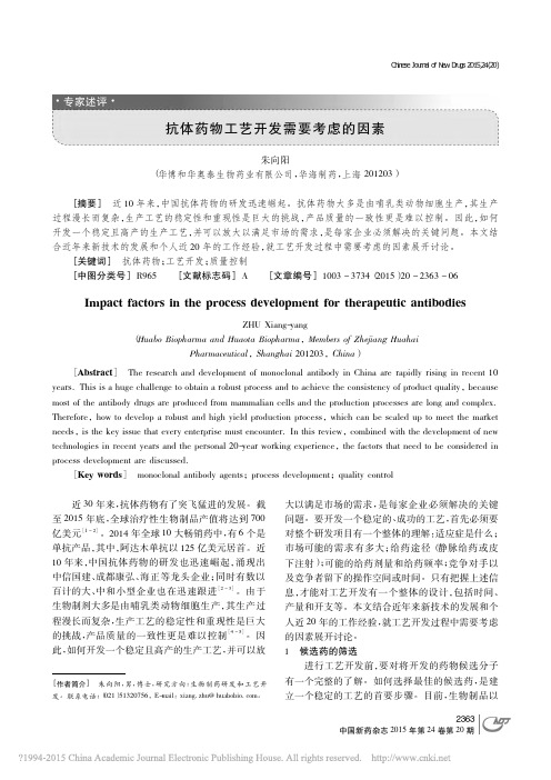
ReviewThe tumor suppressor kinase LKB 1:lessons from mouse modelsSaara Ollila and Tomi P.Ma¨kela ¨*Institute of Biotechnology,University of Helsinki,Viikki Biocenter,Viikinkaari 9B,FIN-00014,Helsinki,Finland*Correspondence to:Tomi P.Ma¨kela ¨,E-mail:tomi.makela@helsinki.fiMutations in the tumor suppressor gene LKB 1are important in hereditary Peutz–Jeghers syndrome,as well as in sporadic cancersincluding lung and cervical cancer.LKB 1is a kinase-activating kinase,and a number of LKB 1-dependent phosphorylation cascades regulate fundamental cellular and organismal processes in at least metabolism,polarity,cytoskeleton organization,and prolifer-ation.Conditional targeting approaches are beginning to demonstrate the relevance and specificity of these signaling pathways in development and homeostasis of multiple organs.More than one of the pathways also appear to contribute to tumor growth fol-lowing Lkb 1deficiencies based on a number of mouse tumor models.Lkb 1-dependent activation of AMPK and subsequent inacti-vation of mammalian target of rapamycin signaling are implicated in several of the models,and other less well characterized pathways are also involved.Conditional targeting studies of Lkb 1also point an important role of LKB 1in epithelial–mesenchymal interactions,significantly expanding knowledge on the relevance of LKB 1in human disease.Keywords:LKB 1,tumor suppressor,mouse model,AMPKIntroductionCancer arises as a result of accumulating genetic and epige-netic changes,which compromise the cell’s ability to control its identity and proliferation.Many identified tumor suppressors play a well-established role in regulation of cell growth and div-ision (e.g.Rb,APC,p 21,PTEN)and genome maintenance (e.g.p 53,BRCA 1-2,ATM,ATR,MLH 1,MSH 2),providing a logical link between the loss of gene product and promotion of carcinogen-esis.An interesting exception is the serine /threonine kinase gene LKB 1(also known as STK 11),which has in recent years taken a prominent position among tumor suppressors.Heterozygous germline mutations in LKB 1predispose to Peutz–Jeghers syndrome (PJS)where patients develop benign polyps in the gastrointestinal (GI)tract and are in high risk of developing malignant tumors in GI tract,breast,and gyneco-logical organs (Giardiello et al .,2000).Importantly,somatic LKB 1mutations are found at least in lung (Ji et al.,2007)and cervical cancer (Wingo et al .,2009).Through phosphoryl-ation of several cellular kinases LKB 1has been implicated in control of cellular and organismal metabolism,cell polarity,and a variety of other functions ranging from proliferation and migration to senescence,apoptosis,DNA damage responseand differentiation (Vaahtomeri and Ma¨kela ¨,2011).Despite these many functions attributed to LKB 1,their specific contri-butions to the maintenance of tissue homeostasis in vivo and tumor growth are only sketchily appearing with thedevelopment of LKB 1mouse models.This work is important to enable rational treatment strategies to LKB 1-deficient tumors.The LKB 1kinase acts in a trimer with a pseudokinase STRAD and the scaffold protein MO 25to phosphorylate at least 14kinases with conserved activation sites (Katajisto et al.,2007).A well-known substrate of LKB 1is AMPK,which is the master reg-ulator of cellular and organismal metabolism,providing a putative downstream pathway to LKB 1-mediated tumor suppression (Shackelford and Shaw,2009).In mouse studies,AMPK requires LKB 1for activation in vivo in most tissues (Sakamoto et al .,2005;Shaw et al .,2005;Contreras et al .,2008;Hezel et al .,2008).AMPK senses the energy state of cells through monitoring AMP levels as a sensitive readout for ATP.AMPK is activated following exercise,hypoxia,or glucose deprivation,after which it phosphor-ylates multiple targets to increase energy uptake and catabolic processes such as glucose uptake and fatty acid oxidation,and suppress anabolic processes such as lipogenesis and cholesterol synthesis (Hardie et al.,2003).AMPK is the potential candidate to mediate LKB 1’s effects in cell growth via the mammalian target of rapamycin (mTOR)signal-ing (Corradetti et al .,2004;Shaw et al .,2004),which is the pathway monitoring the availability of nutrients in regulation of cell size and protein synthesis as well as proliferation (Zoncu et al.,2011).Increased mTOR signaling is common in cancer (Guertin and Sabatini,2007)and also present in at least some Lkb 1-deficient tumors (Shaw et al.,2004;Ji et al.,2007;Contreras et al.,2008;Hezel et al.,2008;Shackelford et al.,2009).An additional link between LKB 1and mTOR pathway#The Author (2011).Published by Oxford University Press on behalf of Journal ofMolecular Cell Biology ,IBCB,SIBS,CAS.All rights reserved.doi:10.1093/jmcb /mjr 016Journal of Molecular Cell Biology (2011),Vol no.0,1–11|1Journal of Molecular Cell Biology Advance Access published September 15, 2011 at Shihezi University on September 27, 2011 Downloaded frommay lie in regulation of PI 3K-Akt pathway inhibitor PTEN by LKB 1(Mehenni et al .,2005).Loss of cell polarity is commonly noted in cancer,and LKB 1is an important factor for cell polarity in different organisms.In C.elegans ,the orthologs for LKB 1(par-4)and MARK s (par-1)were identified in a panel of six partitioning (par )mutants which disrupted the polarity of the early embryos (Kemphues et al.,1988).In Drosophila ,Lkb 1is required for proper oocyte polarity (Martin and St Johnston,2003).In mammalian cells,in both 2D and 3D cell culture models and in vivo ,LKB 1is known to regulate polarity (Baas et al .,2004;Partanen et al .,2007;Hezel et al .,2008).Polarity defects are,however,not seen in all Lkb 1-deficient tumors (Contreras et al.,2008,2010).Several of the LKB 1substrates have been reported to mediate the regulation of cell polarity through regulating the cytoskeleton and formation of cell–cell junctions.MARK kinases are implicated in the stability of microtubules by phosphorylating and thereby dissociation microtubule-associated proteins (MAPs),for example the tau protein,from microtubules (Drewes et al .,1997;Stoothoff and Johnson,2005).Neuronal polarity and axon formation are regu-lated by LKB 1at least partially via BRSK kinases (Kishi et al.,2005;Barnes et al.,2007).To what extent LKB 1acts as a polarity protein in mammalian non-neuronal cells still remains to be deter-mined,although at least in both exo-and endocrine pancreas Lkb 1loss leads to polarity defects in vivo (Hezel et al .,2008;Granot et al .,2009).As formation of stress fibers is essential incell contractility,recent studies associate LKB 1with cell motility via NUAK 1and NUAK 2,which have been implicated in regulation of myosin light chain phosphorylation (Vallenius et al.,2010;Zagorska et al.,2010).For detailed information of the molecular signaling pathways of Lkb 1,the reader is recommended recent reviews more focused on that topic (Katajisto et al.,2007;Hezel and Bardeesy,2008;Vaahtomeri and Ma¨kela ¨,2011).Role of Lkb 1in development and tissue homeostasis in miceAlthough LKB 1is a tumor suppressor,inactivation of Lkb 1through homologous recombination or ‘knock-out’(KO)does not always lead to tumors.This is due partly to essential functions of Lkb 1in development and partly demonstrates the tissue-specificity of Lkb 1functions,where in some cell types biallelic deletion is detrimental to cells or affects specific functions in metabolism as summarized in Figure 1and discussed below.Role of Lkb 1in embryogenesisGeneration of full KO revealed that Lkb 1is essential for embry-ogenesis;no viable Lkb 12/2embryos were seen after E 11.Analysis of the E 8.5–E 9.5embryos revealed severe developmen-tal defects including impaired neural tube closure and somitogen-esis,mesenchymal tissue cell death,and defective vasculature.The extra-embryonic tissues (yolk sac and placenta)were also deformed.VEGF signaling was highly upregulated in theKOFigure 1Non-tumorigenic phenotypes following Lkb 1targeting in mice.Phenotypes (green)are grouped according to tissue type,cell typeaffected /analyzed (blue),and alleles used for targeting.When appropriate,activator of deletor is indicated in purple.Noted signaling change(s)indicated in red.Alleles as displayed in original publications except for Lkb 1flox 2h /flox 2h hypomorphic Lkb 1(Sakamoto et al,2005).(1)Londesborough et al.,2008;(2)Ohashi et al.,2010;(3)Cao et al.,2010;Tamas et al.,2010;(4)Shorning et al.,2009;(5)Woods et al.,2011;(6)Shaw et al.,2005;(7)Sun et al.,2010a ;(8)Sun et al.,2011;(9)Granot et al.,2009;Fu et al.,2009;(10)Koh et al.,2006;(11)Sakamoto et al.,2005;(12)Sakamoto et al.,2006;Jessen,et al.,2010;(13)Ikeda et al.2009;(14)Gurumurthy et al.,2010;Nakada et al.,2010;(15)Gan et al.,2010;(16)Barnes et al.,2007;(17)Ylikorkala et al.,2001.tam,tamoxifen;b -NF,b -naphtoflavone;pIpC,polyinosinic–polycytidylic acid;iv,intravenous.2|Journal of Molecular Cell Biology Ollila and Ma¨kela ¨ at Shihezi University on September 27, 2011 Downloaded fromembryos,possibly relating to the vascular phenotype (Ylikorkala et al .,2001).Embryonic lethality,no embryonic turning,and small somites were also shown in another report of Lkb 1full KO (Jishage et al .,2002).The severe developmental defect was not a result of the abnormal extra-embryonic tissues,since epiblast-specific conditional inactivation of Lkb 1using Mox 1-Cre resulted in very similar embryonic lethal phenotype to full KO (Londesborough et al .,2008).The important role of Lkb 1in development and maintenance of neurons,mesenchymal cells,and vascularization has been recapitulated in tissue-specific Lkb 1KOs.Role of Lkb 1in angiogenesisLondesborough et al .(2008)further dissected the role of Lkb 1in endothelia by deleting Lkb 1in vascular endothelial cells using Tie 1-Cre (Figure 1).The mice died at E 12.5and displayed dilated embryonic vessels and pericardial swelling.The vessels were irre-gular and distorted and suffered from inadequate supportive vas-cular smooth muscle cell layer.Since Tgf b signaling was reduced both in Lkb 1-deficient mouse yolk sacs and human umbilical vein endothelial cells (HUVECs)where LKB 1expression was silenced by siRNA,the vascular phenotype was suggested to result from a loss of supporting vascular smooth muscle cells as a conse-quence of attenuated Tgf b signaling from endothelial cells (Londesborough et al .,2008).Another report also described mice lacking Lkb 1in endothelial cells,deleted using Tie 2-Cre driver (Ohashi et al.,2010)(Figure 1).This study repeated the finding that endothelial Lkb 1is essential for proper embryonic development and no homozygous mutants were born.Analysis of heterozygous Tie 2-Cre;Lkb 1flox /+mice revealed that the mice,including vasculature,seemed phenotypically normal,but displayed reduced revascularization after hind-limb ischemia.Studies in mouse tissues,primary mouse endothelial cells,and HUVECs implemented that the phenotype was mediated via AMPK (Ohashi et al.,2010).In this study,the authors did not address the contribution of Tgf b signaling to the observed phenotype.In the Tie 2-Cre model,Lkb 1–AMPK axis seemed to mediate proangiogenetic signaling as Lkb 1heterozygosity resulted in reduction of revascularization in adult mice (Ohashi et al.,2010).In developing embryo,increased VEGF signaling upon Lkb 1loss would suggest the opposite,antiangiogenic role for Lkb 1(Ylikorkala et al .,2001).Also in the context of PJS polyps where a loss of Lkb 1leads to increased HIF 1a and vasculature,Lkb 1seems to be rather antiangiogenic (Shackelford et al .,2009).However,reduced capillary density was reported in mice where Lkb 1was conditionally deleted from the heart (Ikeda et al .,2009).In 3D culture system where endothelial cells are embedded in Matrigel,both over-expression (Xie et al .,2009)and inhibition (Ohashi et al.,2010)of Lkb 1have been reported to inhibit network formation,suggesting proper expression of LKB 1is essential for angiogenesis.Thus,the precise role of Lkb 1in angiogenesis seems to be dependent on the tissue type and /or the developmental phase,varying from inhibition to promotion.Role of Lkb 1in liverThe finding that Lkb 1functions upstream of AMPK (Shaw et al .,2004)led to interest to study its effects in liver,where many path-ways of carbohydrate and lipid metabolism,including glycogen-esis,glycogenolysis,gluconeogenesis,lipogenesis,and cholesterol synthesis take place.Tail-vain injection of Adeno-Cre to mice carrying conditional Lkb 1allele led to hepatocyte-specific Lkb 1deletion since Adeno-Cre has high tropism for hepatocytes (Shaw et al .,2005)(Figure 1).Lkb 1loss resulted in nearly complete abolishment of AMPK activation in liver,and the glucose metabolism of the mice was impaired demonstrated by elevated blood glucose.CRTC 2phosphorylation was reduced in the livers of the mice,leading to elevated CREB-mediated transcription,including expression of PGC 1a and other gluconeogenetic genes.Also lipogenetic genes were over-expressed.Metformin,the diabetes drug which reduces blood glucose levels via AMPK pathway (Zhou et al .,2001),did not lower blood glucose in the liver-specific Lkb 1KO mice,demonstrating that AMPK activity induced by Lkb 1in liver is required for the effects of metformin in vivo .In another report of liver-specific Lkb 1knockout using Alb-Cre driver,Woods et al.(2011)reported defective bile ducts in liver,leading to accumulation of bile in liver and serum (Figure 1).Bile salt export pump was not located in canalicular membrane of the bile canaliculi,indicating possible defects in cell polarity.The mice also suffered from cholestasis (Woods et al.,2011).These reports of liver-targeted deletions of Lkb 1demonstrate the critical requirement of Lkb 1in glucose,lipid,bile,and cholesterol metab-olism.Furthermore,they show that in liver,Lkb 1is the main acti-vator of AMPK,and its activity is required for the AMPK-mediated suppression of lipogenesis and gluconeogenesis to take place.Role of Lkb 1in muscleMuscles are highly energy-consuming tissues whose glucose homeostasis needs to be regulated both in response to insulin after blood sugar increase,and to exercise-mediated deficiency of glucose storage.Sakamoto et al.(2005)provided the first genetic evidence that Lkb 1is required for AMPK activation in vivo in skeletal muscle.They generated conditional Lkb 1mice in which cDNA of Lkb 1exons 5–7fused with neomycin resistance cassette,surrounded by loxP sites,was inserted between exons 4and 8in the genomic Lkb 1locus.The resulting mice were hypomorphic and expressed only 10%–20%of normal levels of Lkb 1in the absence of Cre -mediated ing MCK-Cre driver to create muscle-specific Lkb 1KO,they found that AMPK a 2(one of the two alternative catalytic subunits of AMPK)activation either by the AMP analog AICAR,muscle con-traction or phenformin,a similar blood glucose lowering drug to metformin,was lost and AMPK a 1activation greatly reduced.Upon contraction,glucose transport to muscle cells was abol-ished (Sakamoto et al.,2005).In another study using the same muscle-specific MCK-Cre with another (non-hypomorphic)con-ditional Lkb 1line,effects of Lkb 1loss in muscle to levels of blood glucose were investigated (Koh et al.,2006)(Figure 1).Interestingly,glucose metabolism seemed to be enhanced in these mice,demonstrated by reduced fasting blood glucose and blood insulin concentrations,improved glucose tolerance,and increased muscle glucose uptake.This phenotype,indicating that Lkb 1in muscle functions as a negative regulator of glucose metabolism,was suggested to be resulting from improved muscle glucose uptake,mediated by increased phosphorylation of Akt and reduced the gene expression of the Akt inhibitor TRB 3.Lkb 1loss abolished the activity of AMPK a 2,but notLessons from LKB 1mouse modelsJournal of Molecular Cell Biology |3at Shihezi University on September 27, 2011 Downloaded fromAMPK a 1in muscle cells.Also MARK 4,but not MARK 2/3activitywas significantly reduced.Based on this study,the metabolic effects mediated by Lkb 1in muscle seem to oppose those of the liver,at least in terms of blood glucose levels (Koh et al.,2006).Recently,the Lkb 1substrate NUAK 2was proposed to be a mediator of contraction-stimulated glucose transport by skel-etal muscle (Koh et al.,2010).Also cardiac muscle lacking Lkb 1has been investigated.Sakamoto et al.(2006)studied the effect of Lkb 1deficiency in heart using the MCK-Cre driver,which deletes Lkb 1in both skel-etal and cardiac myocytes and found that Lkb 1inactivation did not lead to overt cardiac dysfunction,although the weight of the heart was reduced and the atria enlarged;however,the study revealed that cardiac Lkb 1is required for activation of AMPK a 2both in basal conditions and in response to ischemia (Figure 1).Also Jessen et al.(2010)used the MCK-Cre driver but the Lkb 1allele was not hypomorphic as in the Sakamoto et al.(2006)study.They showed that ablation of Lkb 1in heart leads to impaired cardiac function both in basic conditions and post-ischemia and suggested that failure to downregulate mTOR sig-naling by AMPK a 2activation underlined the phenotypes.Ikeda et al.(2009)used a -MHC-Cre to delete Lkb 1specifically from the heart,and a more severe phenotype was observed:the mice displayed hypertrophy and impaired function of the heart,reduction of cardiac capillary density,and increased fibrosis and collagen content and died by 6months of age.The differ-ences between these phenotypes may reflect differences in the timing of Cre activity,specificity of the Cre recombination,and /or the conditional Lkb 1allele used.However,it seems clear that Lkb 1is needed for the normal function of heart both in basal and ischemic conditions.Role of Lkb 1in pancreasPancreatic b -cells secrete insulin and are thus important mediators of whole-body glucose metabolism.As Lkb 1–AMPK axis is important in regulation of liver metabolism and muscle glucose homeostasis,it is of interest to study whether Lkb 1has an effect on the insulin release.Granot et al.(2009)used the Pdx 1-CreER driver to delete pancreatic Lkb 1in 6-week-old mice by tamoxifen injection (Figure 1).In response to glucose injection,the mutant mice secreted more insulin than control mice,which carried the conditional Lkb 1allele but were not subjected to tamoxifen injection.Deletion of Lkb 1led to increased size of b -cells together with disrupted polarity.Increased mTOR signal-ing seemed to mediate the cell size increase,while the polarity defect took place at least partially through MARK 2.Increased insulin secretion was partially dependent on AMPK (Granot et al .,2009).Fu et al .(2009)used the same Pdx 1-CreER system to delete Lkb 1in adult b -cells and also found that the mice showed improved glucose tolerance,b -cells mass had increased,and mTOR pathway was activated (Figure 1).These results place Lkb 1as an important regulator of pancreatic b -cell size,polarity,and function,further highlighting its essence in regulation of organismal metabolism.Sun et al.(2010a)investigated pancreatic b -cells with the Rip 2-Cre driver,which activates Cre -mediated recombination in pancreatic b -cells and some hypothalamic neurons,and found that the mice displayed diminished food intake and weight gain,enhanced insulin secretion,and improved glucose tolerance (Figure 1).Also here,mTOR pathway was activated.However,the study by the same group where both AMPK a subunits were deleted in b -cells using the same Rip 2-Cre showed decreased insulin secretion (Sun et al.,2010b ).This suggests that Lkb 1loss regulates mTOR signaling in b -cells partially independent of AMPK,or that the hypothalamic Lkb 1and AMPK have different functions,impacting the feeding behavior and hormonal balance.Role of Lkb 1in immune systemThree recent studies elegantly demonstrated that Lkb 1regu-lates the quiescence and maintenance of hematopoietic stem cells (HSCs)using conditional Lkb 1alleles with Mx 1-Cre followed by injections of polyinosinic–polycytidylic acid (pIpC),or Rosa 26-CreERt 2followed by tamoxifen injections (Gan et al.,2010;Gurumurthy et al.,2010;Nakada et al.,2010)(Figure 1).Both approaches resulted in a similar phenotype:increased pro-liferation followed soon by decline in HSC number,resulting in loss of all immune cell types (pancytopenia)and death.Transplantation experiments demonstrated that Lkb 1-deficient HSCs were not able to reconstitute the bone marrow of irradiated wild-type (wt)mice,nor were they able to compete with wt donor cells,demonstrating that the effect was cell-autonomous;mito-chondrial defects and decreased ATP levels,as well as altered long-chain fatty acid and nucleotide metabolite levels suggested metabolic defects to underlie the phenotypes noted (Gan et al.,2010;Gurumurthy et al.,2010;Nakada et al.,2010).Interestingly,only minor similarities in mitochondrial phenotypes were found when mice defective for both AMPK a subunits were compared with Lkb 1KO mice (Nakada et al.,2010),implicating other Lkb 1substrates in these phenotypes.Consistent with this,rapamycin or AMPK activators AICAR and A 769662did not rescue the phenotype in any of the studies.Immune cell apopto-sis was increased,and Lkb 1-deficient HSCs also demonstrated increased autophagy in bone marrow,and inhibiting this further decreased immune cell survival (Gan et al.,2010;Gurumurthy et al.,2010;Nakada et al.,2010).This would suggest that Lkb 1in this context is suppressing autophagy,whereas previously it has been reported to activate it following elevation of reactive oxygen species (Alexander et al.,2010).Yet another phenotype potentially decreasing HSC viability was the noted increase in supernumerary centrosomes,aberrant mitotic spindles,and aneuploidy (Nakada et al.,2010),which could be due to compro-mised BRSK 2activity (Alvarado-Kristensson et al.,2009).Recently,two groups generated mice where Lkb 1expression is specifically abolished in the T cell progenitors using the proximal p 56lck-Cre promoter.The studies demonstrate severe deficiency in survival and proliferation of T cell progenitors and mature T cells in the absence of Lkb 1(Cao et al.,2010;Tamas et al.,2010)(Figure 1).Also the survival of isolated peripheral T cells in vitro was dependent on Lkb 1(Tamas et al.,2010).Transfection of thymo-cytes with constitutively active AMPK a 2partially rescued the thy-mocytes from cell death,indicating that thymocyte survival is mediated at least via AMPK pathway (Cao et al.,2010).Thus,the common hematopoietic cell precursors and T cell precursors seem to have different requirement for AMPK signaling,although cell sur-vival is defective in both cell types in the absence of Lkb 1.The studies in hematopoietic cells have revealed an interesting aspect4|Journal of Molecular Cell Biology Ollila and Ma¨kela ¨ at Shihezi University on September 27, 2011 Downloaded fromof Lkb 1biology:although being a tumor suppressor in some tissues,in others Lkb 1is required for survival.Role of Lkb 1in nervous systemLkb 1KO embryos exhibit severe deficiencies in development of neuronal tissues (Ylikorkala et al .,2001).Since LKB 1orthologs in nematodes and fruit flies have been identified through their indis-pensable role in establishing polarity (Kemphues et al .,1988;Martin and St Johnston,2003)and LKB 1regulates polarity also in some mammalian cells (Baas et al .,2004;Partanen et al .,2007),it was of interest to generate models which would reveal the in vivo relevance of Lkb 1in establishing the axon-dendrite polarity in neuronal cells.Barnes et al.(2007)deleted Lkb 1in cer-ebral cortex of developing mice using Emx-Cre driver and showed that Lkb 1and its substrates BRSK 1and BRSK 2are required for axon specification in the studied neurons.This finding confirmed the previously described role of BRSK kinases in neuronal polar-ization (Kishi et al .,2005),and placed Lkb 1as the upstream kinase required for the polarization to take place.Lkb 1-activated BRSKs were shown to modify the cytoskeleton by phosphorylating MAPs (Barnes et al.,2007).Studies in rat hip-pocampal neurons in vitro and developing rat cortical neurons in vivo agreed with the finding that Lkb 1is essential in establishing neuronal polarity;there,lack of either Lkb 1or STRAD prevented axon differentiation (Shelly et al.,2007).Interestingly,over-expression of Lkb 1and STRAD resulted in formation of multiple axons.PKA-mediated phosphorylation of Lkb 1Ser 431was shown to be required for the axon specification (Barnes et al.,2007;Shelly et al.,2007).Thus,Lkb 1activity is modulated by upstream factors in a tissue-and context-specific manner.Not only axon specification but also maintenance seems to be regulated via Lkb 1in some systems.Sun et al.(2011)reported,using the pancreatic and hypothalamic Rip 2-Cre ,that the mice developed hind-limb paralysis due to axon degeneration in thor-acic spinal cord neurons at about 7–8weeks of age (Figure 1).The Rip 2-Cre was found to be active also in spinal cord,especially in the thoracic segments.Deleting both AMPK a subunits did not result in axon degeneration or paralysis,and the authors specu-lated that in the absence of Lkb 1,the neuronal polarization and axon degeneration defects might be mediated by BRSK kinase pathways (Sun et al.,2011).PJS and its mouse modelsLKB 1was linked to human disease when its mutations were found to be causative for PJS (Hemminki et al .,1998;Jenne et al .,1998).A major manifestation in PJS is the appearance of large occluding hamartomatous polyps in the GI tract (Giardiello and Trimbath,2006).Mice carrying one inactivated allele of Lkb 1(Lkb 1+/2)recapitulate PJS by developing hamartomatous GI polyps which are indistinguishable from PJS patient polyps (Bardeesy et al .,2002;Jishage et al .,2002;Miyoshi et al .,2002;Rossi et al .,2002)(Figure 2),although in mice polyps appear more in the stomach and less in the small intestine.Polyps appear at 4–6months (Udd et al.,2010),and lead to lethality at an average age of 11months due primarily to obstructions.Biallelic loss of wt Lkb 1is not a prerequisite for polyp formation,indicating that Lkb 1is a haploinsufficient tumor suppressor at least in the context of PJS polyps (Jishage et al .,2002;Miyoshiet al .,2002;Rossi et al .,2002).Strong up-regulation of COX 2has been identified in the mouse and also PJS patient polyps (Rossi et al .,2002),and COX 2inhibitors have been shown to be efficient suppressors of PJS polyps (Udd et al .,2004).PJS is associated with elevated risk of cancer,especially in the GI tract,and also in breast,pancreas and gynecological cancers (Giardiello and Trimbath,2006;Hearle et al .,2006;Mehenni et al .,2006).Lkb 1+/2mice in turn have been reported to have increased frequency of cancer in liver (Nakau et al .,2002),bones (Robinson et al .,2008),and endometrium (Contreras et al .,2008)(Figure 2).Polyposis in Lkb 1+/2mice is accelerated in a p 53-deficient background (our unpublished data)(Wei et al .,2005;Takeda et al .,2006)(Figure 2),and p 53mutations are detected in the GI cancers of PJS patients (Miyaki et al .,2000).Despite these observations,progression of the benign hamarto-matous polyps to dysplasia or carcinoma is not clearly estab-lished possibly due to the rapid growth of the hamartomatous polyps leading to GI occlusions.As haploinsufficiency of Lkb 1is sufficient for polyp initiation (Jishage et al .,2002;Miyoshi et al .,2002;Rossi et al .,2002)though biallelic loss has been noted (Bardeesy et al .,2002),loss of the remaining allele of Lkb 1may represent a progression step,although it has also been suggested that the loss of Lkb 1is associated with the resist-ance to progression in this context (Bardeesy et al.,2002).Mesenchymal Lkb 1loss leads to PJS-type polyposis in mice PJS polyps are classified as hamartomatous polyps thought to contain all the cell types of the surrounding tissue.However,it was recently noted that epithelial differentiation is disrupted in gastric and small intestinal polyps in Lkb 1+/2mice (Udd et al.,2010),but the model did not enable distinguishing whether this was a cell autonomous function of Lkb 1in epithelial cells.Biallelic disruption of Lkb 1in GI epithelia lead to imbalanced differentiation and positioning of epithelial cells (Shorning et al .,2009)(Figure 1),but was not reported to be associated with tumorigenesis.Polyps in both PJS patients and Lkb 1+/2mice harbor a large component of smooth muscle tissue.Remarkably,in a mouse model,where Lkb 1deficiency was restricted to the smooth muscle lineage by using a tamoxifen-inducible SM 22-CreERt 2line,PJS type polyps appeared in stomachs of the mice with the hetero-and homozygous Lkb 1mutants (Katajisto et al .,2008)(Figure 2).The polyps appeared later than those in the Lkb 1+/2mice,suggesting either that tamoxifen-induced Lkb 1loss at 6weeks of age delayed the poly-posis,or that mesenchymal loss of Lkb 1signaling is sufficient to drive hyperproliferation of epithelial tissue,but that coexisting epithelial mutations accelerate the process.This interesting aspect of Lkb 1signaling in tissue interactions is discussed later.Other Lkb 1tumor mouse modelsInactivating LKB 1mutations are associated with the develop-ment of cancer in several tissues.Various strategies of targeted inactivation of Lkb 1in mice,sometimes in combination of other tumorigenic mutations,have led to the development of various types and grades of tumors in multiple tissues,sometimes mod-eling human cancers in very useful ways as discussed below and summarized in Figure 2.Lessons from LKB 1mouse modelsJournal of Molecular Cell Biology |5at Shihezi University on September 27, 2011 Downloaded from。