Chapter3[1]-Connective Tissue-Chaohong Li
结缔组织---滨州医学院 组织学与胚胎学

1
July 11, 2013
第3章
结缔组织 Connective Tissue
Binzhou Medical College:Wang Dong
Connective Tissue
2
July 11, 2013
上皮组织
Binzhou Medical College:Wang Dong
Dense connective tissue
Adipose tissue(脂肪组织)
Reticular tissue(网状组织e)
血液和淋巴(Blood and Lymph)
Binzhou Medical College:Wang Dong
Binzhou Medical College:Wang Dong
Connective Tissue
10
July 11, 2013
A. 成纤维细胞Fibroblast
Binzhou Medical College:Wang Dong
Connective Tissue
11
July 11, 2013
Binzhou Medical College:Wang Dong
Connective Tissue
25
July 11, 2013
E. 脂肪细胞Fat cell
Structure: Function:
Binzhou Medical College:Wang Dong
Connective Tissue
A. 成纤维细胞Fibroblast
Binzhou Medical College:Wang Dong
Connective Tissue
Bioware
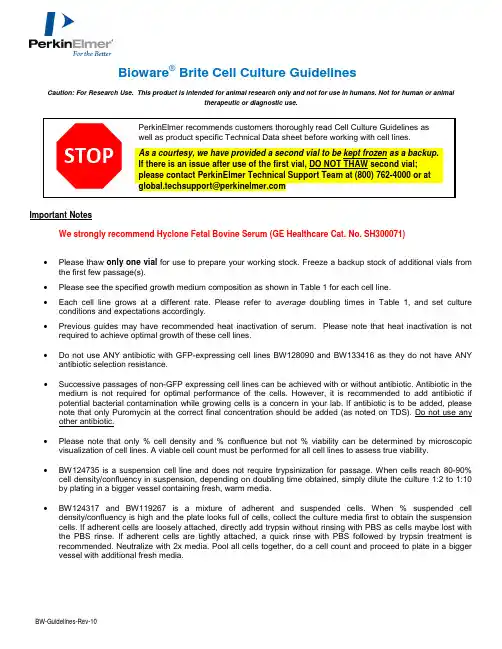
BW-Guidelines-Rev-10 Bioware® Brite Cell Culture GuidelinesCaution: For Research Use. This product is intended for animal research only and not for use in humans. Not for human or animaltherapeutic or diagnostic use.Important NotesWe strongly recommend Hyclone Fetal Bovine Serum (GE Healthcare Cat. No. SH300071) •Please thaw only one vial for use to prepare your working stock. Freeze a backup stock of additional vials from the first few passage(s).•Please see the specified growth medium composition as shown in Table 1 for each cell line.•Each cell line grows at a different rate. Please refer to average doubling times in Table 1, and set culture conditions and expectations accordingly.•Previous guides may have recommended heat inactivation of serum. Please note that heat inactivation is not required to achieve optimal growth of these cell lines.•Do not use ANY antibiotic with GFP-expressing cell lines BW128090 and BW133416 as they do not have ANY antibiotic selection resistance.•Successive passages of non-GFP expressing cell lines can be achieved with or without antibiotic. Antibiotic in the medium is not required for optimal performance of the cells. However, it is recommended to add antibiotic if potential bacterial contamination while growing cells is a concern in your lab. If antibiotic is to be added, please note that only Puromycin at the correct final concentration should be added (as noted on TDS). Do not use any other antibiotic.•Please note that only % cell density and % confluence but not % viability can be determined by microscopic visualization of cell lines. A viable cell count must be performed for all cell lines to assess true viability.•BW124735 is a suspension cell line and does not require trypsinization for passage. When cells reach 80-90% cell density/confluency in suspension, depending on doubling time obtained, simply dilute the culture 1:2 to 1:10 by plating in a bigger vessel containing fresh, warm media.•BW124317 and BW119267 is a mixture of adherent and suspended cells. When % suspended cell density/confluency is high and the plate looks full of cells, collect the culture media first to obtain the suspension cells. If adherent cells are loosely attached, directly add trypsin without rinsing with PBS as cells maybe lost with the PBS rinse. If adherent cells are tightly attached, a quick rinse with PBS followed by trypsin treatment is recommended. Neutralize with 2x media. Pool all cells together, do a cell count and proceed to plate in a bigger vessel with additional fresh media.Table 1.Product Product Description Media Composition* Average Doubling Time (DT)***BW124087 Bioware Brite 4T1-Red-FLuc RPMI+10% Hyclone FBS14BW128090 Bioware Brite 4T1-Red-FLuc-GFP** RPMI+10% Hyclone FBS 14BW124734 Bioware Brite B16F10-Red-FLuc RPMI+10% Hyclone FBS 15BW128444 Bioware Brite PC3-Red-FLuc EMEM+10% Hyclone FBS 24BW133416 Bioware Brite PC3-Red-FLuc-GFP** EMEM+10% Hyclone FBS 24BW124316 Bioware Brite NCI-H460-Red-FLuc RPMI+10% Hyclone FBS 16BW125055 Bioware Brite LNCaP-Red-FLuc RPMI+10% Hyclone FBS 60BW134280 Bioware Brite HepG2-Red-FLuc EMEM+10% Hyclone FBS 30BW124577 Bioware Brite U87MG-Red-FLuc EMEM+10% Hyclone FBS 34BW134246 Bioware Brite GL261-Red-FLuc DMEM+10% Hyclone FBS 26BW128092 Bioware Brite HT1080-Red-FLuc EMEM+10% Hyclone FBS 22BW125058 Bioware Brite BxPC3-Red-FLuc RPMI+10% Hyclone FBS 36BW124353 Bioware Brite HT-29-Red-FLuc McCoy’s 5a +10% Hyclone FBS 24BW124318 Bioware Brite HCT-116-Red-FLuc McCoy’s 5a +10% Hyclone FBS 16BW124735 Bioware Brite K562-Red-FLuc**** RPMI+10% Hyclone FBS 15BW124317 Bioware Brite Colo205-Red-FLuc**** RPMI+10% Hyclone FBS 28BW119262 Bioware Brite MCF7-Red-FLuc EMEM+10% Hyclone FBS 40BW119267 Bioware Brite LL/2-Red-FLuc**** DMEM+10% Hyclone FBS 24BW119276 Bioware Brite SKOV3-Red-FLuc McCoy’s 5a +10% Hyclone FBS 35BW119266 Bioware Brite A549-Red-FLuc RPMI+10% Hyclone FBS 22* Optional: Puromycin at a final concentration of 2 ug/mL for all cell lines listed above except for BW124087 which is at 5ug/mL. ** GFP cell lines (BW128090 and BW133416 do not have any antibiotic selection resistance).*** Doubling time is an average. Actual doubling times will vary based on culture conditions and handling.**** Suspension cell linesThawing a Frozen Cell Vial1. Thaw the vial rapidly by gentle shaking in 37°C water bath by hand. Be careful to keep the cap out of the water.Wipe vial dry.2. Spray the vial and your gloved hands with disinfectant (70% isopropyl alcohol) and wipe dry. Immediately after,open the vial in the hood and transfer contents to 4mL of warm, sterile growth media with serum but no antibiotics. Mix gently. DO NOT CENTRIFUGE.3. Count 1ml of the total cells and immediately plate the remaining cell suspension into a T25 flask. Incubate at37°C, 5-6% CO2, 100% humidity overnight.4. Next day, examine the cells under the microscope. If the cells are confluent, continue to instructions below forpassaging cell lines.If the cells are not confluent:a. Aseptically remove the media and replace with 5mL of the same media warmed to 25ºC-37ºC.b. Continue to incubate the plate(s) for an additional 1-7 days with minimal disturbance. Changemedia every 3-4 days until the cells reach 80-90% confluency; only then proceed to passage thecells.Passaging Cell Lines1. For in vivo use we recommend less than 10 in vitro passages from original vial. However, split cells at leastone time before injecting in vivo.2. When cells are approximately 80-90% confluent, passage cells to vessels with a 1:3 to 1:4 split withoutantibiotic medium.3. To passage the cells, remove media and add 5ml of sterile, room temperature 1X PBS. Gently swirl theadded PBS once over the cells and remove the PBS immediately.4. Next, Add 1ml of 0.05% sterile, warm Trypsin (approximately 1mL for T25; 2ml for T75; 4ml for T150 and 5mlfor T175) to the flask containing cells and gently swirl to allow trypsin to coat the plate. Incubate at 37°C for 1-5 mins to allow cells to dissociate from the plate.5. Examine the flask under a microscope to confirm dissociation. Neutralize with 2x medium, and gentlyre-suspend the cells by pipetting up and down 1-2 times.6. Transfer cells into a bigger flask (T75, T150, T175) at a 1:3-1:7 surface area ratio. Continue to incubate theplate(s) for 1-7 days with minimal disturbance. Change media every 3-4 days until the cells reach 80-90%confluency; only then proceed to passage the cells.Creating Cell Stocks1. When cells have reached 80% confluence, freeze aliquots for 24 hours in -80ºC in 5% DMSO/95% FBS withoutantibiotics. Transfer frozen vials to LN2 tank after 24 hours.2. We recommend that you thaw one test vial to check and confirm viability by cell counting and/or culturing.。
蝴蝶的生命周期 英语作文
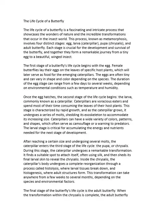
The Life Cycle of a ButterflyThe life cycle of a butterfly is a fascinating and intricate process that showcases the wonders of nature and the incredible transformations that occur in the insect world.This process,known as metamorphosis, involves four distinct stages:egg,larva(caterpillar),pupa(chrysalis),and adult butterfly.Each stage is crucial for the development and survival of the butterfly,and together they form a remarkable journey from a tiny egg to a beautiful,winged insect.The first stage of a butterfly's life cycle begins with the egg.Female butterflies lay their eggs on the leaves of specific host plants,which will later serve as food for the emerging caterpillars.The eggs are often tiny and can vary in shape and color depending on the species.The duration of the egg stage can range from a few days to several weeks,depending on environmental conditions such as temperature and humidity.Once the egg hatches,the second stage of the life cycle begins:the larva, commonly known as a caterpillar.Caterpillars are voracious eaters and spend most of their time consuming the leaves of their host plants.This stage is characterized by rapid growth,and as the caterpillar grows,it undergoes a series of molts,shedding its exoskeleton to accommodate its increasing size.Caterpillars can have a wide variety of colors,patterns, and shapes,which often serve as camouflage or a warning to predators. The larval stage is critical for accumulating the energy and nutrients needed for the next stage of development.After reaching a certain size and undergoing several molts,the caterpillar enters the third stage of the life cycle:the pupa,or chrysalis. During this stage,the caterpillar undergoes a remarkable transformation. It finds a suitable spot to attach itself,often using silk,and then sheds its final larval skin to reveal the chrysalis.Inside the chrysalis,the caterpillar's body undergoes a complete reorganization through a process called histolysis,where larval tissues break down,and histogenesis,where adult structures form.This transformation can take anywhere from a few weeks to several months,depending on the species and environmental factors.The final stage of the butterfly's life cycle is the adult butterfly.When the transformation within the chrysalis is complete,the adult butterflyemerges.This process,known as eclosion,involves the butterfly breaking free from the chrysalis and expanding its wings.Initially,the wings are soft and crumpled,but within a few hours,they harden and become strong enough for flight.The adult butterfly's primary focus is on reproduction and feeding.Butterflies are known for their striking colors and patterns,which serve various purposes,including attracting mates,camouflage,and warning predators.Adult butterflies feed on nectar from flowers,using their long proboscis to reach deep into the blossoms.They play a vital role in pollination, transferring pollen from one flower to another as they feed.This mutualistic relationship benefits both the butterflies and the plants, contributing to the health and diversity of ecosystems.In conclusion,the life cycle of a butterfly is a remarkable journey that highlights the beauty and complexity of nature.From the tiny egg to the vibrant adult butterfly,each stage of metamorphosis is a testament to the adaptability and resilience of these incredible insects.Understanding and appreciating the life cycle of butterflies not only deepens our connection to the natural world but also underscores the importance of conserving their habitats and ensuring the survival of these delicate and enchanting creatures for future generations to enjoy.。
三体123作品梗概作文

三体123作品梗概作文英文回答:The Three-Body Trilogy Synopsis.1. The Three-Body Problem:"The Three-Body Problem" by Liu Cixin is a captivating science fiction novel that blends elements of physics, politics, and human nature into a compelling narrative. The story begins during China's Cultural Revolution, where Ye Wenjie, a young astrophysicist, witnesses her father's death at the hands of Red Guards. Traumatized by the brutality of humanity, she sends a message into space, inviting alien civilizations to invade Earth and cleanse it of human corruption.Fast forward to the present day, where Wang Miao, a nanomaterials researcher, gets drawn into a mysterious virtual reality game called "Three-Body." As he delvesdeeper into the game, he discovers its connection to a secret scientific project investigating strange phenomena occurring in the universe. These phenomena, including the inexplicable suicides of prominent scientists, lead Wang Miao to uncover a shocking truth: Earth is under threat from the Trisolarans, an advanced alien civilization from the Alpha Centauri system.Facing the imminent invasion of the Trisolarans, humanity must grapple with the existential threat posed by an enemy vastly superior in technology and power. The novel explores themes of survival, morality, and the limitations of human knowledge in the face of cosmic uncertainty.2. The Dark Forest:"The Dark Forest," the second installment in the Three-Body Trilogy, delves deeper into the cosmic conflict between humanity and the Trisolarans. With Earth's existence revealed to the Trisolarans, humanity faces a dilemma: should they prepare for war or seek peaceful coexistence? The concept of the "Dark Forest" emerges as ametaphor for the universe's inherent hostility towards civilizations, where revealing one's presence invites destruction.As tensions escalate between Earth and the Trisolarans, a covert organization known as Wallfacer Project isinitiated to devise strategies for humanity's survival. Each Wallfacer is granted near-unlimited resources and authority to formulate their plans, shielded from scrutiny by the Trisolaran sophons, subatomic particles capable of observing human activities.Among the Wallfacers is Luo Ji, a disillusioned astronomer burdened with the responsibility of devising a strategy to deter the Trisolaran threat. Through his interactions with enigmatic figures like the Trisolaran collaborator Zhang Beihai and the mysterious Ye Wenjie, Luo Ji uncovers the true nature of the Trisolaran invasion and the secrets hidden within the cosmos.As the countdown to humanity's confrontation with the Trisolarans ticks away, "The Dark Forest" explores themesof trust, deception, and the precarious balance between hope and despair in the face of an unknowable universe.3. Death's End:"Death's End," the final installment in the Three-Body Trilogy, takes readers on an epic journey across time and space as humanity confronts the ultimate fate of the universe. Following the events of "The Dark Forest,"Earth's fate hangs in the balance as the Trisolaran fleet approaches, poised to annihilate humanity.With the help of advanced technology and strategic ingenuity, humanity manages to stave off immediate destruction. However, the victory proves short-lived as they soon discover a greater threat looming on the horizon: the impending heat death of the universe.As the universe hurtles towards its inevitable demise, humanity faces existential questions about the nature of life, consciousness, and the legacy they leave behind. Against the backdrop of cosmic cataclysm, individuals likeCheng Xin, a cryogenically preserved aerospace engineer,and Yun Tianming, a poet whose consciousness transcends time, grapple with their roles in shaping the destiny of humanity.Through a series of breathtaking twists and revelations, "Death's End" explores themes of sacrifice, redemption, and the eternal quest for meaning in an indifferent universe.Liu Cixin's masterful conclusion to the Three-Body Trilogy leaves readers pondering the profound mysteries ofexistence long after the final page is turned.中文回答:《三体》三部曲梗概。
USP 1132 HCP
Summary, Conclusions, and References (Guidances)
8
HCP Critical Reagents: the Antigen/Standard Design, Preparation, Characterization
Immunogen = (reformulated) standard
2
HCP Expert Panel Charter and Members
Provide stakeholders with best practices (USP chapter <1132>) for preparation and characterization of assay reagents, and development and validation of Host Cell Protein (HCP) measurement procedures (focus on immunoassays)
5
Draft Chapter Terminology, cont’d.
Process-Specific: A proprietary set of standards and antibodies used for a single product that fall in 2 major classes:
– Upstream process-specific: an assay designed from material where the upstream culture process deviates significantly from the platform. This is prior to any purification and may be applied to more than one product if these parameters are similar.
不同酶消化法提取猪原代肝细胞的效果比较

532024.4·试验研究0 引言猪圆环病毒(PCV )是Circoviridae 科Circovirus 属的一种无囊膜的单链环状DNA 病毒。
在已知的4个血清型中,PCV2为猪易感的致病性病毒[1]。
PCV2感染会诱导宿主免疫抑制引起猪圆环病毒病(PCVD ),包括断奶仔猪多系统衰竭综合征、新生仔猪先天性脑震颤、皮炎与肾病综合征、猪呼吸道病综合征、母猪繁殖障碍等,给全世界养猪业带来较大的经济损失,是世界各国的兽医与养猪业者公认的造成重大影响的猪传染病[2]。
PCV2的感染在猪生长发育的不同阶段有不同的组织嗜性。
但无论是胎儿阶段还是出生后,肝细胞都是PCV2感染和复制的靶细胞。
因此,PCV2也被视为一种能够诱导猪肝炎的病毒[3]。
且PCV2诱导的肝细胞凋亡在PCV2引发的相关病变和疾病的发病机制中具有关键性作用[4]。
因此,方便、快捷地获取大量有活性的猪肝细胞对于研究PCVD 的致病机制具有重大意义。
目前获取肝细胞常用的方法主要包括机械分离细胞法、非酶分离细胞法、离体酶消化法和酶灌流法等[5]。
因此,本试验采用简便、经济、无需特殊设备、仅需部分肝组织的离体酶消化法,比较不同酶消化分离猪原代肝细胞的效果,为一般实验室提取分离大量有活性的猪肝细胞提供参考。
1 材料与方法1.1 材料1.1.1 主要试剂新鲜猪肝组织,Hank's 平衡盐溶液(HBSS ),磷酸盐缓冲液(无菌PBS ),4%多聚甲醛(PFA ),收稿日期:2024-01-27基金项目:国家自然科学基金项目:复杂器官与组织在脾脏内的功能性再生(32230056)作者简介:周徐倩(1999-),女,汉族,浙江温州人,硕士在读,研究方向:组织工程与再生医学。
*通信作者简介:董磊(1978-),男,汉族,安徽阜阳人,博士,教授,研究方向:组织工程与再生医学、生物材料。
周徐倩,董磊.不同酶消化法提取猪原代肝细胞的效果比较[J].现代畜牧科技,2024,107(4):53-55. doi :10.19369/ki.2095-9737.2024.04.014. ZHOU Xuqian ,DONG Lei .Comparison of the Effect of Different Enzyme Digestion Methods on Extraction of Porcine Primary Hepatocytes[J].Modern Animal Husbandry Science & Technology ,2024,107(4):53-55.不同酶消化法提取猪原代肝细胞的效果比较周徐倩,董磊*(南京大学,江苏 南京 210023)摘要:猪肝细胞是猪圆环病毒的靶细胞,简单快速地提取猪原代肝细胞对于研究猪圆环病毒病的致病机制具有重要意义。
新世纪医学英语教程 生物医学Unit 3 Reading A
Unit 3 Reading AThe Sense of VisionTo master some important words and phrases in this unitTo grasp the general idea and the specific information in the textsTo learn some basic medical knowledge of the sense of vision in EnglishTeaching AimsWarm-up ActivitiesStructureDetailed StudyImportant Words and PhrasesFurther DiscussionOutlineWarm-up ActivitiesWhat are the five senses of human beings?Warm-up ActivitiesSight or vision is the top sense.How do the eyes work?Each eye is a ball of transparent jelly that works like a _______. Light enters a hole called the _____ and is focus by a ____ onto the _____-- a sheet of light sensitive cells in the back of the eye. These cells detect the color and strength of the light and send signals to the _____, which builds an image.camerapupillensretinabrainPart 1The light coming closer to my eye is piercingly bright, brighter than the white-hot ingots (I’d tonged in an Ohio steel mill 40 years ago).TranslationPara.1ophthalmolosgistophthalmo- or ophthalm-:pref. eye; eyeballE.g. ophthalmoscope: 检眼镜,眼底镜ophthalmology: 眼科学ocul(o)-: word element [L.] eyeE.g. ocularPara.1slit lamp(裂隙灯)an instrument consisting of a high-intensity light source that can be focused to shine a thin sheet of light into the eyeThe lamp facilitates an examination of the anterior segment, or frontal structures and posterior segment, of the human eye, which includes the eyelid, sclera, conjunctiva, iris, natural crystalline lens, and cornea.Para.1minister to sb./sth.to give help to someone who needs it, especially someone who is sick or old:E.g. She spent much time ministering to the sick.E.g. ministering to the needs of other peoplePara.2glaucomaMajor cause?excessively high intraocular pressure (IOP).This increased pressure within the eye, if untreated can lead to optic nerve damage resulting in progressive, permanent vision loss, starting with unnoticeable blind spots at the edges of the field of vision, progressing to tunnel vision, and then to blindness.glauco-: prefix gray or silverglaucomatous: adj.Para.2Para.3Vision: Our Most Important SenseTo be blind is to lose access to the most significant part of our perceptual world.失明也就失去了我们与感性世界中最有意义的那一部份的联系。
缺血性脑卒中后小胶质细胞吞噬作用的研究进展
Journal of China Pharmaceutical University 2023,54(4):399 - 409学 报缺血性脑卒中后小胶质细胞吞噬作用的研究进展汪恒,孙浩,廖红*(中国药科大学新药筛选中心江苏省药效研究与评价服务中心,南京 210009)摘 要 缺血性脑卒中是影响人类健康的重大疾病,目前有关它的病理机制并未完全阐明。
小胶质细胞是中枢神经系统中重要的免疫细胞。
缺血性脑卒中后,大量小胶质细胞激活,并向损伤区域迁移聚集,吞噬坏死的细胞或碎片,释放炎症因子或营养因子,参与了缺血性脑卒中的病理过程。
其中小胶质细胞的吞噬作用在脑缺血损伤以及康复中发挥重要的作用。
本文总结小胶质细胞吞噬作用的分子机制,并综述小胶质细胞吞噬作用在缺血性脑卒中的研究进展,探讨小胶质细胞吞噬作用在脑缺血损伤和康复中的多样性和复杂性,旨在为缺血性脑卒中的治疗和药物研发提供新的思路。
关键词缺血性脑卒中;小胶质细胞;吞噬作用中图分类号R965 文献标志码 A 文章编号1000 -5048(2023)04 -0399 -11doi:10.11665/j.issn.1000 -5048.2023022106引用本文汪恒,孙浩,廖红.缺血性脑卒中后小胶质细胞吞噬作用的研究进展[J].中国药科大学学报,2023,54(4):399–409.Cite this article as:WANG Heng,SUN Hao,LIAO Hong. Reaserch progress of microglial phagocytosis in ischemic stroke[J].J China Pharm Univ,2023,54(4):399–409.Reaserch progress of microglial phagocytosis in ischemic stroke WANG Heng, SUN Hao, LIAO Hong*New Drug Screening Center, Jiangsu Center for Pharmacodynamics Research and Evaluation, China Pharmaceutical University, Nanjing 210009, ChinaAbstract Ischemic stroke is a major disease affecting human health, and its pathological mechanism has not been fully elucidated. Microglia are important immune cells in the central nervous system, and participate in the pathological process of ischemic stroke.Following an ischemic stroke, a surge in activated microglia occurs, migrating and congregating within the afflicted regions.These microglia engulf deceased cells or fragments, releas⁃ing inflammatory or nutritive factors, thereby participating in the pathogenesis of ischemic stroke.The phagocyto⁃sis of microglia plays an important role in cerebral ischemic injury and rehabilitation. This article summarizes the molecular mechanism of microglial phagocytosis and reviews the research progress of microglial phagocytosis in ischemic stroke, and discusses the diversity and complexity of microglial phagocytosis in cerebral ischemic injury and rehabilitation, so as to provide new ideas for the treatment and drug development of ischemic stroke. Key words ischemic stroke; microglia; phagocytosisThis study was supported by the National Natural Science Foundation of China (No.82073831)缺血性脑卒中(ischemia)是全球范围内致残率和致死率最高的疾病之一[1]。
最后一片叶子琼珊知道真相后续写英语作文
最后一片叶子琼珊知道真相后续写英语作文As the last leaf fell, Qiong Shan finally knew the truth. She had been searching for answers for so long, and now, as she stood in the quiet forest, everything became clear. The last leaf was a symbol of hope, of perseverance, and of resilience. It was a reminder that even in the darkest of times, there is always a glimmer of light.Qiong Shan had been on a journey of self-discovery,trying to find her place in the world. She had faced many challenges and obstacles along the way, but she never gave up. She always believed that there was something greater waiting for her, and now she understood what that was.The truth was that the last leaf was a representation of her own inner strength. It was a reflection of her unwavering determination and her refusal to be defeated. It was a testament to her resilience and her ability to overcome even the most difficult of circumstances.As she stood in the forest, Qiong Shan felt a sense of peace wash over her. She knew that she had finally foundwhat she had been searching for. The last leaf had shown her that she was capable of achieving anything she set her mind to, and that no challenge was too great for her to overcome.With a renewed sense of purpose, Qiong Shan set out to continue her journey. She knew that there would still be obstacles and challenges ahead, but she was no longer afraid. She had the strength and resilience of the last leaf, and she was ready to face whatever came her way.中文:当最后一片叶子落下时,琼珊终于知道了真相。
Deep-vein-thrombosis深静脉血栓
Causes
German pathologist Rudolf Virchow postulated the interplay of three processes resulting in venous thrombosis, now known as Virchow's triad:
Causes
√ Swelling…
√ Warmth…
√ Redness or discoloration…
√ Distention of surface veins…
Signs and symptoms
Deep vein thrombosis
In most suspected cases, DVT is ruled out after evaluation, and symptoms are more often due to other causes, such as cellulitis, Baker's cyst, musculoskeletal injury, or lymph edema.
➢ The majority of venous thrombi occur in either the superficial or deep veins of the leg.
➢ A DVT is stationary clotting blood adhered to the deep vein of the pelvis or an extremity and usually occurs in the calf or thigh.
- 1、下载文档前请自行甄别文档内容的完整性,平台不提供额外的编辑、内容补充、找答案等附加服务。
- 2、"仅部分预览"的文档,不可在线预览部分如存在完整性等问题,可反馈申请退款(可完整预览的文档不适用该条件!)。
- 3、如文档侵犯您的权益,请联系客服反馈,我们会尽快为您处理(人工客服工作时间:9:00-18:30)。
Chapter 3: Connective TissueObjectives●List the structural and functional characteristics of connective tissue that distinguish it fromthe epithelial tissue.●Know the functions carried out by connective tissues.●Know the 3 fundamental components found in all connective tissues.●Know the biochemical composition and the sites of synthesis of the components ofextracellular matrix and how they associate with one another.●Know the structure and function of the various cell types found in connective tissue.●Compare the different types of connective tissues in terms of the types, relative amounts, andarrangement of cells, fibers, and ground substance.●Relate the composition of the various connective tissue types to their specific functions. OverviewI. GENERAL FEATURES OF CONNECTIVE TISSUESA. Functions:The functions of connective tissues, determined chiefly by their mechanical properties, include the binding together, compartmentalization, support, and physical and immunologic protection of other tissues and organs, as well as storage.B. Types: The connective tissues include loose and dense collagenous connective tissue (connective tissue proper), reticular connective tissue, elastic connective tissue, and mucous connective tissue, adipose tissue, cartilage, bone and blood.C. Fundamental Components: The types of connective tissue differ in microscopic appearance, but all consist of cells, and extracellular matrix including fibers, and ground substance. Connective tissue types and subtypes are classified according to the amounts, types, and proportions of these components.D. Extracellular Matrix:The fibers and ground substance constitute the extracellular matrix. Connective tissues contain abundant matrix, which, largely determines their mechanical properties. The fibers are of 3 types, collagen, fibers and elastic fibers. The ground substance, in which the fibers and cells are embedded, is composed mainly of glycosaminoglycans (GAGs) dissolved in t issue fluid. Matrix viscosity and rigidity are determined by the amount and types of GAGs, the association of GAGs with core proteins to form proteoglycans, GAG-fiber associations, and GAG-GAG associations. Components of the fibers and ground substance are synthesized and secreted by connective tissue cells. (mostly by fibroblasts), and the fibers are assembled in the extracellular space.E. Embryonic Origin: All connective tissue cell types derive from cells of the embryonic mesenchyme. Mesenchyme derives from embryonic mesoderm, except head mesenchyme, which de rives from the neural crest (mesectoderm).Ⅱ. COMPONENTS OF CONNECTIVE TISSUEA. C ollagen Fibers:The protein collagen is the most abundant in the body. There are many collagen types, some of which form fibers. Collagen fibers often collect to form fiber bundles ranging from 0.5-15 µm in diameter. Collagen fibers include:Type I collagen, type Ⅱcollagen type III col lagen, type IV collagen, type V collagen and type X collagen.●Histologic appearance, mechanical properties and location: Under Light microscopy (LM) collagen occurring in large or small bundles of fibrils or as individual fibrils exhibits acidophitic staining properties in H&E-stained sections. Under electron microscopy (EM) all collagen fibrils and fibers have stripes at intervals of 64 nm along their length. Collagen fiber s,most important mechanical property is their tensile strength, which is(weight for weight) greater than that of steel. Collagen fibers are .found in all connective tissues and in the reticular laminae of certain basement membranes.B. Reticular FIbers: Reticular fibers are chemically identical to collagen, but they are thinner(0.1-1.5 µm) and form delicate silver-staining networks instead of thick bundles. The networks serve as supportive lattices that allow motile cells to move about in loosely arranged tissues such as hematopoietic tissues. Reticular fibers are composed mainly of type Ill collagen and some glycoproteins.C. Elastic Fibers: Elastic fibers consist of an amorphous albuminoid protein called el a stin and numerous proteinaceous microfibrils that become embedded in the elastin. They range in diameter from 0.1-10 µm.●Histologic appearance, mechanical properties and location: Elastin contains few charged amino acids, so it stains poorly with standard ionic dyes. Special stains, such as Verhoeff's stain or Weigerl's resorcinfuchsin stain, are used in light microscopic preparations. In EM preparations, both amorphous elastin and microfibrils can be visualized. Elastic fibers are extremely pliable and elastic. They can be stretched to 150% of their length without breaking and then return to their original length.Elastic fibers are found where their mechanical properties are necessary to allow tissues to stretch or expand and then return to their original shape, e.g. in arterial walls, interalveolar septa, bronchi and bronchioles of the lungs, vocal ligaments, and ligamenta flava of the vertebral column.D. Ground Substance: The ground substance consists mostly of glycoconjugates of 2 classes, proteoglycans and gtycoproteins. Tissue fluids and salts are also present.●Proteoglycans are composed of a core protein to which GAGs are attached. The GAGs of proteoglycans are straight-chain polymers of repeating sugar heterodimers made up of hexosamine (glucosamine or galactosamine) and uronic acid (glucuronic or iduronic acid). Five major classes of GAGs, differing in their sugars, exist in connective tissues: hyaluronic acid (which does not form proteoglycans), chondroitin sulfate,dermatan sulfate, keratan sulfate,and heparan sulfate.●Glyeoproteins are proteins to which shorter, branched oligosaccharide chains are covalently bound. Glycoproteins of ground substance are much smaller than proteoglycans. Examples:fibronectin,which mediates the attachment of cells to the extracellular matrix; laminin,a component of basal laminae that mediates attachment of epithelial cells; and chondronectin, a component of cartilage matrix that mediates the attachment chondrocytes to their matrix.E.Cells:a. Fibroblasts These are the predominant connective tissue cells and are ubiquitous in connective tissue proper. They synthesize, secrete, and maintain all the major components of the extracenular matrix. Structurally, fibroblasts are of 2 types, one of which resembles mesenchymal cells. This type is stellate with long cytoplasmic processes and a large, ovoid, pale-staining nucleus. The cytoplasm contains abundant rough endoplasmic reticulum and Golgi complexes, and this cell type is important in the production of collagen and other matrix components. Cells of the second type are less active and are sometimes termed fibrocytes, because they are believed to be more mature. Fibrocytes are smaller and spindle-shaped, with a dark, elongate nucleus and fewer cytoplasmic organelles. They can revert to the fibroblast state and participate in tissue repair.b. Mast cells These derive from bone marrow precursors and are characterized by abundant basophilic cytoplasmic granules, which are metachromatic. At the EM level, these granules appear as electron-dense granules. Other features of mast cells at this level are many small plasma membrane folds and a well-developed Golgi complex. The granules, which usually obscure the small central nucleus, contain heparin, histamine, and eosinophil chemotactic factor of anaphylaxis (ECF-A). Mast cells also can release leukotrienes or slow-reacting substance of anaphylaxis, so called SRS-A. In addition, mast cells have surface receptors for IgE antibodies and are involved in allergic or immediate hypersensitive reaction.c. Macrophages These are large, stellate cells derived from cells of the blood monocyte lineage that ihfillrate connective tissue and develop into phagocytes. Resident macrophages can proliferate and form additional macrophages. Dye particles injected into the body are engulfed by these cells and accumulate in cytoplasmic granules. Macrophages contain many lysosomes, which aid in digesting phagocytosed materials, and a well-developed Golgi complex. They help maintain the integrity of connective tissues by removing foreign substances and cellular debris, and they participate in the immune response by presenting phagocytosed antigens to lymphocytes. To remove large foreign objects such as splinters, macrophages may fuse to form multinuclear giant cells. Monocyte-derived phagocytes, which together constitute the mononuelear phagocyte system, include the macrophages (lymphoid organs, lungs, serous cavities, and connective tissue), as well as Kupffer cells(liver), osteoclasts (bone)and microgIial cells(central nervous system).d. Plasma cells These differentiate from antigen-stimulated B lymphocytes and are the primary producers of circulating antibodies. They are sparsely distributed throughout the body but abundant in areas susceptible to penetration by bacteria. Plasma cells are large and ovoid, with an eccentric nucleus and abundant rough endoplasmic reticulum. The characteristic “clock face” of the nucleus results from a large, central nucleolus and several large heterochromatin clumps regularly spaced inside the nuclear envelope. These cells usually exhibit a clear juxtanuclear area (cytocenter) containing a well- developed Golgi complex and eentrioles.e. Adipose cells Adipose ceils, or adipocytes,are mesenchymal derivatives specialized as storage depots for llpidse. Mesenchtymal cells These are the precursors of most cells indigenous to connective tissues, including fibrobiasts and adipose cells. Embryonic mesenchyme consists of a loose network ofstellate mesenchymal cells and abundant intercellular fluid. Some mesenchymal cells remain undifferentiated in adult connective tissue and constitute a reserve population of stem cells called adventitial cells, which are difficult to distinguish from some fibroblasts.f. Other blood-derived connective tissue cells Many wandering cell types originate in the bone marrow and are carried to connective tissue by the blood am derived cells found in connective tissues include the leukocytes (white blood cells, i.e. lymphocytes, monocytes,neutrophils, eosinophils,and basophils) whi the immune response and are described in detail in Chapters 12-14.B. Edema: The water in tissue fluid comes from the arterial ends of capillaries in capillary beds, forced out by hydrostatic pressure (arterial pressure). Loss of fluid to the tissues increases the blood solute concentration at the venous end of the capillary; this increased colloid osmotic pressare, along with the lower hydrostatic pressure at the venous end, draws most ofthe tost fluid back into the blood. Any excess fluid remaining in the tissue is normally drained away by lymphatic capillaries, so that there is no net change in the amount of blood or tissue fluid. Edema, or accumulation of excess tissue fluid, accompanies pathological conditions that cause:1. Increased hydrostatic pressure in capillaries by obstructing venous biood flow (eg, congestiveheart failure);2.Decreased colloid osmotic pressure in the blood caused by lack of blood proteins (eg,starvation);3. Increased hydrostatic pressure in the tissue caused by blockage of lymphatic drainage byparasites or tumor cells; ,and4.Increased colloid osmotic paressure in the tissue caused by eacessive accumtfiation ofglycosaminoglycans in the matrix. Edema caused by this camdrtion is called myxedema. Study Focusing QuestionsMono-choice questions3-1. Which one of the following statements regarding collagen is true?(A)It is composed of tropocollagen.(B)Reticular fibers are composed of type II collagen.(C)It is synthesized mostly by mast cells.(D)Elastic fibers are composed of type IV collagen.3-2. Dense regular connective tissue is present in(A)Capsules of organs(B)Basement membrane(C)Tendons(D)Skin3-3. Of the following cell types found in connective tissue, which is most often present along capillaries and resembles fibroblasts?(A)Plasma cell(B)Lymphocyte(C)Macrophage(D)Pericyte3-4. Which one of the following cells arises from monocytes?(A)Plasma cells(B)Fibroblasts(C)Lymphocytes(D)Macrophages3-5. Which of the following cells located in the connective tissue arises from myeloid stem cells?(A)Pericytes(B)Eosinophils(C)Fibroblasts(D)Osteoblasts(E)Adipocytes3-6. Which of the following cells is responsible for anaphylactic shock?(A)Fibroblasts(B)Eosinophils(C)Pericytes(D)Mast cells(E)Macrophages3-7. Which one of the following statements regarding proteoglycans is true?(A)They consist of a core of fibrous protein covalently bound to glycoproteins.(B)They are attached to ribonucleic acid.(C)They are binding sites for deoxyribonucleic acid (DNA).(D)They are composed of a protein core to which glycosaminoglycans are attached. 3-8. Which one of the following statements concerning loose connective tissue is true?(A)It is less abundant than dense connective tissue.(B)It has a lower proportion of cells to fibers than does dense connective tissue.(C)It acts as a medium for exchange of nutrients and wastes between the blood andtissues.(D)It provides structural support for organs.(E)It consists of fibers in which various types of cells are embedded.3-9. Which of the following cells arise from activated B lymphocytes?(A)Macrophages(B)Eosinophils(C)Plasma cells(D)Mast cells(E)Adipose cells3-10. Which of the following cells release histamine and leukotriene C?(A)Macrophages(B)Eosinophils(C)Plasma cells(D)Mast cells(E)Adipose cells3-11. Which of the following cells function in immune response and wound healing?(A)Macrophages(B)Eosinophils(C)Plasma cells(D)Mast cells(E)Adipose cells3-12. Which of the following cells increase in number in hyperplastic obesity?(A)Macrophages(B)Eosinophils(C)Plasma cells(D)Mast cells(E)Adipose cells3-13. Identify the cell that is related to the cell-mediated immune response.(A)Mast cell(B)Adipocyte(C)Fibroblast(D)T lymphocyte(E)Macrophage3-14. Identify the cell that mediates immediate (type I) hypersensitivity reactions.(A)Mast cell(B)Adipocyte(C)Fibroblast(D)T lymphocyte(E)Macrophage3-15. Which cell synthesizes and stores fat?(A)Mast cell(B)Adipocyte(C)Fibroblast(D)T lymphocyte(E)Macrophage3-16. Which of the following is the most abundant cell in connective tissue?(A)Mast cell(B)Adipocyte(C)Fibroblast(D)T lymphocyte(E)MacrophageMultiple-choice questionsFor questions 3-17 through 3- select the right answers.3-17. The ground substance of connective tissue includes all of the following components:A. Hyaluronic acidB. ProteoglycansC. FibronectinD. Collagen fibersE. Glycosaminoglycans3-18.The cell type mainly responsible for producing and maintaining all the components of connective tissue extracellular matrix is the:A, Mesothelial cellB. FibroblastC, Mast cellD. LymphocyteE. Macrophage3-19. The 3 basic components of all types of connective tissue are:A. CellsB, Arteries, veins, and capillariesC. FibersD. Type ll collagen, hyaluronate, and f'ibronectinE, Ground substance3-20. All of the following substances are glycosammoglycans:A. Keratan sulfate ~B, Hyaluronic acidC. Chondroitin sulfateD. Heparan sulfateE. FibronectinTerm explanations3-21.Mast cell3-22.Tissue fluidQuestions for detailed explanations3-23. Discuss the general functions of connective tissues3-24. Which connective tissue cells specifically contribute to the repair of wounds by removing and replacing damaged tissue?Answers and ExplanationsMono-choice questions3-1. A is the correct answer. Collagen is composed of closely packed tropocollagen molecules. Reticular fibers are composed of type III collagen, whereas elastic fibers are composed of elastin microfibrils rather than collagen. Fibrocytes are inactive nonsecreting fibroblasts that synthesize the procollagen molecules. <A, correct>3-2. C is the correct answer. Tendons are composed of dense regular connective tissue containing collagen fibers arranged in a uniform parallel fashion. <C, correct>3-3. D is the correct answer. Pericytes are pluripotential cells that resemble fibroblasts (although they are smaller) and are located adjacent to capillaries. <D, correct>3-4. D is the correct answer. Monocytes leave the bloodstream and migrate into the connective tissue, where they mature into functional macrophages.3-5. B is the correct answer. Eosinophils arise from myeloid stem cells during hemopoiesis and migrate to sites of inflammation within the connective tissue.Pericytes, fibroblasts, osteoblasts, and adipocytes arise from undifferentiated mesenchymal cells.3-6. D is the correct answer. After first exposure to an allergen, plasma cells make immunoglobulin E (IgE) antibodies that bind to Fc (Fc?weirde?RI) receptors on mast cells (and basophils), thus sensitizing them. At the second exposure, the allergen binds to IgE, initiating degranulation of mast cells, thus releasing several mediators that give rise to type I hypersensitivity reaction.3-7. D is the correct answer. Proteoglycans consist of a protein core to which glycosaminoglycans are attached.3-8. C is the correct answer. Both loose and dense connective tissue are composed of three elements: an amorphous ground substance, fibers, and various types of cells.The amorphous ground substance of loose connective tissue is the medium of exchange between the connective tissue cells and the bloodstream.3-9. C is the correct answer. Plasma cells arise from activated B lymphocytes.3-10. D is the correct answer. Mast cells release histamine, leukotriene C, and other mediators.3-11. A is the correct answer. Macrophages function as phagocytes and also secrete substances that assist in the immune response and in wound healing.3-12. E is the correct answer. The number of adipose cells increases in hyperplastic obesity.3-13. D is the correct answer. T lymphocytes are responsible for the cell-mediated immune response.3-14. A is the correct answer. Mast cells are responsible for immediate (type I) hypersensitivity reactions.3-15. B is the correct answer. Adipocytes are responsible for the synthesis and storage of fat.3-16. C is the correct answer. Fibroblasts are the most abundant cell type in connective tissues.Multiple-choice questions3-17. ABCE3-18. B3-19. ACE3-20. ABCDTerm explanations3-21. Mast cells These derive from bone marrow precursors and are characterized by abundant basophilic cytoplasmic granules, which are metachromatic. At the EM level, these granules appear as electron-dense granules. Other features of mast cells at this level are many small plasma membrane folds and a well-developed Golgi complex. The granules, which usually obscure the small central nucleus, contain heparin, histamine, and eosinophilic chemotactic factor of anaphylaxis (ECF-A). Mast cells also can release leukotrienes or slow-reacting substance of anaphylaxis, so called SRS-A. In addition, mast cells have surface receptors for IgE antibodies and are involved in allergic or immediate hypersensitive reaction.3-22. Tissue fluid: The water with small molecules coming from the arterial ends of capillaries in capillary beds, forced out by hydrostatic pressure(arterial pressure) enters the surrounding tissues to form tissue fluid. Loss of fluid to the tissues increases the blood solute concentration at the venous end of the capillary; this increased colloid osmotic pressure,along with the lower hydrostatic pressure at the venous end draws most of the lost fluid back into the blood. Any excess fluid remaining in the tissue is normally drained away by lymphatic capillaries, so that there is no net change in the amount of blood or tissue fluid.Questions for detailed explanations3-23. Discuss the general functions of connective tissues●Support Structural support is the major function of connective tissue, which forms theframework upon which all other body tissues are assembled. Its physical properties allow it to bind, to fill spaces, and to separate functional units of other tissues and organs. It thus maintains functional units in their proper 3-dirnensional relationships, allowing maintenance and coordination of all body functions.●Defense a. Physical The viscosity of the extracellular matrix, due largely to hyaluronicacid, slows the progress of many bacteria and foreign particles. Sheets of tightly packed and often interwoven collagen fibers, as in organ capsules, help to confine local infections.However, some bacteria secrete enzymes that break down matrix components.; b.Immunologic Foreign bodies that successfully penetrate epithelia are intercepted by immunoresponsive cells that inhabit the underlying connective tissue. These cells not only activate a local immune response (inflammation) but mobilize the immune system to supply additional cells via the bloodstream. Recruited cells migrate through capillary and venule walls into the connective tissue.●Repair Rapidly closing any breaches in the body's protective barriers is an importantfunction of connective tissue. Injury stimulates invasion of the site by immunocompetent cells and the proliferation of fibroblasts. Macrophages remove clotted blood, damaged tissue, and foreign material, while fibroblasts secrete intracellular matrix materials to fill the breach.Rapidly formed collagenous matrices that close wounds are often less well organized than the original tissues and form scars. Small sears may eventually be completely remodeled; larger scars are only partially remodeled.●Storage Reserves of water and electrolytes, especially sodium, are stored in theextracellular matrix, owing to the high polyanionic charge density of glycosaminoglycans.Energy reserves in the form of lipids are stored in adipocytes.●Transport Except in the central nervous system, most blood and lymphatic vessels aresurrounded by loose connective tissue, which is thus a crossroads for transporting substances to and from other tissues.3-24. Which connective tissue cells specifically contribute to the repair of wounds by removing and replacing damaged tissue?Rapidly closing any breaches in the body's protective barriers is an important function of connective tissue. Injury stimulates invasion of the site by immunocompetent cells and the proliferation of fibroblasts. Macrophages remove clotted blood, damaged tissue, and foreign material, while fibroblasts secrete intracellular matrix materials to fill the breach. Rapidly formed collagenous matrices that close wounds are often less well organized than the original tissues and form scars. Small sears may eventually be completely remodeled; larger scars are only partially remodeled.(Prof. Chaohong Li)。