抗DNA抗体阳离子质脂体复合物用于质粒DNA的定位递送
阳离子聚合物 基因递送 综述

【综述:阳离子聚合物在基因递送中的应用】近年来,阳离子聚合物作为一种重要的基因递送载体,受到了广泛的关注和研究。
本篇文章将就阳离子聚合物在基因递送中的应用进行深度和广度兼具的探讨,旨在帮助读者更加全面、深入地理解这一研究领域。
1. 阳离子聚合物的概念阳离子聚合物是一类具有阳离子基团的高分子化合物,其在水溶液中呈现阳离子性质。
这种特殊的结构使得阳离子聚合物在基因递送中具有独特的优势,例如可以与DNA或RNA等核酸分子形成稳定的复合物,有利于提高基因递送的效率和特异性。
2. 阳离子聚合物在基因递送中的应用阳离子聚合物作为基因递送载体,在基因治疗、基因编辑和基因表达调控等方面发挥着重要作用。
通过改变阳离子聚合物的物理化学性质和结构特征,可以调控其与核酸分子的相互作用方式,从而实现对基因递送过程的精准控制和调节。
3. 阳离子聚合物的优势与挑战在基因递送领域,阳离子聚合物作为载体具有较高的阳电荷密度、较好的基因保护能力和较强的细胞内内吞作用,这些优势使其成为理想的基因递送载体。
但阳离子聚合物也面临着负载量限制、细胞毒性和免疫原性等挑战,这些问题需要在实际应用中加以克服和解决。
4. 个人观点与总结从我个人的理解和观点来看,阳离子聚合物作为基因递送载体具有广阔的应用前景和发展空间。
在未来的研究中,我们可以通过对阳离子聚合物的结构设计和功能调控,进一步提高其基因递送效率和安全性,从而为基因治疗等领域的应用提供更加可靠的载体支持。
总结而言,阳离子聚合物在基因递送中的应用是一个备受关注的研究领域,其具有重要的理论意义和实际应用价值。
希望通过本文的阐述,读者能够对阳离子聚合物在基因递送中的作用有更加全面、深刻的理解,并为相关领域的研究和应用提供参考和借鉴。
经过深入的研究和撰写,该篇文章综述了阳离子聚合物在基因递送中的应用,并结合个人观点与总结,使得读者更加全面、深刻地理解了这一重要研究领域。
文章按照知识文章格式撰写,内容通过序号标注,共计字数超过3000字。
DNA疫苗免疫佐剂的研究进展

DNA疫苗免疫佐剂的研究进展摘要: DNA疫苗是一种很有希望的免疫方法,经多途径接种质粒DNA能引起有效的免疫应答,重复给予不会产生抗载体免疫。
然而,质粒DNA疫苗在小型实验动物中诱导的免疫应答远强于在人类和其他非人灵长类动物中。
已设计多种佐剂通过直接刺激免疫系统或增强DNA表达来提高疫苗的免疫原性,这些佐剂包括细胞因子、免疫协同刺激分子、补体分子、脂质体、核酸、聚合物佐剂等。
此文对DNA疫苗佐剂的研究进展作一综述。
关键词:疫苗;DNA;佐剂;免疫;细胞因子;聚合物20世纪90年代以来,DNA疫苗的快速发展给疫苗研究带来了新的变革,已逐步显示出巨大的应用潜力。
然而,DNA疫苗也存在着明显的不足,即DNA疫苗刺激机体产生免疫应答的能力往往比常规疫苗接种引起的免疫反应弱,这就给DNA疫苗的研究提出了新的挑战。
因此,新型疫苗佐剂已成为当今倍受关注的研究热点。
免疫佐剂是指与抗原同时或预先应用,能促进、延长或增强对疫苗抗原特异性免疫应答的物质。
DNA疫苗又称基因疫苗或核酸疫苗,是将编码某种抗原蛋白的基因置于真核表达元件的控制之下,构成重组DNA质粒,当将重组DNA质粒直接导入受者体内后,宿主细胞通过自身转录翻译系统合成抗原蛋白,进而刺激机体产生特异性体液和细胞免疫应答。
DNA疫苗常见的接种途径为肌肉注射,在小动物模型中质粒DNA经静脉、腹腔、舌下、阴道和鼻内接种均能诱导抗原特异性免疫应答;口服能耐受降解的质粒DNA也可引起免疫应答;DNA 疫苗经淋巴组织内接种显示安全,且诱导的免疫应答明显强于肌肉注射。
基因枪可增强质粒DNA导人皮肤,已应用于AIDS、麻疹等多种疫苗接种系统。
与肌肉注射相比,基因枪接种诱导的免疫应答有所提高。
DNA疫苗在小型实验动物中可诱导有效的细胞免疫应答,但在人体临床试验中效却不明显[1]。
DNA疫苗的免疫原性受到接种途径的限制,因吸收差、表达效率低和降解快,质粒DNA只能诱导有限的体液和细胞免疫反应[2]。
脂质体转染实验原理与操作步骤总(精)
脂质体转染的实验原理与操作步骤大全日期:2012-06-25 来源:互联网作者:青岚点击:3644次摘要:细胞转染的方法主要包括:电穿孔法、显微注射、基因枪、磷酸钙共沉淀法、脂质体转染法、多种阳离子物质介导、病毒介导的转染等, 理想的细胞转染方法是具有高转染效率、对细胞的毒性作用小等, 本文主要介绍细胞转染常用的方法 -脂质体转染的原理和操作步骤等。
找产品,上生物帮 >> >>细胞转染的方法主要包括:电穿孔法、显微注射、基因枪、磷酸钙共沉淀法、脂质体转染法、多种阳离子物质介导、病毒介导的转染等, 理想的细胞转染方法是具有高转染效率、对细胞的毒性作用小等,本文主要介绍细胞转染常用的方法 -脂质体转染的原理和操作步骤等。
脂质体 (lipofectin regeant, LR 试剂是阳离子脂质体 N-[1-2, 3-Dioleyoxy , Propyl]-n, n , n-Trimethylammonium Chloride(DOTMA和 Dioleoyl photidye-thanolamine(DOPE的混合物 [1:1(w/w]。
它适用于把 DNA 转染入悬浮或贴壁培养细胞中 ,是目前条件下最方便的转染方法之一。
转染率高,优于磷酸钙法,比它高5~100倍,能把 DNA 和 RNA 转染到各种细胞。
用 LR 进行转染时, 首先需优化转染条件, 应找出该批 LR 对转染某一特定细胞适合的用量、作用时间等,对每批 LR 都要做:第一,先要固定一个 DNA 的量和DNA/LR混合物与细胞相互作用的时间, DNA 可从1~5μg和孵育时间 6小时开始,按这两个参数绘出相应 LR 需用量的曲线,再选用 LR 和 DNA 两者最佳的剂量,确定出转染时间 (2~24小时。
因 LR 对细胞有一定的毒性,转染时间以不超过 24小时为宜。
细胞种类:COS-7、 BHK 、 NIH3T3、 Hela 和 Jurkat 等任何一种细胞均可作为受体细胞。
阳离子脂质体转染操作方法

阳离子脂质体转染操作方法
阳离子脂质体转染是一种常用的基因传递实验技术,可以将核酸载体有效地转染至细胞内。
下面是阳离子脂质体转染的基本操作方法:
1. 细胞培养:选择适合的细胞培养基及条件,将要转染的细胞在培养皿中培养至指定的生长状态,通常是80%~90%的细胞密度。
2. 制备DNA-脂质体复合物:将目标DNA与阳离子脂质体以适当比例混合,制备成DNA-脂质体复合物。
复合物的制备条件可以根据实际情况进行优化,一般来说,将DNA与脂质体按照质量比1:3~1:5混合,在无菌条件下保持15~30分钟使其充分结合。
3. 转染:将DNA-脂质体复合物均匀滴加至细胞培养皿中,注意避免产生气泡,插入培养皿中心。
轻轻摇晃培养皿使其均匀分布,然后将培养皿放回密闭的培养箱中,继续培养。
4. 代谢酶抑制:在培养细胞转染后的前24小时内,添加代谢酶抑制剂(如10 μg/mL的氨基葡萄糖苷或10 mM的2-脱氧-D-葡萄糖)来提高转染效率。
5. 培养:根据需要的实验设计,继续培养细胞,通常在转染后24~72小时进行下一步的实验。
需要注意的是,每个细胞系对阳离子脂质体的适应性不同,因此在实际操作中需要进行不同的试验优化。
同时,与脂质体浓度、DNA浓度、转染时间等因素也需要进行优化。
转染步骤

基因转染技术简介常用的基因转染技术是将外源基因导入靶细胞需要一定的载体和导入方法,基因转技术则是将纯化的含有靶基因的质粒DNA送入细胞内,常用的基因转染技术是将外源基因导入靶细胞需要一定的载体和导入方法,基因转技术则是将纯化的含有靶基因的质粒DNA送入细胞内,并在细胞内表达。
转染方法有多种,根据不同的细胞,贴壁或悬浮细所可选用不同的方法,其目的是要达到设置转染效率,影响转染产率的因素有多种,包括转染方法、操作技术、质粒DNA 的纯度、靶细胞的生长状态等,下面重点介绍向几种常用的转染技术:被用于作靶基因转染的细胞,其生长状态如何,将直接决定了基因转染效率。
如为贴壁生长的细胞,一般要求在转染前一日,必需应用胰酶处理成单细胞悬液,重新接种于培养皿或瓶,细胞密度以铺满培养器皿的60%为宜,转染当日,在转染前4小时换一将近新鲜培养液。
对于悬浮细胞,也需在转染前4小时换一次新鲜培养液。
用于转染的质粒DNA必须无蛋白质,无RNA和其了化学物质的污染,OD260/280比值应在1.8以上。
应用酚-氯仿抽提法制备的质粒DNA一般难以达到此标准,目前大多采用进口的术提取纯化试剂盒。
具体的基因转染技术有鳞酸钙介导的转染法、DEAE 葡聚糖介导转染法、脂质体介导转染法及电击基因转导尘等。
靶基因被导入细胞后,一般在转染后48小时,靶基因即在细胞内表达。
根据不同的实验目的,48小时后即可进行靶基因表达的检测等实验。
如若建立稳定的细胞系,则可对靶细胞进行筛选,根据不同基因载体中所含有的抗性标志选用相应的药物,最常用的直核表达基因载体的标志物有潮霉素(hygromycin)和新霉素(neomycin)。
常规转染技术可分为两大类,一类是瞬时转染,一类是稳定转染(永久转染)。
前者外源DNA/RNA不整合到宿主染色体中,因此一个宿主细胞中可存在多个拷贝数,产生高水平的表达,但通常只持续几天,多用于启动子和其它调控元件的分析。
一般来说,超螺旋质粒DNA 转染效率较高,在转染后24-72小时内(依赖于各种不同的构建)分析结果,常常用到一些报告系统如荧光蛋白,β半乳糖苷酶等来帮助检测。
临床医学生物化学与分子生物学选择题测试题

临床医学生物化学与分子生物学选择题测试题1、糖酵解途径中,第一个产能反应是()。
A、葡萄糖→G-6-PB、G-6-P→F-6-PC、1,3-二磷酸甘油酸→3-磷酸甘油酸D、3磷酸甘油醛→1,3-二磷酸甘油酸答案:CA项,葡萄糖→G-6-P为耗能反应;BD两项,G-6-P→F-6-P和3-磷酸甘油醛→1,3-二磷酸甘油酸均无能量释放和消耗。
2、与氨基酸吸收有关的循环是()。
A、甲硫氨酸循环B、核蛋白体循环C、嘌呤核苷酸循环D、γ-谷氨酰基循环答案:D小肠黏膜上γ-谷氨酰基转移酶催化谷胱甘肽上的谷氨酰基与被吸收的氨基酸形成γ-谷氨酰基氨基酸进入细胞,在细胞内释放出相应氨基酸,γ-谷氨酰基经多种酶促反应后与半胱氨酸和甘氨酸重新合成谷胱甘肽,这种转运氨基酸进入细胞内的机制称为γ- 谷氨酰基循环,催化该循环的各种酶除存在于小肠黏膜外,还存在于肾小管细胞和脑细胞中。
3、转基因技术是指()。
A、将目的基因整合入受精卵细胞或胚胎干细胞,导入动物子宫使之发育成个体B、将动物的一个体细胞核导入另一个体的去除了胞核的卵细胞内,使之发育成个体C、采用同源重组技术有目的地去除动物体内某种基因D、将目的基因导入动物体细胞内,使其表达E、采用RNA干扰技术使细胞内的目的基因失活答案:A转基因技术是将目的基因整合入受精卵细胞或胚胎干细胞,然后将细胞导入动物子宫,使之发育成个体,这种个体可以把目的基因继续传给子代。
被导入的目的基因称为转基因,目的基因的受体动物称为转基因动物。
4、属人体营养非必需氨基酸的是()。
A、苯丙氨酸B、甲硫氨酸C、谷氨酸D、色氨酸E、苏氨酸答案:C体内8种必需氨基酸是赖氨酸(Lys)、色氨酸(Trp)、缬氨酸(Val)、亮氨酸(Leu)、异亮氨酸(ILe)、苏氨酸(Thr)、甲硫氨酸(Met)和苯丙氨酸(Phe)。
酪氨酸(Tyr)可以由苯丙氨酸转变而来,半胱氨酸(Cys)可以由甲硫氨酸转变而来, 食物中添加这两种氨基酸可以减少对苯丙氨酸和甲硫氨酸的需要量,故又称为半必需氨基酸。
分子生物学问答题
分⼦⽣物学问答题问答题第⼆章⼀、⽤于基因重组的载体需要具有哪些条件?1)具有⾃主复制能⼒,保证重组DNA分⼦可以在宿主细胞内得到扩增2)具有较多的拷贝数,易与宿主细胞的染⾊体DNA分开,便于分离提纯3)分⼦质量相对较⼩,易与操作,并能够容纳较⼤分⼦质量的⽬的基因4)具多个单⼀限制性内切酶位点,便于⽬的基因克隆5)有⼀个或多个筛选标记(如对抗⽣素的抗性、营养缺陷型或显⾊表型反应等)6)具有较⾼的遗传稳定性⼆、限制性内切酶主要分为⼏个类型?各有什么特点?限制性内切酶主要分为三种类型,即Ⅰ型酶、Ⅱ型酶和Ⅲ型酶。
Ⅰ型和Ⅲ型酶⼀般都是⼤的、多亚基的蛋⽩质复合物,同时具有内切酶活性和甲基化酶活性。
均需ATP ⽔解供能。
Ⅰ型酶能够识别特异的核苷酸序列,但是切割位点是随机的,Ⅲ型限制酶从距离识别位点⼀侧约25bq处单链切割DNA分⼦。
识别序列是⾮对称的,现在已知的酶数量相当少。
Ⅰ型和Ⅲ型酶在DNA重组技术中⽤处不⼤。
Ⅱ型限制性核酸内切酶只有⼀种多肽,通常以同源⼆聚体形式存在,核酸内切酶活性和甲基化作⽤活性是分开的。
⽆需ATP⽔解供能,仅需Mg2﹢参与。
具有序列特异性,可对靶DNA进⾏精确切割,在DNA 重组技术中有特别⼴泛的⽤途。
三、影响限制性内切酶作⽤的因素有哪些?DNA第底物的纯度、DNA的甲基化程度、DNA分⼦的结构、酶切反应的温度、酶切反应的时间、酶切反应的缓冲体系等四、DNA连接酶主要有哪些应⽤?1)作为DNA重组技术的重要⼯具酶,催化两个具有黏性末端或平末端的DNA⽚段5’-P和3’-OH之间形成磷酸⼆酯键,组成新的重组DNA分⼦。
2)在DNA复制中发挥接合缺⼝的作⽤,这种单链缺⼝是由复制叉上的不连续性所产⽣3)在DNA损伤修复、遗传重组、及DNA链的剪接中起缝合缺⼝的作⽤五、反转录病毒载体有哪些优点?1)具有⼴泛的哺乳动物细胞宿主2)可主动感染分裂细胞3)感染细胞后,病毒基因组反转录⽣成双链DNA,可整合到宿主染⾊体中,与染⾊体同时复制,持续表达外源基因4)基因组⾃⾝含有完整⾼效的调控元件六、腺病毒载体有哪些优点?1)可插⼊⼤⽚段外源基因,可达35kb2)宿主范围⼴,尤其⼈类是腺病毒的⾃然宿主3)不仅可感染分裂期细胞,也可感染⾮分裂细胞4)病毒滴度⾼,可达10^9-10^11pfu/ml5)可原位感染组织,如肺等6)重组载体进⼊细胞后并不整合到宿主染⾊体DNA上,⽽是游离于染⾊体外瞬时表达,安全性好七、腺病毒载体有哪些不⾜?①病毒基因组较⼤,构建载体较复杂②⼏乎可感染所有细胞,缺乏特异性③载体不发⽣整合,导致⽬的基因只能短暂表达,因此需要重复应⽤,可能诱发机体的免疫反应第三章①化学合成法已知⽬的基因的核苷酸序列,或根据基因产物的氨基酸序列推导出其核苷酸序列,可利⽤全⾃动DNA合成仪化学合成该⽬的基因。
纳米药物在乳腺癌治疗中的应用
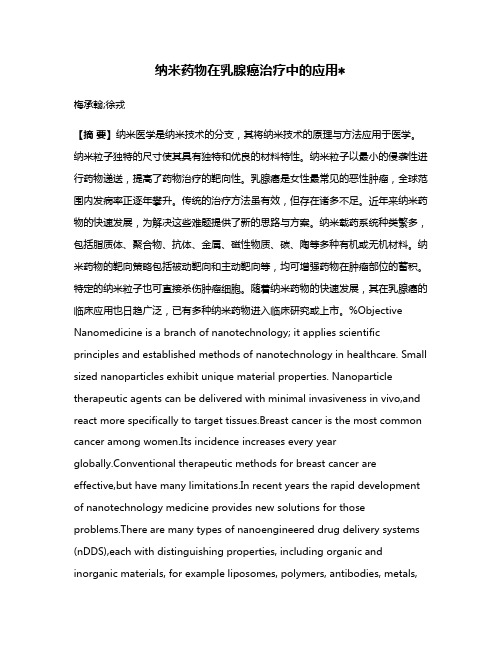
纳米药物在乳腺癌治疗中的应用∗梅承翰;徐戎【摘要】纳米医学是纳米技术的分支,其将纳米技术的原理与方法应用于医学。
纳米粒子独特的尺寸使其具有独特和优良的材料特性。
纳米粒子以最小的侵袭性进行药物递送,提高了药物治疗的靶向性。
乳腺癌是女性最常见的恶性肿瘤,全球范围内发病率正逐年攀升。
传统的治疗方法虽有效,但存在诸多不足。
近年来纳米药物的快速发展,为解决这些难题提供了新的思路与方案。
纳米载药系统种类繁多,包括脂质体、聚合物、抗体、金属、磁性物质、碳、陶等多种有机或无机材料。
纳米药物的靶向策略包括被动靶向和主动靶向等,均可增强药物在肿瘤部位的蓄积。
特定的纳米粒子也可直接杀伤肿瘤细胞。
随着纳米药物的快速发展,其在乳腺癌的临床应用也日趋广泛,已有多种纳米药物进入临床研究或上市。
%Objective Nanomedicine is a branch of nanotechnology; it applies scientific principles and established methods of nanotechnology in healthcare. Small sized nanoparticles exhibit unique material properties. Nanoparticle therapeutic agents can be delivered with minimal invasiveness in vivo,and react more specifically to target tissues.Breast cancer is the most common cancer among women.Its incidence increases every yearglobally.Conventional therapeutic methods for breast cancer are effective,but have many limitations.In recent years the rapid development of nanotechnology medicine provides new solutions for those problems.There are many types of nanoengineered drug delivery systems (nDDS),each with distinguishing properties, including organic and inorganic materials, for example liposomes, polymers, antibodies, metals,magnets, carbons and ceramics. Nanomedicine may have passive and active targeting strategies. Both can enhance the accumulation of the drug in tumor sites. Specific nanoparticles can also kill or damage tumor cells.As its research advances fast,a wide application of nanomedicine in treating breast cancer is getting adopted.Many agents and formulations of nanomedicine are approved for clinical trials or approved for prescription.【期刊名称】《医药导报》【年(卷),期】2015(000)010【总页数】5页(P1267-1271)【关键词】纳米载药系统;癌,乳腺;纳米医学;靶向治疗【作者】梅承翰;徐戎【作者单位】华中科技大学同济医学院药理学系,武汉 430030;华中科技大学同济医学院药理学系,武汉 430030【正文语种】中文【中图分类】R945;R737.9纳米材料有着特殊的尺寸依赖性,在某种尺度时物质会表现出特殊的理化性质。
- 1、下载文档前请自行甄别文档内容的完整性,平台不提供额外的编辑、内容补充、找答案等附加服务。
- 2、"仅部分预览"的文档,不可在线预览部分如存在完整性等问题,可反馈申请退款(可完整预览的文档不适用该条件!)。
- 3、如文档侵犯您的权益,请联系客服反馈,我们会尽快为您处理(人工客服工作时间:9:00-18:30)。
Anti-DNA Antibody/Cationic Lipid Micelles for Plasmid DNA Gene Delivery1Song Cunxian 1, Wang Manyan1, Jin Xu1, Leng Xigang1, Tang Lina1, Sun Hongfan1, and RJ Levy2 (1 Institute of Biomedical Engineering, Peking Union Medical College, Chinese Academy of Medical Sciences, Tianjin, P. R. China 300192;2 The Division of Cardiology, Children’s Hospital of Philadelphia,PA 19104,USA)AbstractWe reported for the first time a new type of plasmid DNA gene delivery systemcharacterized by DNA-(anti-DNA) Antibody-Cationic lipid (DAC) micelles andpermitted the highly efficient entrapment and delivery of plasmid DNA in vitro. Themicelles were characterized with respect to size, charge, and DNA entrapment. DACtriplex system formed a stable suspension of nanospheres, with a mean particlediameter of around 360nm, and a zeta potential of –15mV. Fluorescent DNA studiesdemonstrated greater cell uptake in vitro with DAC compared to the various controlformulations. Confocal microscopy studies confirmed nuclear entry in A10 cells withDAC, while formulations without the antibody demonstrated no nuclear entry. Cellculture studies revealed that DAC gave 4-fold greater level of transfection than theformulation without the antibody. It is concluded that anti-DNA antibodies canmarkedly enhance plasmid DNA gene transfer in A10 cells in culture. This effectappears to be strongly associated with anti-DNA antibody nuclear entry properties.The DAC micelles reported in this study represent one of the most potent plasmidDNA vector delivery systems reported thus far, with efficiencies significantly greaterthan conventional approaches, such as Lipofectin.Keywords: M icelle, DNA-anti-DNA, Nuclear Entry, Gene Delivery1 IntroductionA major problem for current gene therapy strategies is an inability to achieve a high level of expression of the gene product in vivo. Since plasmid DNA vectors have major safety advantages compared to viral vectors, there have been numerous attempts to improve plasmid DNA transfection, and the most promising of these approaches involves using cationic liposome [1,2,3]. In the present studies we investigated anti-DNA antibody-cationic lipid complex as a gene delivery system. It was hypothesized that anti-DNA antibody micelles could enhance DNA delivery through mechanisms involving: Increased levels of DNA incorporation into micelles due to antibody binding, enhanced cell entry via cell membrane-micelle interactions, entry into the cytoplasm of intact DNA-antibody-cationic lipid micelles, associated resistance to nuclease attack, and increased nuclear entry of DNA. Special interest1 Support by the China Specialized Research Fund for the Doctoral Program of Higher Educationunder Grant No. 20030023004, the Key Project Foundation of Tianjin City under Grant No.043803011and the National Natural Science Foundation of China under Grant No. 50473059of this study is the potential possibility of binding a plasmid DNA on such carries as nanoparticle or coronary stent through the antibody bridge for introvascular site-specific gene delivery.2 Materials and Methods2.1 M a t e r ia lsA Mouse anti-Bovine DNA IgM recognizing double- and single- stranded DNA was obtained from US Biological (Swampscott, MA). A β-gal DNA was labeled with Rhodamine and Lipofectin was labeled with fluorescent (BioDIPY) by using the LablelIT™labeling Kits (Mirus-Pan Vera, Madison, WI). Lipofectin, DOPE and DOTMA were purchased from SIGMA Chemical.2.2 Micelle fo rmula t io nFor preparing DAC complex, GFP-DNA in phosphate buffer (PBS) was mixed with the anti DNA IgM and incubated at 37 °C for 1 hour followed by the addition of 5 µl of Lipofectin. Control formulations were prepared in parallel.2.3 M ic e lle c h a r a c t e r iza t io nDAC micelles were assayed with a ZetaPlus Particle Sizer (Brookhaven Instruments Corp., NY). The concentration of DNA entrapped in micelles was determined by spectroscopy after phenol/chloroform extraction of particles separable from suspension by 0.2µm membrane filtration.2.4 Cellula r upta k eA Rhodamine-labeled DNA was used with fluorescent (BioDIPY) Lipofectin to assess the cellular internalization and localization of the DAC complex by confocal microscopy.2.5 In vitro c e ll t r a n sf e c t io nSmooth muscle cells (A10 cells) were incubated at 37°C for 18 hours prior introducing DNA to cells. The DNA complex in serum-free medium was added to replace the medium in cell culture plate. After 24 hours the cell culture medium was replaced with fresh medium. Following 24-48 hours more incubation, the cells were fixed and cover slide mounted using Vectashield Mounting medium with DAPI. The cell was observed under fluorescent microscope using FITC filter for GFP transfection and DAPI filter for total cell numbers. The percent of cells transfected was calculated by flow cytometry and by cell counting.3 Results and Discussion3.1 F o r m u l a t i o n a n d c h a r a c t e r i za t i o n o f D A C m i c e l l e sDAC formed stable micelles by a self-assembly process, when formulated by first combining plasmid DNA and anti-DNA antibody, followed by vortexing with Lipofectin. It is of interest to note that when Lipofectin and plasmid DNA were combined first, and then mixed with the antibody, immune precipitates formed. DAC micelles had an initial mean particle diameter of around 360nm and zeta potential of around -15mV. By comparison, formulation in the same ratio of plasmid DNA/Lipofectin but without anti-DNA (DC formulation), also formed detectable particles with a diameter of 190nm, and had a less electronegative zeta potential (-8.89mV). Despite the addition of cationic lipid to the DAC formulation, there was only a slight reduction in the electro-negative charge (Table 1).Formulation of anti-DNA and Lipofectin without DNA had a markedly large size (>1000nm). It was hypothesized that congeries were formed here for some unknown sake. DAC micelles were investigated for their stability in terms of size and charge, with timed incubations under simulated physiologic conditions (pH 7.4, PBS solution, 37°C). The results revealed that DAC complex had little change in size and zeta potential over the one-week observation period (Figure 1). Using Rhodamine-labeled DNA we observed relatively uniform DNA nanospheres in the DAC preparations (Figure 2).Table 1: Particle size and Zeta potential of various micelle formulations FORMULATION PARTICLE SIZE (nm±se) ZETA POTENTIAL (mV±se) DNA-antiDNA - Lipofectin 360.0 ± 3.1 -15.27 ± 4.45DNA- antiDNA Not stable -17.46 ± 1.42DNA- Lipofectin 191.7.0 ± 2.6 -8.89 ± 5.53AntiDNA-Lipofectin 1363.8 ± 178.2 -6.71 ± 1.86Figure 1: Change of particle size and charge of DAC micelles shake in PBS at 37°CFigure 2: Fluorescence micrograph of the Rhodamine-DACExtraction of DNA from DAC micelles accounted for 28.5±1.5% of the total initial DNA entrapped in the micelles, in comparison with the much lower retaining of DNA in DC formulation (13.6±0.8%) (Figure 3).Figure 3: DNA entrapment in DAC and DC micelles3.2 Cellular uptake of the micellesThe uptake of DAC compared to control formulation (DC) was studied using Rhodamine labeled DNA in a series of A10 cell culture experiments. DAC uptake was observed to be extensive after only 8 hours, with a very prominent cytoplasm-oriented presence at 24 hours. Apparent nuclear entry of DAC was observed with 48 hours cultures. The control experiment revealed qualitatively less amounts of cellular uptake and nuclear entry.In confocal microscopy studies, it was demonstrated that DAC entered cells as an intact antibody-DNA-lipid complex with persistent association of cationic lipid, even in the nuclei of the A10 cells (Figure 4A). By comparison, DNA-cationic lipid without anti-DNA antibody demonstrated little cationic lipid entry in association with fluorescent DNA, and neither fluorescent DNA nor fluorescent lipid were observed in the nuclei (Figure 4B). Thus, DAC is structurally stable under cell culture conditions, and furthermore, our results show that the entire complex can remain intact throughout cellular processing including nuclear entry, indicating that the anti-DNA antibody investigated in our studies has nuclear entry capability.Figure 4: Confocal microscopy studies of A10 cell uptake of (A) DAC consisted of Rhodamine-labeled DNA (red) and BioDIPY labeled cationic lipid (green) and anti-DNA-antibody, colocalized as indicated by yellow in the cytoplasm and nucleus of A10 cells. (B) DC lipoplexes illustrating no nuclear entry3.3 In Vitro A10c e lls t r a n s f e c t io nDAC triplex micelles markedly increased the level of gene transferred to A10 cells, with more than a 4-fold increase in transfection compared to the DC formulation (Figure5A-5B). The DA formulations (GFP DNA with antibody) resulted in no transfection. Flow cytometry studies demonstrated the magnitude of the differences between DAC and other transfection formulations (Figure 5C). It showed that anti-DNA-antibody greatly enhanced DNA transfection compared with same amount of Lipofectinwithout anti-DNA-antibody. Coomassie blue staining of A10 cell cultures following DAC mediated transfection revealed a normal cellular morphology, with no apparent toxicity due to DAC.Since DOTMA is the cationic lipid and DOPE is the neutral lipid in Lipofectin formulation, the question is which one is the contributing ingredient in the DAC system? The two chemicals were further tested separately in A10 cell transfection. The results revealed that DOTMA alone induced almost the same transfection efficiency with Lipofectin. On the other hand, DOPE alone did not show any positive effect (Figure 6). The results suggest that the positive charge of cationic lipid in the triplex micelles played an important role in harmonizing with the anti-DNA antibody.Figure5: Transfection of A10 cells in vitro with GFP-DNA (green fluorescent) heteroplexes formulated with the same amount of DNA. (A) GFP-DAC showed intense fluorescent. (B) GFP-DC without anti-DNA-antibody. (C) Flow cytometry studies demonstrated A10 cells transfection using GFP-DAC was significantly efficient than other control formulationsFigure 6: Results of A10 cell transfection by GFP-DNA-Anti-DNA-antibody with different lipid formulations: (A) DOTMA/DOPE: 2/1, 5µl. (B) DOTMA/DOPE=1:1, 5µl. (C) DOTMA/DOPE=1/2, 5µl. (D) DOPE, 5µl. (E) DOTMA 5µl. (F) DOTMA 10µl. (G) DOTMA 20µl4 ImplicationsThis is the first time to involve antibody in gene vector formulation. Although there were many issues waiting for further exploring, preliminary results from this study revealed greatly potential possibility of using anti-DNA antibody to facilitate high-level gene transfer and expression when harmonized with cationic lipid. With purification of the DAC micelle constructs, and elimination of free antibody, the risk for anti-DNA antibody adverse effects should be hypothetically low. Further investigations with this gene delivery system will need to be mindful of the potential for anti-DNA antibody related diseaseactivity. Furthermore, DNA-antibody can be expediently coupled onto the surface of such medical devices as coronary stent, therefore to deliver therapeutic polynucleotides to an intravascular site achieving efficient gene therapy.A c k n o w l e d g m e n t sThese studies were financially supported by projects from Tianjin Committee of Sciences and Technology and the NSFC. Dr. Levy in Philadelphia, USA is the supervisor and co inventor. References[1] Ouahabi E.I., Thiry M., Pector V., et al. (1997) The role of endosome destabilizing activity in the gene transferprocess mediated by cationic lipids. FEBS Letter 4.4, pp. 187-192[2] Chin-Yi Huang, Tetsuo Uno, John E Murphy, et al. (1998) Lipitoids - novel cationic lipids for cellular deliveryof plasmid DNA in vitro. Chemistry & Biology. Vol. 5, pp. 345-354[3] Katalin Kariko, Alice Kuo, Elliot S. Barnathan, et al. (1998) Phosphate-enhanced transfection of cationiclipid-complexed mRNA and plasmid DNA. Biochimica et Biophysica Acta Vol.1369, pp. 320–334.。