
场致发射显微镜(FEM)
场致发射显微镜(FEM)
一场致发射研究的历史回顾
场致发射发现于1897年。
1992 年LIlienfeld用尖端阴极作x 射线管的电子源。
1828年弥勒和诺德海姆用波动力学解释场致发射, 为场致发射理论奠定了基础。
1940 年电子显微镜发明之后, 弥勒--诺德海姆场致发射公式才得到准确的实验。
证1937年Muller 引进场致发射显微镜的概念, 用它观察尖端的场发射图像, 可以了解晶体排列,研究外来原子在金属表面的吸附和解吸, 以及吸附原子在金属表面的迁移等现象。
1941年Muller 又发现吸附原子在强负电场作用下的解吸现象, 这导致1951年设计出场离子显微镜。
1967 年场离子显微镜发展到原子探针的阶段, 用它可以检查场离子图上任意一点的质谱性质。
在四十年代末期, 为了给积极发展的微波管提供有效的电子发射源, 开始了对场发射阴极的系统研究, 包括对极限电流密度、场发射电流稳定性、“热”场致发射等的研究。
五十年代由于超高真空技术的发展, 在Dyke及其同事的努力下, 稳定的场致发射已经基本上可以实现。
他们采用多尖端阴极, 并将它首先应用于脉冲(闪光)x光管中,以后发展为商用的场发射x光管(Fexirton)和场发射电子注管(Febetron)。
在六十年代初期, 有人提出用大功率电子注来加热原子核使之达到产生热核聚变的温度, 因此开始建立了许多大功率脉冲电子注
加速器。
这种加速器在几百千伏到10兆伏的脉冲加速电压下, 从尖端或刀口阴极发射104A—106A 的电流, 电子注的脉冲功率达1014—1013W。
这种电子注称为相对论性强流电子注, 除了可以产生高强度闪光x射线外, 还可以产生大功率激光、大功率激波、毫米波、亚毫米波, 模拟辐射效应, 还可能用于等离子体加热、约束等离子体、电子注聚变等方面。
这时阴极发射的机理和一般场发射不同, 称等离子体场发射, 苏联称之为爆发电子发射。
随着六十年代末期扫描电子显微镜和表面物理分析仪器的蓬勃发展, 要求有高亮度,细小直径的电子束。
尖端场发射阴极接近一个点源, 电流密度大, 电子初速零散小, 容易聚焦成细束, 逐渐应用于上述仪器的电子枪中。
70年代末,Gomer利用探孔FE从创立了“涨落法”.使FEM在研究表面物理领域中成为重要的研究手段之一。
二场发射显微镜(FEM)原理
图1 是场发射显微镜的结构原理图,泡中尖端状的样品被固定在加热丝上,位于球形玻壳的中心,作为场电子发射阴极,表示为K。
玻壳窗口的内壁先蒸涂上一层透明的导电层(通常为二氧化锡),然后涂敷上荧光粉,制成高发光效率的荧光屏S。
导电层与电极A相连接,作为阳极。
在电极A与K之间加正电压。
若阴极尖端表面是球形,从阴极表面不同位置场致发射的电子将沿着同心球的半径方向运动,最后打在荧光屏上。
这样,在荧光屏上就显示出来阴极(样品)
尖端发射电子能力的像。
而场发射电流密度与逸出功E(φ)和电场强度ε有指数关系,因此场发射体针尖表面的逸出功不同或场强不同,都将引发射电流密度的变化。
除了不同晶面有不同逸出功外,吸附外来原子或存在缺陷的表面态也将引发逸出功的变化;增减表面的粗糙也将引起局部电场的改变。
因此,针尖尖端电子发射将与上述因素有关。
场发射显微镜所用的针尖是用多晶的线状材料,经过腐蚀加工制成。
通常情况下,其场电子发射部分是单晶,由于尖端尺寸很小,往往小于多晶晶粒大小,而且球面暴露部分多是低表面能平台。
实际制成样品的针尖存在柱形或锥形的径部结构,因此针尖表面场强偏离小球体系,可表示为
ε=V/(γr) (1.1)
式中的γ是与针尖结构有关的参数。
由式 1.1可知,对于曲率半径r=1000埃的针尖,阴极加上2—5KV的正电压,在针尖表面就可以得到足够产生场致电子发射的电场强度。
对于金属样品得到更小的曲率半径是容易的。
场致显微镜的理想放大倍数可表示为
M=R/r (1.2)
式中R是荧光屏的半径,r是样品尖端的曲率半径,实际发射体针尖径部的柱形或锥形结构,不仅减小了场强,而且使电力线向轴向压缩,结果实际放大倍数为
M=R/βr (1.3)
式中压缩系数β取值为 1.5—2.通常图像是轴向对称的,在荧光屏上发射体几乎是均匀的。
线性放大倍数的数量级为105—106.
三场发射显微镜结构
对于场发射显微镜研究的两种设备的设计:一种是封闭式显微镜,它整个浸没在液氦或液氢中;另一种是有Muller为场离子发射研究而首先设计的连续抽气管。
1 完全浸没管
2 连续抽气管
蔡司场致发射显微镜场离子显微镜图像
的钨干净的表面上
的位置不同的晶面
方向(110)。
四场发射显微镜的应用
利用场发射显微镜可以判断尖端材料是否纯净,判断的标准是发射图像和尖端的加热处理温度无关。
反之,如尖端材料含有杂质(如C,Si),则尖端在经过高温闪炼后(这时表面杂质暂时都蒸发掉),如适当降低温度,则可观察杂质从内部向表面扩散的发射图像。
从纯金属尖端的发射图像可确定各晶面的逸出功。
场发射显微镜是研究吸附现象的有效方法。
因为吸附现象总是伴
随着逸出功的变化,而逸出功决定发射电流密度。
在场发射显微镜荧光屏上可以看到在单晶上各向异性的吸附图像。
用场发射显微镜研究吸附十分形象化,而且可以分辨到接近原子尺寸。
这个方法还有一个特点,就是吸附的研究可以很方便的在很大的温度范围内进行。
最早研究Ba,Th在钨尖上的吸附,他直接与热阴极的工作有关,研究说明表面迁移对发射机理起很重要的作用。
图6-27给出了在钨表面的发射图像。
碳在钨尖上的吸附具有特征图像。
例如由于分解有机物或蒸发碳丝使碳吸附在钨表面,当把钨尖加热到1000K以上时,由于表面迁移,(110)和(121)面周围发射增加(被认为是钨基底重新排列形成凸面),而(343)面则为暗区,如图6-28所示,这个图像可以判断有无少量的有机物存在。
关于用场发射显微镜观察分子和原子的图像问题。
当尖端表面吸附直径较大的原子或分子时,如表面覆盖率很低,则图像表现出颗粒结构,穆勒认为这是原子或分子的现象。
如钡在钨尖的吸附,当覆盖度小于0.1时可观察到颗粒结构。
根据计算,当钡覆盖度为0.1时,钡原子之间的距离约20A,这和场发射显微镜的分辨率接近。
穆勒把少量的钛青铜分子蒸到钨尖上,钨尖发射图像上除出现单亮点外,还观察到很高的“双极”和“四极”斑点。
如图6-29.后二者以一个单元出现,有事旋转,有时变为另一种形式,有时在离子的轰击下消失。
这些单元很像钛青铜分子的图像。
因为根据X射线分析,钛青铜分子是四极
分子,其结构如图6-30所示。
当然fem也有一定局限性,例如,某些被分析的样品男一直被成曲率半径为0.1um的tip;对原子较为稀疏的晶面或偶极距较大的吸附原子的研究,需考虑tip表面存在强电场的影响。
五空间电荷对FEM图像的影响
众所周知,场发射电流密度大,因而场发射电流在尖端前面所形成的空间电荷比较严重.一般讲场强愈弱,空间电荷层愈薄荧光屏上放大图像就愈能反映尖端表面的真实情况.图2给出我们所摄得的在不同场强及对应场发射电流下一组W(110)尖的高分辨力热场发射显15微图(HRTFEM).从照片1可见,当场强在3.6×10⁷V/cm时,场发射电流仅为0.2LA,此时在照片上吸附在钨上的锆原子清晰可见,分辨力优于0.2nm以上.但随着场强的增加,场发射电流很快上升,空间电荷影响也愈来愈严重,尖端直接表面上清晰图形发生严重畸变(照片2,3).当场强增加到2.5×108V/cm时总发射电流已增到900LA,所得的FEM图形已失去表面结构的特征,只能隐约可见代表几个低指数晶面的黑点与亮区.这就是当前一些资料所发表的FEM图形
参考文献
1 高分辨力热场发射显微镜(HRTFEM)及其应用
陈尔纲杨德清窦菊英孙祥乐(云南大学物理系,云南昆明650091)
2 Muller E.W., Work function of tungsten single crystal planes measured by the field emission microscope // J. Appl. Phys. 1955. V. 26,
№6. P. 732–737.
3 场致发射技术的发展和应用
陆家和张奇
4 “场致发射的实验研究” , 第一次全国电真空器件专业会议论文集1964 ,
陆家和, 张奇
5 “场发射闪光x 射线管” , 电子学通讯 , 第一卷, 第2 期, 于祖哲、刘光治、张奇、王培刚、王月。
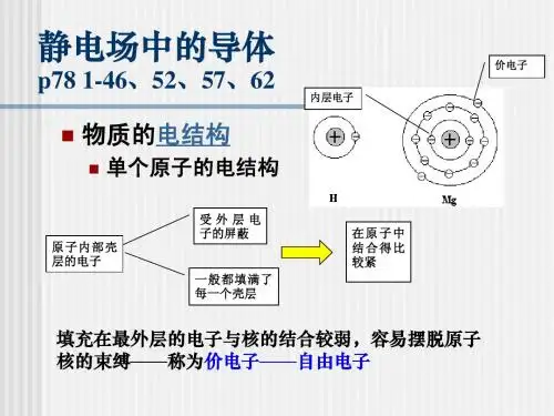
静电场中的导体
说明:
一般情况表面有一定厚度,很复杂如:E=109V,则 感应电荷聚集在表面的厚度为10-10m,本课程不讨论 表面层电荷如何分布。 实际物质内部既有自由电子,又是电介质。如:气体 在一般情况下绝缘(电介质),但加高压气体会被击 穿(导体)——导体是一种理想模型。 对导体只讨论达到静电平衡以后的情况,不讨论加电 以后电荷的平衡过程。
S内
E
E d S 0
内表面不是等势面 ——导 体也不是等势体 ,矛盾
S面内 q 0
内 表面 电 荷代 数和 为 零? 内 表面 无 电荷
q 0
e内 0
空腔内部有带电体 q
导体内表面上所带电荷与腔内电荷的代数 和为零 证明:作Gauss面如图
E内=0 E
力学:只涉及物质的机械性质,对其本身研究甚 少。 电磁学:较多地讨论场,而对物质本身的电磁性 质也涉及得很少。 物质与场是物质存在的两种形式 物质性质非常复杂(要特别注意我们课程中讨论 这种问题所加的限制)
导体静电平衡条件
导体:有足够多的自由电子 ——受电场力会移动.
静电平衡状态:体是一个等势体,导体表 面是等势面 证明:
导体内部E=0
U ab E d l 0
a
b
导体内部任意两点间电势差为零 ——各点等电势——等势体 ——表面为等势面
场强分布
E内 0
表面附近:表 表面 E 表面 : σe 大小: E ε0
导体表面是等势 面,处处与电力 线正交 ?
S内
E d S 0
q 0 q x x q
声学扫描显微镜

扫描声学显微镜
声学扫描显微镜(SAM)是一种多功能、 高分辨率的显微成像仪器,兼具电子 显微术高分辨率和声学显微术非破坏 性内部成像的特点, 被广泛的应用在 ○ 物料检测(IQC)、失效分析(FA)、 质量控制(QC)、质量保证及可靠性 (QA/REL)、研发(R&D)等领域,可以检 测材料内部的晶格结构、杂质颗粒、 内部裂纹、分层缺陷、空洞、气泡、 空隙等,为司法鉴定提供客观公正的 微观依据。
扫描声学显微镜
组员:应物三班 宋 飞326 刘 健303 刘丹跃305
1、显微镜种类介绍 2、扫描声学显微镜的原理 3、扫描声学显微镜的性能及在材料科学中的应用
2
显微镜有很多种类,大致有:
透射电子显微镜(TEM) 扫描电子显微镜(SEM) 场离子显微镜(FIM) 高压电子显微镜(HVEM) 分析电子显微镜(AEM) 场发射电子显微镜(FEEM) 声学显微镜(AM) 扫描隧道显微镜(STM)
原子力显微镜(AFM)
超高压电子显微镜
•
• •
•
主要用途: 观测材料、矿物、生物样品、器件透射 电子显微象及电子衍射图,对样品微观组织、结构、 缺陷等定性、定量分析。 可对样品在加热、拉伸、 电子辐照等条件下微观组织的变化过程进行动态观测。 仪器类别: 03040701 /仪器仪表 /光学仪器 /电子光 学及离子光学仪器 /透射式电子显微镜 指标信息: 加速电压:1000kV 放大倍数:150倍~ 30万倍晶格分辨率:0.27nm 选区衍射相机长:2~ 6m 试样可加热温度:1000℃ 附件信息: 加热台(室温-1000℃),拉伸台加伸台 (室温-1000℃),动态过程摄象录象系统。双倾台 (±45° 该仪器是我国最大型的透射电子显微镜,它主要用于 各种材料的微结构分析,组织特征和相鉴定,缺陷研 究等。与普通电子显微镜相比较,它可以进行微观过 程的动态实验观察、辐照效应研究、厚试样和粗大析 出物的观察分析以及半导体微器件结构研究。同时, 本实验室样品制备、数据结果处理等附属设备齐全, 为科研实验创造了良好条件。
场发射扫描电镜工作原理

场发射扫描电镜工作原理
场发射扫描电镜(FE-SEM,Field Emission Scanning Electron Microscope)工作原理如下:
1. 电子发射:FE-SEM使用一个称为电子枪的器件产生高能电
子束。
电子枪包含一个钨丝或碳纳米管的尖端,通过加热尖端,电子从尖端发射出来。
2. 调制和聚焦:电子束经过一系列的电场调制和聚焦透镜,以使电子束具有足够的能量和焦点来进行扫描。
3. 扫描和信号采集:电子束被聚焦到一个非常小的尖端,称为扫描线圈。
扫描线圈通过控制电子束的位置和速度,将电子束在样品表面上进行快速的扫描。
当电子束与样品表面相互作用时,会有多种信号产生,如二次电子、反射电子、特征X射
线等。
4. 信号检测:上述信号被检测器捕获,并由电子光学系统将其转换成图像或谱图。
典型的信号检测器包括二次电子探测器、透射电子探测器、X射线能谱仪等。
5. 数据显示和处理:捕获的信号通过计算机进行处理和显示,生成高分辨率的扫描电子显微图像。
计算机还可以对图像进行后期处理,如增强、标记、量化等。
总的来说,FE-SEM利用电子束的显微特性来对样品进行高分
辨率的表面观察,并可获取丰富的样品性质信息。
扫描隧道显微镜
扫描隧道显微镜(STM)的原理及其在纳米材料研究中的应用引言透射电子显微镜在观察物质的整体结构方面是很有用的,但在表面结构的分析上却较困难,这是因为透射电子显微镜是由高能电透过样品来获得信息的,反映的是样品物质的内部信息。
扫描电子显微镜(SEM)虽然能揭示一定的表面情况,但由于入射电子总具有一定能量,会穿入样品内部,因此分析的所谓“表面”总在一定深度上,而且分辨率也受到很大限制。
场发射电子显微镜(FEM)和场离子显微镜(FIM)虽然能很好地用于表面研究,但是样品必须特殊制备,只能置于很细的针尖上,并且样品还需能承受高强电场,这样就使它的应用范围受到了限制。
扫描隧道电子显微镜(STM)的工作原理完全不同,它不是通过电子束作用于样品(如透射和扫描电子显微镜)来获得关于样品物质的信息,也不是通过高电场使样品中的电子获得大于脱出功的能量而形成的发射电流成像(如场发射电子显微镜),并以此来研究样品物质,它是通过探测样品表面的隧道电流来成象,从而对样品表面进行研究。
STM技术的最大优势在于可获得原子级的分辨率,通常它的分辨率在平行于表面的方向可达0.1纳米,在垂直于表面的方向可达0.01纳米,此外,STM还可实时地获得材料表面实空间的三维图像;可以观察单个原子层的局部表面结构,而不是整个表面的平均性质;配合扫描隧道谱STS可以得到有关表面电子结构的信息,例如表面不同层次的态密度、表面电子势阱等。
在STM之后衍生出了原子力显微镜、磁力显微镜、近场光学显微镜等一系列新型非接触表面探针技术显微镜,使探针显微镜技术日趋完善,并在纳米科技领域中得到越来越广泛的应用。
关键词:扫描隧道显微镜、量子隧道效应、纳米材料一、扫描隧道显微镜的介绍扫描隧道电子显微镜(scanningtunnelingmicroscope,STM)是一种利用量子理论中的隧道效应探测物质表面结构的仪器,利用电子在原子间的量子隧穿效应,将物质表面原子的排列状态转换为图像信息的。
SPM 基本原理与表面化学方面的应用
扫描隧道显微镜(STM)的发明
Gerd Binnig & Heinrich Rohrer 获1986年诺贝尔物理奖
1982年发明
原始扫描隧道显微镜
由Binnig和Rohrer(IBM Zurich 实验室) 构思和设计的第一台STM如上图所示。其 核心部分包括:针尖( W 或 Pt / Ir 丝); 压电驱动器(压电陶瓷);粗调定位仪; 振动隔离系统(弹簧);计算机控制和图 像采集系统。 第一次在实空间里看到表面原子像: Si(111)-(7×7)的原子图像见右图。 Source: C. Julian Chen
SPM系统 的基本组成部分
•探针 (纳米级,原子级) •进动和扫描系统 •反馈控制回路 •振动隔离系统 •成像系统
http://www.nanoscience.de/group_r/mfm/mfm/
SPM探针
STM Tip
W Si
AFM Tip
Pt/Ir
W
CNT
•其他探针材料: 金,银,铑,镍,碳 管等导电材料 •电化学腐蚀或者机械剪切获得
参考工具书
1. C. J. Chen, “ Introduction to Scanning Tunneling Microscopy ” (Oxford Series in Optical and Imaging Sciences, 4), Oxford University Press, 1993. 2. 《扫描隧道显微学引论》(华中一,朱昂如,金晓峰译,中国轻工业 出版社,1996)。 3. 4. 吴凯,“表面结构与表面分析讲义”,未出版. King Lun Yeung and Nan Yao,“Scanning Probe Microscopy in Catalysis”,J. Nanosci. Nanotech.4 (2004) 1 5. Michael Bowker and Philip R. Davies, “Scanning Tunneling Microscopy in Surface Science, Nanoscience and Catalysis”, WILEY-VCH Verlag GmbH,2009
2018年高一物理竞赛完整讲义(word版)含答案解析第9讲 静电屏蔽 电像法.教师版
第9讲静电屏蔽电像法本讲提纲1.静电感应现象2.静电屏蔽3.电像法以及应用本讲对等效思维运用较多,思考的时候概念转化很快。
部分不适的同学可以先只关注物理现象以及原理的解释,对于负责的运算大家可以根据自己的兴趣尽力跟进老师的讲解,不妨把部分难题当作对自己能力极限的挑战。
知识模块引入:电场中的导体把一块不带电的导体放到电场周围,这个导体就会感应出电荷。
这个现象不难去理解,一般用电场对电荷的力就能解释。
静电感应:导体内自由电子在外电场的作用下定向移动,重新分布的现象.本讲研究的是这个现象的定量规律。
如图演示的是在外电场下金属内部自由电子从移动,到最后稳定分布的过程。
知识点睛一.静电平衡:导体中(包括表面)没有电荷的定向移动的状态.由于感应电荷形成新的电场,最终导致金属内部合场强降为零,静电感应现象才达到了稳定,我们不妨称之为静电平衡。
1.导体内部的场强处处为零.2.整个导体是个等势体,导体的表面是等势面.3.净电荷分布在导体的外表面.4.在导体表面附近,电场线一定与表面垂直.如图是一般形状的金属放入电场后的影响二.静电屏蔽导体壳(网罩)不接地时,可以实现外部对内部的屏蔽,但不能实现内部对外部的屏蔽;导体壳(网罩)接地后,既可实现外部对内部的屏蔽,也可实现内部对外部的屏蔽。
如下图的演示试验,当导体放到静电计周围,并用金属球壳把静电计包起来后,静电计的指针是不偏转的。
如图是静电屏蔽掉超高压的试验示意,在高压电线上工作的工人穿的也是用金属制作的衣服,屏蔽电线周围的强电场。
下面列了另外的一些关于静电屏蔽的应用,具体内容请同学们课后自己查阅资料或者咨询老师。
1.电子仪器,比如示波器的接线都是这样的屏蔽线;2.外的天线;3.电视信号线,外面就有一层金属丝,就为了静电屏蔽,使信号不受干扰;4.务区怕被人打手机,又不能关机,找个金属盒子装进去,就变成了“您拨打的用户不在服务区”;例题精讲【例1】如图所示,把一个带正电的小球放人原来不带电的金属空腔球壳内,其结果可能是()A .只有球壳外表面带正电。
材料表面与界面分析复习题
1表面、界面的定义与理解:表界面是由一个相过渡到另一相的过渡区域。
习惯上把固-气、液-气的过渡区域称为表面,而把固-液、液-液、固-固的过渡区域称为界面。
根据物质的聚集态,表界面通常可以分为以下五类:固-气;液-气;固-液;液-液;固-固;物理表面包括:理想表面、清洁表面、吸附表面表面是一个抽象的概念,实际常把无厚度的抽象表面叫数学表面,把厚度在几个原子层内的表面叫作物理表面,而把我们常说实际的固体表面叫工程表面。
2 理想表面理论前提:①不考虑晶体内部周期性势场在晶体表面中断的影响;②不考虑表面原子的热运动、热扩散、热缺陷等;③不考虑外界对表面的物理-化学作用等;④认为体内原子的位置与结构是无限周期性的,则表面原子的位置与结构是半无限的,与体内完全一样。
3何为清洁表面?清洁表面获得方法?指不存在任何污染的化学春表面,即不存在吸附、催化反应或杂质扩散等一系列物理、化学效应的表面。
获得清洁表面的几种方法: ①在获得超高真空的同时获得清洁的表面。
②用简单的加热方法去除表面的沾污。
③在化学气氛中加热去除那些通过简单加热不能清除的化学吸附沾污。
④对于较顽固的沾污,可以利用惰性气体离子(如Ar+、Ne+)轰击表面而有效地清除污染。
⑤对于一些晶体,可以采用沿特定的晶面自然解理而得到清洁表面。
⑥在适当的基片上通过真空蒸发法获得预想的单晶和多晶薄膜,作为研究对象的清洁表面。
4 根据原子结构的不同清洁表面分哪几种?图示说明(PPT)弛豫表面:指表面层之间以及表面和体内原子层之间的垂直间距d s和体内原子层间距d0相比有所膨胀和压缩的现象。
可能涉及几个原子层。
重构表面:指表面原子层在水平方向上的周期性不同于体内,但在垂直方向上的层间间距d0与体内相同。
台阶结构:表面不是平面,由规则或不规则台阶组成。
由于晶体内部缺陷的存在等因素,使晶体内部应力场分布不均匀,加上在解理晶体对外力情况环境的影响,晶体的解理面常常不能严格地沿所要求的晶面解理,而是伴随着相邻的倾斜晶面的开裂,形成层状的解理表面。
JEOL JSM-7500FA 场发射扫描电子显微镜使用说明书
Instructions f or o perating J EOL J SM-‐7500FA a nalytical field e mission s canning e lectron m icroscope a t Nanomicroscopy C enterVersion 2.1 (April 2011)Juuso K orhonen (***********************)Latest v ersion o f t his d ocument c an a lways b e d ownloaded f rom:k.fi/en/instruments/sem/jsm-‐7500fa/SEM-‐instructions.pdfOfficial i nformationNew u ser t rainingInexperienced u sers h ave a c ouple o f o ptions, l isted b elow i n t he o rder o f preference.1.Ask f or t raining f rom t he m ost e xperienced S EM u ser o f y ourresearch g roup.2.Attend t o t he c ourses T fy-‐125.4313 a nd T fy-‐125.4314 M icroscopyof n anomaterials (5+5 c r). T hey a re l ectured e ach s pring b y P rof.Janne R uokolainen.3.Ask o ne o f t he a dministrators t o a rrange a t raining s ession.a.Small g roups o f 2-‐3 p eople a re p referred f or t he t rainings.Allow s ome t ime t o g ather e nough p eople f or t he g roup.b.Training i s d one u sing a p ractice s ample a nd p ersonalsamples a re u sually n ot i maged.Experienced u sers c an c ontact o ne o f t he a dministrators f or a s hort introduction t o t he e quipment.Every n ew u ser h as t o b e a pproved b y o ne o f t he a dministrators b efore t hey are a llowed t o u se t he S EM o n t heir o wn. T he a dministrator k eeps a s hort (15-‐30 m in) s ession w here t he e ssential s kills o f t he u ser a re c hecked. User a pplicationUser a pplication h as t o b e f illed i n o rder t o g ain r eservation a ccess t o a ny o f the N MC e quipment. T he f orm c an b e f ound f romk.fi/en/documents/nmc_user_application_form.pdf a nd i t i s returned t o o ne o f t he a dministrators f or a pproval.PricingBilling i s d one u sing t he c urrent N MC p rice l ist. C ontact P rof. J anne Ruokolainen f or t he m ost c urrent l ist. P lease n ote t hat i ndividual t raining given b y t he a dministrators w ill a lso b e c harged.Precautions – r ead c arefully•Always c heck t he l iquid n itrogen l evel a nd f ill i f n ecessary.o First u ser o f t he d ay a lways f ills t he t ank.•Keep a ll p arts c lean a nd c lean t hem i f n ecessary w ith e thanol.o Wear g loves w hen h andling h olders.•Fill t he l og b ook o n t he c omputer.o Mark a ny s trange b ehavior o r p roblems t o t he l og b ook.•If s omething i s m issing f rom t he S EM o r f rom t he s ample p reparation room (e.g. g loves, e thanol, h olders, c arbon t ape), p lease i nform o ne o fthe a dministrators (send e mail o r c all).•Use o nly f eatures y ou a re t rained t o u se. F or e xample, d o n ot u se EDS o r R BEI i f y ou d on’t k now h ow t o o perate t hem s afely.•Use o f U SB s ticks i s s trictly p rohibited d ue t o s ecurity i ssues a nd hardware i ncompatibility.•Stay c alm a nd u se y our c ommon s ense.•Contact a dministrators i f y ou a re i n d oubt. C ontact i nformation i s found o n t he l ast p age o f t hese i nstructions.Quick s tartup p rocedure1.Turn o n b oth m onitors a nd c heck t hat S EM s oftware a nd u sage l og(Excel) a re r unning. S tart t hem i f n ecessary. L og i n a s G uest (nopassword).2.Check t he l iquid n itrogen l evel a nd f ill i f n ecessary. T he f irst u serof t he d ay a lways f ills t he t ank.3.Fill t he u sage l og:a.Date, s tart t ime (and e nd t ime).b.Your n ame (and t he n ame o f y our h ost i f y ou d o n ot h avereservation p ermissions).c.Vacuum l evels b efore s tarting.d.Amount o f f illed l iquid n itrogen (write “0” i f y ou o nlychecked t he l evel).e.Write n otes a nd c omments t o t he l ast f ield i s n ecessary.f.Save t he f ile (Ctrl-‐S).4.Prepare y our s ample.5.Insert s ample i nto m icroscope:a.Press E xchange p osition.b.Press a nd h old V ENT f or c a. 1 s ec. O pen s ecuring l atch. W ait.c.Open c hamber a nd i nsert h older a long t he d irection o f t hearrows.d.Close c hamber.e.Press a nd h old E VAC f or c a. 1 s ec. W ait u ntil b linking s tops.f.Operate t he r od t o m ove t he s ample t o t he s tage. I f y ou’re n otabsolutely c ertain h ow t o d o t his, r ead t he d etailedinstructions!g.Take o ut t he r od.6.Wait u ntil v acuum l evel r eaches l ess t han 5⋅10-‐4 P a.7.Set E mission c urrent t o 10 μA.8.Select A cceleration v oltage.9.Press O bservation O N.Shutdown p rocedure1.Press O bservation O FF t o t urn o ff a cceleration v oltage.2.Press E xchange p osition.3.Take o ut h older u sing t he r od.4.Press a nd h old V ENT f or c a. 1 s ec. O pen s ecuring l atch. W ait.5.Open c hamber a nd t ake o ut t he h older.6.Close c hamber.7.Press a nd h old E VAC f or c a. 1 s ec.8.Mark e nding t ime a nd o bservations t o U sage l og a nd s ave f ile (Ctrl-‐S).9.Set S EM M onitor s oftware t o n ormal s ettings:a.Exchange p osition p ressed (green).b.Mode: S EMc.Magnification: m inimum f or b oth S EM a nd L Md.Probe c urrent: 810.Turn o ff s pecial f eatures y ou h ave u sed: I mage r otation, d ynamicfocus, e tc.11.If y ou m ade a ny c hanges i n t he O peration S ettings m enu, c hangethem t o n ormal v alues (scan s peeds, i mage f unction, e tc.).12.Clean t he h olders w ith e thanol i f n ecessary.13.Clean t ables. I f y ou w ant t o s tore y our s amples, m ark t hem w ith y ourname a nd p ut t hem o nto a s helf. T hings l eft o n t he t able a re t hrowninto t he t rash.14.Transfer y our i mages f rom t he s mall c omputer o n t he b ack t able.You c an f ind y our f iles a t t he n etwork d rive c alled H arley.e U SB s tick, S SH, e mail, o r b urn a C D.b.The f iles c annot b e t ransferred d irectly f rom t he S EMcomputer d ue t o s ecurity r easons.15.Turn o ff m onitors. D o n ot l og o ut f rom t he s oftware o r c lose t heExcel l og b ook.Changing s ample1.Press O bservation O FF t o t urn o ff a cceleration v oltage.2.Press E xchange P osition t o m ove t he s tage t o c orrect p osition.3.Take s ample o ut b y o perating t he r od.4.Press a nd h old V ENT f or c a. 1 s ec. t o f lush c hamber a nd o pen l atch.Wait.5.Open c hamber a nd t ake o ut s ample (pull a long t he a rrows, n ot u p).6.Change s ample a nd i nsert h older a long t he a rrows.7.Close c hamber a nd s ecure w ith l atch.8.Press a nd h old E VAC f or c a. 1 s ec. W ait u ntil b linking s tops.9.Insert s ample b y o perating t he r od. T ake o ut r od.10.Wait u ntil c hamber v acuum r eaches 5⋅10-‐4 P a b efore t urning o nacceleration v oltage.Special f eaturesThis i s o nly a q uick r eference. S pecial t raining i s r equired t o u se R BEI o r E DS, because o f s afety i ssues.Infrared c ameraYou c an s ee i nside t he c hamber u sing t he i nfrared c amera.1.Switch c amera o n f rom t he b utton o n t he t able.2.From S EM s oftware s elect N avigator -‐> I nfrared c amera3.Turn c amera o ff w hen u sing R BEI o r E DS.Probe c urrent m eterProbe c urrent m eter c an b e u sed t o c heck t he c urrent g oing t o t he s ample. I t is m ost i mportant i n E DS a nalysis.1.Insert t he d etector b y c hecking P CD f rom t he b ottom r ight c orner o fSEM s oftware.2.Take o ut d etector a fter y ou h ave r ead t he c urrent f rom t he S EMsoftware.Retractable b ackscattering d etector (RBEI)Backscattering d etector i s u sed t o d istinguish b etween e lements o n t he sample.1.Set w orking d istance t o 8 m m o r m ore.a.Inserting R BEI w ith l ess t han 8 m m b etween t he s ample a ndthe o bjective l ens w ill r esult i n s erious d amage.2.Turn o ff i nfrared c amera.3.Insert d etector b y c hecking R BEI f rom t he b ottom r ight c orner o f t heSEM s oftware.4.Select C OMPO o r T OPO f or i mage m ode (same m enu a s S EM a nd L M)and u ser a s low s canning s peed f or o bservation.X-‐ray a nalysis (EDS)This g uide i s n ot a dequate f or p roper o peration o f E DS, b ut i s o nly a q uick reference f or t rained u sers.1.Set w orking d istance t o e xactly 8 m m.a.Focus w ith Z h eight u sing t he r ing o f t he s croll w heel i nsteadof F OCUS.2.Insert R BEI.3.Turn o n b ias v oltage b y c licking t he l ightning i ndicator.a.Wait u ntil c ount r ate s tabilizes.4.Select A nalysis f rom t he r ight s ide o f S EM s oftware.5.Click D T (dead t ime) a nd s elect T4 f rom t he l ist.6.Adjust p robe c urrent s o D T b ecomes g reen (around 20-‐30 %) a ndcount r ate i s c a. 2000-‐3000 c ps.7.Take s pectra, l ine s can, o r m apping u sing t he a ppropriate b uttons.8.When a sked a bout s aving t o a n etwork d rive, s elect O K.9.Save t he a nalysis b efore e xiting a nalysis m ode i n o rder t o b e a ble t oreturn t o t he a nalysis l ater.a.Exporting o nly s aves t he i mage a nd y ou c annot r eturn t omake m ore a nalysis o n t he d ata.10.When y ou a re f inished w ith a nalysis, t urn o ff t he b ias v oltage a ndtake o ut R BEI.Saving E DS s pectraIf y ou w ant t o b e a ble t o p lot y our E DS s pectrum, s elect E xport a nd t hen select M SA f ile. I t w ill s ave t he s pectrum i n a c ompatible f ile f or u se i n O rigin, Excel, o r s ome o ther p lotting p rogram.Detailed i nstructionsOperating t he r od (sample e xchange m echanism)This p rocedure d escribes h ow t o u se t he s ample e xchange m echanism i norder t o e ither r emove o r i nsert a s ample h older i nto/from t he m icroscope.Read t his s ection c ompletely t hrough b efore p roceeding a nd m ake s ure t hatyou u nderstand e very s tep.Precondition: T he e xchange c hamber i s i n v acuum a nd t he d oor s eparatingit f rom t he m icroscope i s o pen. C onfirm t hat E VAC l ight i s l it a nd n otblinking. D epending o n w hether y ou a re i nserting o r t aking o ut a s ample, t heholder m ight b e i n t he e xchange c ompartment (HLDR l ight i s o ff) o r i nsidethe m icroscope (HLDR l ight i s o n).See t he v ideo o n t he c omputer d esktop f or a d emonstration. U PDATE: T hefigures a re f rom a n o ld v ersion o f r od.1.Push t he b ar i nside t he m icroscope b y f ollowing t he p rocedure:a.Lower t he r od t o h orizontal l evel, w hile l ightly h olding i tback.b.Let t he r od b e p ulled i n s lowly.c.Push t he b ar g ently a ll t he w ay i nside u ntil i t s tops (d).•There i s a l ittle r esistance a t t he f inal c ouple o fcentimeters.•The s ample s hould b e n ow e ither r eleased f romthe b ar o r a ttached t o i t (depending o n w hetheryou a re i nserting o r r emoving t he h older).•If y ou h ave n ot p ushed t he s ample a ll t he w ayinside a nd s tart t o p ull b ackwards t here i s adanger t hat t he s ample h older w ill f all t o t hebottom t he s ample c ompartment. I f t his h appens,the w hole s ample c ompartment h as t o b e o pened.Contact S EM a dministrators i n t his c ase.2.Pull t he b ar o ut f rom t he m icroscope u sing t he f ollowingprocedure:a.Pull t he b ar o ut a s f ar a s i t c omes (e).•The t wo a rrows o n t he h older s hould a lign w iththe p ipe e nd.•If y ou h ave n ot p ulled f ar e nough, t he r od m ightbe d amaged d uring t he l ift.b.Lift t he r od u pwards t o v ertical.•Now y ou s hould e ither h ave t he s ample i nside t hemicroscope o r i n t he e xchange c ompartment a ndthe e xchange c ompartment i s i n v acuum.Opening t he s ample e xchange c ompartmentThe f ollowing p rocedure d escribes h ow t o b ring t he e xchangecompartment t o a tmospheric p ressure.Precondition: T here i s n o s ample i nside t he m icroscope o r i t h as b eenbrought t o t he e xchange c ompartment, a nd t he e xchange c ompartment i sin v acuum. F irst c heck t hat H LDR l ight i s o ff o n t he s ample e xchangecompartment (i.e. t here i s n o s ample i nside t he s ample c ompartment).Figure. S ample e xchange r od1.Pressurize t he e xchange c ompartment:a.Press a nd h old (for a bout 1 s econd) t he V ENT b utton o n t heexchange c ompartment.i.The b utton s tarts t o b link a nd y ou h ear s ome s ounds.ii.In a f ew s econds, t he d oor b etween t he e xchangecompartment a nd t he s ample c ompartment c loses.You c an o bserve t his b y e ar a nd b y l ooking a t t hebottom r ight c orner o f t he S EM M onitor.2.Open t he l atch a s s oon a s y ou h ear t he c lick.3.Open t he e xchange c ompartment d oor (it s hould o pen a lmost b yitself).a.You d o n ot n eed t o w ait u ntil t he p umping h as s topped.b.The c ompartment w ill c ontinue p urging f or a f ixed a mount o ftime. Y ou d o n ot h ave t o w ait u ntil i t s tops a nd y ou c anevacuate i t a s s oon a s y ou l ike.4.Now y ou h ave t he s ample c ompartment o pen a nd r eady f orloading/unloading t he s ample h older.Inserting a s amplePrecondition: T here i s n o s ample i nside t he s pecimen c hamber a nd exchange c ompartment i s i n v acuum. F irst c heck t hat H LDR l ight i s o ff o n the e xchange c ompartment (i.e. t here i s n o s ample i nside).1.Move t he s tage t o e xchange p osition:a.Click E xchange P osition o n t he S EM M onitor.i.If b utton i s n ot v isible, c lick "Specimen" f rom t herightmost e dge o f S EM M onitor.ii.Make s ure t hat E XCH P OSN i s l it o n t he e xchangecompartment, b efore p roceeding.2.Bring t he e xchange c ompartment t o a tmospheric p ressure b yfollowing p rocedure i n s ection “Opening t he s ample e xchangecompartment”. Q uick n otes:a.Press a nd h old V ENT f or c a. 1 s ec.b.Open l atch. W ait.c.Open c hamber d oor.3.Put o n g loves i f y ou d o n ot h ave t hem a lready o n.a.Parts t hat a re i n c ontact w ith t he v acuum s hould b e k eptabsolutely c lean. I f y ou h ave t ouched s ome p art, c lean t he p artwith e thanol (not a cetone).4.Insert h older t o t he s pecimen c huck:a.Slide t he s pecimen h older i nto t he s pecimen c huck a long t hearrow d irection o n t he s pecimen h older.5.Check t hat t he O-‐ring s eal o n t he d oor i s O K a nd w ipe i t w ith a c leanglove i f n eeded t o g et r id o f a ny d ust.a.If t he r ing i s r eally d irty, w ipe i t w ith e thanol o r i sopropanol(do n ot u se a cetone o r m ethanol).6.Close t he c hamber d oor a nd s ecure i t w ith t he l atch.7.Evacuate t he c ompartment b y p ressing a nd h olding E VAC (forabout 1 s econd). T he l ight w ill s tart b linking.a.Wait u ntil t he l ight s tops b linking a nd t he d oor s eparatingthe e xchange c ompartment i s c losed. Y ou c an o bserve t hisfrom t he b ottom r ight p art o f t he S EM M onitor.8.Insert t he s ample h older i nside t he m icroscopea.Refer t o s ection “Operating t he r od” i f i n d oubt.9.A p opup w indow s hould a ppear o n t he S EM M onitor. N ow s elect t heappropriate h older a nd s et t he o ffset v alue.a.If p opup d oes n ot a ppear, t ake o ut t he h older a nd i nsert i tagain.10.Wait u ntil t he v acuum l evel r eaches 9.6·10-‐5 P a (if t hat i s n otpossible, w ait a t l east u ntil 5·10-‐4 P a).Taking o ut s amplePrecondition: T here i s a s ample i nside t he m icroscope a nd e xchange compartment i s i n v acuum. F irst c heck t hat H LDR l ight i s o n o n t he s ample exchange c ompartment (i.e. t here i s a s ample i nside) a nd E VAC l ight i s o n and n ot b linking.1.Click O bservation O FF t o t urn o ff a cceleration v oltage.2.Click E xchange P osition t o m ove t he s ample h older t o t he e xchangeposition.a.Make s ure t hat E XCH P OSN i s l it o n t he e xchangecompartment b efore p roceeding.3.Bring t he s ample t o t he e xchange c ompartment b y o perating t herod.a.Refer t o s ection “Operating t he r od” i f y ou a re n ot a bsolutelycertain h ow t o d o t his.4.Pressurize t he e xchange c ompartment:a.Press a nd h old V ENT f or c a. 1 s ec u ntil i t s tarts t o b link.b.Open s ecuring l atch. W ait.c.Open c hamber d oor.5.Now y ou h ave t he s ample c ompartment o pen a nd y ou a re r eady t akeout y our s ample. I f y ou a re d one w ith t he i maging, j ust c lose t heexchange c hamber a nd e vacuate i t o therwise c ontinue w ith i nsertinga n ew s ample. D o n ot l eave t he c hamber o pen f or a l ong p eriod o ftime, b ut e vacuate i t i f n eeded.Sample h oldersSample h olders c onsist o f a b ase p art a nd a n a dapter p art (show o n t he f igure right). T here a re t hree d ifferent a dapters f or d ifferent s pecimen s tubs s hown in t he f igure b elow (a, b , c , d ).The m ost b asic h olders a re t he 12.5 m m (b-‐1) a nd 25 m m (c-‐1) a luminum “JEOL” s tubs . T hey s hould b e u sed w henever p ossible. S tubs s hould a lways b e available a t t he s ample p reparation r oom, b ut y ou c an a lso o rder y our o wn ones e .g. f rom E MS (order n umbers 75730, a nd 75700). T he u se o f r egular holders i s i ncluded i n t he o peration p rice o f t he m icroscope.Also “mini-‐stubs” a re a vailable f or u se w ith a p rovided a dapter. T hey a re preferred f or s mall s amples. T hey c an b e o rdered f rom T ed P ella (order numbers 16180, a nd 16181).For s pecial o ccasions, a H itachi a dapter (a) c an b e u sed. S pecial c are m ust b e taken w hen u sing t hese h olders, b ecause t hey l ack s ome s afety f eatures. Ask a dministrators, i f y ou h ave s pecial r equests f or h olders. T here a re a lso different k inds o f c ross-‐section h olders a vailable. A sk t he a dministrators f or more i nformation.Attaching a dapter t o b ase p arta) Make s ure t hat p arts a re n ot d irty, c lean i f n ecessary. b) Place a dapter o n t he b ase p art.c) Tighten s crew o n t he b ase p art l ightly.Figure. A ttaching a dapter t o base p art.Figure. 12.5 m m a nd 25 m m "JEOL" stubs.Figure. "Mini-‐stubs" a nd 12.5 m m a dapter.Figure. C ross-‐section holders.Sample h eightAlign t he t op o f t he s ample w ith t he g roove i nside t he J EOL a dapter p art. U se the s crew o n b ottom t o r aise o f l ower t he s ample. W hen u sing a nother holder, m ake s ure t hat h eight f rom t able t op l evel i s e xactly 25 m m.CoatingFor n on-‐conductive s amples a c oating i s u sually n eeded f or o bservation i n SEM. T his c an b e e asily p erformed b y u sing s putter c oating o f g old, p latinum, or g old-‐palladium. T here i s a s putter c oater a t N MC, w hich c an b e u sed f or this p urpose. R esolution l imiting f actor i s t he g rain s ize, w hich i s u sually 5-‐20 nm d epending o n t he c onditions o f s puttering.Also c arbon c oating c an b e u sed t o m ake s amples c onductive. I t i s a nappealing m ethod, w hen d oing X -‐ray a nalysis. I t c reates a v ery u niform l ayer without n oticeable grains.Figure. A lign t op o f stub w ith t he g roove on t he s ample holder.Basic m icroscope o perationBasic c onceptsWorking d istance a nd Z v alueWorking d istance (WD) v alue s ets t he e ffective f ocal l ength o f t he o bjective lens.Z h eight v alue s ets t he d istance o f t he (supposed) s urface l evel o f t he s ample from t he o bjective l ens.These t wo v alues a re e qual, w hen t op o f s ample i s a ligned w ith t he t op o f the h older (ie. 25 m m h igh f rom t able l evel, s ee f igure). W D > Z, i f y our sample i s l ower t han t he c orrect l evel a nd v ice v ersa. I f W D < Z y ou n eed t o set t he S ample O ffset v alue a ccordingly.Sample o ffsetThe h eight o f t he t op l evel o f t he s ample m easured f rom t able t op l evel should b e e xactly 25 m m. T he s ample c an b e s et a lso 0-‐4 m m h igher t han t he nominal l evel, b ut t hen t he S ample O ffset v alue h as t o b e s et a fter i nserting sample. I t i s l ocated a t t he b ottom o f t he s ample h older s elect w indow, w hich pops u p a utomatically a fter h older i nsert.Acceleration v oltage, e mission c urrent, p robe c urrent…The f irst t hing t o t hink a bout w hen s tarting i maging i s t he s election o f acceleration v oltage. T he c hoice d epends o n t he t ype o f t he s ample. S ee table b elow f or s ome e xamples.Sample Observation c ondition NotesGold p articles o n conductive s urface 5-‐30 k V, p robe c urrent a t c a. 10, working d istance 1.5-‐8 m mCoated p orous polymer 1-‐5 k V, p robe c urrent 6-‐10, w orking distance 4.5-‐8 m mUncoated p olymer 0.5-‐1 k V, p robe c urrent < 8, g entlebeam m ode, w orking d istance c a. 8mmCoated b iological sample 1-‐5 k V, p robe c urrent c a. 10, w orking distance 4.5-‐25 m m d epending o n feature s izeUncoated p aper 1-‐2 k V, g entle b eam (GB-‐L) m ode,working d istance 4.5 m m, p robecurrent 6-‐10.Coated p aper 5 k V, w orking d istance 4.5-‐25 m m,probe c urrent c a. 10X-‐ray a nalysis o f conductive s ample 15-‐30 k V, w orking d istance e xactly 8mm, h igh p robe c urrentRetractable B EIdetector i nsertedX-‐ray a nalysis o f poorly c onductive sample 5 k V, w orking d istance e xactly 8 m m,probe c urrent a s h igh a s p ossibleRetractable B EIdetector i nsertedThe e mission c urrent i s t he c urrent d rawn f rom t he e mitter. S et i t a lways t o 10 μA. Figure. S ample height s hould b e exactly 25 m m measured f rom t able top l evel.Probe c urrent i s t he c urrent d irected a t t he s ample. H igher v alues g ive better s ignal t o n oise r atio, b ut c ause m ore c harging a rtefacts i n p oorly conducting s amples. V alue o f 8-‐10 i s u sually a g ood c hoice.AligningUsually t he m icroscope i s a ligned w ell e nough f or m icrometer s cale operation. I n t his c ase, o nly f ocusing i s n ecessary. F or h igher m agnification work, t he e lectron b eam n eeds t o b e a ligned a nd a stigmatism o f t he o bjective lens h as t o b e c orrected.FocusThe f irst l evel o f a ligning i s a lways f ocusing. F ocusing i s d one u sing t he FOCUS k nob o n t he o peration c onsole. C lockwise r otation i s u nder f ocus (weaker l ens) a nd c ounterclockwise i s o ver f ocus (stronger l ens).If p ossible s elect s ome f eature, w hich y ou c an u se i n t he m agnification r ange from c a. 1000 t o 20000.Start f rom a l ow m agnification a nd w hen y ou g et g ood e nough i mage m ove on t o h igher m agnification f or f ocusing. I t t he a lignments a re r eally o ff, y ou might n ot g et a c lear i mage a t a ll.Beam a lignBeam a lign i s a lways d one a t p robe c urrent 8. S elect t he c orrect p robe current v alue f rom t he s oftware.Press A LIGN o n o peration c onsole. T he i mage s tarts t o m ove o n t he s creen. Use t he X a nd Y k nobs t o m inimize t he m ovement. P ress A LIGN O FF (STIG) button w hen i mage h as s topped. R epeat f or m agnifications u p t o c a. 20000. Focus t he i mage w henever n ecessary.Astigmatism c orrectionTo c orrect t he o bjective l ens a stigmatism p ress t he S TIG b utton o n t he operation c onsole (it i s u sually a lready s elected a t t his p oint). M ove o n t o a spherical f eature, w hich y ou a re a ble t o o bserve a t m agnification 10000 o r more.Move t he F OCUS k nob s o t hat y ou g o f rom u nderfocus t o o verfocus a nd b ack several t imes. W hen y ou h ave a stigmatism, t he i mage g ets e longated i n diagonal d irections w hen m oving a round t he f ocal p oint. S elect t he f ocal point w here n o e longation o ccurs.Adjust t he X a nd Y k nobs s o t hat y ou g et t he c learest i mage p ossible. F ocus whenever n ecessary.Other c orrectionsThere a re a lso o ther a lignments, s uch a s s ource a lign, c ondenser l ens astigmator, l ow m agnification c enter, a nd s tigmator c enter c orrections. These v alues s hould n ot u sually b e c hanged a nd t heir u se i s n ot d escribed here.Problems a nd t roubleshootingAnswers t o c ommon p roblemsI w ant t o u se U SB s tick t o t ransfer m y f iles!You c an t ransfer y our f iles t o a U SB s tick f rom t he s mall c omputer a t t he b ack wall. Y ou’ll f ind y our f iles u nder t he n etwork d rive H arley.Help! T here i s n o i mage.Follow t he c hecklist t o f ind t he c ause:1.Are y our Z a nd W D v alues t he s ame? I f n ot p ress W D t o s et c orrectdistance.2.What d etector a re y ou u sing? I f W D<8 m m y ou u sually d o n ot g etimage w ith L EI d etector; a nd i f W D>8 m m S EI g ives o nly s tatic n oise.LM m ode s hould w ork f ine i n t his c ase.3.What i s y our p robe c urrent v alue? I f i t i s l ow, t ry i ncreasing i t.4.If n one o f t he a bove i s t rue, t ry r esetting a lignment. G o t o A lignmentpanel a nd c lick R eset A ll.a.In a r eally b ad c ase t he s ource a lignment h as g one b ad.Contact a n a dministrator t o a lign i t.If t here i s n o i mage w hen s tarting o perationFirst, p ress A CB (auto c ontrast & b rightness). I f y ou e ven s ee s ome s tatic noise, y ou o nly n eed t o f ind t he c orrect f ocal p oint. S ee p revious s ection.In c ase y ou h ave c ompletely b lack s creen w hen y ou s tart i maging, f ollow t he list u ntil y ou h ave i mage.1.Restart o f S EM s oftware:a.File-‐>Exit t o g o t o l ogin s creen.b.Close l ogin s creen f rom E xit b utton.c.Wait o ne m inute.d.Start S EM_Monitor s oftware.e.Log i n a s G uest.2.Restart c omputer:a.Close S EM s oftware.b.Save E xcel l og b ook a nd e xit.c.Restart W indows.d.Start S EM s oftware a nd E xcel l og b ook.3.Restart o peration c onsole:a.Read i nstructions b elow.If v acuum b reaks d uring s ample e xchangeVacuum u sually b reaks i f t he l ever i s p ushed o r t wisted d uring t he s ample insertion. T he c omputer w ill r aise a m aintenance w indow s howing e rror messages. T he m icroscope w ill a utomatically s hut d ownsome p arts a nd t he v acuum p umps h ave t o b e r estarted. B ring t he microscope b ack t o i ts n ormal c ondition b efore p roceeding. F or e xample, lift t he r od b ack t o i ts u pright p osition.1.Locate t he t wo V AC S W b uttons b elow t he t able. T here a re a lso M AINSW b uttons, b ut d o n ot t ouch t hem.2.Shut d own v acuum p umps b y p ressing V AC O FF (0=OFF, 1=ON)button. T he p umps s hould n ow s top, i f t hey w ere n ot s hut d ownalready.3.Wait a m oment a nd r estart p umps b y p ushing V AC O N b utton.a.There i s a 20 m in t imer f or s tarting t he p umps s o y ou w illhave t o w ait a t l east 20 m inutes b efore p roceeding.4.After a ll o f t he e rror m essages h ave d isappeared f rom t he d isplay,you c an c ontinue o perating.Turning o ff c omputer1.Log o ut f rom t he S EM s oftware (File-‐>Exit).2.Save t he E xcel l og b ook (File-‐>Save o r C trl-‐S).3.Close t he E xcel l og b ook.4.Select S hutdown f rom S tart m enu.Powering o n c omputer1.Start c omputer f rom t he p ower s witch.2.Log i n a s S EMUser (password: S EMUser).3.Start E xcel l og b ook b y d ouble c licking “SEM U sage L og” o n t he r ightmonitor d esktop.4.Start S EM s oftware b y d ouble c licking S EM_Monitor.5.Log i n a s G uest.Restarting o peration c onsole1.Turn o ff c omputer.2.Press O P S W O FF f rom b elow t he t able.3.Wait c a. 10 s econds.4.Press O P S W O N.5.Turn o f c omputer.6.Wait a c ouple o f m inutes b efore s tarting S EM_Monitor s oftware.。
中科院固体表面物理化学期末考试总结
表界面的分类:气-液;气-固;液-液;液-固;固-固✧表面浓度✧分散度✧表面形貌非均匀性原因:由于固体表面原子的组成、排列、振动状态和体相原子的不同,由于悬挂键导致的化学性质活泼,以及周期性的势场中断导致的表面电子状态差异,固体表面形成很多导致表面形貌非均匀性的元素。
✧位错密度✧表面粗糙度:✧原矢✧米勒指数(miller index)✧晶面间距d hkl✧✧✧表面自由能✧减小表面能的方法✧表面原子重排机理1:表面弛豫作用2:表面相转变3:吸附对纯净底物表面结构的影响层间距的变化;重组的表面结构的变化;吸附原子可以诱导表面重组✧内外表面内表面:多孔或多层材料,孔内或层间的表面比表面积:单位质量材料的表面积;用BET方法测量1.固体表面性质简介固体表面的性质结构特征:不同的位置有不同的性质表面运动:气体分子表面撞击速度;表面扩散系数(爱因斯坦方程):外延生长原子的运动流程:a沉积/吸附在平台上-deposition;b沉积在原子岛上;c 平台上扩散-diffusion;d脱附-desorption;e成核-nucleation;f交互扩散-interdifusion;g粘附在平台上-attachment;h从平台上脱离-detachment;i:粘附在台阶上化学性质:表面浓度依赖于气体分子撞击速度R相界面(Gibbs界面)表面热力学函数其他类推:S,G,G s比表面自由能与温度的关系; ;Van der Waals and Guggenheim Equation:Where: T c为临界温度;为0Kし的表面张力;固体表面能的理论估算金属表面张力估算;偏析作用来自晶体或固溶体中的杂质或溶质在界面聚集的现象表面偏析公式:正规溶液参数扩散扩散:由热运动引起杂质原子、基质原子或缺陷输运的过程原因:原子或离子分布不均匀,存在浓度梯度,产生定向扩散扩散机理:间隙扩散,空位扩散,环形扩散表面扩散靠吸附原子或平台空位的运动实现。
场离子显微镜
场离子显微镜张红敏场离子显微镜原理场离子显微镜(FIM)是一种具有高分辨, 高放大倍数且能直接观察固体表面原子的表面研究装置。
它是从场发射显微镜(FEM)发展而来的,1937年E.W Muller开始研究场发射显微镜,用以研究化学吸附和表面扩散等,但分辨率很差所以不能看到单个原子,而将场离子显微镜与飞行时间质谱连接而构成所谓原子探针场离子显微镜(APFIM), 则既可以直接观察样品表面的原子排列, 又可以鉴别极微区(几个0A)内原子的成分,场离子显微镜已广泛应用于表面科学及材料科学方面的研究。
场离子显微镜将经电化学腐蚀后达到一定曲率半径的针尖样品正对着平板状的荧光屏,为了提高针尖样品的发射稳定性和分辨率, 须将样品冷至液氮、液氢或液氦温度,其次要将镜体内的残余气体抽真空,然后导入He,Ne等稀有气体等或者它们的混合物作为成像气体。
实验时,将针尖加上强正电压,于是以及H2在针尖尖端与萤光屏(接地) 之间就产生放射状的电场。
由于在针尖部分场强很强,由于量子力学的隧道效应, 成像气体原子很容易被离化,即“场离化”,然后, 气体离子就沿着放射状电场加速到达荧光屏, 在荧光屏上产生与表面原子一一对应的辉点。
场离子显微镜的基本原理包括:1.量子力学中电子的隧道效应。
它又称势垒贯穿,是指一个运动的电子遇到一个高于电子能量的势垒,按照经典力学,它是不能越过势垒的,但是按照量子力学,可以解出除了在势垒处的反射外,还有透过势垒的波函数,这表明在势垒的另一边,粒子仍具有一定的概率。
它反映了微观粒子的波动性的存在。
如图1。
2.电学中导体表面电场与其曲率成正比。
即以相同的电压加上相同的导体上,曲率越大,也就是越尖的,导体上电荷越密集,其产生的电场越强。
3.场离化原理。
当气体分子靠近金属或导体样品表面时,强大的正电场改变了气体原子中电荷的分布,气体分子被极化而受电场吸引向针尖飞去。
当气体分子相当靠近具有高电场的表面时,气体分子中电子的位能势垒因受导体表面电场的影响而变形,位能势垒宽度渐渐变窄,气体分子中最外层电子可以有机会穿隧而出至导体样品表面时,气体分子即离化成“气体离子”。