人白介素6IL-6试剂盒使用方法
Human IL-6 ValukineTM ELISA操作手册说明书

PRODUCT INFORMATION&MANUAL Human IL-6Valukine TM ELISAVAL102For the quantitative determination of natural and recombinant human Interleukin6(IL-6)concentrationsFor research use only.Not for diagnostic or therapeutic procedures.Bio-Techne China Co.LtdP:+86(21)52380373P:8009881270F:+86(21)52381001**********************Please refer to the kit label for expiry date.Novus kits are guaranteed for3months from date of receiptVersion202209.5TABLE OF CONTENTSI.BACKGROUND (2)II.OVERVIEW (3)III.ADVANTAGES (4)IV.EXPERIMENT (7)V.KIT COMPONENTS AND STORAGE (8)VI.PREPARATION (10)VII.ASSAY PROCEDURE (12)VIII.REFERENCES (13)I.BACKGROUNDInterleukin6(IL-6)is a pleiotropicα-helical22-28kDa phosphorylated and variably glycosylated cytokine that plays important roles in the acute phase reaction, inflammation,hematopoiesis,bone metabolism,and cancer progression(1-5).Mature human IL-6is183amino acids(aa)in length and shares41%aa sequence identity with mouse and rat IL-6(6).Alternate splicing generates several isoforms with internal deletions,some of which exhibit antagonistic properties(7-10).Cells known to express IL-6include CD8+T cells,fibroblasts,synoviocytes,adipocytes,osteoblasts, megakaryocytes,endothelial cells(under the influence of endothelins),sympathetic neurons,cerebral cortex neurons,adrenal medulla chromaffin cells,retinal pigment cells,mast cells,keratinocytes,Langerhans cells,fetal and adult astrocytes,neutrophils, monocytes,eosinophils,colonic epithelial cells,B1B cells,and pancreatic islet beta cells(2,7,10-33).IL-6production is generally correlated with cell activation and is normally kept in control by glucocorticoids,catecholamines,and secondary sex steroids (2).Normal human circulating IL-6is in the1pg/mL range,with slight elevations during the menstrual cycle,modest elevations in certain cancers,and large elevations after surgery(34-38).IL-6induces signaling through a cell surface heterodimeric receptor complex composed of a ligand binding subunit(IL-6R)and a signal transducing subunit(gp130).IL-6binds to IL-6R,triggering IL-6R association with gp130and gp130dimerization(39).Gp130 is also a component of the receptors for CLC,CNTF,CT-1,IL-11,IL-27,LIF,and OSM (40).Soluble forms of IL-6R are generated by both alternative splicing and proteolytic cleavage(3).In a mechanism known as trans-signaling,complexes of soluble IL-6and IL-6R elicit responses from gp130-expressing cells that lack cell surface IL-6R(1,3). Trans-signaling enables a wider range of cell types to respond to IL-6,as the expression of gp130is ubiquitous,while that of IL-6R is predominantly restricted to hepatocytes,monocytes,and resting lymphocytes(1-3).Soluble splice forms of gp130 block trans-signaling from IL-6/IL-6R but not from other cytokines that use gp130as a co-receptor(3,41).IL-6,along with TNF-αand IL-1,drives the acute inflammatory response,is almost solely responsible for fever and the acute phase response in the liver,and is important in the transition from acute inflammation to either acquired immunity,or chronic inflammatory disease(1-4).It contributes to chronic inflammation in conditions such as obesity,insulin resistance,inflammatory bowel disease,inflammatory arthritis and sepsis when dysregulated,often involving IL-6trans-signaling(1,2).It also plays an important role in the differentiation of naive T cells to Th17inflammatory cells in the presence of TGF-β.IL-6modulates bone resorption and is a major effector of inflammatory joint destruction in rheumatoid arthritis through its promotion of Th17T cell activity(1).It contributes to atherosclerotic plaque development and destabilization(2). However,IL-6can also have anti-inflammatory effects,such as in skeletal muscle where it is secreted in response to exercise(2).It promotes hematopoiesis by being a growth factor for hematopoietic stem cells,induces B cell maturation to plasma cells and perpetuates multiple myeloma(1,42).IL-6also promotes,but probably does not initiate,other types of inflammation-associated carcinogenesis,such as colitis-associated cancer(1).II.OVERVIEWA.PRINCIPLE OF THE ASSAYThis assay employs the quantitative sandwich enzyme immunoassay technique.A monoclonal antibody specific for IL-6has been pre-coated onto a microplate.Standards and samples are pipetted into the wells and any IL-6present is bound by the immobilized antibody.After washing away any unbound substances,an enzyme-linked polyclonal antibody specific for IL-6is added to the wells.Following a wash to remove any unbound antibody-enzyme reagent,a substrate solution is added to the wells and color develops in proportion to the amount of IL-6bound in the initial step.The color development is stopped and the intensity of the color is measured.B.LIMITATIONS OF THE PROCEDURE♦FOR RESEARCH USE ONLY.NOT FOR USE IN DIAGNOSTIC PROCEDURES.♦This kit is suitable for cell culture supernate,serum and plasma.♦The kit should not be used beyond the expiration date on the kit label.♦Do not mix or substitute reagents with those from other lots or sources.♦If samples generate values higher than the highest standard,dilute the samples with Diluent and repeat the assay.♦Any variation in operator,pipetting technique,washing technique,incubation time or temperature,and kit age can cause variation in binding.III.ADVANTAGESA.PRECISIONIntra-assay Precision(Precision within an assay)Three samples were tested twenty times on one plate to assess intra-assay precision. Inter-assay Precision(Precision between assays)Three samples were tested in twenty separate assays to assess inter-assay precision.Intra-assay Precision Inter-assay PrecisionSample123123Mean(pg/mL)20.677.817523.983.3177Standard Deviation 1.20 3.517.33 4.2013.325.5CV% 5.8 4.5 4.217.615.914.4B.RECOVERYThe recovery of human IL-6spiked to levels throughout the range of the assay in various matrices was evaluated.Sample Type Average%Recovery RangeCell culture media(n=4)9581-104%Serum(n=3)9380-99%Plasma(n=4)9681-109%C.SENSITIVITYThe minimum detectable dose(MDD)of IL-6is typically less than1.56pg/mL.The MDD was determined by adding two standard deviations to the mean optical density value of twenty zero standard replicates and calculating the corresponding concentration.D.CALIBRATIONThis immunoassay is calibrated against highly purified E.coli-expressed recombinant human IL-6produced at R&D Systems.The NIBSC/WHO1st International Standard for IL-6(89/548),which was intended as a potency standard,was evaluated in this kit.The NIBSC/WHO standard is a CHO cell-derived recombinant human IL-6.The dose response curve of the International Standard(89/548)parallels the Valukine standard curve.To convert sample values obtained with the Valukine Human IL-6kit to approximate NIBSC89/548units,use the equation below.NIBSC(89/548)approximate value(IU/mL)=0.109×Valukine Human IL-6value (pg/mL)E.LINEARITYTo assess the linearity of the assay,samples were spiked with high concentrations of human IL-6in various matrices and diluted with Diluent1×to produce samples with values within the dynamic range of the assay.Dilution Cell culture media(n=4)Serum(n=3)Plasma(n=4)1:2Average%of Expected11210299 Range(%)105-117101-10289-106 1:4Average%of Expected111106101 Range(%)104-119102-11195-107 1:8Average%of Expected9710899 Range(%)91-105103-11693-104 1:16Average%of Expected8910998 Range(%)81-98102-11790-107 F.SAMPLE VALUESCell Culture Supernates-Human peripheral blood mononuclear cells(1×10⁶cells/mL) were cultured in RPMI supplemented with10%fetal calf serum,50μM β-mercaptoethanol,2mM L-glutamine,100U/mL penicillin,and100μg/mL streptomycin sulfate and stimulated for3days with10μg/mL PHA.An aliquot of the cell culture supernate was removed,assayed for levels of natural IL-6,and measured6640 pg/mL.Serum-Three human serum samples were evaluated for the presence of human IL-6 in this assay.All samples measured ranged from20.5to62.5pg/mL with an average of 48.0pg/mL.Plasma-Four human plasma samples were evaluated for the presence of human IL-6 in this assay.All samples measured ranged from73.5to105pg/mL with an average of 88.6pg/mL.G.SPECIFICITYThis assay recognizes both natural and recombinant human IL-6.The following factors were prepared at50ng/mL and assayed for cross-reactivity.Preparations of the following factors at50ng/mL in a mid-range rhIL-6control were assayed for interference.No significant cross-reactivity or interference was observed.Recombinant human Recombinant mousesgp130IL-6IL-6sRIL-6sR/sgp130IV.EXPERIMENTEXAMPLE STANDARDThe standard curve is provided for demonstration only.A standard curve should be generated for each set of samples assayed.V.KIT COMPONENTS AND STORAGEA.MATERIALS PROVIDEDParts Description SizeHuman IL-6 Microplate 96well polystyrene microplate(12strips of8wells)coated with a mouse monoclonal antibodyagainst human IL-61plateHuman IL-6 Conjugate Solution of polyclonal antibody againsthuman IL-6conjugated to horseradishperoxidase1vialHuman IL-6 Standard recombinant human IL-6in a buffered proteinbase;lyophilized1vialCalibrator Diluent(5×)a5×concentrated buffered protein base1vialWash BufferConcentrate(25×)a25×concentrated solution of buffered surfactant1vial TMB Substrate TMB ELISA Substrate Solution2vials Stop Solution2N sulfuric acid1vial Plate Sealers adhesive strip3stripsB.STORAGEUnopened Kit Store at2-8°C.Do not use past kit expiration date.Opened/ Reconstituted Reagents Diluted Wash BufferMay be stored for up to1month at2-8°C.*Stop SolutionDiluent1×ConjugateTMB SubstrateStandardAliquot and store for up to1month at-20°C in a manual defrost freezer.*Avoid repeated freeze-thaw cycles. Microplate WellsReturn unused wells to the foil pouchcontaining the desiccant pack,resealalong entire edge of zip-seal.May bestored for up to1month at2-8°C.**Provided this is within the expiration date of the kit.C.OTHER SUPPLIES REQUIRED♦Microplate reader capable of measuring absorbance at450nm,with the correction wavelength set at540nm or570nm.♦Pipettes and pipette tips.♦Deionized or distilled water.♦Squirt bottle,manifold dispenser,or automated microplate washer.♦500mL graduated cylinder.D.PRECAUTIONThe Stop Solution provided with this kit is an acid solution.Wear eye,hand,face,and clothing protection when using this materialVI.PREPARATIONA.SAMPLE COLLECTION AND STORAGECell Culture Supernates-Remove particulates by centrifugation and assay immediately or aliquot and store samples at≤-20°C.Avoid repeated freeze-thaw cycles. Samples may require dilution with Calibrator Diluent1×.Serum-Use a serum separator tube(SST)and allow samples to clot for30minutes at room temperature before centrifugation for15minutes at1000x g.Remove serum and assay immediately or aliquot and store samples at≤-20°C.Avoid repeated freeze-thaw cycles.Plasma-Collect plasma using EDTA,heparin,or citrate as an anticoagulant. Centrifuge for15minutes at1000x g within30minutes of collection.Assay immediately or aliquot and store samples at≤-20°C.Avoid repeated freeze-thaw cycles.B.SAMPLE PREPARATIONSerum samples require a5-fold dilution.A suggested5-fold dilution is40μL of sample +160μL of Diluent(1×).Plasma samples require a2-fold dilution.A suggested2-fold dilution is100μL of sample+100μL of Diluent(1×).C.REAGENT PREPARATIONNote:Bring all reagents to room temperature before use.Wash Buffer-If crystals have formed in the concentrate,warm to room temperature and mix gently until the crystals have completely dissolved.Dilute20mL of Wash Buffer Concentrate(25×)into deionized or distilled water to prepare500mL of Wash Buffer. Diluent1×-Add20mL of Calibrator Diluent Concentrate5×into80mL of deionized or distilled water to prepare100mL of Diluent1×.IL-6Standard-Refer to the vial label for reconstitution volume*.This reconstitution produces a stock solution of300pg/mL.Allow the standard to sit for a minimum of15 minutes with gentle agitation prior to making dilutions.*if you have any question,please seek help from our Technical Support.Pipette667μL of Diluent1×into the100pg/mL tube.Pipette500μL of Diluent 1×into each remaining e the stock solution to produce a dilution series (below).Mix each tube thoroughly before the next transfer.The undiluted standard serves as the high standard(300pg/mL).The Diluent1×serves as the zero standard(0 pg/mL).D.TECHNICAL HINTS●When mixing or reconstituting protein solutions,always avoid foaming.●To avoid cross-contamination,change pipette tips between additions of eachstandard level,between sample additions,and between reagent additions.Also, use separate reservoirs for each reagent.●It is recommended that the samples be pipetted within15minutes.●To ensure accurate results,proper adhesion of plate sealers during incubationsteps is necessary.●TMB Substrate should remain colorless until added to the plate.Keep SubstrateSolution protected from light.Substrate Solution should change from colorless to gradations of blue.●Stop Solution should be added to the plate in the same order as the SubstrateSolution.The color developed in the wells will turn from blue to yellow upon addition of the Stop Solution.Wells that are green in color indicate that the Stop Solutionhas not mixed thoroughly with the Substrate Solution.VII.ASSAY PROCEDURENote:Bring all reagents and samples to room temperature before use.It is recommended that all samples and standards be assayed in duplicate.1.Prepare all reagents and working standards as directed in the previous sections.2.Remove excess microplate strips from the plate frame,return them to the foil pouchcontaining the desiccant pack,and reseal.3.Add100μL of Standard,sample,or control per well.Cover with the adhesive stripprovided.Incubate for2hours at room temperature.A plate layout is provided for a record of standards and samples assayed.4.Aspirate each well and wash,repeating the process three times for a total of fourwashes.Wash by filling each well with Wash Buffer(400μL)using a squirt bottle, manifold dispenser,or plete removal of liquid at each step is essential to good performance.After the last wash,remove any remaining Wash Buffer by aspirating or decanting.Invert the plate and blot it against clean paper towels.5.Add200μL of human IL-6Conjugate to each well.Cover with a new adhesive strip.Incubate for2hours at room temperature.6.Repeat the aspiration/wash as in step4.7.Add200μL of TMB Substrate to each well.Incubate for20minutes at roomtemperature.Protect from light.8.Add50μL of Stop Solution to each well.The color in the wells should change fromblue to yellow.If the color in the wells is green or if the color change does not appear uniform,gently tap the plate to ensure thorough mixing.9.Determine the optical density of each well within10minutes,using a microplatereader set to450nm.If wavelength correction is available,set to540nm or570nm.If wavelength correction is not available,subtract readings at540nm or570nm from the readings at450nm.This subtraction will correct for optical imperfections in the plate.Readings made directly at450nm without correction may be higher and less accurate.10.CALCULATION OF RESULTS:Average the duplicate readings for each standard,control,and sample and subtract the average zero standard optical density.Createa standard curve by reducing the data using computer software capable ofgenerating a four parameter logistic(4-PL)curve-fit.As an alternative,construct a standard curve by plotting the mean absorbance for each standard on the y-axis against the concentration on the x-axis and draw a best fit curve through the points on the graph.The data may be linearized by plotting the log of the IL-6 concentrations versus the log of the O.D.and the best fit line can be determined by regression analysis.This procedure will produce an adequate but less precise fit of the data.If samples have been diluted,the concentration read from the standard curve must be multiplied by the dilution factor.VIII.REFERENCES1.Naugler,W.E.and M.Karin(2008)Trends Mol.Med.14:109.2.Schuett,H.et al.(2009)Thromb.Haemost.102:215.3.Jones,S.A.(2005)J.Immunol.175:3468.4.Hodge,D.R.et al.(2005)Eur.J.Cancer41:2502.5.Rose-John,S.et al.(2006)J.Leukoc.Biol.80:227.6.Van Snick,J.et al.(1988)Eur.J.Immunol.18:193.7.Kestler,D.P.et al.(1995)Blood86:4559.8.Kestler,D.P.et al.(1999)Am.J.Hematol.61:169.9.Bihl,M.P.et al.(2002)Am.J.Respir.Cell Mol.Biol.27:48.10.Alberti,L.et al.(2005)Cancer Res.65:2.11.May,L.T.et al.(1986)A83:8957.12.Sad,S.et al.(1995)Immunity2:271.13.Cichy,J.et al.(1996)mun.227:318.14.Miyazawa,K.et al.(1998)Am.J.Pathol.152:793.15.Fried,S.K.et al.(1998)Endocrinology83:847.16.Ishimi,Y.et al.(1990)J.Immunol.145:3297.17.Jiang,S.et al.(1994)Blood84:4151.18.Xin,X.et al.(1995)Endocrinology136:132.19.Marz,P.et al.(1998)A95:3251.20.Ringheim,G.E.et al.(1995)J.Neuroimmunol.63:113.21.Gadient,R.A.et al.(1995)Neurosci.Lett.194:17.22.Kuppner,M.C.et al.(1995)Immunology84:265.23.Gagari,E.et al.(1997)Blood89:2654.24.Cumberbatch,M.et al.(1996)Immunology87:513.25.Fujisawa,H.et al.(1997)J.Interferon Cytokine Res.17:347.26.Lee,S.C.et al.(1993)J.Immunol.150:2659.fortune,L.et al.(1996)J.Neuropathol.Exp.Neurol.55:515.28.Ericson,S.G.et al.(1998)Blood91:2099.29.Melani,C.et al.(1993)Blood81:2744.cy,P.et al.(1998)Blood91:2508.31.Jung,H.C.et al.(1995)J.Clin.Invest.95:55.32.Spencer,N.F.L.and R.A.Daynes(1997)Int.Immunol.9:745.33.Campbell,I.L.et al.(1989)J.Immunol.143:1188.34.D’Auria,L.et al.(1997)Eur.Cytokine Netw.8:383.35.Yamamura,M.et al.(1998)Br.J.Haematol.100:129.36.Angstwurm,M.W.A.et al.(1997)Cytokine9:370.37.Mouawad,R.et al.(1996)Clin.Cancer Res.2:1405.38.Sakamoto,K.et al.(1994)Cytokine6:181.39.Murakami,M.et al.(1993)Science260:1808.40.Muller-Newen,G.(2003)Sci.STKE2003:PE40.41.Mitsuyama,K.et al.(2006)Clin.Exp.Immunol.143:125.42.Cerutti,A.et al.(1998)J.Immunol.160:2145.产品信息及操作手册人IL-6Valukine TM ELISA试剂盒目录号:VAL102适用于定量检测天然和重组人白介素6(IL-6)的浓度科研专用,不可用于临床诊断Bio-Techne China Co.LtdP:+86(21)52380373P:8009881270F:+86(21)52381001**********************有效期详见试剂盒包装标签Novus试剂盒确保在你收货日期3个月内有效目录I.背景 (18)II.概述 (19)III.优势 (20)IV.实验 (23)V.试剂盒组成及储存 (24)VI.实验前准备 (26)VII.操作步骤 (28)VIII.参考文献 (29)白细胞介素-6(IL-6)是一个具有α螺旋结构、22-28kDa的磷酸化和不同程度糖基化的多功能细胞因子,它在疾病急性期反应、炎症、造血、骨代谢以及癌症恶化等方面起重要作用(1-5)。
人白细胞介素6(IL-6)定量检测试剂盒(ELISA)

仅供科研使用,不得用于临床检验。
人白细胞介素6(IL-6)定量检测试剂盒(ELISA)说明书【产品名称】通用名称:人白细胞介素6(IL-6)定量检测试剂盒(ELISA)英文名称:Human Interleukin-6(IL-6)ELISA KIT【包装规格】48人份/盒,96人份/盒【预期用途】仅供科研使用,定量检测血清、血浆、细胞培养上清液中人白细胞介素6(IL-6)的浓度。
【检验原理】本试剂盒采用双抗体夹心酶联免疫吸附试验(ELISA)。
在预包被抗人白细胞介素6(IL-6)抗体(固相抗体)的微孔酶标板中,加入人白细胞介素6(IL-6)校准品和待测样本,再加入另一株HRP标记的抗人白细胞介素6(IL-6)抗体(酶标抗体),经过温育与充分洗涤,去除未结合的组分,在微孔板固相表面形成固相抗体-抗原-酶标抗体的夹心复合物。
加底物A 和B,底物在HRP催化下,产生蓝色产物,在终止液(2M 硫酸)作用下,最终转化为黄色,在酶标仪上测定吸光度(OD值),吸光度(OD值)与待测样品中人白细胞介素6(IL-6)的浓度正相关。
拟合校准品曲线,可以计算出样本中人白细胞介素6(IL-6)的浓度。
【主要组成成分】主要成分校准品浓度依次为:320、160、80、40、20、0 pg/ml。
校准品已经通过测试,结果表明HBs抗原阴性,HIV1、HIV2和HCV抗体阴性,由于不存在一种试验方法能够完全保证没有这些物质,本品必须按照具有潜在的感染性进行处理,处理过程应当遵循通用的安全措施。
需要但未提供的材料及耗材1、酶标仪2、精密移液器及一次性吸头3、蒸馏水4、洗瓶或者自动洗板机5、37℃水浴锅或恒温箱6、500ml量筒7、无粉一次性乳胶手套8、质控品(可从蓝图生物科技产品研发系统中选择)【储存条件及有效期】1、2-8℃保存,切勿冷冻,有效期6个月。
2、开封使用后,包被微孔板放入带有干燥剂的自封袋中,密闭自封袋,并将全部试剂放回2-8℃冰箱。
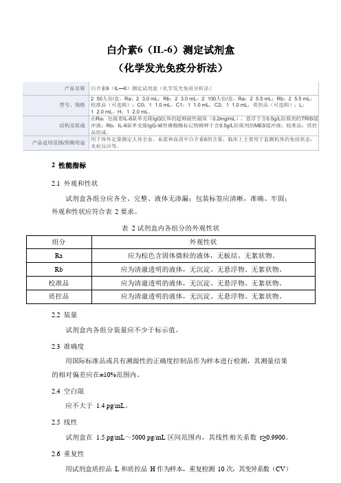
白细胞介素6(IL-6)测定试剂盒(电化学发光免疫分析法)产品技术要求lztk
白细胞介素6(IL-6)测定试剂盒(电化学发光免疫分析法)适用范围:该试剂盒用于体外定量测定人体血清样本中白细胞介素6(IL-6)的含量。
1.1产品型号/规格:50人份/盒、100人份/盒。
1.2主要组成试剂盒由磁分离试剂(M)、试剂a(Ra)、试剂b(Rb)和定标品(IL-6-Cal)(选配)组成。
组成及含量如下:2.1 外观2.1.1 试剂盒各组分应齐全、完整、液体无渗漏;2.1.2 磁分离试剂摇匀后应为棕色含固体微粒的均匀悬浊液,无明显凝集、无絮状物;2.1.3 其它液体组分应澄清,无异物,沉淀物或絮状物;2.1.4 包装标签应清晰、无磨损、易识别。
2.2 空白限应不大于1.5pg/mL。
2.3 准确度用IL-6国际标准品(89/548)进行检测,其测量结果的相对偏差应在±10%范围内。
2.4 线性在[5.0,5000.0]pg/mL范围内,线性相关系数(r)应不小于0.9900。
2.5 精密度2.5.1 分析内精密度在试剂盒的线性范围内,浓度为(30.0±6.0pg/mL)和(500.0±100.0pg/mL)的样品检测结果的变异系数(CV)应不大于8%。
2.5.2 批间精密度在试剂盒的线性范围内,用3个批号试剂盒分别检测浓度为(30.0±6.0pg/mL)和(500.0±100.0pg/mL)的样品,检测结果的变异系数(CV)应不大于15%。
2.6 效期末稳定性本产品效期为15个月,试剂盒在2~8℃下保存至有效期末进行检测,检测结果应符合2.1、2.2、2.3、2.4、2.5.1的要求。
2.7 溯源性依据GB/T21415-2008《体外诊断医疗器械生物样品中量的测量校准品和控制物质赋值的计量学溯源性》的要求,定标品溯源到IL-6国际标准品(89/548)。
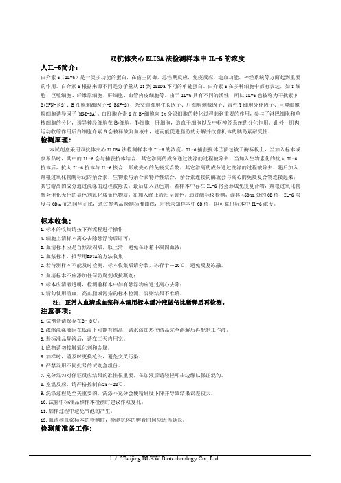
白介素6(IL-6)测定试剂盒产品技术要求深圳市锦瑞生物
白介素6(IL-6)测定试剂盒
(化学发光免疫分析法)
2性能指标
2.1外观和性状
试剂盒各组分应齐全、完整、液体无渗漏;包装标签应清晰,准确、牢固;外观和性状应符合表2 要求。
表 2 试剂盒内各组分的外观性状
2.2装量
试剂盒内各组分装量应不少于标示值。
2.3准确度
用国际标准品或具有溯源性的正确度控制品作为样本进行检测,其测量结果的相对偏差应在±10%范围内。
2.4空白限
应不大于 1.4 pg/mL。
2.5线性
试剂盒在 1.5 pg/mL~5000 pg/mL 区间范围内,其线性相关系数r≥0.9900。
2.6重复性
用试剂盒质控品L 和质控品H 作为样本,重复检测10 次,其变异系数(CV)
应不大于7%。
2.7批间差
用3 个批号试剂盒分别检测同一瓶质控品L 和质控品H,3 个批号试剂盒之间的批间变异系数(CV)应不大于9%。
2.8质控品测定值
用试剂盒配套的校准品校准测量系统后,以试剂盒配套的质控品作为样本进行检测,其测定结果应在标示范围内。
2.9质控品均一性
瓶间变异系数(CV)应不大于10%。
2.10校准品测量准确度
用工作校准品校准测量系统后,以试剂盒配套的校准品作为样本进行检测,其测定结果与标示值的相对偏差应在±10%范围内。
2.11校准品均一性
瓶间变异系数(CV)应不大于10%。
用ELISA试剂盒检测人白介素6IL-6的浓度
双抗体夹心ELISA法检测样本中IL-6的浓度人IL-6简介:白介素6(IL-6)是一类多功能的蛋白,在宿主防御,急性期反应,免疫反应,造血功能,神经系统等方面起到重要的作用。
白介素6根据来源不同是分子量从21到28kDa不同的单链蛋白。
白介素6在多种细胞中都有表达,如T细胞、巨噬细胞、纤维原细胞、肝细胞、血管内皮细胞等。
由于IL-6具有不同的活性,所以IL-6也被称为干扰素β2(IFN-β2)、B细胞刺激因子-2(BSF-2)、杂交瘤细胞生长因子、肝细胞刺激因子、毒性T细胞分化因子、巨噬细胞粒细胞诱导因子(MGI-2A)。
白细胞介素6在B-细胞向Ig分泌细胞的转化过程起到重要的作用,参与了淋巴细胞和单核细胞的分化,诱导神经细胞在B-细胞,T-细胞,肝细胞,造血干细胞以及中枢神经系统的分化作用。
此外,肌肉运动收缩作用后白细胞介素6会被释放到血液中,进而能促进脂肪的分解并改善机体的胰岛素耐受性。
检测原理:本试剂盒采用双抗体夹心ELISA法检测样本中IL-6的浓度。
IL-6捕获抗体已预包被于酶标板上,当加入标本或参考品时,其中的IL-6会与捕获抗体结合,其它游离的成分通过洗涤的过程被除去。
当加入生物素化的抗人IL-6抗体后,抗人IL-6抗体与IL-6接合,形成夹心的免疫复合物,其它游离的成分通过洗涤的过程被除去。
随后加入辣根过氧化物酶标记的亲合素。
生物素与亲合素特异性结合,亲合素连接的酶就会与夹心的免疫复合物连接起来;其它游离的成分通过洗涤的过程被除去。
最后加入显色剂,若样本中存在IL-6将会形成免疫复合物,辣根过氧化物酶会催化无色的显色剂氧化成蓝色物质,在加入终止液后呈黄色。
通过酶标仪检测,读其450nm处的OD值,IL-6浓度与OD450值之间呈正比,通过参考品绘制标准曲线,对照未知样本中OD值,即可算出标本中IL-6浓度。
标本收集:1.标本的收集请按下列流程进行操作:A.细胞上清标本离心去除悬浮物后即可;B.血清标本应是自然凝固后,取上清,避免在冰箱中凝固血液;C.血浆标本,推荐用EDTA的方法收集;D.若待测样本不能及时检测,标本收集后请分装,冻存于-20℃,避免反复冻融。
白介素6检测标准操作程序

白介素6检测标准操作程序1检验目的本试剂盒用于体外定量测定人血清和(或)血浆中的白介素6(IL-6)。
2检验原理及方法夹心法。
测定总时长18分钟。
1.第1次孵育:30L样本、生物素化的白介素6特异性单克隆抗体一起孵育,形成抗原一抗体复合物。
2.第2次孵育:添加包被链霉亲合素的磁珠微粒进行孵育,复合体与磁珠通过生物素和链霉亲合素的作用结合。
3.将反应液吸入测量池中,通过电磁作用将磁珠吸附在电极表面,未与磁珠结合的物质通过ProCell/ProCell M被去除。
给电极加以一定的电压,使复合体化学发光,并通过光电倍增器测量发光强度;4.通过由2点校准生成的分析仪专有的校准曲线和通过试剂条码或电子条形码提供的主曲线来确定结果。
3性能特征3.1检测范围:1.5-5000pg/mL3.2精密度:根据CLSI(临床实验室标准委员会)的方案(EP5-A2),使用Elecsys 试剂、混合人血清和 CLSI(临床和实验室标准化学会)修改协议(EP5-A)规定的质控品确定精密度:每天2次重复共21天;MODULAR ANALYTICSE170分析仪的批内精密度,n=84。
所得结果如下Cobas e 411 分析仪e)可重复性=批内精密度3.3分析特异性/干扰下列物质不会对Elecsys白介素-6检测(浓度范围10-200pg/mL)造成明显的交叉反应。
4标本4.1检测标本:血清和血浆(肝素锂、肝素钠、EDTA和柠檬酸钠抗凝)是推荐使用的样本类型。
尚未验证其他抗凝剂能否适用。
4.2采集及处理:在进行离心操作前需让血清样本完全凝结(凝结时间不小于1小时)。
所有的待测样本在测试前均应进行离心操作。
顶部含脂质的离心样本,需转移澄清、无脂质的部分到新的样本管中。
在测试前,确保已去除了残余的纤维蛋白和细胞类物质。
应小心处理患者样本,避免交叉污染。
在测试前,确保患者样本、校准品、质控品均已平衡至环境温度(20-25°C)。
白介素6_人白介素6_Human IL-6使用说明书
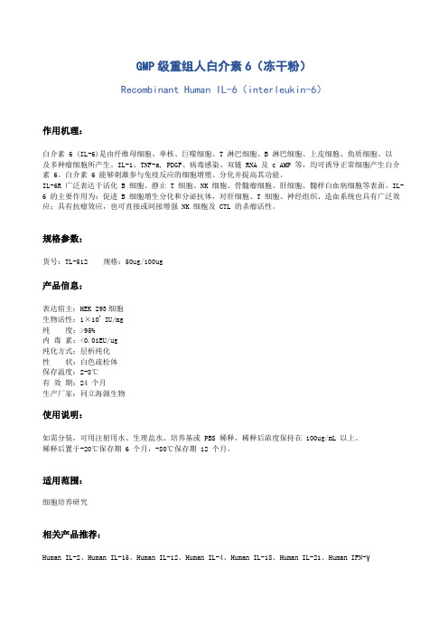
GMP级重组人白介素6(冻干粉)Recombinant Human IL-6(interleukin-6)作用机理:白介素 6 (IL-6)是由纤维母细胞、单核、巨噬细胞、T 淋巴细胞、B 淋巴细胞、上皮细胞、角质细胞、以及多种瘤细胞所产生。
IL-1、TNF-a, PDGF、病毒感染、双链 RNA 及 c AMP 等,均可诱导正常细胞产生白介素 6。
白介素 6 能够刺激参与免疫反应的细胞增殖、分化并提高其功能。
IL-6R 广泛表达于活化 B 细胞、静止 T 细胞、NK 细胞、骨髓瘤细胞、肝细胞、髓样白血病细胞等表面。
IL- 6 的主要作用为:促进 B 细胞增生分化和分泌抗体,对肝细胞、T 细胞、神经组织、造血系统也具有广泛效应;具有抗瘤效应,也可直接或间接增强 NK 细胞及 CTL 的杀瘤活性。
规格参数:货号:TL-512 规格:50ug/100ug产品信息:表达宿主:HEK 293细胞生物活性:1×10⁷ IU/mg纯度:>95%内毒素:<0.01EU/ug纯化方式:层析纯化性状:白色疏松体保存温度:2-8℃有效期:24 个月生产厂家:同立海源生物使用说明:如需分装,可用注射用水、生理盐水、培养基或 PBS 稀释,稀释后浓度保持在 100ug/mL 以上。
稀释后置于-20℃保存期 6 个月,-80℃保存期 12 个月。
适用范围:细胞培养研究相关产品推荐:Human IL-2、Human IL-15、Human IL-12、Human IL-4、Human IL-18、Human IL-21、Human IFN-γ。
白介素6测定试剂盒(化学发光法)产品技术要求丽珠
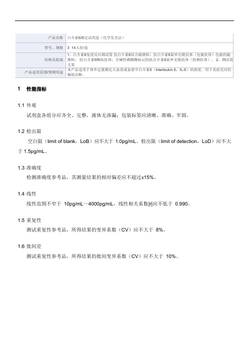
1.1外观
试剂盒各组分应齐全、完整,液体无渗漏;包装标签应清晰、准确、牢固。
1.2检出限
空白限(limit of blank,LoB)应不大于1.0pg/mL。检出限(limiБайду номын сангаас of detection,LoD)应不大于1.5pg/mL。
1.3准确度
检测准确度参考品,其测量结果的相对偏差应不超过±15%。
1.4线性
线性范围不窄于10pg/mL~4000pg/mL,线性相关系数|r|应不低于0.990。
1.5重复性
测试重复性参考品,所得结果的变异系数(CV)应不大于8%。
1.6批间差
测试重复性参考品,所得结果的批间变异系数(CV)应不大于10%。
产品名称
白介素6测定试剂盒(化学发光法)
型号、规格
3×14人份/盒
结构及组成
1、白介素6免疫反应测试筒抗白介素6结合磁微粒:抗白介素6鼠单克隆抗体(包被抗体)包被的磁微粒;抗白介素6酶标抗体:含碱性磷酸酶标记的抗白介素6鼠单克隆抗体(检测抗体)。2、测试筒支架
产品适用范围/预期用途
本产品适用于体外定量测定人血清或血浆中白介素6(Interleukin 6,IL-6)的浓度。用于炎症反应的辅助诊断。
- 1、下载文档前请自行甄别文档内容的完整性,平台不提供额外的编辑、内容补充、找答案等附加服务。
- 2、"仅部分预览"的文档,不可在线预览部分如存在完整性等问题,可反馈申请退款(可完整预览的文档不适用该条件!)。
- 3、如文档侵犯您的权益,请联系客服反馈,我们会尽快为您处理(人工客服工作时间:9:00-18:30)。
人白介素6(IL-6)试剂盒使用方法
检测范围:96T
0-8 ng/L
使用目的:
本试剂盒用于测定人血清、血浆及相关液体样本中白介素6(IL-6)含量。
实验原理
本试剂盒应用双抗体夹心法测定标本中人白介素6(IL-6)水平。
用纯化的人白介素6(IL-6)抗体包被微孔板,制成固相抗体,往包被单抗的微孔中依次加入白介素6(IL-6),再与HRP 标记的白介素6(IL-6)抗体结合,形成抗体-抗原-酶标抗体复合物,经过彻底洗涤后加底物TMB显色。
TMB在HRP酶的催化下转化成蓝色,并在酸的作用下转化成最终的黄色。
颜色的深浅和样品中的白介素6(IL-6)呈正相关。
用酶标仪在450nm波长下测定吸光度(OD 值),通过标准曲线计算样品中人白介素6(IL-6)浓度。
试剂盒组成
1.标本采集后尽早进行提取,提取按相关文献进行,提取后应尽快进行实验。
若不能马上进行试验,可将标本放于-20℃保存,但应避免反复冻融
2.不能检测含NaN3的样品,因NaN3抑制辣根过氧化物酶的(HRP)活性。
操作步骤
1.标准品的稀释:本试剂盒提供原倍标准品一支,用户可按照下列图表在小试管中进行稀
2.加样:分别设空白孔(空白对照孔不加样品及酶标试剂,其余各步操作相同)、标准孔、
待测样品孔。
在酶标包被板上标准品准确加样50μl,待测样品孔中先加样品稀释液40μl,然后再加待测样品10μl(样品最终稀释度为5倍)。
加样将样品加于酶标板孔底部,尽量不触及孔壁,轻轻晃动混匀。
3.温育:用封板膜封板后置37℃温育30分钟。
4.配液:将20倍浓缩洗涤液用蒸馏水20倍稀释后备用
5.洗涤:小心揭掉封板膜,弃去液体,甩干,每孔加满洗涤液,静置30秒后弃去,如此
重复5次,拍干。
6.加酶:每孔加入酶标试剂50μl,空白孔除外。
7.温育:操作同3。
8.洗涤:操作同5。
9.显色:每孔先加入显色剂A50μl,再加入显色剂B50μl,轻轻震荡混匀,37℃避光显色
15分钟.
10.终止:每孔加终止液50μl,终止反应(此时蓝色立转黄色)。
11.测定:以空白空调零,450nm波长依序测量各孔的吸光度(OD值)。
测定应在加终止
液后15分钟以内进行。
操作程序总结:
计算
以标准物的浓度为横坐标,OD值为纵坐标,在坐标纸上绘出标准曲线,根据样品的OD值由标准曲线查出相应的浓度;再乘以稀释倍数;或用标准物的浓度与OD值计算出标准曲线的直线回归方程式,将样品的OD值代入方程式,计算出样品浓度,再乘以稀释倍数,即为样品的实际浓度。
注意事项
1.试剂盒从冷藏环境中取出应在室温平衡15-30分钟后方可使用,酶标包被板开封后如未用完,板条应装入密封袋中保存。
2.浓洗涤液可能会有结晶析出,稀释时可在水浴中加温助溶,洗涤时不影响结果。
3.各步加样均应使用加样器,并经常校对其准确性,以避免试验误差。
一次加样时间最好
控制在5分钟内,如标本数量多,推荐使用排枪加样。
4.请每次测定的同时做标准曲线,最好做复孔。
如标本中待测物质含量过高(样本OD值大于标准品孔第一孔的OD值),请先用样品稀释液稀释一定倍数(n倍)后再测定,计算时请最后乘以总稀释倍数(×n×5)。
5.封板膜只限一次性使用,以避免交叉污染。
6.底物请避光保存。
7.严格按照说明书的操作进行,试验结果判定必须以酶标仪读数为准.
8.所有样品,洗涤液和各种废弃物都应按传染物处理。
9.本试剂不同批号组分不得混用。
10. 如与英文说明书有异,以英文说明书为准。
保存条件及有效期
1.试剂盒保存:;2-8℃。
2.有效期:6个月。