放线菌分类-完整精选.
微生物,放线菌分类

3 基于核酸结构的分析
(1) 16S rRNA / rDNA基因序列的分析
该技术目前在放线菌分类中常被用于放线菌种属的 初步鉴定, 但是值得注意的是仅根据该指标并不能 确定菌株的种属, 必须结合其他的分类指标。
(2)16S-23S rRNA 转录间区序列的分析
16S-23S rRNA转录间区序列( 16S-23S rRNA)的进 化速率比16S rRNA大10倍, 其序列大小比16S r RNA更具直接测序优势, 可鉴别防线菌属与种。但 是16S-23S rRNA的不同拷贝之间大小比较相近且不 易区分。
经典分类学:按微生物 表型特征分类
表型特征:形态学、生理生化学、生 态学等,推断微生物的系统发育。 表型特征结合分子水平上的基因型特 征(如16S rRNA),探讨微生物进化地 位、系统发育关系并进行分类鉴定。
微生物系统学: 按亲缘关系和进 化规律分类
★微生物分类学的三个任务:分类、鉴定及命名 ☆分类是根据微生物的相似性和亲缘关系,将微生物归入不同的分类类群。 ☆鉴定是确定一个新的分离物属于已经确认的哪个分类类群的过程。 ☆命名是根据国际命名法规给每一微生物类群及种类以科学的名称。
5 多相分类系统
由于微生物生理生化学的迅速发展,加速了这趋势 并逐渐打破了原来经典的描述分类,建立了如数值 分类、化学分类、分子分类以至于现在的多相分类 新的标准,大大推进了分类学的发展。多相分类是 将表型、基因型以及系统进化分析数据综合起来作 为分类和进化分析的依据。
3 放线菌的应用
在植物病害防治中的应用 放线菌是人们最早应用到生产中的生防微生 物,但是已投入使用的微生物源杀虫活性物质中 多数是来自真菌,有部分来自细菌如昆虫的病原 菌苏云金杆菌,放线菌来源的较少,从放线菌中 发现的活性物质中,80%是来自链霉菌属。
海洋放线菌的分类鉴定

海洋放线菌的分类鉴定一、放线菌㈠、放线菌的分布放线菌常以孢子或菌丝状态极其广泛地存在于自然界。
不论数量和种类,以土壤中最多。
河流和湖泊中,放线菌数量不多,大多为小单孢菌、游动放线菌和孢囊链霉菌,还有少数链霉菌。
海洋中的放线菌多半来自土壤或生存在漂浮海面的藻体上。
海水中还存在耐盐放线菌。
放线菌有一种土霉味,使水和食物变味,有的放线菌也能和霉菌一样使棉毛制品或纸张霉变。
放线菌主要能促使土壤中的动物和植物遗骸腐烂,最主要的致病放线菌是结核分枝杆菌和麻风分枝杆菌,可导致人类的结核病和麻风病。
放线菌最重要的作用是可以产生、提炼抗生素,目前世界上已经发现的2000多中抗生素中,大约有56%是由放线菌(主要是放线菌属)产生的,如链霉素、土霉素、四环素、庆大霉素等都是由放线菌产生的。
此外有些植物用的农用抗生素和维生素等也是由放线菌中提炼的。
㈡、放线菌的形态与结构放线菌菌体为单细胞,大多由分枝发达的菌丝组成,最简单的为杆状或具原始菌丝。
放线菌是一种革兰氏阳性菌。
放线菌菌丝细胞的结构与细菌基本相同。
根据菌丝形态和功能可分为营养菌丝、气生丝和孢子丝三种。
⑴营养菌丝匍匐生长于培养基内,主要生理功能是吸收营养物,故亦称基内菌丝。
营养菌丝一般无隔膜;直径0.2-0.8微米,但长度差别很大,短的小于100微米,长的可达600微米以上;有的无色素,有的产生黄、橙、红、紫、蓝、绿、褐、黑等不同色素,若是水溶性的色素,还可透入培养基内,将培养基染上相应的颜色,如果是非水溶性色素,则使菌落吨呈现相应的颜色。
色素是鉴定菌种的重要依据。
⑵气生菌丝长出培养基外并伸向空间的菌丝为气生菌丝。
它叠生于营养菌丝上,以至可覆盖整个菌落表面。
在光学显微镜下,颜色较深,直径比营养菌丝粗,约1-1.4微米,其长度则更悬殊。
直形或弯曲而分枝,有的产生色素。
⑶孢子丝气生菌丝上分化出可形成孢子的菌丝即孢子丝。
孢子丝的形状和在气生菌丝上的排列方式,随菌种而异。
孢子丝的形状有直形、波曲和螺旋形。
放线菌属的分类及描述
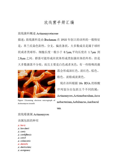
放线菌手册汇编放线菌科概述Actinomycetaceae描述:放线菌科是由Buchanan 在1918年创立的该科的一般特征是:革兰氏染色阳性,分支,偏直条状,大多数成员是属于球杆状或者类球形。
细胞长度一般小于0.5μm,平均长度在1.7μm 到2,9μm 之间。
群落可能形成丝状体形成类似菌丝体的外形,但是大多数菌落不分枝,而且主要是白色或者灰色,有一些特殊的菌落会形成深红色、淡红色、棕色、粉色、淡粉或淡黄色。
现在该科根据16s RNA 的核酸序列划分出包括五个不同的属:Actinomyces,Actionobaculum,Arcanobacterium,Aobiluncus,Aaribaculum.放线霉菌属Actinomyces该属包括的种有A. bovisA. bowdeniiA. canisA. cardiffensisA. catuliA. coleocanisA. dentalisA. denticolensA. europaeusFigure 1Scanning electron micrograph ofActinomyces israeliiA. funkeiA. georgiaeA. gerencseriaeA. graevenitziiA. hongkongensisA. hordeovulnerisA. howelliiA. humiferusA. hyovaginalisA. israeliiA. marimammaliumA. meyeriA. naeslundiiA. nasicolaA. neuiiA. odontolyticusA. oricolaA. radicidentisA. radingaeA. slackiiA. streptomyciniA. suimastitidisA. suisA. turicensisA. urogenitalisA. vaccimaxillaeA. viscosus一、Actinomyces bovisActinomyces bovis is a gram-positive, rod-shaped bacterium of the genus Actinomyces. It is the causative agent of Lumpy jaw in cattle, and occasionally causes infections in humansHistoryActinomyces bovis was first described in 1877 by C. O. Harz, as a microbe within the jaw tissue of cows with lumpy jaw. It was thought to be identical to Actinomyces israelii until 1940, when D. Erikson showed these to be two separate organisms.Figure 2Bovine actinomycosis. Granules (sulfa granules), consisting of colonies of bacteria (Actinomyces bovis) and club-shaped(球棒状的)reaction product, are within a purulent exudate.(脓性渗出物)二、Actinomyces georgiaeActinomyces georgiae is a species in the genus of Actinomyces. It isa part of the human periodontal flora(牙周菌群).三、Actinomyces gerencseriaeActinomyces gerencseriae (Johnson et al. 1990) used to be known asActinomyces israelii serovar II.Actinomyces gerencseriae was named for the bacteriologist, Mary Ann Gerencser.Figure 3蜘蛛状高度分支的微菌落,26h在36°下培养(1500倍放大)Figure 4培养于BHIA上的微菌落,显示了一条带有分支的长丝(24h在36°条件下)四、Actinomyces israeliiActinomyces israelii is a species of Gram-positive, rod-shaped bacteria within the Actinomyces. Known to live commensally on and within humans, A. israelii is an opportunistic pathogen and a cause of actinomycosis. Many physiologically diverse strains of the species are known to exist, though all are strict anaerobes.Actinomycosis is most frequently caused by A. israelii. It is a normal colonizer of the vagina, colon, and mouth. Infection isestablished first by a breach of the mucosal barrier during various procedures (dental, gastrointestinal), aspiration, or pathologies such as diverticulitis. The chronic phase of this disease is also known the "classic phase" because the acute, early phase is often missed by health care providers. This is characterized by slow, contiguous growth that ignores tissue planes and forms a sinus(窦) tract that can spontaneously heal and recur, leading to a densely fibrotic lesion(纤维性病变). This lesion is often characterized as "wooden". Sulfur granules(瘤状颗粒) form in a central purulence(脓)surrounded by neutrophils(嗜中性粒细胞). This conglomeration of organisms is virtually diagnostic of A. israelii.来源:Historically speaking the first written appearance of Actinomyces was made in 1877 when pathologist Otto Bollinger described their presence in cattle. Shortly after that James Israel discovered another specie of actinomyces or actinomycetes that are living in humans and in his honour these actinomyces have name actinomyces israelii. 概念性解释:Anaerobic organism represents any organism that doesn't require oxygen for its growth and in the presence of oxygen it may even die, while facultative anaerobic organism is usually a bacteria that creates adenosine triphosphate (ATP) that is one of the most used enzymes or cabalists or it can turn to the process of fermentation if it is exposed to the oxygen. Gram positive bacteria are those that according to Gram staining are dark blue or violet in colour. If a bacterium is Gram negative it will not be able to retain the violet strain of crystal.There is one difference between Actinomyces and other bacteria and that is that Actinomyces don't form endospores and they form branched networks of hyphae that looks like fungus while otherbacteria are in a shape of rod and they don't make branched networks. In fungi hyphae are the main mode of vegetative growth and their collective name is mycelium.五、Actinomyces naeslundiiActinomyces naeslundii is a Gram-positive, rod-shapedbacterium found in the mouth of humans. The species has beenimplicated in periodontal disease, as well as various tooth cavities.In other cases, A. naeslundii is associated with good oral health. It isone of the first bacteria to occupy the oral cavity and colonize thetooth's surface. It has also been isolated from women with bacterialvaginosis.Any species of the genus Actinomyces, including A. naeslundii, cause a chronic disease called actinomycosis, which is characterized by swelling and formation of an abscess (脓肿) which may exude pus.This can beaccompaniedby tissue fibrosis.六、Actinomyces radicidentisActinomyces radicidentis is a species in the genus of Actinomyces, first isolated from infected root canals of teeth. ITs type strain is CCUG 36733. Once characterized, it has since been found to be present in failed root canal treatments. Its pathogenicity has been suggested to be due to an ability to form cell aggregates, held together by embedding in an extracellular matrix in host tissues. Like other pathogenic Actinomyces, by collectively finding itself in Figure 5Histopathological changes due to en:Actinomycesnaeslundii bacteria. en:Silver stain, brain abscess. Obtained fromthe CDC Public Health Image Library.a protected biofilm(生物膜)environment can evade elimination by host defenses, including phagocytosis.Figure 6 Scanning (a-d) and transmission electron micrographs (e-g) of A radicidentis, strain CCUG 36733. Cells grown on Brucella blood agar exhibit coccoid shape (a), similar to cells grown in serum-supplemented Tryptic soy broth, where fimbriae-like bundles can be seen (b). When cells were grown in RPMI-1640 broth with serum (c, d), they were rod-shaped with intertwining bundles of fimbriae-like cell appendices in a netlike arrangement. In thin section (e), bundle of fimbriae can be observed emerging from cell surface; demarcated area in (e) is magnified in (f). Further thin section (g) shows radially placed fimbriae emerging from fuzzy-coated cell surface. Bars = 1 µm.七、Actinomyces viscosusActinomyces viscosus is a human and animalpathogen/pathobiont which colonizes the mouths of most adult humans.It is Gram-negative, anaerobic, rod-shaped, and filamentous.It causes periodontal disease(牙周病)in animals, and has been isolated from human dental calculus(牙结石)and root surface caries, as well as the oral cavity hamsters and actinomycotic lesions in swine, cats, and dogs. Furthermore, it has been shown to cause endocarditis(心内膜炎).Actinomyces viscosus is aGram-positive, anaerobic,filamentous bacterium that ispart of the human oral flora.This rod shaped filamentousbacteria occur around the teeth,gums(牙龈)and throat inhealthy humans. Species of this bacterium can cause actinomycosis - a granulomatous infection with the formation of abscesses in the mouth, lungs, or the gastrointestinal tract. Oral actinomycosis may occur due to trauma such as a tooth extraction or bleeding gums. Abdominal actinomycosis may follow appendicitis. Treatment is with antibiotics. Actinomyces spp.normally are soil or aquatic fungi feeding off decaying matter (saprophytes,sapro,spoiled). They resemble fungi and form fungi-like mats (mycelia)关于gram-staining:染色效果:Figure 7A Gram stain of mixed Staphylococcus aureus (S. aureus ATCC 25923, gram-positive cocci, in purple) and Escherichia coli (E. coli ATCC 11775, gram-negative bacilli, in red), the most common Gram stain reference bacteria.Gram-positive bacteria havea thick mesh-like cell wallmade of peptidoglycan(50–90% of cell envelope), andas a result are stainedPurple by crystal violet, whereasGram-negative bacteriahave a thinner layer(10% of cell envelope), so donot retain the purple stainand are counter-stained pink by safranin.八、Actinomyces odontolyticusActinomyces odontolyticus is an anaerobic, facultativecapnophilic, gram-positive, nonsporulating, non–acid fast(不耐酸的),non-motile, irregularly staining bacterium. Sometimesshort or medium-sized rods resembling diphtheroids(白喉)areseen. Shorter rods resembling propionibacteria are frequentlyseen with A. odontolyticus and may be arranged inpalisades(栅栏)as well as other diphtheroidal arrangements. Onblood agar, the bacteria develop as small, irregular, whitishcolonies that are smooth to slightly granular and show adark red pigment when mature (2–14 days). This pigmentationis most obvious when the cultures are left standing inair at room temperature after primary anaerobic isolation.The organism also grows well on CNA and Brucella agar. Definitive identification is madeby negative catalaseand oxidasetests, the reduction of nitrate(硝酸盐)to nitrite(亚硝酸盐),filamentationof microcolonies,and absence of growth at pH5.5.Generally, the fermentation(发酵)reactions are variable.。
放线菌分类2

对营养的要求相当弹性,能降解一些杀虫剂和除草剂
嗜皮菌属(Dermatophilus)
寄生于哺乳动物,导致刚果嗜皮菌病的皮肤感染
链霉菌亚目
目前该亚目只有链霉菌科,该科目前有三个属,即 链霉菌属、北里孢菌属和嗜酸链霉菌属。
该科成员均为革兰氏阳性菌好氧菌,产生发育良好、 不易断裂的分支状基内菌丝,大多数产生分支状气
模式种:白色链霉菌
北里孢菌属
该属特征为:基内菌丝发达,类似链霉菌,基内和 气生菌丝上着生常超过20个孢子的长孢子链。基内 菌丝不断裂,无孢囊。细胞壁含甘氨酸、半乳糖,
全细胞水解液含半乳糖,但不含阿拉伯糖、马杜拉
糖和木糖。磷酸类脂类型为P2型, DHA的(G+C) mol%含量范围为66-67
微球菌亚目
包括十四个科,典型科为微球菌科,该科的典型属为微球菌
微球菌属(Micrococcus)
广泛存 在于土壤中,水中和哺乳动物皮肤上,后者更为 常 见,但并无致病性
节杆菌属(Arthrobacter)
杆菌一球菌的生长循环:对数期时,细菌呈不规则,分支
的杆状,以断裂繁殖。当它们进入稳定期,细胞变成球状
பைடு நூலகம்
鱼孢菌科及主要属
该科典型属是鱼孢菌属 鱼孢菌属:菌丝和孢子革兰氏阳性,不抗酸,细胞壁化 学组分Ⅲ型。无基内菌丝。气生菌丝一般短,(0.5~
1.2)×(10~25)微米,少分枝;基部细胞下面形成球冠状
固着器,用以将气生菌丝固定在固体培养基的表面;气生菌 丝断裂成表面光滑的孢子,孢子球形或杆状,有时呈长 达6微米的鱼形,本科因此得名。孢子有颈饰痕迹和1~3 根端生鞭毛,能游动;革兰氏染色不定;细胞壁Ⅰ型。本属 现只发现多形鱼孢菌1种。
什么是放线菌
什么是放线菌引言放线菌(Actinobacteria)是一类广泛存在于自然环境中的细菌,也是一类非常重要的微生物资源。
它们具有丰富的代谢能力和生物活性产物,对于农业、药物开发、环境保护等领域具有重要的应用价值。
放线菌的研究和应用已经成为微生物学和生物技术领域的热点之一。
本文将从分类特征、生物特性、应用领域等方面对放线菌进行详细介绍。
一、分类特征1. 形态特征放线菌是革兰氏阳性细菌,其细胞多为直杆状,长为0.2-2.0μm,直径为0.5-1.0μm。
有的放线菌细胞会形成分枝或丝状结构,使得其菌落呈现放射状生长。
2. 细胞壁特征放线菌的细胞壁主要由多肽聚糖组成,其中N-乙酰葡萄醣胺和N-乙酰半乳葡萄糖胺是其特征性成分。
这些特殊的细胞壁结构使得放线菌对抗生物膜、抗药物和耐酸碱有一定的能力。
3. 分类系统放线菌属于细菌界放线菌纲(Actinobacteria),目前已知的放线菌约有50个属。
根据形态特征、生理和生态习性等分类指标,放线菌可以进一步分为不同的科、属和种。
二、生物特性1. 生长环境放线菌广泛存在于土壤、水体和植物表面等自然环境中。
它们对土壤质地、pH值、湿度和养分含量等因素有一定的适应性,因此在地球生态系统中分布十分广泛。
2. 代谢能力放线菌具有丰富的代谢能力,可以利用多种有机物和无机物作为碳源、氮源和能源。
许多放线菌具有优良的降解能力,能够降解有机污染物和农药,对环境保护具有重要的意义。
3. 生物活性产物放线菌是许多重要天然产物的产生者,其中包括抗生素、抗肿瘤活性物质、抗氧化物质等。
这些生物活性产物对细菌、真菌和肿瘤细胞等具有显著的抑制或杀灭作用,对人类的健康和医疗具有重要意义。
三、应用领域1. 农业应用放线菌具有优良的土壤分解和降解能力,可以降解农药残留、处理农业废弃物等。
此外,放线菌还能够产生一些具有生物肥料作用的物质,可以促进植物的生长和发育。
2. 药物开发放线菌是抗生素的重要来源之一,许多著名的抗生素如链霉素、土霉素等都是由放线菌产生的。
放线菌

气生菌丝发育到一定阶段,其上可分化出形成孢子 二、放线菌 的菌丝,即孢子丝
营养菌丝匍匐生长于 培养基内,吸收营养
营养菌丝发育到一定阶段, 伸向空间形成气生菌丝
二、放线菌
二、放线菌
二、放线菌
二、放线菌
孢子丝释放孢子
繁殖菌丝 (孢子丝)
孢子在适宜 的条件下萌 发,长出1-3 个芽管
气生菌丝
营养菌丝
由于多数链霉菌具有分泌抗生物质或细 胞外酵素的能力,可以有效抑制植物病 原菌。此外,少部分还具有促进植物生 长或诱导植物产生抗病性的效果,因此 链霉菌在生物防治应用上极具潜力。
链霉菌在植物保护上的应用
生物防治法是农业生态系中,植物病原、 昆虫与益菌或天敌等族群间维持均衡的 重要策略之一。就植物病害而言,其定 义是指在自然或人为操控的环境下,透 过一种或多种拮抗微生物,有效降低病 原菌的密度、活力及感染作物的能力, 进而达到防治植物病害的效果。
五、放线菌的主要属
放线菌的主要属
(一)诺卡氏型(Nocardioforms) 特征:好气型,具气生菌丝,多产粉 孢子。 应用:烃类发酵,污水处理 ,产生 抗生素(如万古霉素、头孢菌 素等)
放线菌的主要属
(二)具有气生菌丝特化出的孢囊孢子和分生孢子 1.弗兰克氏放线菌属(Frankia) 可以固氮,存在于非豆科植物根部, (如: 桤木、赤扬等) 2.小单孢菌属(Micromonospora) 可产生多种抗生素,如庆大霉素、利福霉素; 有的种可积累VitB12 3.链霉菌属(Streptomyces) 产生许多著名的抗生素,如链霉素、红霉素、 四环素、土霉素、金菌
A:诺尔斯氏链霉菌; B:皮疽诺卡氏菌; C:酒红指孢囊菌; D:游动放线菌; E:小单胞菌; F:皱双孢马杜拉放线菌
《放线菌分类》课件

表面,主要功能为吸收营养物。 · 显微镜下,基内菌丝比气生菌丝细,多分枝,透明,颜色较浅。直径一般
为0.4-1.2微米,大多数类群不形成横隔也不断裂。少数类群(如诺卡氏菌) 基内菌丝剧烈弯曲成树根状,生长到一定阶段形成横隔,并断裂成不同形
· 5、放线菌次生代谢药物的设计和改造
三、放 线 菌 生 物 技 术 的 发 展
2023/10/14
· (一)分离培养和纯化技术 · 近年来,国内外科学家正在开展一种从自然生态环境中选择性分离放线
菌的分离技术。 · 非链霉菌或稀有放线菌作为新的生理活性物质的重要来源成为关注的热
点。
· Colwell 实验室在1982年提出活的但不可培养微生物的概念,他们发现 将霍乱弧菌和大肠埃希菌(简称大肠杆菌)转到不含营养物质的盐水中, 经常长时间的低温保存,细菌会进入一种数量不减、有代谢活力、但在 正常试验室培养条件下不能生长产生菌落的状态,称为活的但不能培养 (viable but nonculturable, VBNC)
现在
· 一、 国外概况
· 1、历史及著名研究单位 · 最早描述放线菌的学者——Cohn, 他自人泪腺感染病灶中分离到一株丝状病
原菌——链丝菌。而后Harz建立了放线菌属。 · 美国新泽西农业试验站, Rutgers大学微生物研究所 · 1942年发现链霉素。 · 《放线菌的属和种分类鉴定和描述》,放线菌分类学开始形成。 · 整个70年代是放线菌化学分类时代 · 构建放线菌系统发育树,进入分子分类时代
放线菌分类
第一部分总论
2023/10/11
第一节概论
· 一、放线菌在微生物系统学中的地位 · 19世纪前曾将放线菌归入真菌,后将其列于细菌中 · 克拉西里尼科夫首先将放线菌放在植物界,原生植物门,裂殖菌纲,后有人
放线菌
孢子丝发育到一定阶段便分化为分生孢子。在光学显微镜下,孢子呈圆形、椭圆形、杆状、圆柱状、瓜子状、梭状和半月状等,孢子的颜色十分丰富。孢子表面的纹饰因种而异,在电子显微镜下清晰可见,有的光滑,有的褶皱状、疣状、刺状、毛发状或鳞片状,刺又有粗细、大小、长短和疏密之分。
生孢囊放线菌的特点是形成典型孢囊,孢囊着生的位置因种而异。有的菌孢囊长在气丝上,有的菌长在基丝上。孢囊形成分两种形式:有些属菌的孢囊是由孢子丝卷绕而成;有些属的孢囊是由孢囊梗逐渐膨大。孢囊外围都有囊壁,无壁者一般称假孢囊。孢囊有圆形、棒状、指状、瓶状或不规则状之分。孢囊内原生质分化为孢囊孢子,带鞭毛者遇水游动,如游动放线菌属;无鞭毛者则不游动,如链孢囊菌属。
配方二 面粉琼脂培养基
面粉 60克 琼脂 20克
水 1000毫升
把面粉用水调成糊状,加水到500毫升,放在文火上煮30分钟。另取500毫升水,放入琼脂,加热煮沸到溶解后,把两液调匀,补充水分,调整pH值到7.4,分装,灭菌,备用。
战功累累的放线菌
医生常常使用链霉素、红霉素这一类抗生素药物治病,使许多病人转危为安。抗生素的主角就是大名鼎鼎的放线菌。
放线菌培养基
配方一 淀粉琼脂培养基(高氏培养基)
可溶性淀粉 2克 硝酸钾 0.1克
磷酸氢二钾 0.05克 氯化钠 0.05克
硫酸镁 0.05克 硫酸亚铁 0.001克
琼脂 2克 水 1000毫升
先把淀粉放在烧杯里,用5毫升水调成糊状后,倒入95毫升水,搅匀后加入其他药品,使它溶解。在烧杯外做好记号,加热到煮沸时加入琼脂,不停搅拌,待琼脂完全溶解后,补足失水。调整pH值到7.2~7.4,分装后灭菌,备用。
放线菌的形态比细菌复杂些,但仍属于单细胞。在显微镜下,放线菌呈分枝丝状,我们把这些细丝一样的结构叫做菌丝,菌丝直径与细菌相似,小于1微米。菌丝细胞的结构与细菌基本相同。
- 1、下载文档前请自行甄别文档内容的完整性,平台不提供额外的编辑、内容补充、找答案等附加服务。
- 2、"仅部分预览"的文档,不可在线预览部分如存在完整性等问题,可反馈申请退款(可完整预览的文档不适用该条件!)。
- 3、如文档侵犯您的权益,请联系客服反馈,我们会尽快为您处理(人工客服工作时间:9:00-18:30)。
一、酸微菌亚纲(Acidimicrobidae)1 酸微菌目(Acidimicrobiales)酸微菌亚目(Acidimicrobineae)酸微菌科(Acidimicrobiaceae)酸微菌属(Acidimicrobium)典型种:氧化亚铁微酸菌(Acdimicrobium ferrooxidans)二、红色杆菌亚纲(Rubrobacteridae)1 红色杆菌目(Rubrobacterales)红色杆菌亚目(Rubrobacterineae)红色杆菌科(Rubrobacteraceae)红色杆菌属(Rubrobacter)2 土壤红杆菌目(Solirubrobacterales)土壤红杆菌科(Solirubrobacteraceae)扩展杆菌科(Patulibacteraceae)康奈斯氏杆菌科(Conexibacteraceae)康奈斯氏杆菌属(Conexibacter)3 嗜热油菌目(Thermoleophilales)嗜热油菌科(Thermoleophilacceae)嗜热油菌属(Thermoleophilum)三、红蝽杆菌亚纲(Coriobacteride)1 红蝽杆菌目(Coriobacteriales)红蝽杆菌科(Coriobacteriaceae)红蝽杆菌属(Coriobacterium)阿托波菌属(Atopobium)扣林氏菌属(Collinsella)神秘杆菌属(Cryptobacterium)反硝化杆菌属(Denitrobacterium)伊格尔兹氏菌属(Eggerthella)欧陆森氏菌属(Olsenella)斯莱克氏菌属(Slackia)非消化糖杆菌属(Asccharobacter)肠杆菌属(Enterorhabdus)戈登氏杆菌属(Gordonibacter)类伊格尔兹氏菌属(Paraeggerthella)四、腈基降解菌亚纲(Nitriliruptoride)1 腈基降解菌目(Nitriliruptorales)腈基降解菌科(Nitriliruptoraceae)腈基降解菌属(Nitriliruptor)2尤泽比氏菌目(Euzebyales)尤泽比氏菌科(Euzebyaceae)尤泽比氏菌属(Euzebya)五、放线菌亚纲(Actinobacteridae)1 放线菌目(Actinobacterales)放线菌亚目(Actinomycineae)放线菌科(Actinomycetaceae)放线菌属(Actinomyces)放线杆菌属(Actinobaculum)隐秘杆菌属(Arcanobacterium)动弯杆菌属(Mobiluncus)弯曲短杆菌属(Varibaculum)链霉菌亚目(Streptomycineae)链霉菌科(Sterptomycetaceae)链霉菌属(Streptomyces)北里孢菌属(Kitasatospora)链嗜酸菌属(Streptacidiphilus)链孢囊菌亚目(Streptosporangineae)链孢囊菌科(Streptosporangiaceae)链孢囊菌属(Streptosporangium)小双孢菌属(Microbispora)小四孢菌属(Microtetraspora)野野村氏菌属(Nonomuraea)游动单孢菌属(Planomonospora)游动双孢菌属(Planobispora)草孢菌属(Herbidospora)游动四孢菌属(Planotetraspora)高温多孢菌属(Thermopolyspora)果实包囊菌属(Acrocarpospora)球状包囊菌属(Sphaerisporangium)拟诺卡氏菌科(Nocardiopsaceae)拟诺卡氏菌属(Nocardiopsis)高温双岐菌属(Thermobifida)链单孢菌属(Streptomonospora)盐放线孢菌属(Haloactinospora)海洋放线孢菌属(Marinactinospora)高温单孢菌科(Thermomonosporaceae)高温单孢菌属(Thermomonospora)马杜拉放线菌属(Actinomadura)螺孢菌属(Spirillospora)珊瑚放线菌属(Actinocorallia)高温双孢菌属(Thermobispora)小单孢菌亚目(Micromonosporineae)小单孢菌科(Micromonosporaceae)小单孢菌属(Micromonospora)游动放线菌属(Actinoplanes)指孢囊菌属(Dactylosporangium)发仙菌属(Pilimelia)短链孢菌属(Catellatospora)短链游动菌属(Catenuloplanes)库奇游动菌属(Couchiolanes)螺旋游动菌属(Spirilliplanes)疣孢菌属(Verrucosispora)杆状孢囊菌属(Virgisporangium)长孢菌属(Longispora)阿森诺氏菌属(Asanoa)盐孢菌属(Salinispora)放线短链孢菌属(Actinocatenispora)多形态孢菌属(Polymorphospora)克拉西利尼科夫菌属(Krasilnikovia)卢得曼氏菌属(Luedemannella)假孢囊菌属(Pseudosporangium)游动孢囊菌属(Planosporangium)短链球孢囊菌(Catelliglobosispora)滨田氏菌属(Hamadaea)植物放线孢囊菌(Plantactinospora)皱纹单孢菌属(Rugosimonospora)植物栖居菌属(Phytohabitans)放线橘橙孢菌属(Actinaurispora)异短链球孢菌属(Allocatelliglobosispora)糖霉菌亚目(Glycomycineae)糖霉菌科(Glycomycetaceae)糖霉菌属(Glycomyces)斯塔堪布瑞德氏菌属(Stackebrandtia)盐糖霉菌属(Haloglycomyces)细链孢菌亚目(Catenulisporineae)细链孢菌科(Catenulisporaceae)细链孢菌属(Catenulispora)丛生放线菌科(Actinospicaceae)丛生放线菌属(Actinospica)放线多孢菌亚目(Actinopolysporineae)放线多孢菌科(Actinopolysporaceae)放线多孢菌属(Actinopolyspora)动球菌亚目(KIneococcineae)动球菌科(KIneococcaceae)动球菌属(KIneococcus)动孢菌属(Kineosporia)四折叠球菌属(Quadrisphaera)假诺卡氏亚目(Pseudonocardineae)假诺卡氏菌科(Pseudonocardiaceae)假诺卡氏菌属(Pseudonocardia)异壁放线菌菌属(Actinoalloteichus)拟无枝酸菌属(Amycolatopsis)克洛斯氏菌属(Crossiella)拟孢囊菌属(Kibdelosporangium)库兹涅尔氏菌属(Kutzneria)古德飞罗氏菌属(Goodfellowia)普劳斯氏菌属(Prauserella)糖单孢菌属(Saccharomonospora)糖多孢菌属(Saccharopolyspora)异壁链霉菌属(Streptoalloteichus)放线孢菌属(Actinomycetospora)异库兹涅尔氏菌属(Allokutzneria)南海海洋所菌属(Sciscionella)束丝放线菌科(Actinosynnemataceae)束丝放线菌属(Actinosynnema)列舍瓦列氏菌属(Lechevalier)伦兹氏菌属(Lentzea)糖丝菌属(Saccharothrix)动孢放线菌属(Actinokineospora)梅泽宾夫氏菌属(Umezawaea)弗兰克氏菌亚目(Frankineae)弗兰克氏菌科(Frankiaceae)弗兰克氏菌属(Frankia)热酸菌属(Acidothermus)隐孢囊菌科(Cryptosporangiaceae)隐孢囊菌属(Cryptosporangium)矿生菌属(Fodinicola)地嗜皮菌科(Geodermatophilaceae)地嗜皮菌(Geodermatophilus)芽生球菌属(Blastococcus)贫养杆菌属(Modestobacter)中村氏菌科(Nakamurellaceae)潮湿球菌属(Humicoccus)鱼孢菌科(Sporichthyaceae)鱼孢菌属(Sporichthya)棒杆菌亚目(Corynebacterineae)棒杆菌科(Corynebacteriaceae)棒杆菌属(Corynebacterium)苏黎世菌属(Turicella)迪茨氏菌科(Dietziaceae)分枝杆菌科(Mycobacteriaceae)分枝杆菌属(Mycobacterium)诺卡氏菌科(Nocardiaceae)诺卡氏菌属(Nocardia)戈登氏菌属(Gordonia)红球菌属(Rhodococcus)斯科曼氏菌属(Skermania)威廉姆斯氏菌属(Williamsia)米利斯氏菌属(Millisia)慢反应脂肪酸菌科(Segniliparaceae)慢反应脂肪酸菌属(Segniliparus)冢村氏菌科(Tsukamurellaceae)冢村氏菌属(Tsukamurella)微球菌亚目(Micrococcineae)微球菌科(Micrococcaceae)微球菌属(Micrococcus)螨共生菌属(Acaricomes)节杆菌属(Arthrobacter)柠檬球菌属Citricoccus)考克氏菌属(Kocuria)涅斯捷连科氏(Nesterenkonia)肾杆菌属(Renibacterium)罗氏菌属(Rothia)志恒刘菌属(zhihengliuella)中国单孢菌属(Sinomonas)口腔球菌属(Stomatococcus)布登堡菌属(Beutenbergia)乔治菌属(Georgenia)萨勒河菌属(Salana)博戈里亚湖菌科(Bogoriellaceae)博戈里亚湖菌属(Bogoriella)短杆菌科(Brevibacteriaceae)短杆菌属(Brevibacterium)纤维单孢菌科(Cellulomonadaceae)纤维单孢菌属(Cellulomonas)光柱菌属(Actinotalea)去甲基醌菌属(Demequina)厄氏菌属(Oerskovia)吸收不良菌属(Tropheryma)皮杆菌科(Dermabacteraceae)皮杆菌属(Dermabacter)短状杆菌属(Brachybacterium)皮生球菌科(Dermacoccaceae)皮生球菌属(Dermacoccus)肥沃菌属(Demetria)皮革球菌属(Kytococus)嗜皮菌科(Dermatophilaceae)嗜皮菌属(Dermatophilus)动球菌属(Kineosphaera)间孢囊菌科(Intrasporangiaceae)间孢囊菌属(Intrasporangium)砒酸球菌属(Arsenicicoccus)两面神菌属(Janibacter)诺尔士菌属(Knoellia)鸟氨酸球菌属(Ornithinicoccus)鸟氨酸微球菌(Ornithinimicrobium)稻田土壤菌属(Oryzihumus)丝氨酸球菌属(Serinicoccus)地杆菌属(Terrabacter)土壤球菌属(Terracoccus)四球菌属(Tetrasphaera)潮湿芽孢杆菌属(Humibacillus)韩国生工所菌属(Kribbia)矿杆菌属(Fordinibacter)小石球菌属(Lapillicoccus)海藻球菌属(Phycicoccus)海居菌属(Marihabitans)潮湿居菌属(Humihabitans)琼斯菌科(Jonesiaceae)琼斯菌属(Jonesia)微杆菌科(Microbacteriaceae)微杆菌属(Microbacterium)阿格雷氏菌属(Agreia)壤球菌属(Agrococcus)壤霉菌属(Agromyces)棍状杆菌属(Clavibacter)喜冷杆菌属(Cryobaterium)短小杆菌属(Curtobacterium)冷杆菌属(Frigoribacterium)叶居菌属(Frondihabitans)喜珍品杆菌属(Gulosibacter)拉贝达氏菌属(Labedella)利夫森氏菌属(Leifsonia)无色杆菌属(Leucobacter)微孢菌属(Microcella)小土居菌属(Microterricola)栖霉菌属(Mycetocola)奥卡河杆菌属(Okibacterium)盐水杆菌属(Salinibacterium)栖地下菌属(Subtercola)假棍状杆菌属(Pseudoclavibactet)植物杆菌属(Plantibacter)拉氏杆菌属(Rathayibacter)赤球菌属(Rhodoglobus)朴勇河氏菌属(Yonghaparkia)舒曼氏菌属(Schumannella)海藻菌属(Phycicola)克鲁格氏菌属(Klugiella)潮湿杆菌属(Humibacter)冰伽杆菌属(Glaciibacter)齐默曼氏菌属(Zimmermannella)原小单孢菌科(Promicromonospore)原小单孢菌属(Promicromonospora)纤维微菌属(Cellulosimicrobium)栖白蚁菌属(Isoptericola)产丝菌属(Myceligenerans)解木聚糖杆菌属(Xylanibacterium)解木聚糖微菌属(Xylanimicrobium)解木聚糖单孢菌属(Xylanimonas)稀有杆菌科(Rarobacteraceae)稀有杆菌属(Rarobacter)血杆菌科(Sanguibacteraceae)血杆菌属(Sanguibacter)阎氏菌科(Yaniellaceae)阎氏菌属(Yaniella)阮氏菌科(Ruaniaceae)阮氏菌属(Ruania)嗜盐放线杆菌属(Haloactinobacterium)丙酸杆菌亚目(Propionibacterineae)丙酸杆菌科(Propionibacteriaceae)丙酸杆菌属(Propionibacterium)黄球菌属(Luteococcus)小月菌属(Microlunatus)产丙酸菌属(Propioniferax)产丙酸微球菌(Propionimicrobium)四圆形菌属(Tessaracoccus)弗莱德门氏菌属(Friedmanniella)微白霜菌属(Micropruina)产丙酸单孢菌属(Propionicimonas)潮汐滩菌属(Aestuariimicrobium)布克劳氏菌属(Brooklawnia)颗粒球菌属(Granulicoccus)产丙酸细胞菌属(Propionicicella)类诺卡氏菌科(Nocardioidaceae)类诺卡氏菌属(Nocardioides)多形态放线菌属(Actinopolymorpha)气微菌属(Aeromicrobium)姜氏菌属(Jiangella)韩国生工菌属(Kribbella)大理石雕菌属(Marmoricola)2 双栖杆菌目(Bifidobacteriales)双栖杆菌科(Bifidobacteriaceae)双栖杆菌属(Bifidobacterium)加德纳氏菌属(Gardnerella)斯卡多维亚氏菌属(Scardovia)类斯卡多维亚氏菌属(Parascardovia)卡多维亚氏菌属(Aeriscardovia)异斯卡多维亚菌属(Alloscardovia)另类斯卡多维亚氏菌属(Metascardovia)高温放线菌高温放线菌科(Thermoactinomycetaceae)高温放线菌属(Thermoactinomyces)来斯氏菌属(Laceyella)高温黄色微菌属(Thermoflavimicrobium)清野氏菌属(Seinonella)直丝菌属(Planifilum)皂硫-洛克菌属(Mechercharimyces)岛津氏菌属(Schimazuella)最新文件仅供参考已改成word文本。