牙周专业英语2
牙科专业用语中英文对照

牙科专业用语中英文对照通用类颊面 Buccal舌面 Lingual腭面 Palatal近中面 Mesial远中面 Distal牙合面 Oeclusal切端 Incisal颈部 Neck前牙 Anterior中切牙 Central侧切牙 Latral尖牙 Canine/cuspid后牙 Posterior双尖牙 Pre-Molar第一双尖牙 First Prc-Molar第二双尖牙 Second Pre-Molar 磨牙 Molar第一磨牙 First Molar第二磨牙 Second Molar智慧牙 Wisdom覆牙合 Over Bite覆盖 Over Jet缺失牙 Pontic牙模 Model颜色 Shade形态 Shape/Anatomy金属牙合面 Metal Occlusal金属舌面 Metal Backing缓冲(离开) Relief牙合架 Articulator工作模 Master Model对牙合模 Opposite Model参考模 Study Model连接模 Splint/Joint/Connect 分开 Separate烤瓷冠 CMC烤瓷桥 CMB胶托类全口腔胶托 Full / Full Acrylic Denture ,F/F半口胶托 Full Upper or Lower Acrylic Denture 局部胶托 Partial Acrylic Denture ,P/-or -/p钢丝卡环 S.S.Wire Clasp钢丝牙合支托 S.S. Rest个别托盘 Special Tray,S/T蜡堤 Bite Block胶托修理 Repair软胶 Soft Lining衬垫 Reline弹性义齿 Flexible Denture / Valplast胶托加网 Add Mesh钢托类钢托 Cobalt-Chrome Centure/Vatallium舌杆 Partial Denture(美式)UPD/LPD腭板 Lingual Bar连续卡环 Palatal Plate牙合支托 Continue Clasp/Kennedy Clasp舌板 Occlusal Rest上半口网状钢 Lingual Plate半口胶托 Full Upper Mesh钢托上加钢牙 Full Upper or Lower Denture马蹄形 Dummy前后杆 Horse ShoeDouble Bar / Ant .& Post .Bar烤瓷类贵金属烤瓷 Porcelain on Precious Alloy半贵金属烤瓷 Porcelain on S/P(Semi-Precious) 非贵金属烤瓷 Porcelain on N.P.(Non-Precious) 玛利兰桥 Maryland Bridge嵌体 Inlay桩核 Post高嵌体 Onlay瓷贴面 Porcelain Veneer / Porcelain假牙龈/牙肉 Gum Porcelain全瓷冠 Full Ceramic Crown(In-ceram)瓷边 Porcelain Margin/Porcelain Shoulder 涂金粉 Gold Plating金属冠 Full Metal Crown金属边 Metal Margin种植牙 Implant桩连冠 Post Crown/Down Crown通透性的 Transparent透明的 Translucent不透明的 Opacity1.At the registration挂号1. What can I do for you?2. What is wrong with you?3. Do you want to see a dentist?4. Which speciality do you want to register with?您要挂哪个科的号?5. Do you want to have your tooth pulled ( tooth filled )?您要拔牙补牙吗?6. For a filling? A denture? Or a cleaning? 补牙?镶牙?还是洁牙?7. Is this your first visit to this dental clinic?8. May I have your address, telephone number, age and occupation,please?请告诉我您的地址,电话号码,年龄,职业。
牙周专业英语

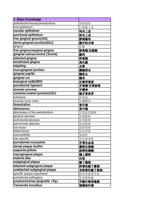
牙周专业英语常用词汇牙周专业英语课文CLINICAL FEATURE OF CHRONIC PERIODONTAL DISEASEChronic gingivitisThe manifestations of gingival inflammation vary considerably between individuals and from one part of the mouth to another. This variation reflects the aetiological factors at work and the tissue response to these factors. This response is essentially a mixture of inflammation and fibrous tissue repair. When the former predominates, signs and symptoms are more obvious; when the fibrous tissue component predominates, clinical manifestations can be much more subtle and recognized only by careful examination.In making a diagnosis it is important to keep in mind the appearance of health, departures from which may indicate disease.Clinical features are:l . Altered gingival appearance.2. Gingival bleeding.3. Discomfort and pain4. Unpleasant taste5. Halitosis.Altered gingival appearanceChanges in appearance are usually described according to color, shape, size, and surface characteristics.Healthy gingivae are pale pink and the margin is knife edged and scalloped; a streamlined papilla is often grooved by a sluice-way and the attached gingiva is stippled.Because the interdental embrasure is the site of greatest plaque stagnation gingival inflammation usually starts in the interdental papilla and spreads around the margin. As the blood vessels dilate the tissue becomes red and swollen with inflammatory exudate. The knife-edged margin becomes rounded, the interdental sluice-way is lost and the surface of the gingiva becomes smooth and glossy. As the gingival fiber the inflammatory process the gingival cuff loses tone and comes away from the tooth surface so that a shallow pocket is formed breaks up bundles. If the inflammation becomes more diffuse and spreads into the attached gingiva the stippling disappears. If inflammation is severe it can spread across the attached gingiva to the alveolar mucosa and so obliterate the normally well-defined mucogingival junction.Usually the most pronounced inflammatory swelling is seen in adolescents and young adults so that false pocketing is formed. It is called false as opposed to real or periodontal pocketing which is formed by apical migration of the crevicular epithelium as the periodontal ligament is destroyed by inflammation. Where several aetiological factors combine, e. g. plaque deposition plus lack of lip-seal plus the endocrinal changes of puberty, gingival swelling, especially papillary swelling, can be pronounced.If plaque irritation is longstanding and low grade, the main tissue reaction will be fibrous tissue production so that the gingiva may remain firm and pink but become thickened and lose its streamlined shape.Gingival bleedingGingival bleeding is probably the most frequent patient complaint. Unfortunately gingival bleeding is so common that people may not take it seriously and even believe it to be normal; however, unless bleeding obviously follows an episode of acute trauma, bleeding is always a sign of pathology. It occurs most frequently on toothbrushing. Bleeding may be provoked by eating hard food, apples, toast, etc. When gingivae are extremely soft and spongy, bleeding can occur spontaneously.Blood may be tasted by the patient and may be smelt on the patient's breath.If the tissue response is fibrous overgrowth, there is no bleeding even with vigorous toothbrushing.Discomfort and painThese are uncommon features of chronic gingivitis and this is probably the main reason for the diseases being overlooked. The gingivae may feel sore when the patient brushes his teeth and because of this he brushes more lightly and less frequently so that plaque accumulates and the condition is perpetuated.This relative absence of pain is one of the symptoms, which differentiates a chronic gingivitis from an acute ulcerative gingivitis.Unpleasant tastePatients may notice the taste of blood, particularly if they suck at an interdental space. Unfortunately the senses are quickly blunted and a disagreeable taste is a relatively infrequent complaint.Halitosis'Bad breath' frequently accompanies gingival disease and is a common cause of a visit to the dentist. The smell derives from blood and poor oral hygiene and must be distinguished from smells from different sources.Halitosis has a number of causes, both intra-oral and extra-oral. Oral disease and residual food deposits, especially those of a volatile nature such as peppermint, garlic, curry, etc., represent the most common cause of halitosis. Pathology of the respiratory tract, nose, sinuses, tonsils and lungs can cause an embarrassing smell, as can disease of the digestive tract. Some items of diet, e.g. garlic, are absorbed by the intestines, taken into the intestinal bloodstream and finally exhaled by the lungs so that they can be smelt a long time after they have been eaten. Mouth odour is common on waking and between meals, when it is associated with food stagnation and reduced salivary flow. Metabolic diseases, diabetes and uraemia give characteristic smells to the breath. Halitosis can increase with age.Chronic periodontitisThe clinical features of chronic periodontitis are:1. Gingival inflammation and bleeding2. Pocketing3. Gingival recession4. Tooth mobility5. Tooth migration6. Discomfort7. Alveolar bone loss8. Halitosis and offensive taste.Of this only pocketing and alveolar bone loss are essential features of chronic periodontitis.Gingival inflammation and bleedingAlthough gingival inflammation is a necessary precursor to periodontitis, obvious manifestations of inflammation become less apparent with the progress of periodontitis. Frequently the gingivae are pink and firm, the contours may be almost normal, there is no bleeding on careful probing and the patient does not complain of bleeding on brushing. It is as though with the development of the pocket the disease has gone underground.The presence and severity of gingival inflammation depends upon oral hygiene status; where this is poor, gingival inflammation is evident and bleeding of brushing, or even spontaneous bleeding, is noticed by the patient. When the patient' s toothbrushing is good enough to control plaque but where subgingival deposits, because of inadequate scaling, persist, the presence of periodontal disease may not be apparent on superficial examination. If a careful history is taken many such patients report a history of past bleeding which stopped when their toothbrushing technique improved. Periodontal destruction in the average adult is the product of past neglect,notthe result of present oral hygiene habits.PocketingPocket measurement is an essential part of periodontal diagnosis but must be interpreted together with gingival inflammation and swelling and radiographic evidence of alveolar bone loss. Theoretically, if there is no gingival swelling a pocket over 2 mm deep indicates some apical migration of crevicular epithelium but inflammatory swelling is so common especially in the younger individual that pocketing of 3-4mm may be entirely gingival or 'false'. Pocketing of 4mm is likely to indicate an early chronic periodontitis.The precise measurement of pockets is difficult because:1. Probing the pocket can be uncomfortable and even painful if there is frank inflammation.2. Pocket depth is extremely variable around a tooth. Interproximal pocketing is usually deepest because that is the site of greatest plaque accumulation, while pocketing on the facial aspect of the tooth is usually most shallow as this is where the toothbrush makes the greatest impact and may even produce gingival recession. This means that four or more measurements may be required on each tooth to give an accurate picture.3. Where present oral hygiene is good the gingival cuff may be so tight around the neck of the tooth as to resist the insertion of an ordinary periodontal probe without causing pain. The measurement of pockets in anaesthetized tissue often produces quite different results from previous measurement made in sentient tissue.4. Tooth contour and angulation, subgingival calculus or restorations, as well as carious cavities, may impede the insertion of the probe.There are many designs of pocket-measuring probe, some of, which are too thick to provide accurate measurement and some of which are sharp so that the tissue is penetrated unless great care is taken. It has been shown that pockets of over 3mm are measured with diminishing reliability, and it is unfortunate that much periodontal research is based upon such an unreliable criterion. Sometimes a purulent discharge can be expressed from the pocket by pressure on the pocket wall.Gingival recessionGingival recession and root exposure may accompany chronic periodontitis but are not necessarily a feature of the disease. Where recession occurs pocket depth measurement is only a partial representation of the total amount of periodontal destruction.Tooth mobilitySome tooth mobility in a labiolingual plane can be elicited in healthy, single-rooted teeth, especially lower incisors, being more mobile than multirooted teeth. Increasing tooth mobility is produced by,l. Spread of inflammation from the gingiva into the deeper tissues2. Loss of supporting tissue3. Occlusal trauma.Mobility also increases after periodontal surgery and in pregnancy. In periodontal pathology tissue destruction is always accompanied by inflammation and frequently by occlusal trauma. Mobility, which is produced by inflammation and occlusal trauma, is reversible, as demonstratedby the reduction in mobility following scaling and occlusal adjustment; mobility associated with destruction of supporting tissue is not reversible.Assessment of mobility for research purposes can be made using special apparatus but clinical assessment is usually subjective. It is elicited by exerting pressure on one side of the tooth under examination with an instrument or finger tip while placing a finger of the other hand on the other side of the tooth and its neighbour which is used as a fixed point so that relative movement can be discerned. Another way of eliciting mobility (although not assessing it) is to place fingers over the facial surfaces of the teeth while the patient grinds the teeth.The degree of mobility may be graded as follows:Grade l. Just discernibleGrade 2. Easily discernible and up to l mm labiolingual displacementGrade 3. Over l mm labiolingual displacement, mobility of the tooth up and down in an axial direction.Tooth migrationMovement of a tooth (or teeth) out of its original position in the arch is a common feature of periodontal disease and one which alerts the patient to the problem. A balance of tongue, lip and occlusal forces maintains tooth position in health. Once supporting tissue is lost these forces determine the pattern of tooth migration. The incisors move most frequently in a labial direction but teeth may move in any direction or become extruded. Once a tooth migrates the force on that tooth changes and this may promote further stress and further migration. If an upper incisor migrates labially the lower lip may come to lie lingual to the incisal edge of the tooth and produce further migration.DiscomfortOne of the most important features of chronic periodontitis is the almost total absence of discomfort or pain unless acute inflammation supervenes. This is one of the main distinctions between periodontal and pulp disease. Discomfort or pain on percussion of the tooth indicates some active inflammation of the supporting tissues, which is at its most acute in abscess formation when the tooth becomes exquisitely sensitive to touch. Sensitivity to hot and cold is sometimes present when there is gingival recession and root exposure. Indeed one common clinical experience is the appearance of sensitivity, especially to cold, when roots once covered in calculus are cleaned. On occasion pulp pathology may be a complication of advanced periodontal disease and severe pain may then develop.Alveolar bone lossResorption of alveolar bone and the associated destruction of periodontal ligament are the most important feature of chronic periodontitis, and the one, which leads to tooth loss. There is considerable variation in both the form and rate of alveolar bone resorption and in constructing a treatment plan the amount of bone loss, the rate at which resorption is progressing and the pattern of bone loss need to be accurately established. Radiographic examination is an essential part of periodontal diagnosis and with certain limitations provides evidence of the alveolar bone height, the form of bone destruction, the width of the periodontal ligament space and the density of cancellous trabeculation. Serial radiographs taken over a period of time can provideinformation about the rate of bone loss. However, radiographic examination without careful clinical examination can be very misleading. A periodontal diagnosis cannot be made from radiographs alone as there is no way of distinguishing on the radiograph past bone destruction from current bone resorption.Because the images of the facial and lingual plates of bone are largely obscured by the dense image of the tooth, diagnosis depends upon obtaining a clear image of the interdental bone. Careful angulation of the X-ray beam and a standardized routine of exposure and processing the radiographic film is essential.The first radiographic sign of periodontal destruction is loss of density of the alveolar margin. This is most clearly seen between posterior teeth where in health the broad interdental septum projects a dense and well-defined image of the alveolar margin. The image of the narrow interdental septa between anterior teeth is less well defined in health and early pathological changes are less easy to see. With continuing bone resorption the height of the alveolar bone is further reduced.Even correctly angulated the radiographs may not disclose the true state of interdental resorption, e. G. An interdental crater between molars can be masked by the images of the facial and lingual walls of the defect. Bone defects, which lie over the facial or lingual aspects of the teeth, e. G. Marginal gutters, may be completely obscured and revealed only when flaps are raised at surgery.Moreover, distinguishing between facial and lingual defects may not be possible from radiographic evidence alone. Two radiographs taken at slightly different angles often reveal defects undetected by one. This is especially true in the diagnosis of furcation defects. These are usually revealed by radiographic examination but the exact form of the defect may not be discernible. The thick palatal root of an upper molar may mask a trifurcation defect. Widening of the periodontal space in the furcation provides evidence of an early lesion. Widening of the periodontal space on one side or all around a tooth frequently indicates excessive occlusal stress. This is sometimes accompanied by widening or funnelling of the coronal aspect of the socket. All departures from the normal radiographic appearance must be checked against other clinical features, in particular pocket depth and mobility patterns, and if these do not correspond reexamination should be carried out. Clinical features taken together should make a reasonable fit, which sheds light on both the pathological condition and its aetiology. Thus, where radiographic examination of a mobile tooth reveals that the supporting bone is virtually intact, careful examination of the occlusion is essential. There must always be an identifiable reason for any pathological change.Halitosis and offensive tasteAn offensive taste and smell frequently accompany periodontal disease especially when oral hygiene is poor. Acute inflammation, with the production of pus, which exudes from pockets on pressure, also causes halitosis. A source of constant surprise is the lack of awareness of affected individuals and their spouses to the powerful fetor, which like a malignant wind escapes from their mouths when they speak. Lack of sensibility and unconcern about dental health seem to go hand in hand, and as patient cooperation is essential to the success of periodontal treatment this sensibility, or lack of it, can provide a clue to prognosis.Diagnosis, prognosis and treatment planMaking a diagnosisThe diagnosis should not be limited to giving a name to the condition. If periodontal disease is to be treated and its recurrence prevented, a diagnosis should include the identification of all aetiological factors, i.e. (i) those factors which predispose to plaque deposition and retention, and (ii) those factors, local or systemic, which influence adversely the behavior of the tissue. It should go without saying that you cannot remove or control factors, which have not been identified, yet all too frequently treatment is reduced to the control of signs and symptoms, and inevitably disease recurs.At the time of the initial examination some attempt should be made to assess the patient's attitude to dental health. Patient cooperation is essential to the success of periodontal treatment and it is this fact which makes the treatment of periodontal disease different from that of caries and other dental diseases when the patient can take a more passive attitude.Patient examinationThe examination should be methodical and comprehensive and should follow the standard pattern of the classic case history.Present complaint and its historyA patient with periodontal disease may have no complaint at all and the obvious to the presence of any disease in the mouth; indeed, the patient may be suspicious of any suggestion that disease is present! The most common complaints are bleeding gums, loose teeth, drifting of the teeth (usually the upper incisors), nasty taste, halitosis, swelling of the gums, discomfort and occasionally acute pain.Few patients at the initial consulation provide concise and completely relevant information. All too often, the necessary information has to be elicited by abstraction from a long, sometimes rambling, and account which must be listened to with patience and close attention. In addition, Pertinent questions should be asked:Are you in pain?Where is the pain?Is it a throbbing or dull pain?Does the pain keep you awake?What brings on the pain - - hot, cold, sweet, biting?Have you had pain in the past or is this the first time?What treatment have you received for pain?Do your gums ever bleed?When you brush your teeth?When you eat hard food?Did your gums bleed in the past?What treatment did you receive?Do any of your teeth feel loose?Have you always had that space between your front teeth?Have you had any swelling in your mouth? Where, when, etc.?Dental historyDo you go to the dentist regularly?What was the last treatment you received?When did you last have a scaling, i. e. Cleaning by your dentist?Do you have any dentures (false teeth that you can take out) - - how long have you had them? Have you any false teeth that are fixed in - - how long have you had them?At this stage questioning about home care can be a waste of time.Answers to such questions as 'How often do you clean your teeth?"are often suspect, as the patient is likely to say what he imagines he is supposed to say, i.e. twice a day, night and morning. Even if this happens to be the truth, it gives no indication of the quality of the performance; only an examination of the mouth provides information about that.At this time, some idea about habits should be gleaned, e.g. smoking, clenching, and night grinding, and biting pencils and so on.Medical historyAlthough a medical history may not seem relevant to some patients, it is essential to obtain one for a number of reasons:l. The patient may be suffering from some condition, e.g. Cardiovascular disease, renal disease, etc., which win require special precautions and/or modification of the treatment and will necessitate communication with the patient's physician.2. Systemic conditions, e.g. Pregnancy, diabetes, win alter the way in which the periodontal tissues behave and may demand medical attention before periodontal treatment can be carried out.3. The mouth may be the site of some manifestation of a systemic condition, e. g. Anaemia, which could affect any periodontal treatment.4. The patient may be receiving medication, e. G. Monoamine oxidase inhibitors for depression, which may connect with medication involved in the periodontal treatment, e.g. General anaesthetics.A medical history should record any present illness and medication; any past serious illness and medication, e.g. Steroids taken in the recent past, allergies, especially any history of penicillin sensitivity, abnormal bleeding tendencies, in particular excessive bleeding after injury or tooth extraction.The use of a questionnaire may be helpful.Where some systemic problem exists, communication with the patient's physician is essential.Patient appraisalWhile taking the history, a general appraisal of the patient should be made, and such features as obesity, general posture, pallor, skin rash, heavy breathing, lip posture should be noted.Oral examinationThe examination of the mouth should be carried out in a methodical and thorough manner; this is the dentist' s special area. Halitosis is noted, as the mouth is opened or even earlier when the patient is giving a history.l. The oral mucosa, cheeks, lips, tongue, palate, floor of' mouth and vestibules, are examined for ulceration, vesicles, swelling, eroded patches, abnormal colour and white lines or patches.Tooth indentations in the margin of the tongue and interdental keratosis, i. e. A white line in the cheek at the level of the occlusion, often indicates a clenching or grinding habit.Aphthous ulcers frequently occur in the labial or lingual vestibule or inside the lips. Lichen planus may be seen as fine, interlacing white lines on the cheeks or alveolar mucosa. Vesicles or eroded patches should be fully investigated.A sinus on the alveolar mucosa with or without the discharge of pus on pressure, indicates the presence of an alveolar abscess.In the older individual, a squamous-cell carcinoma may appear as a painless swelling, ulcer or eroded white patch in any part of the oral mucosa, but especially in the vestibules. Oral lesions of primary, secondary or tertiary syphilis may appear on the lips, tongue, palate and even the gingivae; widespread candida lesions in a young male could be indicative of HIV infection.Any departure from the norm must be examined carefully, and if infection or malignant disease is suspected, an examination of the submandibular and cervical lymph nodes will help with a diagnosis. Immediate referral to the physician or appropriate specialist is essential.2. Removable appliances, if present, should be examined for their fit, design and relationship to any inflammation of the oral mucosa and gingiva.3. Oral hygiene. Note presence and position of plaque, supragingival and subgingival calcalus. Subginigival calculus can be detected with a sharp probe or a Cross calculus probe but may also be seen as a dark blue shadow in the gingival margin. The use of a disclosing agent will help to identify plaque and demonstrate its presence to the patient. Sometimes the location of plaque and calculus points to a predisposing factor, e. G. Better oral hygiene on the left side is usually associated with right-handed tooth brushing; interproximal deposits and gingival inflammation may be caused by the overhanging margins of restorations or poor contact relations.4. Teeth are charted and cavities, restorations and malalignments recorded. Attrition may indicate a grinding habit; abrasion a vigorous and damaging toothbrushing technique.5. Gingivae are examined for colour, shape, size and consistency, keeping in mind the picture of health, pink, knife-edged, streamlined and firm, any departure from which could indicate pathology.6. Pocket measurement should be carried out on each tooth and recorded. Ideally, true mesial, distal, facial and lingual measurements are required, but this is possible only where teeth are missing, so that unimpeded access to these surfaces is possible. Where proximal teeth are present, measurement is made at the line angles, and on facial and lingual surfaces. Taking six readings on each tooth is ideal but may be very time consuming, and if diagnosis is made at a reasonably early stage in periodontal breakdown, only one or two measurements made at the mesiobuccal and mesiolingual line angles may be sufficient. Where there appears to be furcation involvement of molars, or drifting of incisors, facial and lingual measurements on these teeth are essential.A pocket-measuring probe must be fine enough to enter a narrow pocket, but must have a blunt end so that the tissue is not perforated. The sharp-ended probe used for the detection of caries should not be used. The pocket-measuring probe must be inserted into the pocket, as near parallel to the axis of the tooth as possible; if inserted obliquely, a false reading will be obtained. Great care has to be taken to manipulate the probe so that the true depth of the pocket is recorded. Delicate handling of the probe mast is employed to negotiate subgingival deposits without impacting against the root surface. Vigorous probing is not only painful but also likely to give an inaccurate reading; even gentle probing of inflamed gingivae can be painful. Theproblems of pocket measurement can be demonstrated by the fact that pocket measurement after local anaesthesia usually gives greater readings than in the unanaesthetized tissue.Gutta-percha or silver points, which may be calibrated, may be left in situ during radiographic examination of suspected infrabony pockets.In addition to recording pocket depth, it is important to assess the clinical attachment level (amelocemental junction, CEJ). Where there is considerable gingival hyperplasia pocketing may be fairly deep; say 5-7mm, but attachment loss may be small or nil. Where there has been considerable gingival recession, a shallow pocket may be associated with considerable destruction of the periodontal tissues. Therefore, in order to interpret pocket measurement, one must also note (a) the position of the gingival margins on the tooth surface, and (b) the position of the alveolar crest as seen on the radiograph.7. Radiographic examination will demonstrate the position of the alveolar margin and the condition of the alveolar bone. In a child or adolescent, radiographic examination may not be essential but if any doubt exists about the integrity of the alveolar margin, bitewing films of posterior teeth and periapical films of the incisors should provide adequate information. If there is evidence of established bone loss, further radiographic examination can then be undertaken. In the adult, full mouth examination may be necessary. The long-cone paralleling technique provides the most reliable radiographic evidence. The bisecting angle technique is more likely to give a distorted picture of the relationship of the alveolar margin to the CEJ. Vertical bitewing radiographs are useful for posterior teeth; the orthopantomograph (OPG) provides an overall picture, but detail of the alveolar margin is frequently ill declined. Repeat radiographs may be necessary; at intervals (not less than 3 years) determined by patient susceptibility, to show progression.8. Occlusion. The examination of occlusion should include:(a)The Angle' s classification(b) Overbite and overjet(c) Tooth relation in protrusive and lateral positions and movements(d) Any deviation from the normal path of opening and closure(e) Any temporomandibular joint (TMJ) discomfort or clicking(t) Any history of habits, e.g. clenching or grinding the Teeth. The occlusion needs to be examined closely where:(a)Teeth are mobile or sensitive(b) There is discomfort, clicking, deviation of the mandible on opening and closing, or limitation of movement(c) Radiographs show widening of the periodontal spaces or vertical bone defects, i.e. Possible signs of excessive occlusal stress.Special testsIf the severity of the inflammation or the degree of periodontal destruction appears to be out of proportion to the observed aetiological factors, or if general appraisal of the patient suggests that some systemic factor may be operating, then blood and urine examination or other special tests may be required. In such cases it is imperative to communicate with the patient' s physician prior to the start of treatment.Making a prognosis.。
口腔医学专业英语修改版
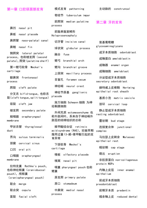
第一章口腔颌面部发育鼻凹nasal pit鼻板nasal placode鼻腭管naso-palatal canal鼻鳍nasal fin侧腭突lateral palatal process,也称继发腭(second palate),腭突(palatine shelf)第一鳃弓软骨Meckel's cartilage额鼻突frontonasal process腭裂cleft palate分叉舌 bifid tongue,也称舌裂(cleft tongue,split tongue)颌裂cleft jaw继发腭secondary palate颊咽膜orapharyngeal membrane甲状舌管thyroglossal duct界沟sulcus terminalis颈窦cervical sinus口凹oral pit口咽膜orapharyngeal membrane拉特克囊Rathke's pouch,也称神经颊囊(craniobuccal pouch),颅颊囊(craniopharyngeal pouch)联合merge联合突copula面裂facial cleft模式发育patterning奇结节tuberculum impar前腭突median palatineprocess前脑单脑室畸形Holoprosencephaly切牙管 incisive canal球状突globular process融合fuse鳃弓branchial arch鳃沟branchial groove上颌突maxillary process舌盲孔foramen cecum神经嵴neural crest神经节原基ganglionicplacode施万细胞 Schwann细胞为神经鞘膜细胞外间充质 ectomesenchyme 也称外胚间叶,系来自于神经嵴外胚层的结缔组织的总称维甲酸综合征retinoicacid syndrome(RAS),妊娠早期服用过量13-顺-维甲酸引起的发育异常下颌软骨Meckel'scartilage嗅板olfactory placode嗅窝nasal pit咽囊 pharyngeal pouch也称鳃囊原发腭 primary palate原口stomadeum中鼻突medial nasalprocess主动脉的conotruncal第二章牙的发育氨基葡萄糖glycosaminoglycans成牙本质细胞odontoblast成釉蛋白 ameloblastin成釉器enamel organ成釉细胞ameloblast分泌型成牙本质细胞secretory odontoblast赫特威上皮根鞘Hertwingepithelial root sheath基质小泡matrix vesicle颈环cervical loop静止型成牙本质细胞resting odontoblast蕾状期bud stage连接复合体junctionalcomplex马拉瑟上皮剩余Malassezepithelial rest帽状期cap stage萌出eruption非胶原蛋白 non-collagenousprotein NSPs内釉上皮层inner enamalepithelium前成牙本质细胞preodontoblast前期牙本质predentin缩余釉上皮reduced dentalepithelium托姆斯突Tomes processe脱落 shedding 交替外釉上皮层outer enamal epithelium星网状层stellate reticulum牙板dental lamina牙囊dental sac牙乳头dental papilla引导管gubernacular canal釉丛蛋白tuftelin釉结enamel knot釉龛enamel niche釉索enamel cord釉质形成amelogenesis原发性上皮带primary epithelial band罩牙本质mantle dentin中间层stratum intermedium终棒terminal web钟状期bell stage第三章牙体组织玻璃粘连蛋白vitronectin,细胞外粘附蛋白,副纤维粘连蛋白成纤维细胞fibroblast成牙本质细胞odontoblast成牙本质细胞突起odontoblastic process,也称"Tomes' fiber"成牙本质细胞突周间隙periodontoblastic space成釉蛋白ameloblastin穿通纤维perforatingfiber蛋白酶proteinases第三期牙本质tertiarydentin多细胞层cell-rich zone乏细胞层cell-free zone,the zone of Weil,Weil层反应性牙本质reactiondentin反转线reversal line非釉原蛋白non-amelogenins缝隙连接gap junction骨钙素osteocalcin骨连接素osteonectin骨桥蛋白osteopontin骨样牙本质osteodentin固有牙髓pulp proper管间牙本质intratubulardentin管周牙本质peritubulardentin核心蛋白聚糖decorin横纹cross striations基质金属蛋白酶20 matrixmetalloproteinases 20,MMP20继发性牙本质secondarydentin,牙发育完成之后形成的牙本质腱蛋白tenascin绞釉gnarled enamel紧密连接tight junction科尔夫纤维Korff's fiber,最先形成的牙本质纤维类牙骨质cementoid流体动力学说hydrodynamictheory磨损abrasion, attrition纳米球nanospheres耐龋潜能cariostaticpotential脑啡肽enkephalin诺伊曼鞘Neumann sheath欧文线Owen line(加重的牙本质生长线),也称"contourline of Owen"前期牙本质predentin桥粒desmosome球间牙本质interglobulardentin芮氏线lines of Retzius,釉质生长线沙比纤维Sharpey's fiber,穿通纤维神经壁层parietal layer ofnerves,Raschkow 丛神经传导学说directinnervation theory生长激素抑制素somatostatin生长线incremental lines,同芮氏线施雷格线Schreger line树突状细胞dendriticcells双糖链蛋白聚糖biglycan丝氨酸蛋白酶serine proteinases(kallikrein-4)死区dead tract髓核pulp core髓周牙本质circumpulpal dentin糖胺聚糖glycosaminoglycans透明层hyaline layer透明牙本质scleroticdentin透明牙本质transparent dentin托姆斯颗粒层Tomes' granular layer托姆斯突凹Tomesprocesses pit,TPP微孔micropore未分化间充质细胞undifferentiated mesenchymal cell无细胞固有纤维牙骨质acellular intrinsic fiber cementum, AIFC无细胞外源性纤维牙骨质acellular extrinsic fiber cementum, AEFC无细胞无纤维牙骨质acellular afibrillar cementum, AAC无细胞牙骨质acellular cementum无釉柱牙釉质rodless enamel 细胞牙骨质cellularcementum纤维粘连蛋白fibronectin 限制板lamina limitans楔状缺损wedge shaped defect新生线neonatal line修复性牙本质reparative dentin牙本质dentin牙本质磷蛋白dentin phosphophoryn;dentin phosphoproteins,DPP牙本质生长线von Ebner1ine牙本质涎蛋白dentin sialoprotein,DSP牙本质涎磷蛋白dentin sialophosphoproteins牙本质小管dentinal tubule牙本质牙骨质界dentino-cemental junction 牙骨质cementum牙骨质生长因子cementum growth factor牙骨质粘附蛋白cementum adhesion protein牙面平行线perikymata,也称釉面横纹牙髓dental pulp牙髓牙本质复合体pulpo-dentinal complex牙釉质enamel牙釉质溶解蛋白enamelysin牙釉质牙本质界enamel-dentinal junction (EDJ)牙釉质牙骨质界enamelo-cemental junction油炸圈样appearance of doughnut有细胞固有纤维牙骨质cellular intrinsic fiber cementum, CIFC有细胞混合性分层牙骨质cellular mixed stratified cementum, CMSC釉板enamel lamellae釉丛enamel tufts釉丛蛋白tuftelin釉蛋白enamelin釉帽enamel cap釉面横纹perikymata,也称牙面平行线釉梭enamel spindle釉小皮enamel cuticle釉牙本质界enamel-dentinal junction (EDJ)釉原蛋白amelogenins釉柱enamel rod釉柱鞘enamel rod sheath原发性牙本质primary dentin,牙发育时期形成的牙本质原胶原tropocollagen灶性孔focal hole, FH罩牙本质mantle dentin中间连接intermediate junction中间牙骨质intermediate cementum重塑remodeling转导学说transduction theory组织细胞histiocyte第四章牙周组织不成熟的弹力纤维oxytalan fibers弹力纤维elastin fibers点彩stippling凋亡apoptosis附着龈attached gingiva根尖组apical group根间组interradicular group固有牙槽骨alveolar bone proper环行组circular group结合上皮junctional epithelium束骨bundle bone水平组horizontal group斜行组oblique group牙槽骨alveolar bone牙槽嵴组alveolar crest group牙槽突alveolar process牙槽龈组alveologingival group牙骨膜组dentoperiostealgroup牙间乳头interdentalpapilla牙龈gingiva牙龈结合dentogingivaljunction牙龈上皮gingvalepithelium第六章涎腺半月板demilune胞吐exocytosis杯状细胞goblet cell储备细胞reserve cell锇酸osmic acid分泌单位secretory unit分泌管secretory duct富脯氨酸蛋白proline-richprotein富酪氨酸蛋白tyrosine-rich protein干细胞stem cell高血糖素样蛋白glucogon-like protein颌下腺submandibulargland混合性腺泡mixed acinus肌动蛋白actin肌球蛋白myosin肌上皮细胞myoepithelialcell肌微丝myofilament浆液性腺泡serous acinus浆粘液细胞seromucouscell晶样体crystalloid局浆分泌merocrine抗蛋白溶解蛋白antiproteolytic protein篮细胞basket cell连接复合体junctionalcomplex酶原颗粒zymogen granule排泄管excretory duct粘多糖mucin粘液性腺泡mucous acinus全浆分泌holocrine-typesecretion乳铁蛋白lactoferrin闰管intercalated duct腮腺parotid gland腮腺导管Stensen duct腮腺素parotin舌下腺sublingual gland舍格伦综合征Sj?grensyndrome肾素rennin嗜酸粒细胞瘤oncocytoma嗜酸细胞oncocyte嗜酸性腺瘤oxiphilicadenoma唾液saliva唾液腺salivary gland味觉素gustin纹管striated duct涎液素ptyatin腺泡acinus小涎腺minor salivary gland原始多潜能涎腺导管细胞primitive pluripotential salivary duct cells致密小体dense body组氨酸histidine第七章颞下颌关节多细胞带cellularrich zone钙化软骨带zone of calcified cartilage关节囊capsule关节盘disc滑膜synovial membrane髁突condyle颞下颌关节temporo-mandibular join,TMJ纤维软骨带fibrocartilaginous zone纤维性关节表面带fibrous articular surface第八章牙发育异常II型牙本质结构不良dentin dysplasia type II ,冠部牙本质结构不良I型牙本质结构不良dentindysplasia type I白兰地型brandywineisolate,牙本质形成缺陷的一种斑釉mottled enamel半面过度增生hemifacialhyperplasia成熟不全型hypomatuaration type低磷酸酯酶症hypophosphatasia多生牙hyperdontia;supernumerary teeth非氟性牙釉质浑浊症non-fluoride enamel opacities氟牙症dental fluorosis附加牙supplemental teeth副磨牙paramolar钙化不全型hypocalcifiedtype过早脱落premature loss畸形舌侧尖lingual cuspdeformity畸形舌侧窝lingual fossadeformity畸形中央尖central cuspdeformity结合牙concrescence巨牙macrodontia壳状牙shell-teeth矿化不全型hypomineralized type牛牙症taurodontism前牙的牙外突densevaginatus of anterior teeth区域性牙发育不良regionalodontodysplasia,阴影牙融合牙fusion乳牙滞留persistence ofdeciduous teeth桑椹牙mulberry molar少汗外胚层发育不良hypohidrotic ectodermaldysplasia少牙hypodontia双生牙gemination水流围绕圆石waterstreaming round bouldings,根部牙本质结构不良的表现四环素牙tetracyclinestained teeth锁骨颅骨发育不全征cleidocranial dysplasia;也有称锁骨头颅发育不良胎生牙natal teeth,出生时即已萌出的牙特纳牙Turner's teeth,与乳牙有关的感染或创伤引起继生恒牙成釉细胞的损伤,导致继生恒牙牙釉质形成不全或矿化不全弯曲牙dilacerations无牙adontia先天性梅毒牙congenitalsyphilis小牙microdontia新生牙noenatal teeth,出生后30天内萌出的牙形成不全型hypoplastictype雪帽型snow-capped牙本质基质蛋白1 dentinmatrix protein 1, DMP1牙本质结构不良dentin dysplasia牙本质涎蛋白dentin sialoprotein, DSP牙本质形成缺陷症II型dentinogenesis imperfecta type II牙变色discoloration of teeth牙骨质发育不全hypocementosis牙骨质过度增生hypercementosis牙内陷dens invaginatus牙釉质混浊症enamel opacities牙釉质矿化不全hypomineralized enamel牙釉质形成不全enamel hypoplasia牙釉质形成缺陷症amelogenesis imperfecta牙釉质延伸cervical enamel extension牙中牙dens in dente牙阻生impaction of teeth延迟萌出retarded eruption遗传性乳光牙本质hereditary opalescent dentin阴影牙ghost teeth,区域性牙发育不良鹰爪尖talon cusp釉珠enamel pearl远中磨牙distomolar早萌premature eruption正中牙mesiodens第九章龋病暗层dark zone变形链球菌mutansstreptococci; S. mutans表层surface zone病损体部body of thelesion蛋白溶解-螯合学说proteolysis-chelation theory蛋白溶解学说proteolytictheory发酵乳杆菌L. fermentus放线菌属actinomyces干酪乳杆菌L. casei根龋root caries化学寄生学说chemico-parasitic theory化学细菌学说chemico-bacterial theory坏死崩解层zone ofdestruction获得性薄膜acquiredpellicle,唾液薄膜急性龋acute caries静止性龋arrested caries菌斑bacterial plaque慢性龋chronic caries猛性龋rampant caries内氏放线菌 A. naeslundii平滑面龋smooth surfacecaries轻链球菌S. mitis龋病dental caries乳杆菌属Lactobacilli三联因素学说threeprimary factors theory嗜酸乳杆菌L. acidophilus酸原学说acidogenictheory透明层translucent zone ,硬化层脱矿层zone ofdemineralization唾液薄膜salivarypellicle,获得性薄膜窝沟龋pit and fissurecaries细菌侵入层zone ofbacterial invasion血链球菌S. sanguis牙本质龋dentin caries牙骨质龋cementum caries牙釉质龋enamel caries远缘链球菌S. sobrinus粘性放线菌 A. viscosus第十章牙髓病白三烯leukotrienes,LTs白细胞介素interleukin不可逆性牙髓炎irreversible pulpitis残髓炎residual pulpitis成牙本质细胞层空泡变性vacuolar degeneration of theodontoblastic layer急性化脓性牙髓炎acutesupurative pulpitis急性浆液性牙髓炎acute serous pulpitis急性牙髓炎acute pulpitis可复性牙髓炎reversible pulpitis慢性闭锁性牙髓炎chronic closed pulpitis慢性溃疡型牙髓炎chronic ulcerative pulpitis慢性牙髓炎chronic pulpitis慢性增生性牙髓炎chronic hyperplastic pulpitis逆行性牙髓炎retrograde pulpitis前列腺素prostaglandins,PGs特发性吸收idiopathic resorption牙内吸收internal tooth resorption牙髓变性pulp degeneration牙髓充血pulp hyperemia牙髓钙化pulpcalcification牙髓坏疽pulp gangrene牙髓坏死pulp necrosis牙髓渐进性坏死necrobiosis牙髓网状萎缩reticular atrophy of the pulp牙髓纤维性变pulpfibrosis牙髓炎pulpitis牙体吸收tooth resorption牙外吸收external toothresorption转化生长因子transfergrowth factor,TGF第十一章根尖周炎蜂窝织炎cellulitis根尖肉芽肿periapicalgranuloma根尖周炎periapicalperiodontitis急性根尖周炎acuteperiapical periodontitis急性牙槽脓肿acutealveolar abscess磷脂壁酸lipoteichoicacids慢性根尖脓肿chronicperiapical abscess慢性根尖周炎chronicperiapical periodontitis慢性牙槽脓肿chonicalveolar abscess肽葡聚糖peplidoglyans致密性骨炎condensingosteoitis肿瘤坏死因子tumornecrosis factor,TNF第十二章牙周组织病白细胞介素interleukins,IL白血病性龈增大gingivalenlargement associated withleukemia伴白血病性龈炎gingivitiswith leukemia伴有牙髓病变的牙周炎periodontitis associated withendodontic lesions边缘性龈炎marginalgingivitis变性degeneration病损确立期establishedlesion剥脱性龈病损desquamativelesion of gingival创伤trauma创伤性咬合traumaticocclusion蛋白酶proteinases发育性或获得性异常及其状况developmental or acquireddeformities and conditions反应全身疾病的牙周炎periodontitis as amanifestation of systemicdiseases放线共生放线杆菌Actinobacillus actinomycetemcomitans,Aa非菌斑性牙龈病损non-plaque-induced gingivallesions非炎症性noninflammatory奋森龈炎Vincentgingivitis福赛类杆菌Bcteroidesforsythus,Bf共聚coaggregation骨内袋intrabony pocket骨上袋supragingival pocket护骨因子osteoprotegerin,OPG坏死性牙周病necrotizing periodontal diseases基质金属蛋白酶matrix metalloprotinases,MMP激素性龈炎steroid hormone-influencedgingivitis急性坏死性溃疡性龈炎acute necrotizing ulcerative gingivitis急性坏死性龈炎acute necrotizing gingivitis继发性咬合创伤secondary occlusal trauma家族性龈纤维瘤病congenital familial fibromatosis浆细胞龈炎plasma cell gingivitis胶原酶collagenase金属蛋白酶metalloproteinases进展期advanced lesion聚集aggregation菌毛fimbriae老年性萎缩senile atrophy硫酸软骨素酶chondrosulphatase慢性牙周炎chronic periodontitis慢性龈炎chronicgingivitis密螺旋体属Treponema破骨细胞分化因子osteoclast differentationfactor,ODF前列腺素E2prostglandinE2,PGE2侵袭性牙周炎aggressiveperiodontitis青春期龈炎pubertalgingivitis人类白细胞抗原humanleucocyte antigen,HLA妊娠期龈炎pregnancygingivitis始发期initial stage嗜麦芽糖密螺旋体T.maltophilum梭螺菌龈炎fusospirochetal gingivitis特发性浆细胞龈口炎idiopathic plasma-cellgingivostomatitis特发性龈增生idiopathicgingival hyperplasia透明质酸酶hyaluronidase唾液粘蛋白mucin维生素C缺乏性龈炎vitamin C deficeintgingivitis萎缩atrophy细胞因子cytokine细胞粘附分子cellularadhesion m olecules,CAM细菌性生物膜dental plaque biofilm先天性家族性纤维瘤病congenital familialfibromatosis牙槽骨弥漫性萎缩diffuseatrophy of alveolar bone牙菌斑性牙龈病dentalplaque-induced gingivaldisease牙龈病gingival diseases牙龈卟啉单胞菌Porphyromonas gingivalis,牙龈二氧化碳嗜纤维菌Capno gingivalis牙龈乳头炎papillarygingivitis牙龈退缩gingivalrecession牙周变性periodontaldegeneration牙周病periodontaldisease牙周脓肿abcesses of theperiodontium牙周炎periodontitis牙周症periodontosis炎症inflammation咬合创伤occlusal trauma药物性龈炎medication-influencedgingivitis遗传性牙龈纤维瘤病hereditary gingivalfibromatosis遗传性龈增生hereditarygingival hyperplasia龈袋gingival pocket龈沟液gingivalcrevicular fluid,GCF龈裂gingival cleft;Stillman'S cleft龈增生gingival hyperplasia营养不良dystrophy营养不良性dystrophic早老性萎缩presenile atrophy早期病变early lesion粘附adhesion粘性放线菌Actinomyces viscosus,Av战壕口炎trench mouth脂多糖lipopolysaccharedes,LPS中间密螺旋体中性多形核白细胞polymorphonuclear leukocytes,PMN肿瘤neoplasia肿瘤坏死因子-αtumor necrosis factor-α,TNF-α组织金属蛋白酶的抑制剂tissue inhibitors of metalloproteinase,TIMP第十三章口腔粘膜病艾滋病AIDS白斑leukoplakia白塞综合征Behcet syndrome白色海绵状斑痣white sponge nevus白色念珠状菌candida albicans白色水肿leukoedema白皱折病white foldeddisease斑macule扁平苔藓lichen planus,LP程序化细胞死亡programmedcell death大疱bulla单纯性疱疹herpes simplex地图舌geographic tongue淀粉样变性amyloidosis多形渗出性红斑erythemamultiforme exsudativum非典型性atypia非霍奇金淋巴瘤non-Hodgkin lymphoma复发性阿弗他口炎recurrent aphthousstomatitis,RAS复发性阿弗他溃疡recurrent aphthous ulcer,RAU复发性坏死性粘膜腺周围炎periadenitis mucosa necroticarecurrens,PMNR过度不全角化hyperparakeratosis过度角化hyperkeratosis过度正角化hyperorthokeratosis海绵形成spongiosis红斑erythroplakia红色增殖性病变erythroplastic lesion获得性免疫缺陷综合征acquired immunodeficiencysyndrome, AIDS ,艾滋病基底细胞空泡性变及液化vaculation and liquefactionof hasal cell棘层松解acantholysis棘层增生acanthosis痂crusts假膜pseudomembrane,伪膜间杂型红斑interspersederythroplakia胶样小体colloid body,Civatte小体角化不良dyskeratosis结节病sarcoidosis均质型红斑homogenouserythroplakia皲裂rhagade抗核抗体antinuclearantibody,ANA颗粒型红斑granularerythroplakia口腔 Kaposi肉瘤oralKaposi sarcoma口腔非霍其金淋巴瘤oralnon-Hodgkin lymphoma口腔毛状白斑oral hairyleukoplakia,OHL口腔念珠菌病oralcandidiasis口腔粘膜下纤维化oralsubmucous fibrosis溃疡ulcer类天疱疮样扁平苔藓lichenPlanus Pemphigoides,LPP良性游走性舌炎benignmigratory glossitis良性粘膜类天疱疮benignmucous membrane pemphigoid硫黄素T thioflavine T慢性盘状红斑狼疮chronic discoid lupus erythematosus梅一罗综合征Melkersson-Rosenthal syndrome糜烂erosion念珠菌病candidiasis疱vesicle疱疹性口炎herpetic stomatitis气球变性ballooning degeneration桥粒芯胶粘蛋白desmocollins桥粒芯糖蛋白desmogleins丘疹papule区域剥脱性舌炎glossitis areata exfoliativa人免疫缺陷病毒human immunodeficiency virus,HIV肉芽肿性唇炎cheilitis granulomatosa上皮萎缩epithelial atrophy上皮异常增生epithelial dysplasia舌乳头炎lingualpapillitis嗜碱性变性basophilic degeneration噬黑色素细胞melanophages天疱疮pemphigus天疱疮细胞Tzanck cell网状变性reticulardegeneration韦格内肉芽肿Wegenergranulomatosis萎缩atrophy细胞凋亡cell apoptosis腺性唇炎cheilitisglandularis肖曼小体Schauann bodies,多核巨细胞内的包涵体眼、口、生殖器三联综合征oculo-oral-genital syndrome增殖性红斑erythroplasia粘膜良性淋巴组织增生病benign lymphoadenosis ofmucosa周缘扩展现象Nikolsky征第十四章颌骨疾病板层骨lamellar bone表皮树突状细胞dermaldendrocyte伯基特淋巴瘤Burkitt'slymphoma不确定细胞indeterminatecell成软骨细胞瘤chondroblastoma虫蚀状moth-eatenappearance出血性骨囊肿hemorrhagicbone cyst穿凿性吸收tunnelingresorption or dissectingresorption穿凿样punched-outappearance单纯性骨囊肿simple bonecyst单骨性骨纤维异常增殖症monostotic bibrous dysplasia也称单骨性骨纤维结构不良单核基质细胞mononucleatestromal cell单核巨噬细胞系统mononuclear phagocyte system动脉瘤性骨囊肿aneurysmalbone cyst多发性骨髓瘤multiplemyeloma多发性软骨瘤病multiplechondromatosis多骨性骨纤维异常增殖症polyostotic fibrous dysplasia也称多骨性骨纤维结构不良恶性成骨细胞瘤malignantosteoblastoma腭隆突torus palatinus放射性骨坏死osteoradionecrosis非钙化骨样组织unmineralized osteoid富巨细胞性病变giant cellrich lesion钙化骨mineralized bone孤立性骨囊肿solitarybone cyst骨化性骨膜炎periostitisossificans骨化性纤维瘤ossifyingfibroma骨巨细胞瘤giant celltumor of bone,GCT骨壳involucrum骨瘤osteoma骨膜骨肉瘤periosteal osteosarcoma骨膜下骨吸收subperiosteal bone resorption骨旁骨肉瘤paraosteal osteosarcoma骨肉瘤osteosarcoma骨软骨瘤osteochondroma骨软骨性外生骨疣osteocartilaginous exostosis骨髓瘤myeloma骨外骨肉瘤extraosseous osteosarcoma骨外型软骨肉瘤extraosseous chondrosarcoma骨纤维结构不良fibrous dysplasia of bone 也称骨纤维异常增殖症骨小梁周围纤维化peritrabecular fibrosis骨样骨瘤osteoid osteoma海绵型骨瘤cancellous osteoma汉-许-克病Hand-Schiiller-Christian disease颌骨放线菌病actinomycosis of jaws颌骨骨髓炎osteomyelitis of jaws颌骨结核tuberculosis of jaws颌骨巨细胞病变giant cell granuloma颌骨梅毒syphilis of jaws弧立性骨髓瘤solitarymyeloma化牙骨质纤维瘤cementifying fibroma急性化脓性颌骨骨髓炎acute suppurativeosteomyelitis of jaws继发软骨secondarycartilage继发性甲状旁腺功能亢进secondaryhyperparathyroidism家族性颌骨多囊性病familial mulitilocular cysticdisease of jaws家族性颌骨纤维异常增殖症familial fibrous dysplasia ofthe jaws家族性巨颌症cherubism甲状旁腺功能亢进Hyperparathyroidism假囊肿pseudocyst间叶型软骨肉瘤mesenchymal chondrosarcoma浆细胞瘤plasmacytoma结核性颌骨骨髓炎tuberculous osteomyelitis ofjaws静止性骨腔static bonecavity巨大型骨样骨瘤giantosteoid osteoma巨细胞肉芽肿giant celllesions of the jaws巨细胞修复性肉芽肿giantcell reparative granuloma郎格汉斯细胞病Langerhanscell disease,同郎格汉斯细胞组织细胞增生症(Langerhanscell histiocytosis)郎格汉斯细胞组织细胞增生症Langerhans cellhistiocytosis,同郎格汉斯细胞病(Langerhans cell disease)朗汉斯巨细胞Langhansgiant cell勒-雪病Letterer-Siwedisease良性成骨细胞瘤benignoseoblastoma淋巴结的交错突细胞interdigitating dendriticcell硫磺颗粒sulfur granule滤泡树枝状细胞folliculardendritic cell慢性非化脓性硬化性骨炎Garré's chronicnonsuppurative sclerosingostitis Garre慢性骨髓炎伴增生性骨膜炎chronic osteomyelitis withproliferative periostitis慢性化脓性颌骨骨髓炎chronic suppurativeosteomyelitis of jaws慢性局灶性硬化性颌骨骨髓炎chronic focal sclerosingosteomyelitis of jaws慢性弥漫性硬化性颌骨骨髓炎chronic diffuse sclerosingosteomyelitis of jaws慢性硬化性颌骨骨髓炎chronic sclerosingosteomyelitis of jaws磨砂玻璃样groundglassappearance内生型软骨瘤enchondroma破骨细胞瘤osteoclastoma侵袭性成骨细胞瘤aggressive osteoblastoma侵袭性骨化性纤维瘤aggressive ossifying fibroma青少年骨化性纤维瘤juvenile ossifying fibroma 日光放射状(影像) sun-ray肉芽肿性炎症granulomatous inflammation 软骨瘤chondroma软骨颅chondrocranium软骨肉瘤chondrosarcoma软骨粘液样纤维瘤chondromyxoid fobroma上皮样细胞epithelioid cell嗜酸性肉芽肿eosinophilic granuloma树突状细胞系统dendritic cell system死骨sequestrum髓外浆细胞瘤extramedullary plasmacyfoma髓外性尤文肉瘤extramedullarg Ewing sarcoma图顿巨细胞Touton giant cell外伤性骨囊肿traumatic bone cyst外生骨疣exostosis下颌隆突torus mandibularis纤维-骨病变fibro-osseous lesion新生儿上颌骨骨髓炎neonatal maxillitis牙骨质骨化性纤维瘤cemento-ossifying fibroma洋葱皮onion-skin硬骨板lamina dura原发性甲状旁腺功能亢进primary hyperparathyroidism致密型骨瘤compactosteoma致密性骨炎condensingostitis中心型central type周围型peripheral type周围型软骨瘤peripreralchondroma周围性巨细胞肉芽肿peripheral giant cellgranuloma转化亢进high-turnoverstate棕色瘤brown tumor组织细胞增生症XhistiocytosisX第十五章颞下颌关节病(染色体)三体trisomy垂直裂vertical cleft错颌malocclusion骨剥露denudation骨关节病osteoarthrosis骨关节炎osteoarthritis,OA骨赘性唇状突出osteophytic lipping滑膜软骨瘤病synovialosteochondromatosis髁突增生condylarhyperplasia类风湿肉芽肿rheumatoidgranuloma类风湿性关节炎rheumatoidarthritis,RA类风湿性小结rheumatoidnodule颞下颌关节紊乱病temporomandibular jointdisorders切线裂tangential cleft绒毛状突起villousprojection色素性绒毛结节性滑膜炎pigmented villonosdularsynovitis退行性关节病degeneratitive joint disease纤维素样变fibrinoidchange象牙化eburnation (骨质)血管翳pannus蚓状小体vermiform bodies游离小体loose body原纤维化fibrillation第十六章涎腺非肿瘤性疾病与涎腺肿瘤PAS periodic acid schiff的缩写,PAS反应即过碘酸雪夫反应,用于对糖原的特殊染色癌胚抗原carcinoembryonicantigen,CEA癌在多形性腺瘤中carcinoma ex pleomorphic adenoma; carcinoma in pleomorphic adenoma艾滋病病毒相关性涎腺疾病HIV-associated salivary gland disease暗细胞dark cells奥辛兰Alcian blue半多能双储备细胞理论semipluripotentialbicellular reserve cell theory伴淋巴间质的未分化癌undifferentiated carcinoma with lymphoid stroma变性型涎腺肿大症degenerative sialosis波形丝蛋白vimnentin成涎细胞瘤 sialoblastoma大细胞癌 large cell carcinoma导管发育异常developmental anomalies of ducts导管内乳头状瘤intraductal papilloma导管乳头状瘤ductal papilloma导管腺泡单位ductoacinar unit淀粉酶amylase顶浆分泌apocrine多动脉周围炎polyarteritis多发性肌炎polymyositis多囊腮腺polycysticparotid gland多能单储备细胞理论pluripotential unicellularreserve cell theory多细胞理论multicellulartheory多形性低度恶性腺癌polymorphous low-gradeadenocarcinoma多形性腺瘤pleomorphicadenoma恶性多形性腺瘤malignantpleomorphic adenoma恶性淋巴上皮病变malignant lymphoepitheliallesion发育性舌侧下颌涎腺陷入evelopmental lingual salivarygland depression放射线损伤radiant impair非皮脂淋巴腺瘤non-sebaceous lymphadenoma非特异性透明细胞癌 clearcell carcinoma, not otherwisespecified非特异性腺癌adenocarcinoma not otherwisespecified,NOS分泌组份secretorycomponent副涎腺accessory salivarygland干燥综合征sicca syndrome管状腺瘤canalicularadenoma; tubular adenoma过氧化物酶抗过氧化物酶peroxidase anti-proxidase,PAP汗腺瘤syringoma合胞体syncytium坏死性涎腺化生necrotizing sialometaplasia混合瘤mixed tumor获得性免疫缺陷综合征acquired immune deficiencysyndrome AIDS霍奇金淋巴瘤 Hodgkinlymphoma肌上皮瘤myoepithelioma基底储备细胞理论basalreserve cell theory基底鳞状的旋涡状basisquamous whorling基底细胞腺瘤basal celladenoma急性化脓性腮腺炎 acutepyogenic paratitis急性涎腺炎acutesialadenitis挤压假象crush artifact甲状腺球蛋白thyroglobulin浆液细胞腺癌serous cellcarcinoma结外边缘带B细胞淋巴瘤extranodal marginal zoneB-cell lymphoma静止骨腔static bonycavity巨细胞包涵体病cytomegalic inclusion disease口腔干燥症xerostomia链亲和素过氧化物酶streptavidin peroxidase,S-P链亲和素生物素复合物streptavidin-biotin complex SABC良性淋巴上皮病变benign lymphoepithelial lesion亮细胞light cells淋巴上皮癌lymphoepithelial carcinoma淋巴上皮囊肿lymphoepithelial cyst鳞状细胞癌squamous cell carcinoma流行性腮腺炎epidemic parotitis; mumps慢性复发性腮腺炎chronic recurrent parotitis慢性涎腺炎chronic sialadenitis慢性硬化性颌下腺炎chronic sclerosing sialadenitis of submandibular gland; Küttner瘤弥漫性大B细胞淋巴瘤diffuse large B-cell lymphoma迷走涎腺aberrantsalivary glands免疫球蛋白A immunoglobulin A免疫细胞化学immunocytochemistry免疫组织化学immunohistochemistry囊腺癌 cystadenocarcinoma囊腺瘤 cystadenoma内翻性乳头状瘤inverted ductal papilloma皮脂淋巴腺癌 sebaceous lymphadenocarcinoma皮脂淋巴腺瘤sebaceouslymphadenoma皮脂腺瘤sebaceousadenoma前淋巴瘤prelymphoma桥本甲状腺炎Hashimoto'sthyroiditis亲和素生物素过氧化物酶复合体avidin biotin-peroxidasecomplex, ABC人类免疫缺陷病毒hunmanimmunodeficiency HIV溶菌酶lysozyme乳铁蛋白lactoferrin,LF乳头状淋巴囊腺瘤papillary cystadenomalymphomatosum乳头状囊腺癌papillarycystadenocarcinoma乳头状囊腺瘤papillarycystadenoma乳头状涎腺瘤sialadenomapapilliferum上皮-肌上皮癌epithelio-myoepithelialcarcinoma上皮肌上皮岛epi-myoepithelial island上皮膜抗原epithelialmembrane antigen,EMA舍格伦综合征Sj?gren'ssyndrome施墨试验Schirmer test,检查泪液分泌情况的一种试验嗜酸细胞瘤oncocytoma嗜酸性腺瘤oxyphilicadenoma透明细胞癌clear cellcarcinoma透明细胞瘤clear celltumor未分化癌undifferentiatedcarcimma细胞角蛋白cytokeratin纤维连接蛋白fibronectin涎石病sialolithiasis涎腺病毒病salivary glandvirus disease涎腺导管癌salivary ductcarcinoma涎腺导管结石salivaryduct stone涎腺导管囊肿salivaryduct cyst涎腺发育不全aplasia ofsalivary gland涎腺发育异常developmentanomalies of salivary gland涎腺放线菌病actinomycosis of salivaryglands涎腺结核tuberculosis ofsalivary gland涎腺囊肿salivary glandcyst涎腺退行性肿大degenerative swelling ofsalivary gland涎腺先天性缺失congenitalabsence of salivary gland涎腺炎sialadenitis涎腺异位hetrotopic ofsaIivary gland涎腺症sialadenosis腺癌adenocarcinoma腺淋巴瘤adenolymphoma; Warthin瘤腺鳞癌adenosquamous carcinoma腺泡细胞癌acinic cell carcinoma腺样囊性癌adenoid cystic carcinoma小管-腺泡复合体tubelo-acinae-complexs小细胞癌small cell carcinoma小叶状癌lobular carcinoma血管瘤 haemangioma血型物质blood group substances粘液表皮样癌mucoepidermoid carcinoma粘液腺腺瘤样增生adenomatoid hyerplasia of mucous glands植物血凝素受体lectin receptors终末导管癌teminal duct carcinoma转移性多形性腺瘤Metastasizing pleomorphic adenoma组织多肽抗原tissuce peptide antigen,TPA第十七章口腔颌面部囊肿鼻唇囊肿nasolabial cyst 鼻腭管囊肿nasopalatine duct cyst鼻牙槽囊肿nasoalveolarcyst残余囊肿residual cyst成人龈囊肿gingival cystof adults发育性根侧囊肿lateralperiodontal cyst根尖囊肿radicular cyst含牙囊肿dentigerous cyst畸胎样囊肿teratoid cyst即婴儿龈囊肿Bohn结节甲状舌管囊肿thyroglossaltract cyst假性囊肿pseudocyst颈部淋巴上皮囊肿cervicallymphoepithelial cyst口腔淋巴上皮囊肿orallymphoepithelial cyst滤泡囊肿follicular cyst锚纤维anchoring fibrils萌出囊肿eruption cyst皮样囊肿dermoid cyst皮样样囊肿发epidermoid切牙管囊肿incisive canalcyst切牙窝incisive fossa球状上颌囊肿globlo-maxillary cyst鳃裂囊肿branchial cleftcyst上皮斑epithelial plaque舌下囊肿ranula透明小体Rushton body外渗性粘液囊肿mucousextravasation cyst下颌感染性颊囊肿mandibular infected buccalcyst下颌正中囊肿medianmandibular cyst涎腺牙源性囊肿sialo-odontogenic cyst腺牙源性囊肿glandularodontogenic cyst新生儿牙板囊肿dentallamina cyst of the newborn牙旁囊肿paradental cyst牙源性产粘液囊肿mucusproducing odontogenic cyst牙源性角化囊肿odontogenic keratocyst牙源性囊肿odontogeniccyst炎症性根侧囊肿inflammatory collateral cyst异位口腔胃肠囊肿heterotopic oralgastrointestinal cyst婴儿龈囊肿 gingival cyst ofinfants釉突 enamel spur粘液囊肿mucocele痣样基底细胞癌综合征Gorlin 综合征痣样基底细胞癌综合征naevoid basal cell carcinomasyndrome潴留性粘液囊肿mucousretention cyst。
牙周病学专业英文单词整理
牙周病学医学英语

10
③The asynchronous multiple burst theory that implies that periodontal disease activity, and resultant destruction occurs within a specific period of life and is followed by remission.
Unit 10 Periodontal Disease
Zeng sujuan Dental Hospital of Guangzhou Medical College
1
The characteristic feature of periodontitis is a loss of the connective tissue and alveolar stucture, termed a loss of attachment.
21
Each of these major forms has subclassifications.
subclassification:亚型
22
Diagnosis was made on the basis of clinical parameters documented in a thorough periodontal assessment, as well as consideration of the age of onset, rapidity of progression, and extend∕pattern of alveolar bone loss.
Destruction:破坏、消灭
牙周专业英语

牙周专业英语常用词汇牙周专业英语课文CLINICAL FEATURE OF CHRONIC PERIODONTAL DISEASEChronic gingivitisThe manifestations of gingival inflammation vary considerably between individuals and from one part of the mouth to another. This variation reflects the aetiological factors at work and the tissue response to these factors. This response is essentially a mixture of inflammation and fibrous tissue repair. When the former predominates, signs and symptoms are more obvious; when the fibrous tissue component predominates, clinical manifestations can be much more subtle and recognized only by careful examination.In making a diagnosis it is important to keep in mind the appearance of health, departures from which may indicate disease.Clinical features are:l . Altered gingival appearance.2. Gingival bleeding.3. Discomfort and pain4. Unpleasant taste5. Halitosis.Altered gingival appearanceChanges in appearance are usually described according to color, shape, size, and surface characteristics.Healthy gingivae are pale pink and the margin is knife edged and scalloped; a streamlined papilla is often grooved by a sluice-way and the attached gingiva is stippled.Because the interdental embrasure is the site of greatest plaque stagnation gingival inflammation usually starts in the interdental papilla and spreads around the margin. As the blood vessels dilate the tissue becomes red and swollen with inflammatory exudate. The knife-edged margin becomes rounded, the interdental sluice-way is lost and the surface of the gingiva becomes smooth and glossy. As the gingival fiber the inflammatory process the gingival cuff loses tone and comes away from the tooth surface so that a shallow pocket is formed breaks up bundles. If the inflammation becomes more diffuse and spreads into the attached gingiva the stippling disappears. If inflammation is severe it can spread across the attached gingiva to the alveolar mucosa and so obliterate the normally well-defined mucogingival junction.Usually the most pronounced inflammatory swelling is seen in adolescents and young adults so that false pocketing is formed. It is called false as opposed to real or periodontal pocketing which is formed by apical migration of the crevicular epithelium as the periodontal ligament is destroyed by inflammation. Where several aetiological factors combine, e. g. plaque deposition plus lack of lip-seal plus the endocrinal changes of puberty, gingival swelling, especially papillary swelling, can be pronounced.If plaque irritation is longstanding and low grade, the main tissue reaction will be fibrous tissue production so that the gingiva may remain firm and pink but become thickened and lose its streamlined shape.Gingival bleedingGingival bleeding is probably the most frequent patient complaint. Unfortunately gingival bleeding is so common that people may not take it seriously and even believe it to be normal; however, unless bleeding obviously follows an episode of acute trauma, bleeding is always a sign of pathology. It occurs most frequently on toothbrushing. Bleeding may be provoked by eating hard food, apples, toast, etc. When gingivae are extremely soft and spongy, bleeding can occur spontaneously.Blood may be tasted by the patient and may be smelt on the patient's breath.If the tissue response is fibrous overgrowth, there is no bleeding even with vigorous toothbrushing.Discomfort and painThese are uncommon features of chronic gingivitis and this is probably the main reason forthe diseases being overlooked. The gingivae may feel sore when the patient brushes his teeth and because of this he brushes more lightly and less frequently so that plaque accumulates and the condition is perpetuated.This relative absence of pain is one of the symptoms, which differentiates a chronic gingivitis from an acute ulcerative gingivitis.Unpleasant tastePatients may notice the taste of blood, particularly if they suck at an interdental space. Unfortunately the senses are quickly blunted and a disagreeable taste is a relatively infrequent complaint.Halitosis'Bad breath' frequently accompanies gingival disease and is a common cause of a visit to the dentist. The smell derives from blood and poor oral hygiene and must be distinguished from smells from different sources.Halitosis has a number of causes, both intra-oral and extra-oral. Oral disease and residual food deposits, especially those of a volatile nature such as peppermint, garlic, curry, etc., represent the most common cause of halitosis. Pathology of the respiratory tract, nose, sinuses, tonsils and lungs can cause an embarrassing smell, as can disease of the digestive tract. Some items of diet, e.g. garlic, are absorbed by the intestines, taken into the intestinal bloodstream and finally exhaled by the lungs so that they can be smelt a long time after they have been eaten. Mouth odour is common on waking and between meals, when it is associated with food stagnation and reduced salivary flow. Metabolic diseases, diabetes and uraemia give characteristic smells to the breath. Halitosis can increase with age.Chronic periodontitisThe clinical features of chronic periodontitis are:1. Gingival inflammation and bleeding2. Pocketing3. Gingival recession4. Tooth mobility5. Tooth migration6. Discomfort7. Alveolar bone loss8. Halitosis and offensive taste.Of this only pocketing and alveolar bone loss are essential features of chronic periodontitis.Gingival inflammation and bleedingAlthough gingival inflammation is a necessary precursor to periodontitis, obvious manifestations of inflammation become less apparent with the progress of periodontitis. Frequently the gingivae are pink and firm, the contours may be almost normal, there is no bleeding on careful probing and the patient does not complain of bleeding on brushing. It is as thoughwith the development of the pocket the disease has gone underground.The presence and severity of gingival inflammation depends upon oral hygiene status; where this is poor, gingival inflammation is evident and bleeding of brushing, or even spontaneous bleeding, is noticed by the patient. When the patient' s toothbrushing is good enough to control plaque but where subgingival deposits, because of inadequate scaling, persist, the presence of periodontal disease may not be apparent on superficial examination. If a careful history is taken many such patients report a history of past bleeding which stopped when their toothbrushing technique improved. Periodontal destruction in the average adult is the product of past neglect, notthe result of present oral hygiene habits.PocketingPocket measurement is an essential part of periodontal diagnosis but must be interpreted together with gingival inflammation and swelling and radiographic evidence of alveolar bone loss. Theoretically, if there is no gingival swelling a pocket over 2 mm deep indicates some apical migration of crevicular epithelium but inflammatory swelling is so common especially in the younger individual that pocketing of 3-4mm may be entirely gingival or 'false'.Pocketing of 4mm is likely to indicate an early chronic periodontitis.The precise measurement of pockets is difficult because:1. Probing the pocket can be uncomfortable and even painful if there is frank inflammation.2. Pocket depth is extremely variable around a tooth. Interproximal pocketing is usually deepest because that is the site of greatest plaque accumulation, while pocketing on the facial aspect of the tooth is usually most shallow as this is where the toothbrush makes the greatest impact and may even produce gingival recession. This means that four or more measurements may be required on each tooth to give an accurate picture.3. Where present oral hygiene is good the gingival cuff may be so tight around the neck of the tooth as to resist the insertion of an ordinary periodontal probe without causing pain. The measurement of pockets in anaesthetized tissue often produces quite different results from previous measurement made in sentient tissue.4. Tooth contour and angulation, subgingival calculus or restorations, as well as carious cavities, may impede the insertion of the probe.There are many designs of pocket-measuring probe, some of, which are too thick to provide accurate measurement and some of which are sharp so that the tissue is penetrated unless great care is taken. It has been shown that pockets of over 3mm are measured with diminishing reliability, and it is unfortunate that much periodontal research is based upon such an unreliable criterion. Sometimes a purulent discharge can be expressed from the pocket by pressure on the pocket wall.Gingival recessionGingival recession and root exposure may accompany chronic periodontitis but are not necessarily a feature of the disease. Where recession occurs pocket depth measurement is only a partial representation of the total amount of periodontal destruction.Tooth mobilitySome tooth mobility in a labiolingual plane can be elicited in healthy, single-rooted teeth, especially lower incisors, being more mobile than multirooted teeth. Increasing tooth mobility is produced by,l. Spread of inflammation from the gingiva into the deeper tissues2. Loss of supporting tissue3. Occlusal trauma.Mobility also increases after periodontal surgery and in pregnancy. In periodontal pathology tissue destruction is always accompanied by inflammation and frequently by occlusal trauma. Mobility, which is produced by inflammation and occlusal trauma, is reversible, as demonstrated by the reduction in mobility following scaling and occlusal adjustment; mobility associated with destruction of supporting tissue is not reversible.Assessment of mobility for research purposes can be made using special apparatus but clinical assessment is usually subjective. It is elicited by exerting pressure on one side of the tooth under examination with an instrument or finger tip while placing a finger of the other hand on the other side of the tooth and its neighbour which is used as a fixed point so that relative movement can be discerned. Another way of eliciting mobility (although not assessing it) is to place fingers over the facial surfaces of the teeth while the patient grinds the teeth.The degree of mobility may be graded as follows:Grade l. Just discernibleGrade 2. Easily discernible and up to l mm labiolingual displacementGrade 3. Over l mm labiolingual displacement, mobility of the tooth up and down in an axial direction.Tooth migrationMovement of a tooth (or teeth) out of its original position in the arch is a common feature of periodontal disease and one which alerts the patient to the problem. A balance of tongue, lip and occlusal forces maintains tooth position in health. Once supporting tissue is lost these forces determine the pattern of tooth migration. The incisors move most frequently in a labial direction but teeth may move in any direction or become extruded. Once a tooth migrates the force on that tooth changes and this may promote further stress and further migration. If an upper incisor migrates labially the lower lip may come to lie lingual to the incisal edge of the tooth and produce further migration.DiscomfortOne of the most important features of chronic periodontitis is the almost total absence of discomfort or pain unless acute inflammation supervenes. This is one of the main distinctions between periodontal and pulp disease. Discomfort or pain on percussion of the tooth indicates some active inflammation of the supporting tissues, which is at its most acute in abscess formation when the tooth becomes exquisitely sensitive to touch. Sensitivity to hot and cold is sometimes present when there is gingival recession and root exposure. Indeed one common clinical experience is the appearance of sensitivity, especially to cold, when roots once covered in calculus arecleaned. On occasion pulp pathology may be a complication of advanced periodontal disease and severe pain may then develop.Alveolar bone lossResorption of alveolar bone and the associated destruction of periodontal ligament are the most important feature of chronic periodontitis, and the one, which leads to tooth loss. There is considerable variation in both the form and rate of alveolar bone resorption and in constructing a treatment plan the amount of bone loss, the rate at which resorption is progressing and the pattern of bone loss need to be accurately established. Radiographic examination is an essential part of periodontal diagnosis and with certain limitations provides evidence of the alveolar bone height, the form of bone destruction, the width of the periodontal ligament space and the density of cancellous trabeculation. Serial radiographs taken over a period of time can provide information about the rate of bone loss. However, radiographic examination without careful clinical examination can be very misleading. A periodontal diagnosis cannot be made from radiographs alone as there is no way of distinguishing on the radiograph past bone destruction from current bone resorption.Because the images of the facial and lingual plates of bone are largely obscured by the dense image of the tooth, diagnosis depends upon obtaining a clear image of the interdental bone. Careful angulation of the X-ray beam and a standardized routine of exposure and processing the radiographic film is essential.The first radiographic sign of periodontal destruction is loss of density of the alveolar margin. This is most clearly seen between posterior teeth where in health the broad interdental septum projects a dense and well-defined image of the alveolar margin. The image of the narrow interdental septa between anterior teeth is less well defined in health and early pathological changes are less easy to see. With continuing bone resorption the height of the alveolar bone is further reduced.Even correctly angulated the radiographs may not disclose the true state of interdental resorption, e. G. An interdental crater between molars can be masked by the images of the facial and lingual walls of the defect. Bone defects, which lie over the facial or lingual aspects of the teeth, e. G. Marginal gutters, may be completely obscured and revealed only when flaps are raised at surgery.Moreover, distinguishing between facial and lingual defects may not be possible from radiographic evidence alone. Two radiographs taken at slightly different angles often reveal defects undetected by one. This is especially true in the diagnosis of furcation defects. These are usually revealed by radiographic examination but the exact form of the defect may not be discernible. The thick palatal root of an upper molar may mask a trifurcation defect. Widening of the periodontal space in the furcation provides evidence of an early lesion. Widening of the periodontal space on one side or all around a tooth frequently indicates excessive occlusal stress. This is sometimes accompanied by widening or funnelling of the coronal aspect of the socket.All departures from the normal radiographic appearance must be checked against other clinical features, in particular pocket depth and mobility patterns, and if these do not correspond reexamination should be carried out. Clinical features taken together should make a reasonablefit, which sheds light on both the pathological condition and its aetiology. Thus, where radiographic examination of a mobile tooth reveals that the supporting bone is virtually intact, careful examination of the occlusion is essential. There must always be an identifiable reason for any pathological change.Halitosis and offensive tasteAn offensive taste and smell frequently accompany periodontal disease especially when oral hygiene is poor. Acute inflammation, with the production of pus, which exudes from pockets on pressure, also causes halitosis. A source of constant surprise is the lack of awareness of affected individuals and their spouses to the powerful fetor, which like a malignant wind escapes from their mouths when they speak. Lack of sensibility and unconcern about dental health seem to go hand in hand, and as patient cooperation is essential to the success of periodontal treatment this sensibility, or lack of it, can provide a clue to prognosis.Diagnosis, prognosis and treatment planMaking a diagnosisThe diagnosis should not be limited to giving a name to the condition. If periodontal disease is to be treated and its recurrence prevented, a diagnosis should include the identification of all aetiological factors, i.e. (i) those factors which predispose to plaque deposition and retention, and (ii) those factors, local or systemic, which influence adversely the behavior of the tissue. It should go without saying that you cannot remove or control factors, which have not been identified, yet all too frequently treatment is reduced to the control of signs and symptoms, and inevitably disease recurs.At the time of the initial examination some attempt should be made to assess thepatient's attitude to dental health. Patient cooperation is essential to the success of periodontal treatment and it is this fact which makes the treatment of periodontal disease different from that of caries and other dental diseases when the patient can take a more passive attitude.Patient examinationThe examination should be methodical and comprehensive and should follow the standardpattern of the classic case history.Present complaint and its historyA patient with periodontal disease may have no complaint at all and the obvious to thepresence of any disease in the mouth; indeed, the patient may be suspicious of any suggestion that disease is present! The most common complaints are bleeding gums, loose teeth, drifting of the teeth (usually the upper incisors), nasty taste, halitosis, swelling of the gums, discomfort and occasionally acute pain.Few patients at the initial consulation provide concise and completely relevantinformation. All too often, the necessary information has to be elicited by abstraction from a long, sometimes rambling, and account which must be listened to with patience and close attention. In addition, Pertinent questions should be asked:Are you in pain?Where is the pain?Is it a throbbing or dull pain?Does the pain keep you awake?What brings on the pain - - hot, cold, sweet, biting?Have you had pain in the past or is this the first time?What treatment have you received for pain?Do your gums ever bleed?When you brush your teeth?When you eat hard food?Did your gums bleed in the past?What treatment did you receive?Do any of your teeth feel loose?Have you always had that space between your front teeth?Have you had any swelling in your mouth? Where, when, etc.?Dental historyDo you go to the dentist regularly?What was the last treatment you received?When did you last have a scaling, i. e. Cleaning by your dentist?Do you have any dentures (false teeth that you can take out) - - how long have you had them?Have you any false teeth that are fixed in - - how long have you had them?At this stage questioning about home care can be a waste of time.Answers to such questions as 'How often do you clean your teeth?"are often suspect, as the patient is likely to say what he imagines he is supposed to say, i.e. twice a day, night and morning. Even if this happens to be the truth, it gives no indication of the quality of the performance; only an examination of the mouth provides information about that.At this time, some idea about habits should be gleaned, e.g. smoking, clenching, and night grinding, and biting pencils and so on.Medical historyAlthough a medical history may not seem relevant to some patients, it is essential to obtain one for a number of reasons:l. The patient may be suffering from some condition, e.g. Cardiovascular disease, renal disease, etc., which win require special precautions and/or modification of the treatment and will necessitate communication with the patient's physician.2. Systemic conditions, e.g. Pregnancy, diabetes, win alter the way in which the periodontal tissues behave and may demand medical attention before periodontal treatment can be carried out.3. The mouth may be the site of some manifestation of a systemic condition, e. g. Anaemia, which could affect any periodontal treatment.4. The patient may be receiving medication, e. G. Monoamine oxidase inhibitors for depression, which may connect with medication involved in the periodontal treatment, e.g. General anaesthetics.A medical history should record any present illness and medication; any past serious illness and medication, e.g. Steroids taken in the recent past, allergies, especially any history of penicillin sensitivity, abnormal bleeding tendencies, in particular excessive bleeding after injury or tooth extraction.The use of a questionnaire may be helpful.Where some systemic problem exists, communication with the patient's physician is essential.Patient appraisalWhile taking the history, a general appraisal of the patient should be made, and such features as obesity, general posture, pallor, skin rash, heavy breathing, lip posture should be noted.Oral examinationThe examination of the mouth should be carried out in a methodical and thorough manner; this is the dentist' s special area. Halitosis is noted, as the mouth is opened or even earlier when the patient is giving a history.l. The oral mucosa, cheeks, lips, tongue, palate, floor of' mouth and vestibules, are examined for ulceration, vesicles, swelling, eroded patches, abnormal colour and white lines or patches.Tooth indentations in the margin of the tongue and interdental keratosis, i. e. A white line in the cheek at the level of the occlusion, often indicates a clenching or grinding habit.Aphthous ulcers frequently occur in the labial or lingual vestibule or inside the lips. Lichen planus may be seen as fine, interlacing white lines on the cheeks or alveolar mucosa. Vesicles or eroded patches should be fully investigated.A sinus on the alveolar mucosa with or without the discharge of pus on pressure, indicates the presence of an alveolar abscess.In the older individual, a squamous-cell carcinoma may appear as a painless swelling, ulcer or eroded white patch in any part of the oral mucosa, but especially in the vestibules. Oral lesions of primary, secondary or tertiary syphilis may appear on the lips, tongue, palate and even the gingivae; widespread candida lesions in a young male could be indicative of HIV infection.Any departure from the norm must be examined carefully, and if infection or malignant disease is suspected, an examination of the submandibular and cervical lymph nodes will help with a diagnosis. Immediate referral to the physician or appropriate specialist is essential.2. Removable appliances, if present, should be examined for their fit, design and relationship to any inflammation of the oral mucosa and gingiva.3. Oral hygiene. Note presence and position of plaque, supragingival and subgingival calcalus. Subginigival calculus can be detected with a sharp probe or a Cross calculus probe but may also be seen as a dark blue shadow in the gingival margin. The use of a disclosing agent will help to identify plaque and demonstrate its presence to the patient. Sometimes the location of plaque and calculus points to a predisposing factor, e. G. Better oral hygiene on the left side is usually associated with right-handed tooth brushing; interproximal deposits and gingival inflammation may be caused by the overhanging margins of restorations or poor contact relations.4. Teeth are charted and cavities, restorations and malalignments recorded. Attrition may indicate a grinding habit; abrasion a vigorous and damaging toothbrushing technique.5. Gingivae are examined for colour, shape, size and consistency, keeping in mind the picture of health, pink, knife-edged, streamlined and firm, any departure from which could indicate pathology.6. Pocket measurement should be carried out on each tooth and recorded. Ideally, true mesial, distal, facial and lingual measurements are required, but this is possible only where teeth are missing, so that unimpeded access to these surfaces is possible. Where proximal teeth are present, measurement is made at the line angles, and on facial and lingual surfaces. Taking six readings on each tooth is ideal but may be very time consuming, and if diagnosis is made at a reasonably early stage in periodontal breakdown, only one or two measurements made at the mesiobuccal and mesiolingual line angles may be sufficient. Where there appears to be furcation involvement of molars, or drifting of incisors, facial and lingual measurements on these teeth are essential.A pocket-measuring probe must be fine enough to enter a narrow pocket, but must have a blunt end so that the tissue is not perforated. The sharp-ended probe used for the detection of caries should not be used. The pocket-measuring probe must be inserted into the pocket, as near parallel to the axis of the tooth as possible; if inserted obliquely, a false reading will be obtained.Great care has to be taken to manipulate the probe so that the true depth of the pocket is recorded. Delicate handling of the probe mast is employed to negotiate subgingival deposits without impacting against the root surface. Vigorous probing is not only painful but also likely to give an inaccurate reading; even gentle probing of inflamed gingivae can be painful. The problems of pocket measurement can be demonstrated by the fact that pocket measurement after local anaesthesia usually gives greater readings than in the unanaesthetized tissue.Gutta-percha or silver points, which may be calibrated, may be left in situ during radiographic examination of suspected infrabony pockets.In addition to recording pocket depth, it is important to assess the clinical attachment level (amelocemental junction, CEJ). Where there is considerable gingival hyperplasia pocketing may be fairly deep; say 5-7mm, but attachment loss may be small or nil. Where there has been considerable gingival recession, a shallow pocket may be associated with considerable destruction of the periodontal tissues. Therefore, in order to interpret pocket measurement, one must also note (a) the position of the gingival margins on the tooth surface, and (b) the position of the alveolar crest as seen on the radiograph.7. Radiographic examination will demonstrate the position of the alveolar margin and the condition of the alveolar bone. In a child or adolescent, radiographic examination may not be essential but if any doubt exists about the integrity of the alveolar margin, bitewing films of posterior teeth and periapical films of the incisors should provide adequate information. If there is evidence of established bone loss, further radiographic examination can then be undertaken.In the adult, full mouth examination may be necessary. The long-cone paralleling technique provides the most reliable radiographic evidence. The bisecting angle technique is more likely to give a distorted picture of the relationship of the alveolar margin to the CEJ. Vertical bitewing radiographs are useful for posterior teeth; the orthopantomograph (OPG) provides an overall picture, but detail of the alveolar margin is frequently ill declined. Repeat radiographs may be necessary; at intervals (not less than 3 years) determined by patient susceptibility, to show progression.。
口腔医学专业英语修改版

第一章口腔颌面部发育鼻凹nasal pit鼻板nasal placode鼻腭管naso-palatal canal鼻鳍nasal fin侧腭突lateral palatal process,也称继发腭(second palate),腭突(palatine shelf)第一鳃弓软骨Meckel's cartilage额鼻突frontonasal process腭裂cleft palate分叉舌 bifid tongue,也称舌裂(cleft tongue,split tongue)颌裂cleft jaw继发腭secondary palate颊咽膜orapharyngeal membrane甲状舌管thyroglossal duct界沟sulcus terminalis颈窦cervical sinus口凹oral pit口咽膜orapharyngeal membrane拉特克囊Rathke's pouch,也称神经颊囊(craniobuccalpouch),颅颊囊(craniopharyngeal pouch)联合merge联合突copula面裂facial cleft模式发育patterning奇结节tuberculum impar前腭突median palatineprocess前脑单脑室畸形Holoprosencephaly切牙管 incisive canal球状突globular process融合fuse鳃弓branchial arch鳃沟branchial groove上颌突maxillary process舌盲孔foramen cecum神经嵴neural crest神经节原基ganglionicplacode施万细胞 Schwann细胞为神经鞘膜细胞外间充质 ectomesenchyme 也称外胚间叶,系来自于神经嵴外胚层的结缔组织的总称维甲酸综合征retinoicacid syndrome(RAS),妊娠早期服用过量13-顺-维甲酸引起的发育异常下颌软骨Meckel'scartilage嗅板olfactory placode嗅窝nasal pit咽囊 pharyngeal pouch也称鳃囊原发腭 primary palate原口stomadeum中鼻突medial nasalprocess主动脉的conotruncal第二章牙的发育氨基葡萄糖glycosaminoglycans成牙本质细胞odontoblast成釉蛋白 ameloblastin成釉器enamel organ成釉细胞ameloblast分泌型成牙本质细胞secretory odontoblast赫特威上皮根鞘Hertwingepithelial root sheath基质小泡matrix vesicle颈环cervical loop静止型成牙本质细胞resting odontoblast蕾状期bud stage连接复合体junctional complex马拉瑟上皮剩余Malassez epithelial rest帽状期cap stage萌出eruption非胶原蛋白 non-collagenous protein NSPs内釉上皮层inner enamal epithelium前成牙本质细胞preodontoblast前期牙本质predentin缩余釉上皮reduced dental epithelium托姆斯突Tomes processe脱落 shedding 交替外釉上皮层outer enamal epithelium星网状层stellate reticulum牙板dental lamina牙囊dental sac牙乳头dental papilla引导管gubernacular canal釉丛蛋白tuftelin釉结enamel knot釉龛enamel niche釉索enamel cord釉质形成amelogenesis原发性上皮带primaryepithelial band罩牙本质mantle dentin中间层stratumintermedium终棒terminal web钟状期bell stage第三章牙体组织玻璃粘连蛋白vitronectin,细胞外粘附蛋白,副纤维粘连蛋白成纤维细胞fibroblast成牙本质细胞odontoblast成牙本质细胞突起odontoblastic process,也称"Tomes' fiber"成牙本质细胞突周间隙periodontoblastic space成釉蛋白ameloblastin穿通纤维perforatingfiber蛋白酶proteinases第三期牙本质tertiarydentin多细胞层cell-rich zone乏细胞层cell-free zone,the zone of Weil,Weil层反应性牙本质reactiondentin反转线reversal line非釉原蛋白non-amelogenins缝隙连接gap junction骨钙素osteocalcin骨连接素osteonectin骨桥蛋白osteopontin骨样牙本质osteodentin固有牙髓pulp proper管间牙本质intratubulardentin管周牙本质peritubulardentin核心蛋白聚糖decorin横纹cross striations基质金属蛋白酶20 matrixmetalloproteinases 20,MMP20继发性牙本质secondarydentin,牙发育完成之后形成的牙本质腱蛋白tenascin绞釉gnarled enamel紧密连接tight junction科尔夫纤维Korff's fiber, 最先形成的牙本质纤维类牙骨质cementoid流体动力学说hydrodynamic theory磨损abrasion, attrition纳米球nanospheres耐龋潜能cariostatic potential脑啡肽enkephalin诺伊曼鞘Neumann sheath欧文线Owen line(加重的牙本质生长线),也称"contour line of Owen"前期牙本质predentin桥粒desmosome球间牙本质interglobular dentin芮氏线lines of Retzius,釉质生长线沙比纤维Sharpey's fiber,穿通纤维神经壁层parietal layer of nerves,Raschkow 丛神经传导学说direct innervation theory生长激素抑制素somatostatin生长线incremental lines,同芮氏线施雷格线Schreger line树突状细胞dendriticcells双糖链蛋白聚糖biglycan丝氨酸蛋白酶serineproteinases(kallikrein-4)死区dead tract髓核pulp core髓周牙本质circumpulpaldentin糖胺聚糖glycosaminoglycans透明层hyaline layer透明牙本质scleroticdentin透明牙本质transparentdentin托姆斯颗粒层Tomes'granular layer托姆斯突凹Tomesprocesses pit,TPP微孔micropore未分化间充质细胞undifferentiated mesenchymalcell无细胞固有纤维牙骨质acellular intrinsic fibercementum, AIFC无细胞外源性纤维牙骨质acellular extrinsic fibercementum, AEFC无细胞无纤维牙骨质acellular afibrillar cementum,AAC无细胞牙骨质acellularcementum无釉柱牙釉质rodlessenamel细胞牙骨质cellularcementum纤维粘连蛋白fibronectin限制板lamina limitans楔状缺损wedge shapeddefect新生线neonatal line修复性牙本质reparativedentin牙本质dentin牙本质磷蛋白dentinphosphophoryn;dentinphosphoproteins,DPP牙本质生长线von Ebner1ine牙本质涎蛋白dentinsialoprotein,DSP牙本质涎磷蛋白dentinsialophosphoproteins牙本质小管dentinal tubule牙本质牙骨质界dentino-cemental junction牙骨质cementum牙骨质生长因子cementum growth factor牙骨质粘附蛋白cementum adhesion protein牙面平行线perikymata,也称釉面横纹牙髓dental pulp牙髓牙本质复合体pulpo-dentinal complex牙釉质enamel牙釉质溶解蛋白enamelysin牙釉质牙本质界enamel-dentinal junction (EDJ)牙釉质牙骨质界enamelo-cemental junction油炸圈样appearance of doughnut有细胞固有纤维牙骨质cellular intrinsic fiber cementum, CIFC有细胞混合性分层牙骨质cellular mixed stratified cementum, CMSC釉板enamel lamellae釉丛enamel tufts釉丛蛋白tuftelin釉蛋白enamelin釉帽enamel cap釉面横纹perikymata,也称牙面平行线釉梭enamel spindle釉小皮enamel cuticle釉牙本质界enamel-dentinal junction(EDJ)釉原蛋白amelogenins釉柱enamel rod釉柱鞘enamel rod sheath原发性牙本质primarydentin,牙发育时期形成的牙本质原胶原tropocollagen灶性孔focal hole, FH罩牙本质mantle dentin中间连接intermediatejunction中间牙骨质intermediatecementum重塑remodeling转导学说transductiontheory组织细胞histiocyte第四章牙周组织不成熟的弹力纤维oxytalanfibers弹力纤维elastin fibers点彩stippling凋亡apoptosis附着龈attached gingiva根尖组apical group根间组interradiculargroup固有牙槽骨alveolar boneproper环行组circular group结合上皮junctionalepithelium束骨bundle bone水平组horizontal group斜行组oblique group牙槽骨alveolar bone牙槽嵴组alveolar crestgroup牙槽突alveolar process牙槽龈组alveologingivalgroup牙骨膜组dentoperiosteal group牙间乳头interdental papilla牙龈gingiva牙龈结合dentogingival junction牙龈上皮gingval epithelium第六章涎腺半月板demilune胞吐exocytosis杯状细胞goblet cell储备细胞reserve cell锇酸osmic acid分泌单位secretory unit 分泌管secretory duct富脯氨酸蛋白proline-rich protein富酪氨酸蛋白tyrosine-rich protein干细胞stem cell高血糖素样蛋白glucogon-like protein颌下腺submandibular gland混合性腺泡mixed acinus肌动蛋白actin肌球蛋白myosin肌上皮细胞myoepithelialcell肌微丝myofilament浆液性腺泡serous acinus浆粘液细胞seromucouscell晶样体crystalloid局浆分泌merocrine抗蛋白溶解蛋白antiproteolytic protein篮细胞basket cell连接复合体junctionalcomplex酶原颗粒zymogen granule排泄管excretory duct粘多糖mucin粘液性腺泡mucous acinus全浆分泌holocrine-typesecretion乳铁蛋白lactoferrin闰管intercalated duct腮腺parotid gland腮腺导管Stensen duct腮腺素parotin舌下腺sublingual gland舍格伦综合征Sjgrensyndrome肾素rennin嗜酸粒细胞瘤oncocytoma嗜酸细胞oncocyte嗜酸性腺瘤oxiphilicadenoma唾液saliva唾液腺salivary gland味觉素gustin纹管striated duct涎液素ptyatin腺泡acinus小涎腺minor salivarygland原始多潜能涎腺导管细胞primitive pluripotentialsalivary duct cells致密小体dense body组氨酸histidine第七章颞下颌关节多细胞带cellularrichzone钙化软骨带zone of calcified cartilage关节囊capsule关节盘disc滑膜synovial membrane髁突condyle颞下颌关节temporo-mandibular join,TMJ纤维软骨带fibrocartilaginous zone纤维性关节表面带fibrous articular surface第八章牙发育异常II型牙本质结构不良dentin dysplasia type II ,冠部牙本质结构不良I型牙本质结构不良dentin dysplasia type I白兰地型brandywine isolate,牙本质形成缺陷的一种斑釉mottled enamel半面过度增生hemifacial hyperplasia成熟不全型hypomatuaration type低磷酸酯酶症hypophosphatasia多生牙hyperdontia;supernumerary teeth非氟性牙釉质浑浊症non-fluoride enamel opacities氟牙症dental fluorosis附加牙supplemental teeth副磨牙paramolar钙化不全型hypocalcifiedtype过早脱落premature loss畸形舌侧尖lingual cuspdeformity畸形舌侧窝lingual fossadeformity畸形中央尖central cuspdeformity结合牙concrescence巨牙macrodontia壳状牙shell-teeth矿化不全型hypomineralized type牛牙症taurodontism前牙的牙外突densevaginatus of anterior teeth区域性牙发育不良regionalodontodysplasia,阴影牙融合牙fusion乳牙滞留persistence ofdeciduous teeth桑椹牙mulberry molar少汗外胚层发育不良hypohidrotic ectodermaldysplasia少牙hypodontia双生牙gemination水流围绕圆石waterstreaming round bouldings,根部牙本质结构不良的表现四环素牙tetracyclinestained teeth锁骨颅骨发育不全征cleidocranial dysplasia;也有称锁骨头颅发育不良胎生牙natal teeth,出生时即已萌出的牙特纳牙Turner's teeth,与乳牙有关的感染或创伤引起继生恒牙成釉细胞的损伤,导致继生恒牙牙釉质形成不全或矿化不全弯曲牙dilacerations无牙adontia先天性梅毒牙congenitalsyphilis小牙microdontia新生牙noenatal teeth,出生后30天内萌出的牙形成不全型hypoplastictype雪帽型snow-capped牙本质基质蛋白1 dentin matrix protein 1, DMP1牙本质结构不良dentin dysplasia牙本质涎蛋白dentin sialoprotein, DSP牙本质形成缺陷症II型dentinogenesis imperfecta type II牙变色discoloration of teeth牙骨质发育不全hypocementosis牙骨质过度增生hypercementosis牙内陷dens invaginatus牙釉质混浊症enamel opacities牙釉质矿化不全hypomineralized enamel牙釉质形成不全enamel hypoplasia牙釉质形成缺陷症amelogenesis imperfecta牙釉质延伸cervical enamel extension牙中牙dens in dente牙阻生impaction of teeth延迟萌出retarded eruption遗传性乳光牙本质hereditary opalescent dentin阴影牙ghost teeth,区域性牙发育不良鹰爪尖talon cusp釉珠enamel pearl远中磨牙distomolar早萌premature eruption正中牙mesiodens第九章龋病暗层dark zone变形链球菌mutansstreptococci; S. mutans表层surface zone病损体部body of thelesion蛋白溶解-螯合学说proteolysis-chelation theory蛋白溶解学说proteolytictheory发酵乳杆菌L. fermentus放线菌属actinomyces干酪乳杆菌L. casei根龋root caries化学寄生学说chemico-parasitic theory化学细菌学说chemico-bacterial theory坏死崩解层zone ofdestruction获得性薄膜acquiredpellicle,唾液薄膜急性龋acute caries静止性龋arrested caries菌斑bacterial plaque慢性龋chronic caries猛性龋rampant caries内氏放线菌 A. naeslundii平滑面龋smooth surfacecaries轻链球菌S. mitis龋病dental caries乳杆菌属Lactobacilli三联因素学说threeprimary factors theory嗜酸乳杆菌L. acidophilus酸原学说acidogenictheory透明层translucent zone ,硬化层脱矿层zone ofdemineralization唾液薄膜salivarypellicle,获得性薄膜窝沟龋pit and fissurecaries细菌侵入层zone of bacterial invasion血链球菌S. sanguis牙本质龋dentin caries牙骨质龋cementum caries牙釉质龋enamel caries远缘链球菌S. sobrinus粘性放线菌 A. viscosus第十章牙髓病白三烯leukotrienes,LTs白细胞介素interleukin不可逆性牙髓炎irreversible pulpitis残髓炎residual pulpitis成牙本质细胞层空泡变性vacuolar degeneration of the odontoblastic layer急性化脓性牙髓炎acute supurative pulpitis急性浆液性牙髓炎acute serous pulpitis急性牙髓炎acute pulpitis可复性牙髓炎reversible pulpitis慢性闭锁性牙髓炎chronic closed pulpitis慢性溃疡型牙髓炎chroniculcerative pulpitis慢性牙髓炎chronicpulpitis慢性增生性牙髓炎chronichyperplastic pulpitis逆行性牙髓炎retrogradepulpitis前列腺素prostaglandins,PGs特发性吸收idiopathicresorption牙内吸收internal toothresorption牙髓变性pulpdegeneration牙髓充血pulp hyperemia牙髓钙化pulpcalcification牙髓坏疽pulp gangrene牙髓坏死pulp necrosis牙髓渐进性坏死necrobiosis牙髓网状萎缩reticularatrophy of the pulp牙髓纤维性变pulpfibrosis牙髓炎pulpitis牙体吸收tooth resorption牙外吸收external toothresorption转化生长因子transfergrowth factor,TGF第十一章根尖周炎蜂窝织炎cellulitis根尖肉芽肿periapicalgranuloma根尖周炎periapicalperiodontitis急性根尖周炎acuteperiapical periodontitis急性牙槽脓肿acutealveolar abscess磷脂壁酸lipoteichoicacids慢性根尖脓肿chronicperiapical abscess慢性根尖周炎chronicperiapical periodontitis慢性牙槽脓肿chonicalveolar abscess肽葡聚糖peplidoglyans致密性骨炎condensingosteoitis肿瘤坏死因子tumornecrosis factor,TNF第十二章牙周组织病白细胞介素interleukins,IL白血病性龈增大gingival enlargement associated with leukemia伴白血病性龈炎gingivitis with leukemia伴有牙髓病变的牙周炎periodontitis associated with endodontic lesions边缘性龈炎marginal gingivitis变性degeneration病损确立期established lesion剥脱性龈病损desquamative lesion of gingival创伤trauma创伤性咬合traumatic occlusion蛋白酶proteinases发育性或获得性异常及其状况developmental or acquired deformities and conditions反应全身疾病的牙周炎periodontitis as a manifestation of systemic diseases放线共生放线杆菌Actinobacillus actinomycetem comitans,Aa非菌斑性牙龈病损non-plaque-induced gingivallesions非炎症性noninflammatory奋森龈炎Vincentgingivitis福赛类杆菌Bcteroidesforsythus,Bf共聚coaggregation骨内袋intrabony pocket骨上袋supragingivalpocket护骨因子osteoprotegerin,OPG坏死性牙周病necrotizingperiodontal diseases基质金属蛋白酶matrixmetalloprotinases,MMP激素性龈炎steroidhormone-influencedgingivitis急性坏死性溃疡性龈炎acute necrotizing ulcerativegingivitis急性坏死性龈炎acutenecrotizing gingivitis继发性咬合创伤secondaryocclusal trauma家族性龈纤维瘤病congenital familialfibromatosis浆细胞龈炎plasma cellgingivitis胶原酶collagenase金属蛋白酶metalloproteinases进展期advanced lesion聚集aggregation菌毛fimbriae老年性萎缩senile atrophy硫酸软骨素酶chondrosulphatase慢性牙周炎chronicperiodontitis慢性龈炎chronicgingivitis密螺旋体属Treponema破骨细胞分化因子osteoclast differentationfactor,ODF前列腺素E2prostglandinE2,PGE2侵袭性牙周炎aggressiveperiodontitis青春期龈炎pubertalgingivitis人类白细胞抗原humanleucocyte antigen,HLA妊娠期龈炎pregnancygingivitis始发期initial stage嗜麦芽糖密螺旋体T. maltophilum梭螺菌龈炎fusospirochetal gingivitis特发性浆细胞龈口炎idiopathic plasma-cell gingivostomatitis特发性龈增生idiopathic gingival hyperplasia透明质酸酶hyaluronidase唾液粘蛋白mucin维生素C缺乏性龈炎vitamin C deficeint gingivitis萎缩atrophy细胞因子cytokine细胞粘附分子cellular adhesion m olecules,CAM细菌性生物膜dental pla que biofilm先天性家族性纤维瘤病congenital familial fibromatosis牙槽骨弥漫性萎缩diffuse atrophy of alveolar bone牙菌斑性牙龈病dental plaque-induced gingival disease牙龈病gingival diseases牙龈卟啉单胞菌Porphyromonas gingivalis,牙龈二氧化碳嗜纤维菌Capno gingivalis牙龈乳头炎papillarygingivitis牙龈退缩gingivalrecession牙周变性periodontaldegeneration牙周病periodontaldisease牙周脓肿abcesses of theperiodontium牙周炎periodontitis牙周症periodontosis炎症inflammation咬合创伤occlusal trauma药物性龈炎medication-influencedgingivitis遗传性牙龈纤维瘤病hereditary gingivalfibromatosis遗传性龈增生hereditarygingival hyperplasia龈袋gingival pocket龈沟液gingivalcrevicular fluid,GCF龈裂gingival cleft;Stillman'S cleft龈增生gingivalhyperplasia营养不良dystrophy营养不良性dystrophic早老性萎缩presenileatrophy早期病变early lesion粘附adhesion粘性放线菌Actinomycesviscosus,Av战壕口炎trench mouth脂多糖lipopolysaccharedes,LPS中间密螺旋体中性多形核白细胞polymorphonuclearleukocytes,PMN肿瘤neoplasia肿瘤坏死因子-αtumornecrosis factor-α,TNF-α组织金属蛋白酶的抑制剂tissue inhibitors ofmetalloproteinase,TIMP第十三章口腔粘膜病艾滋病AIDS白斑leukoplakia白塞综合征Behcet syndrome白色海绵状斑痣white sponge nevus白色念珠状菌candida albicans白色水肿leukoedema白皱折病white folded disease斑macule扁平苔藓lichen planus,LP程序化细胞死亡programmed cell death大疱bulla单纯性疱疹herpes simplex地图舌geographic tongue淀粉样变性amyloidosis多形渗出性红斑erythema multiforme exsudativum非典型性atypia非霍奇金淋巴瘤non-Hodgkin lymphoma复发性阿弗他口炎recurrent aphthous stomatitis,RAS复发性阿弗他溃疡recurrent aphthous ulcer,RAU复发性坏死性粘膜腺周围炎periadenitis mucosa necrotica recurrens,PMNR过度不全角化hyperparakeratosis过度角化hyperkeratosis过度正角化hyperorthokeratosis海绵形成spongiosis红斑erythroplakia红色增殖性病变erythroplastic lesion获得性免疫缺陷综合征acquired immunodeficiencysyndrome, AIDS ,艾滋病基底细胞空泡性变及液化vaculation and liquefactionof hasal cell棘层松解acantholysis棘层增生acanthosis痂crusts假膜pseudomembrane,伪膜间杂型红斑interspersederythroplakia胶样小体colloid body,Civatte小体角化不良dyskeratosis结节病sarcoidosis均质型红斑homogenouserythroplakia皲裂rhagade抗核抗体antinuclearantibody,ANA颗粒型红斑granularerythroplakia口腔 Kaposi肉瘤oralKaposi sarcoma口腔非霍其金淋巴瘤oralnon-Hodgkin lymphoma口腔毛状白斑oral hairyleukoplakia,OHL口腔念珠菌病oralcandidiasis口腔粘膜下纤维化oralsubmucous fibrosis溃疡ulcer类天疱疮样扁平苔藓lichenPlanus Pemphigoides,LPP良性游走性舌炎benignmigratory glossitis良性粘膜类天疱疮benignmucous membrane pemphigoid硫黄素T thioflavine T慢性盘状红斑狼疮chronicdiscoid lupus erythematosus梅一罗综合征Melkersson-Rosenthalsyndrome糜烂erosion念珠菌病candidiasis疱vesicle疱疹性口炎herpeticstomatitis气球变性ballooning degeneration桥粒芯胶粘蛋白desmocollins桥粒芯糖蛋白desmogleins丘疹papule区域剥脱性舌炎glossitis areata exfoliativa人免疫缺陷病毒human immunodeficiency virus,HIV肉芽肿性唇炎cheilitis granulomatosa上皮萎缩epithelial atrophy上皮异常增生epithelial dysplasia舌乳头炎lingualpapillitis嗜碱性变性basophilic degeneration噬黑色素细胞melanophages天疱疮pemphigus天疱疮细胞Tzanck cell网状变性reticular degeneration韦格内肉芽肿Wegener granulomatosis萎缩atrophy细胞凋亡cell apoptosis腺性唇炎cheilitisglandularis肖曼小体Schauann bodies,多核巨细胞内的包涵体眼、口、生殖器三联综合征oculo-oral-genital syndrome增殖性红斑erythroplasia粘膜良性淋巴组织增生病benign lymphoadenosis ofmucosa周缘扩展现象Nikolsky征第十四章颌骨疾病板层骨lamellar bone表皮树突状细胞dermaldendrocyte伯基特淋巴瘤Burkitt'slymphoma不确定细胞indeterminatecell成软骨细胞瘤chondroblastoma虫蚀状moth-eatenappearance出血性骨囊肿hemorrhagicbone cyst穿凿性吸收tunnelingresorption or dissectingresorption穿凿样punched-outappearance单纯性骨囊肿simple bonecyst单骨性骨纤维异常增殖症monostotic bibrous dysplasia也称单骨性骨纤维结构不良单核基质细胞mononucleatestromal cell单核巨噬细胞系统mononuclear phagocyte system动脉瘤性骨囊肿aneurysmalbone cyst多发性骨髓瘤multiplemyeloma多发性软骨瘤病multiplechondromatosis多骨性骨纤维异常增殖症polyostotic fibrous dysplasia也称多骨性骨纤维结构不良恶性成骨细胞瘤malignantosteoblastoma腭隆突torus palatinus放射性骨坏死osteoradionecrosis非钙化骨样组织unmineralized osteoid富巨细胞性病变giant cellrich lesion钙化骨mineralized bone孤立性骨囊肿solitary bone cyst骨化性骨膜炎periostitis ossificans骨化性纤维瘤ossifying fibroma骨巨细胞瘤giant cell tumor of bone,GCT骨壳involucrum骨瘤osteoma骨膜骨肉瘤periosteal osteosarcoma骨膜下骨吸收subperiosteal bone resorption骨旁骨肉瘤paraosteal osteosarcoma骨肉瘤osteosarcoma骨软骨瘤osteochondroma骨软骨性外生骨疣osteocartilaginous exostosis骨髓瘤myeloma骨外骨肉瘤extraosseous osteosarcoma骨外型软骨肉瘤extraosseous chondrosarcoma骨纤维结构不良fibrous dysplasia of bone 也称骨纤维异常增殖症骨小梁周围纤维化peritrabecular fibrosis骨样骨瘤osteoid osteoma海绵型骨瘤cancellousosteoma汉-许-克病Hand-Schiiller-Christiandisease颌骨放线菌病actinomycosis of jaws颌骨骨髓炎osteomyelitisof jaws颌骨结核tuberculosis ofjaws颌骨巨细胞病变giant cellgranuloma颌骨梅毒syphilis of jaws弧立性骨髓瘤solitarymyeloma化牙骨质纤维瘤cementifying fibroma急性化脓性颌骨骨髓炎acute suppurativeosteomyelitis of jaws继发软骨secondarycartilage继发性甲状旁腺功能亢进secondaryhyperparathyroidism家族性颌骨多囊性病familial mulitilocular cysticdisease of jaws家族性颌骨纤维异常增殖症familial fibrous dysplasia ofthe jaws家族性巨颌症cherubism甲状旁腺功能亢进Hyperparathyroidism假囊肿pseudocyst间叶型软骨肉瘤mesenchymal chondrosarcoma浆细胞瘤plasmacytoma结核性颌骨骨髓炎tuberculous osteomyelitis ofjaws静止性骨腔static bonecavity巨大型骨样骨瘤giantosteoid osteoma巨细胞肉芽肿giant celllesions of the jaws巨细胞修复性肉芽肿giantcell reparative granuloma郎格汉斯细胞病Langerhanscell disease,同郎格汉斯细胞组织细胞增生症(Langerhanscell histiocytosis)郎格汉斯细胞组织细胞增生症Langerhans cellhistiocytosis,同郎格汉斯细胞病(Langerhans cell disease)朗汉斯巨细胞Langhansgiant cell勒-雪病Letterer-Siwedisease良性成骨细胞瘤benign oseoblastoma淋巴结的交错突细胞interdigitating dendritic cell硫磺颗粒sulfur granule滤泡树枝状细胞follicular dendritic cell慢性非化脓性硬化性骨炎Garré's chronic nonsuppurative sclerosing ostitis Garre慢性骨髓炎伴增生性骨膜炎chronic osteomyelitis with proliferative periostitis慢性化脓性颌骨骨髓炎chronic suppurative osteomyelitis of jaws慢性局灶性硬化性颌骨骨髓炎chronic focal sclerosing osteomyelitis of jaws慢性弥漫性硬化性颌骨骨髓炎chronic diffuse sclerosing osteomyelitis of jaws慢性硬化性颌骨骨髓炎chronic sclerosing osteomyelitis of jaws磨砂玻璃样groundglass appearance内生型软骨瘤enchondroma破骨细胞瘤osteoclastoma侵袭性成骨细胞瘤aggressive osteoblastoma侵袭性骨化性纤维瘤aggressive ossifying fibroma青少年骨化性纤维瘤juvenile ossifying fibroma日光放射状(影像) sun-ray肉芽肿性炎症granulomatous inflammation软骨瘤chondroma软骨颅chondrocranium软骨肉瘤chondrosarcoma软骨粘液样纤维瘤chondromyxoid fobroma上皮样细胞epithelioidcell嗜酸性肉芽肿eosinophilicgranuloma树突状细胞系统dendriticcell system死骨sequestrum髓外浆细胞瘤extramedullary plasmacyfoma髓外性尤文肉瘤extramedullarg Ewing sarcoma图顿巨细胞Touton giantcell外伤性骨囊肿traumaticbone cyst外生骨疣exostosis下颌隆突torusmandibularis纤维-骨病变fibro-osseouslesion新生儿上颌骨骨髓炎neonatal maxillitis牙骨质骨化性纤维瘤cemento-ossifying fibroma洋葱皮onion-skin硬骨板lamina dura原发性甲状旁腺功能亢进primary hyperparathyroidism致密型骨瘤compactosteoma致密性骨炎condensingostitis中心型central type周围型peripheral type周围型软骨瘤peripreralchondroma周围性巨细胞肉芽肿peripheral giant cellgranuloma转化亢进high-turnoverstate棕色瘤brown tumor组织细胞增生症XhistiocytosisX第十五章颞下颌关节病(染色体)三体trisomy垂直裂vertical cleft错颌malocclusion骨剥露denudation骨关节病osteoarthrosis骨关节炎osteoarthritis,OA骨赘性唇状突出osteophytic lipping滑膜软骨瘤病synovial osteochondromatosis髁突增生condylar hyperplasia类风湿肉芽肿rheumatoid granuloma类风湿性关节炎rheumatoid arthritis,RA类风湿性小结rheumatoid nodule颞下颌关节紊乱病temporomandibular joint disorders切线裂tangential cleft绒毛状突起villous projection色素性绒毛结节性滑膜炎pigmented villonosdular synovitis退行性关节病degeneratitive joint disease纤维素样变fibrinoidchange象牙化eburnation (骨质)血管翳pannus蚓状小体vermiform bodies游离小体loose body原纤维化fibrillation第十六章涎腺非肿瘤性疾病与涎腺肿瘤PAS periodic acid schiff的缩写,PAS反应即过碘酸雪夫反应,用于对糖原的特殊染色癌胚抗原carcinoembryonicantigen,CEA癌在多形性腺瘤中carcinoma ex pleomorphicadenoma; carcinoma inpleomorphic adenoma艾滋病病毒相关性涎腺疾病HIV-associated salivary glanddisease暗细胞dark cells奥辛兰Alcian blue半多能双储备细胞理论semipluripotentialbicellular reserve celltheory伴淋巴间质的未分化癌undifferentiated carcinomawith lymphoid stroma变性型涎腺肿大症degenerative sialosis波形丝蛋白vimnentin成涎细胞瘤 sialoblastoma大细胞癌 large cellcarcinoma导管发育异常developmental anomalies ofducts导管内乳头状瘤intraductal papilloma导管乳头状瘤ductalpapilloma导管腺泡单位ductoacinarunit淀粉酶amylase顶浆分泌apocrine多动脉周围炎polyarteritis多发性肌炎polymyositis多囊腮腺polycysticparotid gland多能单储备细胞理论pluripotential unicellularreserve cell theory多细胞理论multicellulartheory多形性低度恶性腺癌polymorphous low-grade adenocarcinoma多形性腺瘤pleomorphic adenoma恶性多形性腺瘤malignant pleomorphic adenoma恶性淋巴上皮病变malignant lymphoepithelial lesion发育性舌侧下颌涎腺陷入evelopmental lingual salivary gland depression放射线损伤radiant impair非皮脂淋巴腺瘤non-sebaceous lymphadenoma非特异性透明细胞癌 clear cell carcinoma, not otherwise specified非特异性腺癌adenocarcinoma not otherwise specified,NOS分泌组份secretory component副涎腺accessory salivary gland干燥综合征sicca syndrome管状腺瘤canalicular adenoma; tubular adenoma过氧化物酶抗过氧化物酶peroxidase anti-proxidase,PAP汗腺瘤syringoma合胞体syncytium坏死性涎腺化生necrotizing sialometaplasia混合瘤mixed tumor获得性免疫缺陷综合征acquired immune deficiencysyndrome AIDS霍奇金淋巴瘤 Hodgkinlymphoma肌上皮瘤myoepithelioma基底储备细胞理论basalreserve cell theory基底鳞状的旋涡状basisquamous whorling基底细胞腺瘤basal celladenoma急性化脓性腮腺炎 acutepyogenic paratitis急性涎腺炎acutesialadenitis挤压假象crush artifact甲状腺球蛋白thyroglobulin浆液细胞腺癌serous cellcarcinoma结外边缘带B细胞淋巴瘤extranodal marginal zoneB-cell lymphoma静止骨腔static bonycavity巨细胞包涵体病cytomegalic inclusion disease口腔干燥症xerostomia链亲和素过氧化物酶streptavidin peroxidase,S-P链亲和素生物素复合物streptavidin-biotin complexSABC良性淋巴上皮病变benignlymphoepithelial lesion亮细胞light cells淋巴上皮癌lymphoepithelial carcinoma淋巴上皮囊肿lymphoepithelial cyst鳞状细胞癌squamous cellcarcinoma流行性腮腺炎epidemicparotitis; mumps慢性复发性腮腺炎chronicrecurrent parotitis慢性涎腺炎chronicsialadenitis慢性硬化性颌下腺炎chronic sclerosingsialadenitis of submandibulargland; Küttner瘤弥漫性大B细胞淋巴瘤diffuse large B-cell lymphoma迷走涎腺aberrantsalivary glands免疫球蛋白A immunoglobulin A免疫细胞化学immunocytochemistry免疫组织化学immunohistochemistry囊腺癌 cystadenocarcinoma 囊腺瘤 cystadenoma内翻性乳头状瘤inverted ductal papilloma皮脂淋巴腺癌 sebaceous lymphadenocarcinoma皮脂淋巴腺瘤sebaceous lymphadenoma皮脂腺瘤sebaceous adenoma前淋巴瘤prelymphoma桥本甲状腺炎Hashimoto's thyroiditis亲和素生物素过氧化物酶复合体avidin biotin-peroxidase complex, ABC人类免疫缺陷病毒hunman immunodeficiency HIV溶菌酶lysozyme乳铁蛋白lactoferrin,LF乳头状淋巴囊腺瘤papillary cystadenomalymphomatosum乳头状囊腺癌papillarycystadenocarcinoma乳头状囊腺瘤papillarycystadenoma乳头状涎腺瘤sialadenomapapilliferum上皮-肌上皮癌epithelio-myoepithelialcarcinoma上皮肌上皮岛epi-myoepithelial island上皮膜抗原epithelialmembrane antigen,EMA舍格伦综合征Sjögren'ssyndrome施墨试验Schirmer test,检查泪液分泌情况的一种试验嗜酸细胞瘤oncocytoma嗜酸性腺瘤oxyphilicadenoma透明细胞癌clear cellcarcinoma透明细胞瘤clear celltumor未分化癌undifferentiatedcarcimma细胞角蛋白cytokeratin纤维连接蛋白fibronectin涎石病sialolithiasis涎腺病毒病salivary glandvirus disease涎腺导管癌salivary ductcarcinoma涎腺导管结石salivaryduct stone涎腺导管囊肿salivaryduct cyst涎腺发育不全aplasia ofsalivary gland涎腺发育异常developmentanomalies of salivary gland涎腺放线菌病actinomycosis of salivaryglands涎腺结核tuberculosis ofsalivary gland涎腺囊肿salivary glandcyst涎腺退行性肿大degenerative swelling ofsalivary gland涎腺先天性缺失congenitalabsence of salivary gland涎腺炎sialadenitis涎腺异位hetrotopic ofsaIivary gland涎腺症sialadenosis腺癌adenocarcinoma腺淋巴瘤adenolymphoma; Warthin瘤腺鳞癌adenosquamous carcinoma腺泡细胞癌acinic cell carcinoma腺样囊性癌adenoid cystic carcinoma小管-腺泡复合体tubelo-acinae-complexs小细胞癌small cell carcinoma小叶状癌lobular carcinoma血管瘤 haemangioma血型物质blood group substances粘液表皮样癌mucoepidermoid carcinoma粘液腺腺瘤样增生adenomatoid hyerplasia of mucous glands植物血凝素受体lectin receptors终末导管癌teminal duct carcinoma转移性多形性腺瘤Metastasizing pleomorphic adenoma组织多肽抗原tissuce peptide antigen,TPA 第十七章口腔颌面部囊肿鼻唇囊肿nasolabial cyst鼻腭管囊肿nasopalatineduct cyst鼻牙槽囊肿nasoalveolarcyst残余囊肿residual cyst成人龈囊肿gingival cystof adults发育性根侧囊肿lateralperiodontal cyst根尖囊肿radicular cyst含牙囊肿dentigerous cyst畸胎样囊肿teratoid cyst即婴儿龈囊肿Bohn结节甲状舌管囊肿thyroglossaltract cyst假性囊肿pseudocyst颈部淋巴上皮囊肿cervicallymphoepithelial cyst口腔淋巴上皮囊肿orallymphoepithelial cyst滤泡囊肿follicular cyst锚纤维anchoring fibrils萌出囊肿eruption cyst皮样囊肿dermoid cyst皮样样囊肿发epidermoid切牙管囊肿incisive canalcyst切牙窝incisive fossa球状上颌囊肿globlo-maxillary cyst鳃裂囊肿branchial cleftcyst上皮斑epithelial plaque舌下囊肿ranula透明小体Rushton body外渗性粘液囊肿mucousextravasation cyst下颌感染性颊囊肿mandibular infected buccalcyst下颌正中囊肿medianmandibular cyst涎腺牙源性囊肿sialo-odontogenic cyst腺牙源性囊肿glandularodontogenic cyst新生儿牙板囊肿dentallamina cyst of the newborn牙旁囊肿paradental cyst牙源性产粘液囊肿mucusproducing odontogenic cyst牙源性角化囊肿odontogenic keratocyst牙源性囊肿odontogeniccyst炎症性根侧囊肿inflammatory collateral cyst异位口腔胃肠囊肿heterotopic oral gastrointestinal cyst婴儿龈囊肿 gingival cyst of infants釉突 enamel spur粘液囊肿mucocele痣样基底细胞癌综合征Gorlin 综合征痣样基底细胞癌综合征naevoid basal cell carcinoma syndrome潴留性粘液囊肿mucous retention cyst第十八章牙源性肿瘤壁成釉细胞瘤mural ameloblastoma标准或经典型骨内成釉细胞瘤classic intraosseous ameloblastoma成牙骨质细胞瘤cementoblastoma成牙组织dentalformative tissue成釉细胞瘤 ameloblastoma成釉细胞肉瘤 ameloblasticsarcoma成釉细胞纤维瘤ameloblastic fibroma成釉细胞纤维肉瘤ameloblatic fibrosarcoma成釉细胞纤维牙本质瘤ameloblastic fibrodentinoma成釉细胞纤维牙本质肉瘤ameloblasticfibrodentinsarcoma成釉细胞纤维-牙肉瘤ameloblasticfibro-odontosarcoma丛状型plexiform pattern丛状型单囊成釉细胞瘤plexiform unicysticameloblastoma促结缔组织增生型成釉细胞瘤desmoplastic ameloblastoma单囊性成釉细胞瘤unicystic ameloblastoma恶性成釉细胞瘤malignantameloblastoma反折线reversal line非肿瘤性牙滤泡增生non-neoplastic thickenedfollicle骨异常增殖症 osseousdysplasias混合性牙瘤complexodontoma基底细胞型成釉细胞瘤basal cell ameloblastoma棘皮瘤型acanthomatoustype畸形malformation极性倒置reversedpolarity角化成釉细胞瘤keratoameloblastoma颗粒细胞型granular celltype良性成牙骨质细胞瘤benigncementoblastoma滤泡型follicular pattern玫瑰花样结构rosette-likestructure明显的玻璃样变markedhyalinization前成釉细胞preameloblast乳头状角化成釉细胞瘤papilliferouskeratoameloblastoma牙本质生成性影细胞癌dentinogenic ghost cellcarcinoma牙本质生成性影细胞瘤dentinogenic ghost cell tumor牙骨质样组织 cementoid牙瘤 odontoma。
口腔专业英语

dental plaque 牙菌斑S.sanguis 血链球菌S.mitis 轻链球菌S.mutans 变形链球菌aerobic 好氧的,氧气的;依靠氧气的anaerobic 没有空气而能生活得,厌氧的1.In contrast to mucosal surface,the surface of teeth are not constantly renewed byshedding of colonized epithelial c ells.较之粘膜,牙面不能稳定地通过上皮脱落而更新。
2.Selective manner in which bacteria attach to the tooth surface is thought to reflectthe fact that bacteria on their surface contain a recognition system which enables components on the bacteria surface (adhesions) to blind to complementary molecules (receptors) in the pellicle.细菌附着与牙面的选择性方式反映了这一事实,即细菌表面具有识别系统,该系统能使细菌表面的附着器与获得性膜中的互补手提分子结合。
ecology 生态学;个体生态学ecosystem 生态系统(学)mutanism 突变的S.sanguis 血链球菌S.mutans 变形链球菌1.Colonization is a complex process, as it involves not only interaction between bacteria and their environment but aslo bacterial interactions.定植是一个复杂的过程,它不仅包括细菌和环境间的相互作用,也包括细菌之间的相互作用。
- 1、下载文档前请自行甄别文档内容的完整性,平台不提供额外的编辑、内容补充、找答案等附加服务。
- 2、"仅部分预览"的文档,不可在线预览部分如存在完整性等问题,可反馈申请退款(可完整预览的文档不适用该条件!)。
- 3、如文档侵犯您的权益,请联系客服反馈,我们会尽快为您处理(人工客服工作时间:9:00-18:30)。
牙周生态系(periodontal ecosystem)生态平衡(dynamic equilibrium) 口腔正常菌群(oral normal flora)或固有菌丛(indigenous flora)。
牙菌斑生物膜(dental plaque biofilm)获得性薄膜(acquired pellicle)细菌粘附(adhesion)和共聚(coaggregation)龈上菌斑(supragingival plaque)龈下菌斑(subgingival plaque)人巨细胞病毒(human cytomegalovirus, HCMV); EB病毒(Epstein-Barrvirus,EBV);单纯疱疹病毒(herpes simple virus,HSV)线性牙龈红斑(linear gingival erythema, LGE)HIV相关性牙周炎(坏死性溃疡性牙周炎,necrotizing ulcerative periodontitis)内毒素(endotoxin)脂多糖(lipopolysaccharide)脂磷壁酸(lipoteichoic acid,LTA)外膜蛋白(outer membrane proteins,OMP)纤毛蛋白(fimbriae proteins)膜泡(vesicles)•胶原酶(collagenase)•蛋白酶(proteinase)•透明质酸酶(hyaluronidase)•链激酶(strptokinase)•硫酸软骨素酶(chondrosulfatase)•神经氨酸苷酶(neuralminidase)•白细胞毒素(leukotoxin)•抗中性粒细胞因子(anti-neutrophil factor)代谢产物(metabolic end products)牙周致病菌(periodontal pathogen)证据充分的致病菌伴放线放线杆菌(Aa)牙龈卟啉单胞菌(Pg)福赛坦氏菌(Tf)中等证据的致病菌直肠弯曲菌(Cr) 微小微单胞菌(P.micros)缠结优杆菌(E.nodaum) 中间链球菌(Si)具核梭杆菌(Fn) 齿垢密螺旋体(Td)中间普氏菌(Pi)/变黑普氏菌(Pn)牙龈卟啉单胞菌(Porphyromonas gingivalis, Pg)牙龈素(gingipains)福赛坦氏菌(Tannerella forsythia, Tf)伴放线放线杆菌(Actinobacillus actinomycetemcomitans, Aa)中间普氏菌(Prevotella intermedia,Pi)具核梭杆菌(Fusobacterium nucleatum, Fn)粘放线菌(Actinomyces viscosus, Av)齿垢密螺旋体(Treponema denticola, Td)牙周组织的防御机制(Defense Mechanisms of Periodontal Tissues)♦上皮屏障(Epithelial Barrier)♦吞噬细胞(Phagocytic Cells)♦龈沟液(Gingival Crevicular fluid)♦唾液(Saliva)上皮附着(epithelial attachment)吞噬细胞(Phagocytic Cells)♦中性多形核白细胞(polymorphonuclear leukocyte)♦单核/巨噬细胞(monocytes / macrophage)龈沟液(gingival crevicular fluid ,GCF)唾液(Saliva)宿主的免疫炎症反应(Host Immunoinflammatory Responses in Periodontal Disease)♦先天性免疫反应(Innate Immune Response)♦获得性免疫反应(Acquired Immune Response)♦补体(complement)♦急性期蛋白(acute phase protein)♦干扰素(interferon)♦中性多形核白细胞(polymorphonuclear leukocytes,PMNs,neutrophil)中性多形核白细胞(polymorphonuclear leukocytes,PMNs,neutrophil)牙周炎发病中宿主和微生物的相互作用(Host-parasite Interaction in Periodontitis) ♦牙周炎的组织破坏(tissue destruction in periodontitis)♦牙周炎的愈合过程(healing processes in periodontitis)蛋白酶(proteinases):基质金属蛋白酶(matrix metalloproteinases,MMPs),细胞因子(cytokines)前列腺素(Prostaglandin)正常组织更新(normal tissue turnover)牙周炎的愈合过程(healing processes in periodontitis) 再生(regeneration)修复(repair):牙周病的全身促进因素(Host Contributing Factors in Periodontal Diseases)易感性(susceptibility)♦遗传因素(Genetic Factors)♦性激素(Hormone)♦吸烟(Smoking)♦有关的系统病(Systemic Diseases)糖尿病(Diabetes Mellitus)中性粒细胞异常(neutrophil abnormalities)艾滋病(acquired immune deficiency syndrome,AIDS)骨质疏松症(osteoporosis)精神压力(Stress)牙周病的局部促进因素(Local contributing factors)一、牙石(Dental Calculus)二、解剖因素(Anatomic Factors)三牙齿位置异常、拥挤和错合畸形(Malalignment, Crowding and Malocclusion)四、医源性损伤因素(Iatrogenic Factors)五、he创伤(Traumatic Occlusion)六、食物嵌塞(Food Impaction)七、不良习惯(Habits)八、牙面着色(Dental Stains)膜龈异常(mucogingival deformities)医源性因素(iatrogenic):充填体悬突(overhangs)1. 磨牙症(bruxism)[brvksizm]/紧咬牙(clenching)2. 吐舌习惯(tongue thrusting)3. 刷牙创伤(toothbrush trauma)4. 口呼吸(mouth breathing)5.其他(others):咬硬物、咬唇颊等。
牙面着色(dental stains)牙周病的主要症状(main symptoms)及临床病理(clinic pathology)牙龈出血和炎症bleeding and inflammation in gingiva牙周袋的形成periodontal pocket being formed牙槽骨吸收alveolar bone resorption牙松动和移位tooth mobility and tooth migration牙周病的活动性periodontal disease activity1. 初期牙龈炎病损(initial lesion)2. 早期牙龈炎病损(early lesion)3. 确立期牙龈炎病损(established lesion)4. 晚期牙周炎病损(advanced lesion)探诊深度(probing depth,PD)及附着水平(attachment level,AL,或临床附着水平clinical attachment level, CAL)龈袋(gingival pocket)牙周袋(periodontal pocket)牙周袋形成的机制(mechanism of pocket formation)牙周袋的病理(histopathology)炎性软组织壁(soft tissue wall)根面壁(root surface wall)袋内容物(pocket contents)结构改变(structrual ) 化学改变(chemical)细胞毒性改变(cytotoxic)脱矿(demineralization)再矿化(remineralization)牙周袋的类型(Types of Periodontal Pocket)1 . 骨上袋(suprabony pocket)2 . 骨下袋(infrabony pocket)/骨内袋(intrabony pocket)1 . 单面袋(simple pocket)2 . 复合袋(compound pocket)3 . 复杂袋(complex pocket)一、吸收的机制(mechanism of resorption)二、吸收的病理(pathology of resorption)三、破坏的形成(formation of destruction)四、牙槽骨吸收的临床表现(clinical symptom of resorption)1. 水平型吸收(horizontal resorption)2. 垂直型吸收(vertical resorption)或角形吸收(angular resorption)3. 凹坑状吸收(osseous crater)牙周病的活动性(periodontal disease activity)静止期(quiescence) 加重期(exacerbation)♦病史收集(Health History)♦牙周组织检查(Periodontal tissue examination)♦合与咬合功能的检查(Occlusion examination)♦X线片检查(Radiographic Aids)♦牙周病历的特点及书写要求(Periodontal Case History)♦牙周炎的辅助诊断方法(Advanced Diagnostic Techniques)系统病史(Medical History)口腔病史(Dental History)牙周病史(Periodontal History)口腔卫生状况(Oral Hygiene Status)二、牙龈情况(Gingival Inflammation)三、牙周探诊(Periodontal Probing)四、牙的松动度(Tooth Mobility)牙龈炎症状况(Gingival Inflammation)1.牙龈指数(gingival index,GI)2.出血指数(Bleeding Index, BI)3.龈沟出血指数(sulcus bleeding index,SBI)4.探诊出血(bleeding on probing,BOP)探诊深度(probing depth)和附着水平(attchment level)牙尖交错(intercuspal occlusion,ICO)紧咬牙(clenching),磨损的小平面(wearing facet)早接触(premature contact)Florida Probe System病因学(Etiology)始动因子(initial factor)牙龈病(Gingival Diseases) 慢性龈炎(chronic gingivitis) 边缘性龈炎(marginal gingivitis),单纯性龈炎(simple gingivitis) 青春期龈炎(puberty gingivitis) 增生性龈炎(hyperplastic gingivitis,puberty-associated gingivitis)妊娠期龈炎(pregnancy gingivitis)白血病的牙龈病损(gingival lesion associated leukemia)药物性牙龈增生(drug-induced gingival hyperplasia)牙龈纤维瘤病(gingival fibromatosis)牙龈瘤(epulis)急性坏死性溃疡性龈炎(acute necrotizing ulcerative gingivitis, ANUG)急性龈乳头炎(acute gingival papillitis)急性多发性龈脓肿( gingival abscess)慢性牙周炎(chronic periodontitis , CP)牙位特异性(tooth-specific)位点特异性(site-specific) 局限型(localized)广泛型(generalized)维护期治疗(maintenance phase therapy)或牙周支持治疗(supportive periodontal therapy)消除局部致病因子(Removal ofCausative Factors)控制菌斑(plaque control)洁治术(supragingival scaling)根面平整术(root planing)新附着(new attachment)药物治疗(pharmacotherapy) 建立平衡的合关系(Restore of Balanced Occlusion)拔牙(Tooth Extraction)消除危险因素(Elimination /Correction of risk Factors)维护期的牙周支持疗法(Ssupportive Periodontal Therapy in Maintenance Phase ) "Five A's" strategy with ALL patients.• ASK patients about their tobacco habits• ADVISE them on the importance of giving up• AGREE with them a quit date• ASSIST them in achieving this goal• ARRANGE follow-up反映全身疾病的牙周炎(periodontitis as a manifestation of systemic diseases)侵袭性牙周炎(aggressive periodontitis,AgP)早发性牙周炎(early-onset periodontitis, EOP):青少年牙周炎(juvenile periodontitis, JP)快速进展性牙周炎(rapidly progressive periodontitis,RPP)青春前期牙周炎(pre-pubertal periodontitis,PPP)局限型侵袭性牙周炎(localized aggressive periodontitis,LAgP)广泛型侵袭性牙周炎(generalized aggressive periodontitis, GAgP)反映全身疾病的牙周炎(Periodontitis as a Manifestation of Systemic disease)。