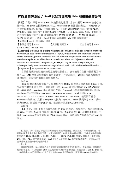
shRNA SNAIL 导致的细胞表型改变
锌指蛋白转录因子Snail沉默对宫颈癌Hela细胞侵袭的影响

锌指蛋白转录因子Snail沉默对宫颈癌Hela细胞侵袭的影响摘要】目的:探讨Snail对Hela细胞侵袭的作用。
方法:采用Hilymax法进行细胞转染,RT-qPCR法检测mRNA表达,Western blot检测蛋白表达,Transwell小室检测细胞侵袭。
结果:与对照组相比,干扰组Snail mRNA表达下降约72.3%(P<0.01);Snail蛋白水平下降约54.2%(P<0.05)。
在24h,48h,72h,干扰组较对照组细胞侵袭能力下降,其抑制率约为17.6%(P<0.05),31.5%(P<0.01),36.1%(P<0.05)。
结论:Snail下调可显著抑制Hela细胞的侵袭能力。
【关键词】Snail;宫颈癌;侵袭【中图分类号】R73-3 【文献标识码】A 【文章编号】2095-1752(2017)07-0267-02【Abstract】Objective To explore whether Snail influences Hela cell invasion. Methods Hilymax was used for cell transfection. RT-qPCR, Western blot and Transwell were for mRNA detection, protein detection and cell invasion, respectively. Results Snail mRNA was downregulated 72.3% while the protein was about 54.2%(P<0.05).The cell invasion was inhibited 17.6%(P<0.05),31.5%(P<0.01),36.1%(P<0.05) at 24h,48h,72h,respectively. Conclusion Down regulation of Snail could inhibit Hela cell invasion. . 【Key words】Snail;Cervical cancer;Invasion宫颈癌是威胁女性健康的常见的恶性肿瘤[1],患者的死亡往往与肿瘤复发转移有关。
RNA剪接错误对细胞功能影响深入探究

RNA剪接错误对细胞功能影响深入探究细胞是生物体的基本单位,其正常功能对于维持生物体的正常生理功能至关重要。
细胞中的基因表达调控是一系列精密的过程,其中RNA剪接是一种关键的调控机制。
然而,当RNA剪接出现错误时,将对细胞功能产生深远的影响。
本文将深入探讨RNA剪接错误对细胞功能的影响以及可能的影响机制。
首先,我们需要了解什么是RNA剪接错误。
RNA剪接是转录过程中的一个关键步骤,它将前体mRNA中的内含子(Intron)剪除,并将外显子(Exon)重新连接,形成成熟的mRNA分子。
这个过程由剪接体(spliceosome)中的snRNA和蛋白质共同完成。
然而,由于基因组的复杂性和剪接过程的多样性,剪接错误时有发生。
RNA剪接错误包括外显子剪接失误、内含子保留以及剪接位点错误等。
RNA剪接错误对细胞功能的影响可以从几个方面来探究。
首先,错误的剪接会导致编码蛋白质序列的改变。
这可能导致蛋白质结构的改变或功能的丧失。
例如,外显子剪接失误可能导致蛋白质结构中关键的结构域缺失,从而影响其正常功能。
内含子保留则可能导致非功能性或具有负面效应的肽段的插入,进而影响蛋白质的结构和功能。
其次,错误的剪接还会影响细胞内的转录调控网络。
正常的剪接是一种重要的转录后调控机制,它可以通过剪接形式的选择性调节基因的表达量和多样性。
当RNA剪接发生错误时,会打乱正常的基因表达调控,导致异常的蛋白质表达和信号传导。
这对于细胞的正常功能会产生重大影响。
此外,RNA剪接错误还可以导致转录产物的稳定性降低。
正常的剪接可保证编码蛋白质的mRNA的稳定性,而剪接错误可能导致mRNA的降解增加。
这进一步降低了细胞中正常编码蛋白质的表达水平,从而对细胞功能产生直接的不利影响。
除了影响基因表达调控和蛋白质功能外,RNA剪接错误还被广泛认为是许多疾病的潜在原因。
近年来的研究表明,RNA剪接错误与多种疾病如帕金森病、癌症和神经系统疾病等相关联。
GSK-3β通过调节 Snail 参与 Fas 诱导的EMT过程

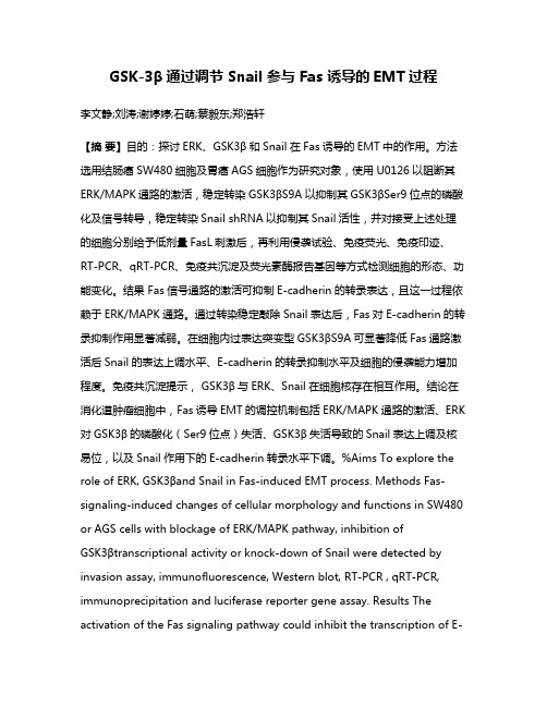
GSK-3β通过调节 Snail 参与 Fas 诱导的EMT过程李文静;刘涛;谢婷婷;石萌;蔡毅东;郑浩轩【摘要】目的:探讨ERK、GSK3β和Snail在Fas诱导的EMT中的作用。
方法选用结肠癌SW480细胞及胃癌AGS细胞作为研究对象,使用U0126以阻断其ERK/MAPK通路的激活,稳定转染GSK3βS9A以抑制其GSK3βSer9位点的磷酸化及信号转导,稳定转染Snail shRNA以抑制其Snail活性,并对接受上述处理的细胞分别给予低剂量FasL刺激后,再利用侵袭试验、免疫荧光、免疫印迹、RT-PCR、qRT-PCR、免疫共沉淀及荧光素酶报告基因等方式检测细胞的形态、功能变化。
结果 Fas信号通路的激活可抑制E-cadherin的转录表达,且这一过程依赖于ERK/MAPK通路。
通过转染稳定敲除Snail表达后,Fas对E-cadherin的转录抑制作用显著减弱。
在细胞内过表达突变型GSK3βS9A可显著降低Fas通路激活后Snail的表达上调水平、E-cadherin的转录抑制水平及细胞的侵袭能力增加程度。
免疫共沉淀提示,GSK3β与ERK、Snail在细胞核存在相互作用。
结论在消化道肿瘤细胞中,Fas诱导EMT的调控机制包括ERK/MAPK通路的激活、ERK 对GSK3β的磷酸化(Ser9位点)失活、GSK3β失活导致的Snail表达上调及核易位,以及Snail作用下的E-cadherin转录水平下调。
%Aims To explore the role of ERK, GSK3βand Snail in Fas-induced EMT process. Methods Fas-signaling-induced changes of cellular morphology and functions in SW480 or AGS cells with blockage of ERK/MAPK pathway, inhibition ofGSK3βtranscriptional activity or knock-down of Snail were detected by invasion assay, immunofluorescence, Western blot, RT-PCR , qRT-PCR, immunoprecipitation and luciferase reporter gene assay. Results The activation of the Fas signaling pathway could inhibit the transcription of E-cadherin in an ERK/MAPK pathway dependent manner. The inhibition of E-cadherin transcription by Fas signaling was significantly reduced after stable transfection of Snail shRNA. The overexpression of mutantGSK3βS9A significantly reduced Fas-induced Snail upregulation, E-cadherin downregulation, and cellular motility enhancement. The result of co immunoprecipitation demonstrated the interaction between GSK3β, ERK and Snail in nucleus. Conclusion In gastrointestinal cancer cells, Fas signaling can induce EMT through the activation of ERK/MAPK pathway, the phosphorylation and inactivation of GSK3βby ERK, the upregula-tion and nuclear translocation of Snail resulting from GSK3βinactivation, as well as Snail-induced downregu-lation of E-cadherin.【期刊名称】《现代消化及介入诊疗》【年(卷),期】2013(000)005【总页数】6页(P267-272)【关键词】Fas信号通路;GSK3β;上皮间质转化;转移;消化道肿瘤【作者】李文静;刘涛;谢婷婷;石萌;蔡毅东;郑浩轩【作者单位】510515广东省胃肠疾病重点实验室,南方医科大学南方医院消化内科;510515广东省胃肠疾病重点实验室,南方医科大学南方医院消化内科;510515广东省胃肠疾病重点实验室,南方医科大学南方医院消化内科;510515广东省胃肠疾病重点实验室,南方医科大学南方医院消化内科;100091 中国中医科学院西苑医院消化科;510515广东省胃肠疾病重点实验室,南方医科大学南方医院消化内科【正文语种】中文消化道肿瘤是目前致死率最高的肿瘤之一[1]。
shRNA-resistant Gankyrin质粒的构建及鉴定

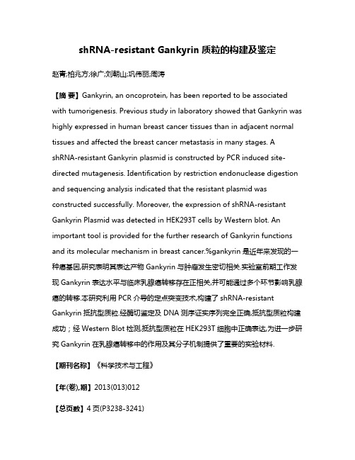
shRNA-resistant Gankyrin质粒的构建及鉴定赵青;柏兆方;徐广;刘朝山;巩伟丽;周涛【摘要】Gankyrin, an oncoprotein, has been reported to be associated with tumorigenesis. Previous study in laboratory showed that Gankyrin was highly expressed in human breast cancer tissues than in adjacent normal tissues and affected the breast cancer metastasis in many stages. A shRNA-resistant Gankyrin plasmid is constructed by PCR induced site-directed mutagenesis. Identification by restriction endonuclease digestion and sequencing analysis indicated that the resistant plasmid was constructed successfully. Moreover, the expression of shRNA-resistant Gankyrin Plasmid was detected in HEK293T cells by Western blot. An important tool is provided for the further research of Gankyrin functions and its molecular mechanism in breast cancer.%gankyrin是近年来发现的一种癌基因,研究表明其表达产物Gankyrin与肿瘤发生密切相关.实验室前期工作发现Gankyrin表达水平与临床乳腺癌转移存在正相关,并可能通过多个环节影响乳腺癌的转移.本研究利用PCR介导的定点突变技术,构建了shRNA-resistant Gankyrin抵抗型质粒.经酶切鉴定及DNA测序证实序列完全正确,抵抗型质粒构建成功;经Western Blot检测,抵抗型质粒在HEK293T细胞中正确表达,为进一步研究Gankyrin在乳腺癌转移中的作用及其分子机制提供了重要的实验材料.【期刊名称】《科学技术与工程》【年(卷),期】2013(013)012【总页数】4页(P3238-3241)【关键词】Gankyrin;定点突变;抵抗型质粒;RNA干扰回复【作者】赵青;柏兆方;徐广;刘朝山;巩伟丽;周涛【作者单位】国家生物医学分析中心,北京100850【正文语种】中文【中图分类】Q343.17每年大约有90%的肿瘤患者死于肿瘤转移[1,2]。
Snail蛋白诱导EMT在肝癌中的研究进展

Snail蛋白诱导EMT在肝癌中的研究进展[摘要]:Snail为锌指蛋白超家族的第一个成员,在转录调控、细胞信号和发育过程中发挥积极作用。
有研究表明,Snail 可促使上皮-间充质转化及E-cadherin的降解,在许多肿瘤组织中呈中高表达,被认为是促进肿瘤侵袭转移的因素。
对 Snail的深入研究不仅能更进一步阐明 Snail的作用机制,并且为进一步研究以 Snail 为靶点的肿瘤治疗策略提供理论依据。
[关键词]:Snail; 上皮-间充质转化;肿瘤1.Snail的结构Snail家族是富含锌指结构的转录因子,是锌指蛋白家族的一个分支,包括Snail(Snail1)、Slug(Snail2)、Smuc(Snail3)。
它们主要在人体的胎盘、胚胎中胚层、成人的心、肝、骨骼肌以及未分化的组织中表达。
Snail基因位于染色体20q13.2,分子量为29100,由264个氨基酸构成[1]。
它的羧基端由高度保守的包含4~6个锌指结构组成,其中锌指结构域中(C2H2)的半胱氨酸和组氨酸的残基能与锌离子形成共价键,可以结合在特异的DNA序列上。
羧基端通常与靶基因启动子的CAGGTG序列(E-box)结合;氨基端是相对保守的SNAG功能结构域,可以抑制靶基因的转录。
2.Snail的功能Snail转录因子家族在与细胞存活和分化相关的多个过程中发挥着关键作用。
在转录水平上,Snail-1活性可能通过其他能够直接与Snail-1启动子相互作用的多功能因子进行调节,例如HIF-1α、NF-κB、Notch胞内结构域、IKKα、SMAD、HMGA2、Egr-1、PARP-1或STAT3。
一般来说,Snail-1扮演着转录阻遏子的角色,该阻遏子与包含称为E-box序列的调控区和启动子结合。
然而,由于E-box序列存在于许多不同基因的启动子中,Snail-1具有非常广泛的活性,同时也是参与致癌的关键基因的调节器。
Snail-1的主要功能无疑是抑制E-钙粘蛋白,但也抑制其他特征性上皮标记物。
Wnt信号通路诱导肿瘤细胞上皮间质转化的研究进展

·905·
Wnt / β-catenin 信号 通 路 诱 导 EMT 发 生 的 重 要 性。Prasad 等[11]又进一步通过对 98 例临床浸润性乳腺导管癌样本中 Wnt / β-catenin 信号通路的 表 达 方 式 以 及 关 键 组 分 E 钙 粘 素、Slug 和 GSK3β 之间关系的分析发现,Slug 作为介导 EMT 发生的重要分子,可以通过激活 Wnt / β-catenin 信号通路降 低 E 钙粘素,首次提供了临床证据支持在浸润性乳腺导管癌 的 EMT 中,Wnt / β-catenin 信号通路的表达上调。Zhao 等[12] 研究发现,使用缺氧诱导因子-1α( HIF-1α) 可以诱导前列腺 癌细胞( LNCaP) 发生 EMT。细胞的间质标志物呈现高表达, 上皮标记物则低表达; 而转染 β-catenin 的短发卡 RNA( shRNA) 后,细胞上皮标记物 E 钙粘素表达增加,间质标记物 N钙粘素( N-cadherin) ,波形蛋白( vimentin) 以及基质金属蛋白 酶-2( MMP-2) 的表达则明显下降,通过 β-catenin 的 shRNA 作用,LNCaP 细胞的 EMT 发生了逆转,证明了 Wnt / β-catenin 信号通路作为一个必要的内源性信号,可能直接控制 HIF1α 诱导 EMT 的发生。Stemmer 等[13]研究发现,Wnt / β-catenin 信号激活可以使 Slug、Snail、Twist 等表达增加,降低 E 钙 粘素并形成 EMT。而在结肠癌细胞系的 EMT 过程中,Snail 过表达可以增加 Wnt 信号靶基因的表达,Snail 通过其 N 端 与 β-catenin 相互作用,进一步激活 Wnt 信号下游靶基因的 表达,从而形成 Wnt 信号激活的正反馈。
Snail1 siRNA 对高糖诱导肾小管上皮细胞表型转变的影响
Snail1 siRNA 对高糖诱导肾小管上皮细胞表型转变的影响方开云;石明隽;肖瑛;桂华珍;郭兵;张国忠【期刊名称】《中国病理生理杂志》【年(卷),期】2009(025)012【摘要】目的:观察Snail1 siRNA对高糖诱导的肾小管上皮细胞向间充质细胞转变(TEMT)的影响.方法: 原代培养肾小管上皮细胞分为5组:(1)对照组(含糖5.5 mmol/L);(2)高糖组(含糖25 mmol/L);(3)Snail1 siRNA处理组,转染Snail1 siRNA,6 h后更换为高糖(含糖25 mmol/L)培养;(4)control siRNA处理组,转染control siRNA作为siRNA阴性对照,6 h后换为高糖(含糖25 mmol/L)培养;(5)高渗组(含D-manntio19.5 mmol/L);72 h后收集细胞,用Western blotting和半定量RT-PCR检测Snail1、TGF-β_1、α-平滑肌肌动蛋白(α-SMA)、vimentin和E-cadherin蛋白和mRNA表达.结果: 与高糖组比较,肾小管上皮细胞转染Snail1 siRNA后,Snail1 mRNA和蛋白表达水平分别下降62%和68%(P<0.01).同时,Snail1 siRNA处理组α-SMA和vimentin蛋白和mRNA表达显著下调(P<0.01),而E-cadherin蛋白和mRNA表达显著上调(P<0.01).结论: Snail1参与了高糖诱导TEMT的调节.【总页数】6页(P2424-2429)【作者】方开云;石明隽;肖瑛;桂华珍;郭兵;张国忠【作者单位】贵州省人民医院麻醉科,贵州,贵阳,550002;贵阳医学院病理生理学教研室,贵州,贵阳,550004;贵阳医学院病理生理学教研室,贵州,贵阳,550004;贵阳医学院病理生理学教研室,贵州,贵阳,550004;贵阳医学院病理生理学教研室,贵州,贵阳,550004;贵阳医学院病理生理学教研室,贵州,贵阳,550004【正文语种】中文【中图分类】R363【相关文献】1.Snail1/IGF-1信号通路介导高糖诱导的肾小管上皮细胞EMT [J], 潘祉谕;达静静;董蓉;吴静;皮明婧;俞佳丽;孙翼;聂瑛洁;查艳2.Akt/GSK-3β介导高糖上调肾小管上皮细胞Snail1的表达 [J], 余红;石明隽;肖瑛;刘瑞霞;王圆圆;郭兵;张国忠3.siRNA沉默CTGF表达对高糖诱导人肾小管上皮细胞转分化的影响 [J], 汤珣;蔡德鸿;曾莉;章俊4.去甲斑蝥素对TGF-β1诱导的人近端肾小管细胞上皮间质转分化以及转录因子Snail1表达的影响 [J], 刘伏友;夏运成;孙岩;孙林;凌光辉;李瑛;刘健;袁曙光;刘映红;张东山5.针对结缔组织生长因子的siRNA对高糖诱导人肾小管上皮细胞肥大的影响 [J], 章俊;杜庆生;蔡德鸿;曾莉;汤珣因版权原因,仅展示原文概要,查看原文内容请购买。
上皮间质转化与肿瘤侵袭、转移
上皮间质转化与肿瘤侵袭、转移朱娓【摘要】肿瘤转移是肿瘤发展的重要阶段,是多数恶性肿瘤患者的主要致死因素.上皮间质转化(transformation of epithelial mesenchymal,EMT)作为肿瘤发生转移的第一步,是一个动态的多基因、多步骤的生物学过程,与恶性肿瘤的侵袭、转移关系密切.研究EMT发生发展机制,寻找控制肿瘤侵袭、转移的有效途径或肿瘤治疗新靶点,是目前肿瘤研究的热点之一.该文仅就EMT、相关因子及信号转导通路与肿瘤侵袭、转移的关系作一综述.【期刊名称】《外科研究与新技术》【年(卷),期】2014(003)001【总页数】5页(P55-59)【关键词】上皮间质转化;肿瘤;转移【作者】朱娓【作者单位】徐汇区大华医院普外科上海200237【正文语种】中文【中图分类】R730上皮间质转化(epithelial mesenchymal transition,EMT)是Greenburg等[1]于1982年发现晶状体上皮细胞在胶原凝胶中可转变为间质细胞样细胞后提出的概念。
人们发现,EMT不但在胚胎发育过程中对组织器官形成至关重要,还参与创伤愈合、组织重建、肿瘤侵袭转移过程。
近年,关于EMT与恶性肿瘤侵袭转移的关系及其作用机制研究,已成为肿瘤领域的研究热点之一。
深入了解EMT 的发生机制,可为进一步认识肿瘤、治疗肿瘤提供新的理论依据和研究切入点。
故本文就EMT及其相关因子、信号转导通路与肿瘤侵袭转移的关系作一综述。
EMT是指上皮细胞在特定的生理和病理环境下向间充质细胞转变分化的现象,是细胞失去上皮细胞表型并逐渐获得间质细胞表型的过程,是一个复杂、有序、多基因、可调控的生物学过程。
其分子标志物的特征性变化为上皮 E-钙粘蛋白(epithelium-cadherin,E-cad)、α-连环蛋白(α-Catenin)、β-连环蛋白(β-Catenin)、桥粒蛋白、细胞角蛋白等上皮细胞标志物表达下降;N-钙粘蛋白(N-cadherin,N-cad)、成纤维细胞特异蛋白、纤连蛋白(fibronectin)、波形蛋白(vimentin,Vim)等间质细胞标志物表达上升。
Snail在IgA肾病组织中的表达及其与肾小管上皮-间质转化的关系
Snail在IgA肾病组织中的表达及其与肾小管上皮-间质转化的关系李静;高慧敏;王弦;秦蓉【摘要】目的探讨在组织和细胞水平上Snail的表达与肾小管上皮-间质转化(epithelial-mesenchymal transition,EMT)及肾小管间质纤维化(tubulointerstitial fibrosis,TIF)的关系;观察转染Snail基因后人肾小管上皮细胞(HK-2)miRNA表达谱的变化,以深入阐明EMT机制中miRNA的重要性.方法采用免疫组化法检测Snail及EMT相关蛋白vimentin、SMA、E-cadherin在40例IgA肾病患者肾穿刺组织中的表达.采用RT-PCR及Western blot法检测Snail、E-cadherin、vimentin、SMA在HK-2细胞正常对照组、空转染组、Snail基因转染组中的表达,进一步借助基因芯片筛选出差异表达的miRNA.结果免疫组化结果显示,IgA肾病组织中Snail与vimentin及SMA蛋白的表达呈正相关,与E-cadherin蛋白的表达呈负相关,且TIF程度越高,Snail蛋白表达越强.RT-PCR及Western blot检测结果显示,与对照组相比,Snail转染组Snail、vimentin、SMA 在基因和蛋白水平表达均升高,E-cadherin蛋白表达降低,差异具有统计学意义(P<0.05).基因芯片结果表明,Snail转染HK-2细胞后,筛选出5个明显差异表达的miRNA,预测出5 026个可能的潜在靶基因.结论 Snail表达与肾小管EMT及TIF 关系密切,可作为新靶点,在EMT防治中起重要作用;差异表达的miRNAs可能参与Snail促进EMT及TIF过程的发生、发展.%To investigate the relationship between Snail and renal tubular epithelial-mesenchymal transition (EMT) or tubulointerstitial fibrosis (TIF) at tissue and cellular levels and to observe the changes of miRNA profile after transfecting Snail gene into human renal tubular epithelial cells (HK-2),to further elucidate the importance ofmiRNA in the pathogenesis of renal fibrosis.Methods The expression of Snail and EMT-related proteins vimentin,SMA,E-cadherin was detected by immunohistochemistry in renal tissues of 40 patients with IgA nephropathy.The expression of Snail,E-cadherin and SMA in normal control group,empty transfection group and Snail gene transfection group was detected by Western blot and RT-PCR.Furthermore,differentially expressed miRNAs were screened by gene chip.Results By immunohistochemistry,Snail expression was positively correlated with vimentin and SMA,negatively correlated with E-cadherin in IgA nephropathy.The higher degree of the TIF,the stronger the expression of pared with the control group,the expression of Snail,vimentin and SMA in the snail transfected group increased.However,E-cadherin decreased at gene and protein level by the RT-PCR and Western blot (P <0.05).The difference was statistically significant.Five distinctly different miRNAs were screened by gene chip after Snail gene was transfected into HK-2 cells,and then 5 026 potential target genes werepredicted.Conclusion Snail expression is closely related with renal tubular epithelial mesenchymal transition and tubulointerstitial fibrosis,and it may be used as a new target in EMT prevention.Differentially expressed miRNAs may be involved in the development of EMT and TIF.【期刊名称】《临床与实验病理学杂志》【年(卷),期】2017(033)006【总页数】7页(P629-635)【关键词】肾病;肾小管上皮细胞;Snail;上皮-间质转化;免疫组织化学;miRNA;基因芯片【作者】李静;高慧敏;王弦;秦蓉【作者单位】安徽医科大学病理学教研室,合肥230032;安徽医科大学病理学教研室,合肥230032;安徽医科大学病理学教研室,合肥230032;安徽医科大学病理学教研室,合肥230032【正文语种】中文【中图分类】R692肾小管间质纤维化(tubulointerstitial fibrosis, TIF)是各种慢性肾脏疾病进展为终末期肾病的最终共同途径[1]。
转录因子Snail的作用机制及其生理功能
转录因子Snail的作用机制及其生理功能
胡士军;马兴红;杨增明
【期刊名称】《细胞生物学杂志》
【年(卷),期】2006(28)2
【摘要】Snail为起负调节作用的锌指转录因子,其序列和功能在不同种属动物中十分保守。
Snail超家族成员在胚胎着床、胚胎发生、肿瘤发生、细胞命运决定、细胞周期调控、左右不对称发育及创伤愈合等生理或病理过程发挥重要作用。
对Snail的进一步研究,不仅可以阐明Snail超家族的作用机制,而且可以为探究Snail 相关的肿瘤治疗策略提供重要的理论基础。
【总页数】5页(P160-164)
【关键词】Snail;锌指;上皮细胞-间充质细胞转换;胚胎发生;肿瘤发生
【作者】胡士军;马兴红;杨增明
【作者单位】东北农业大学生命科学学院
【正文语种】中文
【中图分类】Q753;TS202.3
【相关文献】
1.转录因子Snail对胃癌MKN-28细胞的增殖、凋亡及侵袭的影响 [J], 武平
2.锌指转录因子SNAIL调控和功能 [J], 毕嫣然; 吴照球; 傅蓉
3.Snail转录因子在肝癌中的表达及干扰Snail对肝癌转移的影响 [J], 钱明;张士群;朱嘉
4.鼠双微体基因2和锌指转录因子Snail在甲状腺癌组织的表达及其临床意义 [J], 王宏;金志巍;王成燕;王帅;张贺
5.转录因子SNAIL在小鼠植入前胚中的表达及作用 [J], 吕瑞敏;刘玥;许颂华;林建闽;莫开恩;Eman Nageeb Elsayed Draz;王世鄂
因版权原因,仅展示原文概要,查看原文内容请购买。
- 1、下载文档前请自行甄别文档内容的完整性,平台不提供额外的编辑、内容补充、找答案等附加服务。
- 2、"仅部分预览"的文档,不可在线预览部分如存在完整性等问题,可反馈申请退款(可完整预览的文档不适用该条件!)。
- 3、如文档侵犯您的权益,请联系客服反馈,我们会尽快为您处理(人工客服工作时间:9:00-18:30)。
The effects of shRNA-mediated gene silencing of transcription factor SNAI1on the biological phenotypes of breast cancer cell line MCF-7Yan Lu •Lina Yu •Minlan Yang •Xiangshu Jin •Zhijing Liu •Xiaowei Zhang •Liping Wang •Dongjing Lin •Yuanyuan Liu •Min Wang •Chengshi QuanReceived:18August 2013/Accepted:15November 2013/Published online:29November 2013ÓSpringer Science+Business Media New York 2013Abstract To research the effects of silencing transcrip-tion factor SNAI1on the in vitro biological phenotypes of breast cancer cell line MCF-7,based on the gene sequence of SNAI1,we linked shRNA with the green fluorescent protein-expressing eukaryotic expression vector pGCsi-lencer TM U6/Neo/GFP,and transfected it into MCF-7cells.The SNAI1gene-silencing effect was authenticated by RT-PCR and immunofluorescence.We then examined the effect of gene silencing on the expression of epithelial and mesenchymal markers and on their biological phenotypes of the target cells.Finally,we explained that SNAI1was bound to E-cadherin in MCF-7cells by ChIP.Silencing SNAI1upregulated the expression of epithelial markers claudin-4,claudin-7,and E-cadherin,while expression of the mesenchymal marker matrix metalloproteinase-2was downregulated.The capacity for proliferation,migration,and invasion was diminished.SNAI1binds to the E-cad-herin gene promoter and inhibits its transcription.We can conclude that silencing gene SNAI1inhibits expression of properties that are associated with the malignant phenotype of MCF-7cells and reverses the epithelial–mesenchymaltransition process by regulating relevant target gene E-cadherin.Keywords SNAI1ÁGene silencing ÁBiological phenotypes ÁMCF-7IntroductionDuctal carcinoma of the breast is one of the most common malignant tumors in women,and its incidence is increasing year by year [1,2].Tumor metastasis is the main reason for mortality from this disease [3].In breast cancer,the expression level of SNAI1(snail homolog 1),a transcription factor,correlates with tumor grade and lymph node metas-tasis [4].SNAI1can regulate the expression of a target gene by E-box (CAGGTG\CACCTG)which can specifically bind upstream of the target promoter to inhibit the expression of the gene,resulting in initiation of epithelial–mesenchymal transition [5–11]which is also mainly characterized by downregulation of E-cadherin [12,13].SNAI1induces epithelial–mesenchymal transition which increases metas-tasis through loss of apical–basolateral polarity and gain of mesenchymal characteristics to enhance motility [14].Consequently,we have postulated that SNAI1plays a key role in the invasion and metastasis of breast cancer through binding to E-cadherin gene promoter.To test this hypothesis,we designed the plasmid pGCsi-lencer TM U6/Neo/GFP,which can silence the expression of SNAI1.The plasmid was transfected into MCF-7cells which have high endogenous SNAI1expression.We then examined the expression of epithelial and mesenchymal markers as well as the effect of SNAI1silencing on proliferation,migration,and invasion in vitro.SNAI1binding to E-cadherin gene promoter in MCF-7cells was authenticated by ChIP.Yan Lu and Lina Yu have contributed equally to this paper.Y.Lu ÁM.Yang ÁX.Jin ÁZ.Liu ÁX.Zhang ÁL.Wang ÁD.Lin ÁY.Liu ÁM.Wang ÁC.Quan (&)The Key Laboratory of Pathobiology,Ministry of Education,Department of Pathology,College of Basic Medical Sciences,Jilin University,126Xinmin Avenue,130021Changchun,China e-mail:quancs@L.YuThe First Hospital Cancer Center of Jilin University,Changchun,ChinaMol Cell Biochem (2014)388:113–121DOI 10.1007/s11010-013-1903-4Materials and methodsCell cultureHuman mammary epithelial cell line HBL-100,human breast cancer cell lines MCF-7and MDA-MB-231,and mousefibroblast cell line NIH3T3were cultured in high glucose DMEM medium(GIBCO,USA)supplemented with10%fetal bovine serum(FBS;Hyclone,USA)at 37°C,in the presence of5%CO2.Transient transfectionBased on Ding et al.[15],shRNA(containing sense and antisense sequences linked by a hairpin loop:TTCAAGA-GA)was designed and constructed into the vector pGCsi-lencer TM U6/Neo/GFP(GeneChem,China).The MCF-7cells were then transfected with SuperFectÒTransfection Reagent(QIAGEN,USA).The negative control cell line was generated by infecting cells with the vector pGCsi-lencer TM U6/Neo/GFP constructed with oligonucleotides, which has no homology to the human gene.The control group was the MCF-7cell line that has not been treated. Reverse transcription-polymerase chain reaction(RT-PCR)Total RNA was isolated with TRIZOL(Invitrogen,USA) and reverse transcribed with M-MLV reverse transcriptase (TaKaRa,Japan).The primers(Sangon,China)used for RT-PCR were devised by software Prime premier5.0and are listed in Table1.Western blot analysisCells were harvested and lysed in300l l ice-cold Radio Immunoprecipitation Assay Lysis Buffer supplemented with 1%phenylmethylsulfonylfluoride,then centrifuged at 12,0009g,at4°C for30min to remove cell debris.Their concentration was determined by a BCA Protein Assay Kit (Pierce Chemical Co.,Rockford,IL,USA).Cell lysates were separated in10%SDS-PAGE and then transferred onto a nitrocellulose membrane(Millipore,Temecular,CA,USA). The membrane was then incubated with5%nonfat milk (Applichem)at37°C for1h,and incubated with1:1,000 diluted primary antibody SNAI1(ENOGENE,CHINA)at 4°C overnight and1:1,000diluted secondary antibody for 1h at room temperature thereafter.The secondary antibody was appropriate IgG,conjugated to horseradish peroxidase. The blots werefinally stained using an ECL Western blotting system(GE,Fairfield,CT,USA).ImmunofluorescenceThe assay was performed to evaluate the expression of SNAI1as previously described[16].In our study,cells were incubated with primary SNAI1antibody(1:300),at4°C overnight.The secondary antibody was Alexafluor488anti-rabbit IgG(1:250dilution;CST,USA).Cells were visualized with a laser scanning confocal microscope(Olympus,Japan).Cell proliferation analysisIn order to measure cell proliferation,cells were seeded in 24-well plates at a low density(29104cells/well)and cul-tured as described above.The number of cells was countedTable1Primers and information for RT-PCR MMP-2matrix metalloproteinase-2Gene Primers Length(bp)Annealingtemperature(°C)CyclesSNAI150-GCCTAGCGAGTGGTTCTTCTG-301685430 50-TAGGGCTGCTGGAAGGTAAA-30Claudin-450-TGCACTCTGCGAACGTTAAG-301415635 50-GCGATGCCCATTACCTGTAG-30Claudin-650-TTCATCGGCAACAGCATCGT-303455635 50-GGTTATAGAAGTCCCGGATGA-30Claudin-750-GGAGATCCCAGGTCACACAT-301405030 50-CAGGGTCTGCCCTAGTCATC-30E-cadherin50-CGGTGGTCAAAGAGCCCTTACT-301715832 50-TGAGGGTTGGTGCAACGTCGTTA-30MMP-250-CTTCCAAGTCTGGAGCGATGT-301185230 50-TACCGTCAAAGGGGTATCCAT-30GAPDH50-TGTTGCCATCAATGACCCCTT-301875525 50-CTCCACGACGTACTCAGCG-30every24h over the next7days.Three replicate counts were performed in each of three independent experiments.Wound healing assay19105cells were seeded per well into a24-well plate and cultured as described above.When cells had grown to confluence,a straight wound was created in each mono-layer by dragging a10-l l pipette tip through the mono-layer.Images were taken under a microscope(Olympus, Japan)at0and24h after wounding to determine the width of the wounded area.The relative migration distance(%of recovery)was calculated as(W0-W24)/W09100%.In each experiment,three replicate wells were measured.Invasion assayWe cultured NIH3T3cells as described above and replaced the medium with serum-free DMEM when the cells were at logarithmic growth phase.The supernatant was collected after 24h and passed through a0.22-l mfilter to remove the cell debris.Cells of three groups were harvested from monolayer culture by trypsinization,centrifuged to collect them into a 1.5-ml eppendorf tube,and stained with0.5ml Rhodamine (1mg/ml)for30min.We planted59104stained cells on a Matrigel membrane which was in the upper compartment of the Millipore chamber.The supernatant was placed into the lower compartment as a chemoattractant.The chambers were then incubated for5h at37°C in the presence of5%CO2. The Matrigel membranes were scanned by a laser scanning confocal microscope(Olympus,Japan)to see how many cells had migrated through the Matrigel.Two replicates were performed in each of three independent experiments. Chromatin immunoprecipitation assayThe experiment was performed as described by the EZ-ChIP kit protocol(MILLOPORE,USA).Briefly,the steps were as fol-lows:Cells were cultured to confluence in10-cm dishes (19107–59107cells per dish).Proteins were cross-linked to DNA,by adding formaldehyde drop-wise directly to the media for afinal concentration of1%,and then incubated at37°C for 10min.Incubated with2.5M glycine for5min at room temperature to terminate formaldehyde cross-linking.The cells were removed mechanically in cold PBS and transferred into 15-ml tubes.They were then harvested by centrifugation and resuspended in lysis buffer(contains protease inhibitors).The lysate was sonicated to shear the DNA to an average fragment size of200–1,000bp.The fragment size was fol-lowed on a1.5%agarose gel.The reaction mixture was cen-trifuged,the supernatant was transferred to a new tube,and 100l l of each sonicated sample was removed.900l l ChIP dilution buffer and20l l509Protease Inhibitor Compounds were added to the each of sonicated samples.The antibody was diluted to a concentration of1:500.RNA polymerase II anti-body was added as a positive control and rabbit IgG antibody was used as a negative control.60l l of protein A/G beads and salmon protease were added to all samples which were then immunoprecipitated overnight with rotation at4°C.The pro-tein A/G beads were harvested by centrifugation(2,0009g)and washed with buffer.The DNA was then eluted and PCR ana-lysis was performed to detect the promoter regions of E-cad-herin(promoter,5’-ACTCCAGGCTAGAGGGTCAC-3’and 5’-CCGCAAGCTCACAGGTGCTTTGCAGTTCC-3’). Statistical analysisStatistical analysis was performed using the statistical software SPSS12.0for t test and one-way ANOVA.The criterion for statistical significance was P\0.05. ResultsSNAI1expression in mammary cell linesRT-PCR and western blot analysis showed that there was lower SNAI1expression in human mammary epithelial cell line HBL-100and higher expression in the two human breast cancer cell lines.MCF-7cells had higher expression levels than MDA-MB-231(Fig.1a,b).Therefore,we chose MCF-7as the focus of our study.SNAI1gene-silencing effectRT-PCR demonstrated that after transfection with shRNA, SNAI1was significantly downregulated incomparison Fig.1Expression of SNAI1in different cell lines.We evaluated the expression of SNAI1in mammary epithelial cell line HBL-100and breast cancer cell lines MCF-7and MDA-MB-231(a,RT-PCR;b, Western blot).SNAI1expression was higher in MCF-7cells than in the other two cell lineswith the control and negative control group between which there was no significant difference in SNAI1expression (Fig.2a,b).Figure2c shows that following transfection with shRNA,SNAI1expression was downregulated,as the greenfluorescence was weaker in the nucleus compared with the other two groups,and that SNAI1was located in the nucleus.Expression of epithelial and mesenchymal markersRT-PCR analysis shows that,after silencing of SNAI1,the epithelial markers claudin-4,claudin-7,and E-cadherin were upregulated.At the same time,the mesenchymal marker matrix metalloproteinase-2was downregulated (Fig.3a,b).Thus,we postulate that SNAI1silencing can lead to epithelial–mesenchymal transition.Effect of SNAI1silencing on cell behaviorsAfter growing the cells for7days in24-well plates,we counted the cell number each day and found that cells in the shRNA group proliferated significantly more slowly than the other two groups(Fig.4a).After wounding the monolayer, we measured the relative migration distance of the cells in each group and calculated their relative mobility at0and 24h(Fig.4b,c).At24h,the in vitro migration rate of the shRNA group was significantly lower than the rate of the other two groups.After SNAI1silencing,the migration distance(Fig.4d lower panel)and the number(Fig.4d upper panel)of cells that went through Matrigel to the polycar-bonate membrane level were less than the other two groups. There was no significant difference between the control and the negative control group cells(Fig.4).Therefore,we concluded that SNAI1can promote the proliferation, migration,and invasion of MCF-7cells.SNAI1is bound to the E-cadherin geneFigure2c shows that SNAI1was located in the nucleus and may have some function related to epithelial–mesenchymal transition in the nucleus.Chromatin immunoprecipitation demonstrated that SNAI1was bound to the E-cadherin gene promoter in MCF-7cells(Fig.5b).Furthermore, SNAI1silencing was associated with upregulated E-cad-herin(Fig.3a,b).Therefore,we can concluded that SNAI1 is a transcriptional factor,which can bind to the E-cadherin gene and inhibits its transcription.DiscussionTranscription factor SNAI1is a member of the snail tran-scription inhibitor super-family,with more than50family members which have been characterized in metazoans.In humans,it is distributed in the placenta,heart,lung,brain, liver,and skeletal muscles[17].At the carboxy terminus of the SNAI1gene,there are four highly conserved zincfingers which specifically bind to the E-box sequence(CAGGTG\CAGGTG)of the upstream target gene promoter region and have the capacity of initi-ating the epithelial–mesenchymal transition process by inhibiting the activity of the target gene promoter[18]. Epithelial–mesenchymal transition is characterized by the loss of apical–basolateral polarity,gain ofmesenchymal Fig.2Expression of SNAI1in different groups.RT-PCR(a,b)andimmunocytofluorescent analysis(c)show that SNAI1expression waslower in the shRNA group than the other two groups.The data areexpressed as mean±SD,N=3;*P\0.01versus control andnegative group(b).After silencing,SNAI1expression wasdownregulated,as the greenfluorescence was weaker in the nucleuscompared with the other two groups(c).Three independent exper-iments were done(M marker,C control,N negative control,S SNAI1-shRNA)characteristics,extracellular matrix degradation,weakened intercellular adhesion,and enhanced cell motility [8,19,20].The epithelial–mesenchymal transition process is critical for embryonic development,wound healing,cell differentia-tion,cell migration,and tumor metastasis [21,22].On the contrary,mesenchymal cells can acquire characteristics of epithelial cells through transformation,namely,mesenchy-mal–epithelial transition.Therefore,we have postulated that reversing the epi-thelial–mesenchymal transition phenotype might reduce the invasive and metastatic capabilities of early cancers and prolong the survival of tumor-bearing animals.In this study,we used RNAi technology to silence SNAI1gene expression and studied the resulting biological phenotypes of MCF-7cells.After silencing SNAI1,the epithelial markers E-cad-herin,claudin-4,and claudin-7were upregulated and the mesenchymal cell marker matrix metalloproteinase-2was downregulated.The results show that silencing SNAI1activated the epithelial cell-associated genes.The cells then regained their epithelial polarity and capacities for cell–cell adhesion,thus demonstrating the unique morphology of epithelial cells.Silencing SNAI1induced downregulation of matrix metalloproteinase-2and reduced the ability of the cells to degrade extracellular matrix and collagen.In other words,the cells lost the ability of migration and invasion,in concert with the loss of mesenchymal to epithelial transformation.Other in vitro experiments have obtained results that are consistent with our data.After antisense SNAI1was transfected into colon cancer cell line HT-29,the cell pseudopodia became shortened and the cells became round,suggesting mesenchymal cell-like phenotype transition to an epithelioid phenotype [23].In the present study,we also examined the proliferative capacity of the test cells in vitro.After silencing SNAI1,the proliferation rate of the MCF-7cells was diminished.In a similar experiment,Olmeda et al.[24]silenced the SNAI1gene in MDA-MB-231cells which are typically poorly differentiated and easily transplanted.60days after inoculating the silenced cells and control cells into mice,both the number of metastases and the tumor size in the SNAI1-silenced group were significantly less than the control group,indicating that SNAI1inhibited cell prolif-eration in vivo.This observation is also consistent with our experimental data.Olmeda et al.postulated that SNAI1might be a downstream molecule of TGF-b ,PI3K,or a signal pathway and suggested that the reduction of SNAI1might diminish cell proliferation capacity through a signal transduction pathway.In our study,silencing SNAI1inhibited cell proliferation in association with upregulation of E-cadherin,claudin-4,and claudin-7,suggesting that the connections between cells and their anchorage in local position may inhibit mitosis.This concept is supported by the work of St Croix et al.[25]who found that E-cadherin is not only a mediator of epithelial adhesion but also an important contact cell growth inhibitor.By upregulating cell cycle inhibitor p27,E-cadherin acted on the cell cycle to inhibit cell prolifer-ation.Thus,it may be that SNAI1affects cell proliferation indirectly through E-cadherin expression.Silencing SNAI1appears to inhibit MCF-7cell migra-tion in several ways.First,in our study,SNAI1directly lowered the expression of E-cadherin by binding to its promoter and inhibiting its transcription and translation.E-cadherin is the main component for epithelial adhesion connection [26].It can be connected to b -catenin to form an E-cadherin/b -catenin complex,causing the cells to be connected to each other and restricting their migration or invasion [27].In the process of tumorigenesis and pro-gression,the decline in expression of E-cadherin can weaken cell adhesion,thus causing tumor cells to exitfromFig.3Expressions of epithelial and mesenchymal markers.mRNA level of the expression of epithelial markers claudin-4,claudin-6,claudin-7,and E-cadherin and mesenchymal marker MMP-2(a ,b ).Except claudin-6,expression of epithelial markers in the experimental group was upregulated in comparison with the control and negative control groups.Expression of the mesenchymal marker MMP-2was downregulated.Data represent one of at least three independent experiments.Each value represents the mean ±SD.*P \0.01versus the parent control cells (M marker,C control,N negative control,S SNAI1-shRNA)the primary tumor,invade the surrounding tissue,and subsequently diffuse through blood vessels [28].Second,we found that silencing SNAI1upregulated the expression of key components of the tight junction—claudin-4and claudin-7—which play a decisive role in the constitution of epithelial and endothelial tissue [29].Tight junction com-ponents may strengthen the connections between cells in order to maintain organizational structure and the local steady state.Therefore,we postulate that the diminished capacity of cell migration in vitro after silencing SNAI1may be related to the upregulation of E-cadherin,claudin-4,and claudin-7.Similar to our results,Fabre-Guillevin [30]also found that in the breast cancer cell line MDA-MB-231,the ability of cells to migrate in vitro was significantly weakened when SNAI1was inhibited.Moreover,in human colorectal cancer cell lines HCT-116and HT29[31]and hepatoma cell line HepG2[32],the results were also consistentwithFig.4Effect of SNAI1silencing on cell behaviors in MCF-7cells.a Cell proliferation curve.There was a significant difference between the numbers of cells in each group from the second day,but no significant difference in cell proliferation between the control group and the negative control group.SNAI1transfection of MCF-7cells significantly inhibited their proliferative ability (The data are expressed as mean ±SD,N =6,*P \0.01versus control and negative group).b Wound healing assay to detect cell migration ability.After wounding the monolayer,we measured the relative migration distance of the cells in each group and calculated the relative mobility at 0and 24h.In the shRNA group,the migration rate at 24h was significantly lower than the other two groups (N =6,*P \0.01).Mobility of control group and negative group was notsignificantly different (c ).d Invasion assay to detect cell invasion ability.Cells were planted on a Matrigel surface in the upper well of a Boyden chamber,and then incubated at 37°C for 5h.Conditioned medium was used as a chemoattractant.After incubation of the chamber,we used confocal microscopy to scan the cells in each group layer by layer.Cells that had migrated through the Matrigel layer to the polycarbonate membrane level were counted (d ,upper panels ).Cell migration distance is shown in (d lower panels ).Invasive ability of the shRNA group was significantly less than the other two groups;the shRNA group had no detectable cells that had passed through the Matrigel layer (e ,N =6,**P \0.001);M polycarbonate membrane,C control,N negative control,S SNAI1-shRNAthe present experiment that cell migration was significantly weakened.Tumor invasion influence factors include reduction of intercellular adhesion,increased mobility of the tumor cells,and enzymatic degradation of the basement membrane and extracellular matrix by enzymes such as matrix metallo-proteinases,cathepsins,and plasminogen activators [33].In our experiments,silencing SNAI1significantly downregu-lated the expression of matrix metalloproteinase-2and inhibited the cellular invasive ability in vitro.Silencing SNAI1also downregulated the expression of matrix metalloproteinase-9.Matrix metalloproteinase-2and matrix metalloproteinase-9are gelatinases [34].Their substrates are degenerated collagen and the main constituent of base-ment membrane,type-IV collagen.Matrix metalloprotein-ase-2can also degrade fibronectin and laminin,which contribute to the process of tumor invasion [35].We postulate the existence of a unique mechanism by which these genes are regulated through silencing SNAI1.In our experiments,after gene silencing,SNAI1was mainly expressed in cytoplasm,instead of in both nucleus and cytoplasm,and it upregulated E-cadherin.Further-more,SNAI1was bound to E-cadherin in MCF-7cells;we concluded that SNAI1,as a transcriptional factor,works in nucleus to bind to E-cadherin gene promoter and inhibitsits transcription,resulting in epithelial–mesenchymal transition.In the promoter region upstream of E-cadherin [36],claudin-4,and claudin-7[37],there are three,eight,and eight E-box structures,respectively,to which SNAI1could directly bind,thus playing a transcriptional repressor role.Thus,silencing SNAI1could reduce its normal inhibitory role,causing upregulation of E-cadherin,claudin-4,and claudin-7.Although claudin-6has three E-boxes as well,its expression did not change significantly.This shows that though the gene claudin-6also consists of E-boxes,SNAI1did not directly bind to them.This is similar to the regulation of claudin-1.Ohkubo and Ozawa [38]found that there are two E-boxes in the claudin-1promoter region and RT-PCR results showed that the transferred exogenous SNAI1gene did not cause alteration of claudin-1expression.Luciferase reporter experiments also indicated that claudin-1gene promoter activity was not changed.These showed that E-box was not the unique gene region for SNAI1to bind to the gene promoter region,which can inhibit transcription.Furthermore,SNAI1gene silencing also led to downregu-lation of MMP-2,while the promoter region of the matrix metalloproteinase gene family does not have an E-box [39,40].Therefore,SNAI1did not directly act on the tran-scription phase of matrix metalloproteinases and further research is needed.Since current research has shown that the expression level of SNAI1also influences occludin,claudin-3,and ZO-1[41],we will include analysis of these mole-cules in our next experiments.In summary,we have found that silencing SNAI1,which is a transcriptional inhibitor that can bind to E-cadherin gene promoter and inhibits its transcription,leads to changes in proliferation,migration,and invasion,resulting in transformation of the malignant phenotype to a more benign state in breast cancer cell line MCF-7.Acknowledgments This study was supported by the National Nat-ural Science Foundation of China (Code:81172499)and Science and Technology Development Plan of the Office of Science and Tech-nology Project in Jilin Province (Code:201115113).We thank Dr.William Orr,Department of Pathology,University of Manitoba,Canada,for his help in the preparation of the manuscript.References1.Jung KW,Won YJ,Kong HJ,Oh CM,Seo HG,Lee JS (2013)Cancer statistics in Korea:incidence,mortality,survival and prevalence in 2010.Cancer Res Treat 45:1–14.doi:10.4143/crt.2013.45.1.12.Cowin P,Rowlands TM,Hatsell SJ (2005)Cadherins and cate-nins in breast cancer.Curr Opin Cell Biol 17:499–508.doi:10.1016/j.ceb.2005.08.0143.May CD,Sphyris N,Evans KW,Werden SJ,Guo W,Mani SA (2011)Epithelial-mesenchymal transition and cancer stemcells:Fig.5Association of SNAI1with E-cadherin promoter region.a Genomic DNA was sheared by sonication to an average fragment size of 100–2,000bp.b Cross-linked chromosomal DNA was immunoprecipitated from HBL-100cell and MCF-7cell nuclear extracts using a ChIP-grade SNAI1antibody.SNAI1was bound to E-cadherin promoter in MCF-7cells,but not in HBL-100cells (input positive control,IgG negative control)a dangerously dynamic duo in breast cancer progression.BreastCancer Res13:202.doi:10.1186/bcr27894.Blanco MJ,Moreno-Bueno G,Sarrio D,Locascio A,Cano A,Palacios J,Nieto MA(2002)Correlation of Snail expression with histological grade and lymph node status in breast carcinomas.Oncogene21:3241–3246.doi:10.1038/sj.onc.12054165.Geradts J,de Herreros AG,Su Z,Burchette J,Broadwater G,Bachelder RE(2011)Nuclear Snail1and nuclear ZEB1protein expression in invasive and intraductal human breast carcinomas.Hum Pathol42:1125–1131.doi:10.1016/j.humpath.2010.11.004 6.Peinado H,Ballestar E,Esteller M,Cano A(2003)Snail mediatese-cadherin repression by the recruitment of the Sin3A/histone deacetylase1(HDAC1)/HDAC2complex.Mol Cell Biol 24:306–319.doi:10.1128/mcb.24.1.306-319.20047.Foubert E,De Craene B,Berx G(2010)Key signalling nodes inmammary gland development and cancer.The Snail1-Twist1 conspiracy in malignant breast cancer progression.Breast Cancer Res12:206.doi:10.1186/bcr25858.van der Wurff AA,Vermeulen SJ,van der Linden EP,Mareel MM,Bosman FT,Arends JW(1997)Patterns of alpha-and beta-catenin and E-cadherin expression in colorectal adenomas and carcinomas.J Pathol182:325–330.doi:10.1002/(SICI)1096-9896(199707)182: 3\325:AID-PATH865[3.0.CO;2-Y9.Dang H,Ding W,Emerson D,Rountree CB(2011)Snail1induces epithelial-to-mesenchymal transition and tumor initiating stem cell characteristics.BMC Cancer11:396.doi:10.1186/1471-2407-11-39610.Schwartz B,Melnikova VO,Tellez C,Mourad-Zeidan A,BlehmK,Zhao YJ,McCarty M,Adam L,Bar-Eli M(2007)Loss of AP-2alpha results in deregulation of E-cadherin and MMP-9and an increase in tumorigenicity of colon cancer cells in vivo.Onco-gene26:4049–4058.doi:10.1038/sj.onc.121019311.Mauro L,Catalano S,Bossi G,Pellegrino M,Barone I,Morales S,Giordano C,Bartella V,Casaburi I,Ando S(2007)Evidences that leptin up-regulates E-cadherin expression in breast cancer:effects on tumor growth and progression.Cancer Res67:3412–3421.doi:10.1158/0008-5472.CAN-06-289012.Thiery JP,Sleeman JP(2006)Complex networks orchestrateepithelial-mesenchymal transitions.Nat Rev Mol Cell Biol 7:131–142.doi:10.1038/nrm183513.Peinado H,Olmeda D,Cano A(2007)Snail,Zeb and bHLHfactors in tumour progression:an alliance against the epithelial phenotype?Nat Rev Cancer7:415–428.doi:10.1038/nrc2131 14.Singh A,Settleman J(2010)EMT,cancer stem cells and drugresistance:an emerging axis of evil in the war on cancer.Oncogene29:4741–4751.doi:10.1038/onc.2010.21515.Ding JX,Feng YJ,Yao LQ,Yu M,Jin HY,Yin LH(2006)Thereinforcement of invasion in epithelial ovarian cancer cells by17 beta-Estradiol is associated with up-regulation of Snail.Gynecol Oncol103:623–630.doi:10.1016/j.ygyno.2006.04.02316.Peng X,Mehta R,Wang S,Chellappan S,Mehta RG(2006)Pro-hibitin is a novel target gene of vitamin D involved in its antipro-liferative action in breast cancer cells.Cancer Res66:7361–7369.doi:10.1158/0008-5472.CAN-06-100417.Moreno-Bueno G,Portillo F,Cano A(2008)Transcriptional regu-lation of cell polarity in EMT and cancer.Oncogene27:6958–6969.doi:10.1038/onc.2008.34618.Ferrari-Amorotti G,Fragliasso V,Esteki R,Prudente Z,Soliera AR,Cattelani S,Manzotti G,Grisendi G,Dominici M,Pieraccioli M, Raschella G,Chiodoni C,Colombo MP,Calabretta B(2013)Inhib-iting interactions of lysine demethylase LSD1with snail/slug blocks cancer cell invasion.Cancer Res73:235–245.doi:10.1158/0008-5472.CAN-12-173919.Peinado H,Ballestar E,Esteller M,Cano A(2004)Snail mediatesE-cadherin repression by the recruitment of the Sin3A/histonedeacetylase1(HDAC1)/HDAC2complex.Mol Cell Biol 24:306–31920.Shimokawa M,Haraguchi M,Kobayashi W,Higashi Y,Mats-ushita S,Kawai K,Kanekura T,Ozawa M(2013)The tran-scription factor Snail expressed in cutaneous squamous cell carcinoma induces epithelial-mesenchymal transition and down-regulates COX-2.Biochem Biophys Res Commun 430:1078–1082.doi:10.1016/j.bbrc.2012.12.03521.Wang H,Wang HS,Zhou BH,Li CL,Zhang F,Wang XF,ZhangG,Bu XZ,Cai SH,Du J(2013)Epithelial-mesenchymal transi-tion(EMT)induced by TNF-alpha requires AKT/GSK-3beta-mediated stabilization of snail in colorectal cancer.PLoS ONE 8:e56664.doi:10.1371/journal.pone.005666422.Micalizzi DS,Ford HL(2009)Epithelial-mesenchymal transitionin development and cancer.Future Oncol5:1129–1143.doi:10.2217/fon.09.9423.Batlle E,Sancho E,Franci C,Dominguez D,Monfar M,BaulidaJ,Garcia De Herreros A(2000)The transcription factor snail is a repressor of E-cadherin gene expression in epithelial tumour cells.Nat Cell Biol2:84–89.doi:10.1038/3500003424.Olmeda D,Moreno-Bueno G,Flores JM,Fabra A,Portillo F,Cano A(2007)SNAI1is required for tumor growth and lymph node metastasis of human breast carcinoma MDA-MB-231cells.Cancer Res67:11721-11731.doi:10.1158/0008-5472.CAN-07-231825.St Croix B,Sheehan C,Rak JW,Florenes VA,Slingerland JM,Kerbel RS(1998)E-Cadherin-dependent growth suppression is mediated by the cyclin-dependent kinase inhibitor p27(KIP1).J Cell Biol142:557–57126.Wang Z,Gong Q,Fan Q(2012)Expression of E-cadherin inangiomyolipoma.Hum Pathol43:2348–2353.doi:10.1016/j.humpath.2012.04.01127.Kitase Y,Shuler CF(2013)Microtubule disassembly pre-vents palatal fusion and alters regulation of the E-cadherin/ catenin complex.Int J Dev Biol57:55–60.doi:10.1387/ijdb.120117yk28.Chen D,Wang Y,Zhang K,Jiao X,Yan B,Liang J(2012)Antisense oligonucleotide against clusterin regulates human hepatocellular carcinoma invasion through transcriptional regu-lation of matrix metalloproteinase-2and e-cadherin.Int J Mol Sci 13:10594–10607.doi:10.3390/ijms13081059429.Matter K,Aijaz S,Tsapara A,Balda MS(2005)Mammalian tightjunctions in the regulation of epithelial differentiation and prolifer-ation.Curr Opin Cell Biol17:453–458.doi:10.1016/j.ceb.2005.08.00330.Fabre-Guillevin E,Malo M,Cartier-Michaud A,Peinado H,Moreno-Bueno G,Vallee B,Lawrence DA,Palacios J,Cano A, Barlovatz-Meimon G,Charriere-Bertrand C(2008)PAI-1and functional blockade of SNAI1in breast cancer cell migration.Breast Cancer Res10:R100.doi:10.1186/bcr220331.Fan F,Samuel S,Evans KW,Lu J,Xia L,Zhou Y,Sceusi E,Tozzi F,Ye XC,Mani SA,Ellis LM(2012)Overexpression of Snail induces epithelial-mesenchymal transition and a cancer stem cell-like phenotype in human colorectal cancer cells.Cancer Med1:5–16.doi:10.1002/cam4.432.Hu CT,Wu JR,Chang TY,Cheng CC,Wu WS(2008)Thetranscriptional factor Snail simultaneously triggers cell cycle arrest and migration of human hepatoma HepG2.J Biomed Sci 15:343–355.doi:10.1007/s11373-007-9230-y33.Kevans D,Wang LM,Sheahan K,Hyland J,O’Donoghue D,Mulcahy H,O’Sullivan J(2011)Epithelial-mesenchymal transi-tion(EMT)protein expression in a cohort of stage II colorectal cancer patients with characterized tumor budding and mismatch repair protein status.Int J Surg Pathol19:751–760.doi:10.1177/ 1066896911414566。
