关节假体周围感染(PJI)
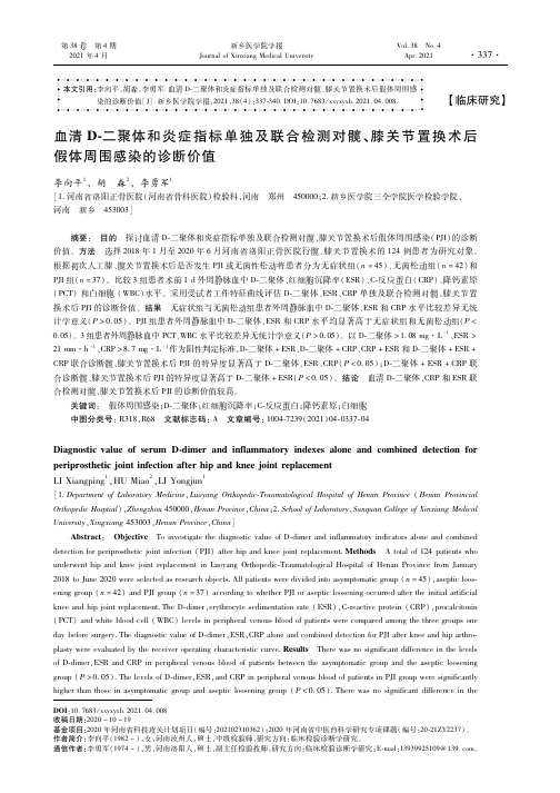
血清D-二聚体和炎症指标单独及联合检测对髋、膝关节置换术后假体周围感染的诊断价值

DOI:10.7683/xxyxyxb.2021.04.008收稿日期:2020-10-19基金项目:2020年河南省科技攻关计划项目(编号:202102310362);2020年河南省中医药科学研究专项课题(编号:20 21ZY2237)。
作者简介:李向平(1982-),女,河南汝州人,硕士,中级检验师,研究方向:临床检验诊断学研究。
通信作者:李勇军(1974-),男,河南洛阳人,硕士,副主任检验技师,研究方向:临床检验诊断学研究;E mail:13939925109@139.com欁欁欁欁欁欁欁欁欁欁欁欁欁欁欁欁欁欁欁欁欁欁欁欁欁欁欁欁欁欁欁欁欁欁欁欁欁欁欁欁欁欁欁欁欁欁欁欁欁欁欁欁欁欁欁欁欁欁欁欁欁欁欁欁欁欁欁欁欁欁欁欁欁欁欁欁氉氉氉氉。
本文引用:李向平,胡淼,李勇军.血清D 二聚体和炎症指标单独及联合检测对髋、膝关节置换术后假体周围感染的诊断价值[J].新乡医学院学报,2021,38(4):337 340.DOI:10.7683/xxyxyxb.2021.04.008.【临床研究】血清D 二聚体和炎症指标单独及联合检测对髋、膝关节置换术后假体周围感染的诊断价值李向平1,胡 淼2,李勇军1[1.河南省洛阳正骨医院(河南省骨科医院)检验科,河南 郑州 450000;2.新乡医学院三全学院医学检验学院,河南 新乡 453003]摘要: 目的 探讨血清D 二聚体和炎症指标单独及联合检测对髋、膝关节置换术后假体周围感染(PJI)的诊断价值。
方法 选择2018年1月至2020年6月河南省洛阳正骨医院行髋、膝关节置换术的124例患者为研究对象。
根据初次人工膝、髋关节置换术后是否发生PJI或无菌性松动将患者分为无症状组(n=45)、无菌松动组(n=42)和PJI组(n=37)。
比较3组患者术前1d外周静脉血中D 二聚体、红细胞沉降率(ESR)、C 反应蛋白(CRP)、降钙素原(PCT)和白细胞(WBC)水平。
长期使用抗生素抑制细菌感染完整版

长期使用抗生素抑制细菌感染完整版当患者的感染被假定为无法通过确定的疗程或病源控制治愈时,医生就会开具抑制性抗生素治疗。
接受抑制性抗生素疗法的患者通常合并症较多,感染通常涉及残留的假体材料。
部分由于缺乏关于明确的抑制性抗生素使用指南,部分由于相关感染的复杂性,医生通常会给患者开具极长的、甚至无限期的抑制性抗生素治疗。
在这种情况下,长期抗生素暴露的风险尚未完全确定,但其中包括了从轻微到严重的药物不良反应、抗生素耐药菌的产生以及肠道微生物群的紊乱。
在这篇叙述性综述中,我们介绍了4种常见适应症中使用抑制性抗生素疗法的现有证据,审视了当前文献中的空白,并探讨了这种疗法已知和潜在的风险。
我们还就如何提高未来研究的证据质量提出了建议,特别是强调了需要一个标准化术语来描述使用长疗程抗生素来抑制难以治疗的感染。
抗生素是临床医生和许多患者熟悉的治疗药物。
2021年,美国开出了超过2.11亿张抗生素处方。
本综述侧重于抗生素使用的一个子部分:长期使用抗生素治疗被认为无法治愈的感染。
虽然这只占抗生素总使用量的一小部分,但就其性质而言,它可能代表着抗菌药物管理的重要机会。
我们小组之前的一项研究发现,在长期使用抗生素的3大适应症中,使用抗生素来抑制被认为无法治愈的感染的证据最不充分。
预防性和非感染性用途的其他广泛适应症不在本综述的讨论范围内。
长期或无限期使用抗生素已被认为是一种治疗策略,适用于不适合采用侵入性手术方法的患者。
这种方法通常不是为了治愈感染,而是为了改善症状,防止病情恶化或复发,从而达到具有临床意义的程度。
我们之前的研究发现,在澳大利亚一家大型医院网络中,最常采用抑制性抗生素疗法(SAT)治疗的感染有四类:即假体周围关节感染(PJI)、血管移植物感染(VGI)和其他血管感染,包括感染性心内膜炎和霉菌性动脉瘤、心脏植入式电子设备感染(CIEDI),以及骨髓炎和其他骨科硬件相关感染。
针对这些广泛感染的指南通常建议采用药物和手术相结合的治疗方法。
《医学检验》项目学习答案

项目学习《流式细胞术的原理及临床应用介绍》答案:"显微镜术可以研究()" "A""()是一种自动分析细胞的高技术,其原理是悬浮在液体中的分散细胞一个个分别通过测量区,当每个细胞通过测量区时产生电信号,这些信号可以代表荧光、光散射,光吸收或细胞的阻抗等" "A""流式细胞仪是集()单克隆抗体技术于一体的先进科学技术设备。
以下哪项不正确()" "C""侧向散射光,又称()散射,其信号的强弱与细胞内颗粒的强弱与细胞内颗粒结构的质量成正比,用于检测细胞内部结构属性" "D""以下哪项不属于细胞荧光化学技术的组成部分()" "D""():用于筛检抗体(抗特定抗原成分的特异性抗体)。
此法是测定抗体最常用的方法" "A""()检测是判断机体免疫功能的一个重要指标" "A""以下哪项不是细胞因子()" "B""()是一种微珠多用途检测分析技术,它由一系列的微珠组合来捕获并结合流式细胞仪技术检测细胞培养液、EDTA血浆、血清样本中被检测物质的量" "C""():用于检测目的抗原的量。
其优点是避免了对特异性抗体的直接标记,但增加了操作步骤和测定时间" "D""设置()是为了检测荧光抗体是否有效,并不是每次分析时都必须设置" "A" "流式细胞术对高速流过检测区的单个细胞或生物颗粒进行()的一种技术" "A""实验中,常利用()这两种参数的组合,区分不同的细胞群体,去除碎片、死细胞和粘连细胞的干扰" "B""当细胞携带两种以上荧光素激发出不同波长荧光时,理论上可选择()使每种荧光仅被相应的通道检测到" "C""SSC方向与激光束和液流形成的平面相垂直,亦称(),其信号强度反映细胞内部颗粒度和精细结构的变化" "C""淋巴细胞一般可分为()三个亚群,即“TBNK淋巴细胞亚群”;其中T细胞还可分为CD4细胞和CD8细胞。
人工关节置换术后关节感染演示ppt课件

本文首先介绍人工关节置换术及术后关节感染的相关概念和 背景知识,然后分析术后关节感染的流行病学特征和危险因 素,接着探讨术后关节感染的诊断方法和治疗策略,最后提 出预防措施和展望未来的研究方向。
02
人工关节置换术概述
手术定义与分类
手术定义
人工关节置换术是一种通过手术将病 变的关节部分或全部切除,代之以人 造关节,以恢复关节功能、缓解疼痛 、提高生活质量的外科手术方法。
愈。
并发症的预防与处理
预防措施
术前充分评估患者身体状况,选择合适的手术时机和手术方式;术中严格遵守无菌操作原则,减少手术创伤;术 后加强护理和营养支持,提高患者免疫力。
处理措施
一旦出现并发症,如假体松动、骨折、深静脉血栓等,应立即采取相应治疗措施,如手术修复、抗凝治疗等。同 时,加强患者心理护理和康复锻炼,促进患者全面康复。
03
术后关节感染现状及危害
感染发生率与流行病学特点
感染发生率
人工关节置换术后关节感染的发生率 因手术类型、患者因素、医疗环境等 多种因素而异。一般来说,感染率较 低,但仍然是术后严重的并发症之一 。
流行病学特点
术后关节感染多发生在手术后的早期 阶段,其中革兰氏阳性菌是最常见的 致病菌。此外,高龄、肥胖、糖尿病 等患者因素也会增加感染的风险。
的认知度和配合度。
术后护理指导
02
指导患者进行正确的术后护理,如保持伤口清洁、避免剧烈运
动等,降低感染风险。
心理支持与疏导
03
关注患者心理状态,提供必要的心理支持和疏导,帮助患者树
立战胜疾病的信心。
07
总结与展望
本次汇报内容回顾
01
人工关节置换术后关节感染现状
纤维蛋白原和D-二聚体对关节假体周围感染的诊断价值

中平创伤11•科杂志202丨彳丨•- 5月第23卷第5期Chin J Orlhop Trauma. May 202丨,Vol. 23,No. 5• 383 ••关节感染•纤维蛋白原和D-二聚体对关节假体周围感染的诊断价值商广前项帅黄辉季丰张海宁王英振徐浩青岛大学附属医院关节外科 266000通信作者:徐浩,Email:186****6627@163. com【摘要】目的探讨纤维蛋白原和D-二聚体对髋、膝关节置换术后(T M)假体周围感染(P JI)的诊断价值。
方法回顾性分析自2013年8月至20丨9年6月在青岛大学附属医院关节外科进行人工髋、膝关节翻修术的175例患者资料。
其中59例被诊断为PJI(PJ丨组)(膝31例,髋28例),男33例,女26例;年龄(67.4 ±11.7)岁;体重指数(BMI)为(26. 1±3.6)kg/m2。
丨16例被诊断为无菌性松动(AL)(AL 组)(膝19 例,髋 97 例),男67 例,女 49 例;年龄(70. 3 ±8. 9)岁;BMI 为(25. 0 ±3. 6) k g/m2。
统计分析两组患者血浆C反应蛋白(C R P)、红细胞沉降率(E S R)、纤维蛋白原和D-二聚体的水平,以受试者工作特征(ROC)曲线计算每个指标的灵敏度与特异度,并依据曲线下面积(AUC)比较其诊断价值。
结果 PJ1组和A L组患者在性别、年龄、BM I等方面差异均无统计学意义(P>0.05),但在关节类型方面差异有统计学意义(户<0.05)。
与人1_组比较,丨>_|丨组〇^上5丨{、纤维蛋白原和0-二聚体均显著升高,差异有统计学意义(/5<0.05)。
〇^、£31纤维蛋白原和1>二聚体的曲线下面积分别为0. 830、0. 850、0. 848、0. 664。
根据约登指数,C R P、ESR、纤维蛋白原和D-二聚体的最佳临界值分别为8. 06 m g/L、17. 60 m m/h、3. 73 g/L、685. 00 ng/m L,敏感度分别为79. 2%、85. 4%、81. 3%、64. 6%,特异性分别为85. 7% ,76. 2%、79. 8%、61. 9%。
髋关节置换术后关节假体周围感染的诊断与治疗进展

髋关节置换术后关节假体周围感染的诊断与治疗进展胡冰涛;李瑞延;杨帆;刘贺;李照彦;秦彦国【期刊名称】《临床误诊误治》【年(卷),期】2018(031)002【总页数】4页(P110-113)【关键词】关节成形术,置换,髋;关节假体周围感染;文献综述【作者】胡冰涛;李瑞延;杨帆;刘贺;李照彦;秦彦国【作者单位】130000长春,吉林大学第二医院骨科;130000长春,吉林大学第二医院骨科;130000长春,吉林大学第二医院骨科;130000长春,吉林大学第二医院骨科;130000长春,吉林大学第二医院骨科;130000长春,吉林大学第二医院骨科【正文语种】中文【中图分类】R687.42;R619全髋关节置换术(total hip arthroplasty, THA)是常见的骨科手术,主要用于治疗退行性髋关节疾病、伴有移位的股骨颈骨折以及髋关节表面软骨损伤等疾病[1]。
尽管THA可以提高患者生活质量,延长患者寿命,但研究表明THA易出现并发症,如术后脱位、关节假体周围感染(prosthetic joint infection, PJI)、切口感染、假体周围骨折及血肿等[2]。
有文献报道初次髋关节置换术后PJI发病率为1%~2%[3],如果不能得到有效治疗,将会导致髋部剧烈疼痛、运动受限、残疾,严重者可导致死亡。
PJI会导致灾难性后果,并增加再手术难度。
因此,PJI的预防和管理尤为重要。
肥胖、吸烟史、糖尿病、营养不良、免疫系统缺陷、髋关节既往手术史及THA手术时间较长等均是PJI的危险因素[4]。
患者年龄、术前血清白蛋白及血红蛋白含量低、引流时间也是THA后感染的危险因素[4]。
假体感染的常见途径包括术中感染、血源性感染及其他感染病灶蔓延[5]。
有学者将PJI分为早期感染(术后3个月内)、迟发感染(术后3~24个月)和晚期慢性感染(术后≥24个月),不同时期感染临床表现和治疗措施不相同[6]。
初次THA患者PJI早期通常是由高毒性细菌所致,主要表现为髋部持续性疼痛,髋关节周围皮肤红肿、渗出及流脓[3];而晚期慢性感染临床症状与髋关节假体无菌性松动相似,常表现为局部活动性疼痛,影像学检查可见假体松动、感染性骨破坏,血C反应蛋白(CRP)或红细胞沉降率(ESR)值可能是正常的,术前关节液培养结果可能为阴性[7]。
假体周围感染的诊断和治疗

标题:Culture negative PJI periprosthetic joint infection: Strategies to improve yield正文:Cultures of tissue or fluid obtained from an affected joint constitute an important component of the diagnosis and treatment of periprosthetic joint infection.According to its new definition, the sole isolation of a pathogen by culture from two separate tissue or fluid samples that are obtained from the affected prosthetic joint are used to diagnose a periprosthetic joint infection (PJI). Additionally, when a pathogen is identified, the antibiotic treatment can be tailored using specific sensitivity. Unfortunately, in 7% to 12% of PJI cases, cultures may be negative even in the presence of clear signs of infection. It is also important to note that cultures are used to confirm and not to screen for PJI.Negative cultures may be caused due to various reasons, including the use of antimicrobial therapy prior to the collection of fluid or tissue, inappropriate collection of tissue samples (i.e., excessive cauterization) and the use of short periods of incubation, especially when low virulence pathogens cause the infection. Some pathogens are difficult to identify using traditional tissue cultures, particularly if they are encapsulated in biofilm, or when fungi ormycobacteria are the culprits and a specific medium culture for these pathogens is not used. Negative cultures limit the tailoring of the antibiotic treatment and, moreover, hinder the justification for revision surgery if other components of the PJI definition are not present. Lack of identification of the infecting organism also poses challenge regarding the selection of appropriate antibiotics for addition to the cement spacer and also in the postoperative period. In order to cover all possible infecting organisms, often a broad spectrum antibiotic or multiple antibiotics are administered that place the patient at systemic risk and carry the disadvantage of not treating fungi and rare organisms.The most effective strategy to improve the culture yield is to withhold aspiration of the joint for at least 2 weeks after the last dose of antibiotic. The latter is based on the available evidence evaluated by the work group that was convened by the American Academy of Orthopaedic Surgeons. The other recommendation of the same work group was to withhold administration of antibiotic to any patient with suspected PJI until appropriate tests, such as joint aspiration, for diagnosis of PJI are performed.Another strategy to improve yield of culture isto prolong the period of incubation of culturesamples for at least 14 days and possibly 21 days. The tissue or fluid samples obtained from the joint should also be transferred to the laboratory for immediate processing in all cases, in general, and in culture negative cases in particular. It is our practice to call the microbiology department and inform them of the challenge of isolating the infecting organism and “warn” them about the sample being sent. The appropriate use of culture media is paramount to obtain accurate positive cultures. If fungi ormycobacteria infection is suspected, the proper media should be used. Cultivation of samples should be performed under sterile conditions, ideally using a laminar flow workbench. Someauthorities also propose the use of enriched medium for all cultures, such as trypticase-soy agar containing sheep blood, chocolate agar, MacConkey agar and brain-heart infusion broth.Another reason for false negative PJI cases relates to aggregation of the infecting organism in a biofilm. In the biofilm, the organisms are able to communicate through sophisticated molecularmechanisms, such as quorum sensing, and avoid destruction by the host’s immun e system.Javad Parvizi Carlos HigueraBiofilm disruption techniques to “mobilize” the infecting organism have been described. Sonication of the retrieved prosthesis is one such method that is reported to improve yield of culture. However, not all centers are equipped to perform sonication of the retrieved prostheses limiting the use of this technique. The advantages of this technique include the fact that it is simple, reproducible and it yields viable microorganisms that can be subjected to antimicrobial susceptibility testing. The sonication fluid also typically yields a high number of organisms, which may help to distinguish between infected and contaminated prostheses.Role of molecular techniquesThe introduction of molecular techniques offers another option when negative cultures are present in patients where PJI is suspected. Polymerase chain reaction (PCR)-based assays increase the sensitivity of cultures. There are multiple PCR-based techniques using either species-specific primers or broad-range primer pair to 16S rRNA gene to amplify DNA. However, these techniques carry extreme sensitivity, leading to a higher incidence of false positive cultures. We have been evaluating the role of multiplex PCR (Ibis T5,000) that relies on pan-genomic amplification, and mass spectrometry for culture negative cases. The results, so far, have been encouraging. Following amplificationprocess and isolation of the organism, fluorescent in situ hybridization (FISH) has also been used to confirm that the organisms were indeed real and not contaminants.Other alternatives for improving high false-positive results include the use of ribosomal RNA-based PCR, which has shown a specificity close to 100% and a sensitivity equivalent to intraoperative cultures. This technique retains the cellviability-sensitive degradation patterns of RNA even after 7 days of sterilization, while retaining the ability to identify infections. This technology is still in the process of becoming fully available in the clinical practice.The slow transition of molecular techniques into clinical applications has shown promising results. A progressive decrease in cost and increase in the availability of this technology may provide the clinician with another tool to enhance the identification of pathogens in patients with high suspicion of having PJI, where a confirmation of the diagnosis would guide the appropriate treatment and improve outcomes. Further research with large, multicenter cohorts is needed to understand the practicality of these techniques.In the presence of negative cultures in patients with high suspicion of PJI, it is paramount to understand its potential causes, and to expand the type and incubation period of the cultures in order to rule out infection with low virulence pathogens, fungi or mycobacteria.References:∙Berbari EF, Marculescu C, Sia I, et al. Culture-negative prosthetic joint infection. Clin Infect Dis.2007;45:1113-1119.∙Bergin PF, Doppelt JD, Hamilton WG, et al. Detection of periprosthetic infections with use of ribosomalRNA-based polymerase chain reaction. J Bone JointSurg Am. 2010;92(3):654-663.∙Della Valle C, Parvizi J, Bauer TW, et al. Diagnosis of periprosthetic joint infections of the hip and knee. J AmAcad Orthop Surg. 2010;18(12):760-770.∙Parvizi J, Ghanem E, Menashe S, et al. Periprosthetic infection: What are the diagnostic challenges? J BoneJoint Surg Am. 2006;88 (4):138-147.∙Parvizi J, Zmistowski B, Berbari EF, et al. Newdefinition for periprosthetic joint infection: From theWorkgroup of the Musculoskeletal Infection Society.Clin Orthop Relat Res. 2011;469:2992-2994.∙Javad Parvizi, MD, FRCS, editor of Infection Watch, can be reached at the Rothman Institute, 925 Chestnut St., 5th Floor, Philadelphia, PA 19107; 267-339-3617;email: parvj@.∙Carlos A. Higuera, MD, can be reached atcarhiguera75@.∙Disclosures: Parvizi is a consultant to Stryker. Higuera has no relevant financial disclosures.。
2018版假体周围感染和手术部位感染专家共识ppt

有新的证据表明情感障碍,像抑郁和焦虑,会增加PJIs的风险。虽然已 经提供了这种联系的生理和心理上的解释,但目前还不清楚在手术前调节或 者治疗这种疾病是否可以降低PJIs的风险。 证据水平:强 代表投票:同意:88%,不同意:4%,弃权:8%(超级多数,强烈共识)
问题14: 可以通过哪些术前优化贫血提高血红蛋白浓度?
共识: 文献表明,铁和/或促红细胞生成素(EPO)的使用可提高术前血红蛋白浓度,
降低术后输血的需要。然而,铁可能只对已经存在铁缺陷的患者有效,并与许 多副作用相关。考虑到EPO的高成本,术前给药去避免单靠输血还没有被发现 是有成本效益。需要进一步的研究来评估术前异体输血的风险和益处。 证据水平:有限 代表投票:同意:92%,不同意:3%,弃权:5%(超级多数,强烈共识)
问题8: 术前是否需要停用免疫调节疾病修饰药物(如甲氨蝶呤或抗肿瘤坏死因子(抗-TNF)药物)
以减少后续手术部位感染/假体周围关节感染(SSI/PJI)的风险? 共识:
1.适用于成人风湿性关节炎(类风湿性关节炎(RA),银屑病关节炎(PsA),青少年特发 性关节炎(JA)、强直性脊柱炎(AS)或全身性红斑狼疮(SLE)),所有生物抗风湿性药物包括 TNF 抑 制 剂 和 IL-6 阻 滞 剂 ( 见 表 1 的 完 整 清 单 ) 应 在 全 髋 ( THA ) 和 膝 关 节 置 换 术 (TKA),之前进行完整的给药周期。手术时间应为保持剂量后的一周。如果伤口愈合良 好,所有缝线都已拆除,且没有非手术部位的感染,这些药物在手术两周后可以重新开始。
问题10: 阿片类药物的摄入是否与手术部位感染/假体周围关节感染(SSI/PJI)
