大鼠尾部血管的解剖结构与鼠尾的生理功能探讨
解剖老鼠的实验报告(3篇)

第1篇一、实验目的1. 了解哺乳动物内部结构的基本组成;2. 掌握解剖实验的基本方法和步骤;3. 提高对动物解剖学知识的理解和掌握。
二、实验材料1. 老鼠一只(已处死,并去除内脏);2. 解剖器械一套;3. 解剖盘、解剖剪、镊子、解剖刀、解剖针、解剖剪等;4. 纱布、棉球、生理盐水等。
三、实验步骤1. 准备工作(1)将老鼠放置在解剖盘上,用解剖剪剪开老鼠的皮肤,使其暴露出肌肉和内脏。
(2)用解剖刀沿脊柱两侧向腹部切开肌肉,暴露出内脏。
2. 解剖过程(1)心脏解剖用解剖剪剪开心脏周围的肌肉,暴露出心脏。
观察心脏的形状、大小、颜色和心房、心室的结构。
(2)肝脏解剖用解剖剪剪开肝脏周围的肌肉,暴露出肝脏。
观察肝脏的形状、大小、颜色和表面结构。
(3)肺脏解剖用解剖剪剪开胸腔,暴露出肺脏。
观察肺脏的形状、大小、颜色和表面结构。
(4)肾脏解剖用解剖剪剪开腹壁,暴露出肾脏。
观察肾脏的形状、大小、颜色和表面结构。
(5)胃、小肠、大肠解剖用解剖剪剪开腹壁,暴露出胃、小肠、大肠。
观察它们的形状、大小、颜色和表面结构。
(6)生殖器官解剖用解剖剪剪开腹壁,暴露出生殖器官。
观察生殖器官的形状、大小、颜色和结构。
(7)神经系统解剖用解剖剪剪开头部,暴露出大脑、脊髓等神经系统结构。
观察它们的形状、大小、颜色和结构。
3. 清洗与观察(1)用生理盐水清洗解剖过的内脏,观察其颜色、质地和结构。
(2)将解剖过的内脏逐一编号,以便观察和记录。
四、实验结果与分析1. 心脏解剖心脏呈椭圆形,颜色鲜红,心房和心室结构明显。
2. 肝脏解剖肝脏呈红褐色,表面光滑,质地柔软。
3. 肺脏解剖肺脏呈粉红色,质地柔软,表面有明显的支气管和肺泡。
4. 肾脏解剖肾脏呈红褐色,表面光滑,质地柔软。
5. 胃、小肠、大肠解剖胃呈囊状,小肠呈管状,大肠呈弯曲状。
6. 生殖器官解剖雄性生殖器官包括睾丸、阴茎等;雌性生殖器官包括卵巢、输卵管、子宫等。
7. 神经系统解剖大脑呈椭圆形,脊髓呈圆柱形。
[超详细]大鼠的解剖图谱
![[超详细]大鼠的解剖图谱](https://img.taocdn.com/s3/m/b7793e767f21af45b307e87101f69e314332fa0d.png)
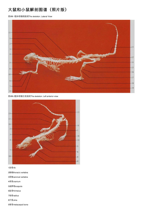
[超详细]大鼠[ 超详细]大鼠的解剖图谱1图Ⅷ-1 整体骨骼侧面观The skeleton. Lateral View图Ⅷ-2 整体骨骼左前面观The skeleton. Left anterior view1肋骨rib2胸椎thoracic vertebra3 颈椎cervical vertebra4 颅骨cranium5肩胛骨scapula6肱骨hiimeius7桡骨radius8尺骨ulna9掌骨metacarpal bone10 指骨digital bone11 腰椎lumbar vertebra12 髂骨ilium13尾骨coccyx14股骨femur15髌骨patella16腓骨fubula17胫骨tibia18跖骨metatarsal bone19 趾骨digital bone图Ⅷ-4 头骨侧面The skull. Lateral aspect 1枕骨occipital bone2顶间骨interparietal bone3 矢状缝sagittal suture4颧骨malar bone5上颌骨maxillary bone6前颌骨premaxillary bone7枕外嵴external occipital creat8 顶骨parietal bone9 额骨frontal bone10 鼻骨nasal bone11 鼻间缝internasal suture12 前筛孔preethmoid pore13 蝶腭孔sphenopalatine foramen14 门齿incisor tooth15下颌骨mandible16视神经孔optic foramen17 枕骨occipital bone18茎突styloid process19外耳道external acoustic meatus 20 颞骨temporal bone图Ⅷ-6 下颌骨侧面The mandible. Lateral aspect 4颧骨malar bone5上颌骨maxillary bone6前颌骨premaxillary bone14 门齿incisor tooth17 枕骨occipital bone19 外耳道external acoustic meatus21 腭裂patoschisis22 臼齿molar tooth23 腭骨palatine bone24 翼孔pterygoid apertures25 破裂孔foramen lacerum26 枕大孔foramen magnum27 腭后孔posterior palatine foramen28 鼻后孔posterior nasal apertures29 卵圆孔foramen ovale30鼓骨tympanic bone31舌下神经孔hypoglossal foramen32 下颌联合mandibular symphysis33 颏孔mental foramen34 冠状突coronoid process35 下颌支ramus of mandible36 角状突process of horn37 下颌孔mandibular foramen38 翼肌窝pterygoid fossa39 髁突condylar process图Ⅷ-7 颈椎与胸椎背面The cervical 图Ⅷ-9 前肢骨背面The bone of vertebra and thoracic vertebra.Dorsal aspect anterior 1imb.Dorsal aspect图Ⅷ-8 前肢骨外侧面The bone of anteriorlimb. Lateral aspect1 枢椎axis2 第4 颈椎4th cervical vertebra3 第6 颈椎6th cervical vertebra4 第1 胸椎1st thoracic vertebra5 第4 胸椎4th thoracic vertebra6 寰椎axis7 第3 颈椎3rd cervical vertebra 8 第5 颈椎5th cervical vertebra9 第7 颈椎7th cervical vertebra 10 第3 胸椎3rd thoracic vertebral1 肱骨体shaft of humcrus 12 桡骨radius13 尺骨ulna 14 尺骨茎突styloid process of ulna15 掌骨metacarpal bone 16 肱骨头head of hurnerusl7 肘突cubital process 18 腕骨carpal bone19 指骨digital bone图Ⅷ-10 股骨前面The femur. 图Ⅷ-11 胫骨与腓骨前面The tibia Anterior aspect and fibula. Anterior aspect1 大转子greater trochanter2 股骨颈neck of femur3 第三转子third trochanter4 髌骨patella5股骨头femoral head6小转子lesser trochanter7 股骨体shaft of femur8 股骨内侧髁medial condyle of femur9 外侧髁lateral condyle10腓骨fibula11外踝lateral malleolus12 跗骨tarsal bone13 胫骨内侧髁medial condyle of tibia14 径骨粗隆tibial tuberosity15 胫骨tibiaI6 内踝medial mallcolus17 跖骨metatarsal bone18斜方肌trapezius muscle19臀浅肌glutoeus super(icialis muscle20 股直肌rectus femoris muscle21 腹外斜肌external oblique muscle of abdomen22 背阔肌latissimus dorsi muscle23 肩斜方肌shoulder-trapezius muscle24 大圆肌teres major muscle25 肩三角肌shoulder-deltoid muscle26 前锯肌serratus anterior muscle1 锁骨提肌levator clavicle muscle2 肩三角肌shoulder-deltoid muscle3 肱三头肌triceps brachii muscle4 臀浅肌gluteus suferficidlis muscle5 股直肌rectus femoris muscle6 腓肠肌gastrocnemius muscle7 夹肌splenius muscle 8颈菱形肌rhomboideus cervicis muscle9胸菱形肌rhomboideus pectoralis muscle10 背阔肌latissimus dorsi muscle11 腹外斜肌external oblique muscle of abdomen12 股二头肌biceps femoris muscle13 半腱肌semitendinosus muscle3 肱三头肌triceps brachii muscle5 股直肌rectus femoris muscle11 腹外斜肌external oblique muscle of abdomen 13半腱肌semitendinosus muscle14二腹肌前腹anterior belly of digastric muscle15 二腹肌后腹posterior belly of digastric muscle16 胸骨舌骨肌sternohyoid muscle17 肱二头肌biceps brachii muscle18 腹直肌rectus abdominis muscle19 股动、静脉femoral artery and femoral vein20 阴囊scrotum21 咬肌masseter muscle 22 胸乳突肌sternomastoideus muscle23 肩三角肌shoulder-deltoid muscle24桡侧腕伸肌extensor carpi radialis muscle25指总伸肌extensor digitorum communis muscle26 尺侧腕伸肌extensor carpi ulnaris muscle27 胸浅肌pectoral superficialis muscle28 前锯肌serratus anterior muscle29 股内侧肌vastus medialis muscle30 耻骨肌pectineus muscle31 长收肌adductor longus muscle32 股薄肌gracilis muscle图Ⅷ-15 头、颈部侧面 The head and neck. Lateral aspect1 颞浅动、静脉 superficial temporal artery and vein2 眶上静脉 supraorbitalvein3 面横动、静脉 transverse facial artery and vein4 下颌缘神经marginalmandibularenerv 5 面后静脉posterior facial vein 6 面前静脉 anterior facial vein 图Ⅷ-16 口腔(1)The oral cavity (1)7 颈外静脉external jugular vein1 颞浅动、静脉 superficial temporal artery and vein2 眶上静脉 supraorbitalvein3 面横动、静脉 transverse facial artery and vein4 下颌缘神经 marginalmandibular nerve5 面后静脉posterior facial vein6 面前静脉 anterior facial vein7 颈外静脉 external jugular vein8 硬腭褶 fold of hard palate9 臼齿 molar tooth 10 门齿 incisor tooth 11 软腭 soft palate 12 舌根 root of tongue 13 舌体 body of tongue14 舌尖apex of tongue图Ⅷ-17 口腔(2)The oral cavity (2)1 喉口aperture of larynx2 舌根root of tongue3 舌尖apex of tongue 5 软腭soft palate7 臼齿molar tooth 4 硬腭褶fold of hard palate 6 会厌epiglottis8 舌体body of tongue图Ⅷ-18 脑与脊髓背面The brain and 图Ⅷ-19 脑与脊髓腹面The brain and spinal cord.Dorsal aspect spinal cordVentral aspect9 嗅球olfactory bulb1l 绒球flocculus13 腰膨大lumbar enlargement 15 大脑cerebrum17 脊髓圆锥courts medullaris 19 脑桥pons21 嗅束olfactory tract23 三叉神经trigeminal nerve 10 中央纵裂central longitudinal fissue12 颈膨大cervical enlargement14 外侧纵沟lateral longitudinal sulcus16 小脑(中央部)cerebellum (central part)18 脑垂体pituitary gland20 延髓myelencephalon22 视神经optic nerve图Ⅷ-20 磁共振冠状面定位像(1)The scout view of coronal images(1)图Ⅷ-21 磁共振冠状面定位像(2)The scout view of coronal images(2)1眼球eyeball2嗅脑rhinencephalon3大脑皮层cerebral cortex4 大脑镰cerebral faix5 鼻旁窦paranasal sinus6 纵裂longitudinal fissure冠状面T1加权像,颞颌关节线圈,SE 序列,层厚3.0mm,无间隔,TR=500rns,TE=20ms。
大鼠和小鼠解剖图谱(照片版)
⼤⿏和⼩⿏解剖图谱(照⽚版)图Ⅷ-1整体⾻骼侧⾯观The skeleton. Lateral View图Ⅷ-2整体⾻骼左前⾯观The skeleton. Left anterior view1肋⾻rib2胸椎thoracic vertebra3颈椎cervical vertebra4颅⾻cranium5肩胛⾻scapula6肱⾻hiimeius7桡⾻radius8尺⾻ulna9掌⾻metacarpal bone10指⾻digital bone11腰椎lumbar vertebra12髂⾻ilium13尾⾻coccyx14股⾻femur15髌⾻patella16腓⾻fubula17胫⾻tibia18跖⾻metatarsal bone19趾⾻digital bone图Ⅷ-4头⾻侧⾯The skull. Lateral aspect图Ⅷ-6下颌⾻侧⾯The mandible. Lateral aspect1枕⾻occipital bone2顶间⾻interparietal bone3⽮状缝sagittal suture4颧⾻malar bone5上颌⾻maxillary bone6前颌⾻premaxillary bone7枕外嵴external occipital creat8顶⾻parietal bone9额⾻frontal bone10⿐⾻nasal bone11⿐间缝internasal suture12前筛孔preethmoid pore13蝶腭孔sphenopalatine foramen 14门齿 incisor tooth15下颌⾻mandible16视神经孔optic foramen17枕⾻occipital bone18茎突styloid process19外⽿道external acoustic meatus 20颞⾻temporal bone21 腭裂patoschisis22⾅齿molar tooth23腭⾻palatine bone24翼孔pterygoid apertures25破裂孔foramen lacerum26枕⼤孔foramen magnum27腭后孔posterior palatine foramen 28⿐后孔posterior nasal apertures 29卵圆孔foramen ovale30⿎⾻tympanic bone31⾆下神经孔hypoglossal foramen 32下颌联合mandibular symphysis 33颏孔mental foramen34冠状突coronoid process35下颌⽀ramus of mandible36⾓状突process of horn37下颌孔mandibular foramen38翼肌窝pterygoid fossa39髁突condylar process图Ⅷ-7颈椎与胸椎背⾯The cervical 图Ⅷ-9前肢⾻背⾯The bone of vertebra and thoracic vertebra.Dorsal aspect anterior 1imb.Dorsal aspect图Ⅷ-8前肢⾻外侧⾯The bone of anteriorlimb. Lateral aspect1枢椎axis 2第4颈椎4th cervical vertebra3第6颈椎6th cervical vertebra 4第1胸椎1st thoracic vertebra5第4胸椎4th thoracic vertebra 6寰椎axis7第3颈椎3rd cervical vertebra 8第5颈椎5th cervical vertebra9第7颈椎7th cervical vertebra 10第3胸椎3rd thoracic vertebral1肱⾻体shaft of humcrus 12桡⾻radius13尺⾻ulna 14尺⾻茎突styloid process of ulna15掌⾻metacarpal bone 16肱⾻头head of hurnerusl7肘突cubital process 18腕⾻carpal bone19指⾻digital bone图Ⅷ-10股⾻前⾯The femur. 图Ⅷ-11胫⾻与腓⾻前⾯The tibia Anterior aspect and fibula. Anterior aspect1⼤转⼦greater trochanter2股⾻颈neck of femur3第三转⼦third trochanter4髌⾻patella5股⾻头femoral headG⼩转⼦lesser trochanter7股⾻体shaft of femur8股⾻内侧髁medial condyle of femur9外侧髁lateral condyle10腓⾻fibula11外踝 lateral malleolus12跗⾻tarsal bone13胫⾻内侧髁medial condyle of tibia14径⾻粗隆tibial tuberosity15胫⾻tibiaI6内踝medial mallcolus17跖⾻metatarsal bone18斜⽅肌trapezius muscle19臀浅肌glutoeus super(icialis muscle20股直肌rectus femoris muscle21腹外斜肌external oblique muscle of abdomen 22背阔肌latissimus dorsi muscle 23肩斜⽅肌shoulder-trapezius muscle24⼤圆肌teres major muscle25肩三⾓肌shoulder-deltoid muscle26前锯肌serratus anterior muscle1锁⾻提肌levator clavicle muscle 2肩三⾓肌shoulder-deltoid muscle 3肱三头肌triceps brachii muscle4臀浅肌gluteus suferficidlis muscle 5股直肌rectus femoris muscle6腓肠肌gastrocnemius muscle7夹肌splenius muscle8颈菱形肌rhomboideus cervicis muscle9胸菱形肌rhomboideus pectoralis muscle10背阔肌latissimus dorsi muscle11腹外斜肌external oblique muscle of abdomen 12股⼆头肌biceps femoris muscle13半腱肌semitendinosus muscle14⼆腹肌前腹anterior belly of digastric muscle 15⼆腹肌后腹posterior belly of digastric muscle 16胸⾻⾆⾻肌sternohyoid muscle 17肱⼆头肌biceps brachii muscle18腹直肌rectus abdominis muscle19股动、静脉femoral artery and femoral vein 20阴囊scrotum21咬肌masseter muscle22胸乳突肌sternomastoideus muscle23肩三⾓肌shoulder-deltoid muscle24桡侧腕伸肌extensor carpi radialis muscle25指总伸肌extensor digitorum communis muscle 26尺侧腕伸肌extensor carpi ulnaris muscle27胸浅肌pectoral superficialis muscle28前锯肌serratus anterior muscle29股内侧肌vastus medialis muscle30耻⾻肌pectineus muscle31长收肌adductor longus muscle32股薄肌gracilis muscle图Ⅷ-15头、颈部侧⾯The head and neck. Lateral aspect上静脉supraorbitalveine 7颈外静脉external jugular vein 1颞浅动、静脉superficial temporal artery and vein 2眶3⾯横动、静脉transverse facial artery and vein 4下颌缘神经marginalmandibular nerv 5⾯后静脉posterior facial vein 6⾯前静脉anterior facial vein1颞浅动、静脉superficial temporal artery and vein 2眶上静脉supraorbitalvein3⾯横动、静脉transverse facial artery and vein 4下颌缘神经marginalmandibular nerve 5⾯后静脉posterior facial vein 6⾯前静脉anterior facial vein 7颈外静脉external jugular vein 8硬腭褶fold of hard palate9⾅齿molar tooth 10门齿incisor tooth11软腭soft palate 12⾆根root of tongue13⾆体body of tongue 14⾆尖apex of tongue图Ⅷ-18脑与脊髓背⾯The brain and 图Ⅷ-19脑与脊髓腹⾯The brain and spinal cord.Dorsal aspect spinal cordVentral aspect1 喉⼝aperture of larynx 2⾆根root of tongue 3⾆尖apex of tongue4硬腭褶fold of hard palate 5软腭soft palate6会厌epiglottis 7⾅齿molar tooth8⾆体body of tongue 9嗅球olfactory bulb10中央纵裂central longitudinal fissue 1l 绒球flocculus 12颈膨⼤cervical enlargement13腰膨⼤lumbar enlargement14外侧纵沟lateral longitudinal sulcus 15⼤脑cerebrum 16⼩脑(中央部)cerebellum (central part )17脊髓圆锥courts medullaris18 脑垂体 pituitary gland 19脑桥pons 20延髓 myelencephalon21嗅束olfactory tract22视神经optic nerve 23三叉神经trigeminal nerve图Ⅷ-20磁共振冠状⾯定位像(1)The scout view of coronal images(1)图Ⅷ-21磁共振冠状⾯定位像(2)The scout view of coronal images(2)1眼球eyeball2嗅脑rhinencephalon3⼤脑⽪层cerebral cortex4⼤脑镰cerebral faix5⿐旁窦paranasal sinus6纵裂longitudinal fissure冠状⾯T1加权像,颞颌关节线圈,SE序列,层厚3.0mm,⽆间隔,TR=500rns,TE=20ms。
解剖大鼠的实验报告

一、实验目的1. 掌握解剖学的基本方法与技能;2. 熟悉大鼠的解剖结构,了解其器官的形态和功能;3. 培养学生的观察能力和实验操作能力。
二、实验材料与仪器1. 实验动物:成年雄性大鼠一只;2. 仪器:解剖盘、解剖剪、镊子、解剖刀、解剖针、解剖显微镜、解剖显微镜光源、解剖显微镜载物台、解剖显微镜镜头等;3. 药品:生理盐水、10%甲醛溶液、70%酒精、5%碘酊、5%氯化钠溶液等。
三、实验步骤1. 准备工作(1)将大鼠处死,解剖前应确保大鼠已死亡,以免造成实验者伤害。
(2)用生理盐水清洗大鼠体表,去除污物。
(3)将大鼠放在解剖盘上,用解剖剪沿背部正中线剪开皮肤,暴露肌肉和内脏。
2. 解剖顺序(1)从头至尾解剖:1)剪开颅骨,暴露大脑、小脑和脑干;2)剪开胸腔,暴露心脏、肺、气管、食管、胃、肝脏、脾脏、肾脏、肾上腺等器官;3)剪开腹腔,暴露小肠、大肠、胃、肝脏、脾脏、肾脏、肾上腺等器官;4)剪开盆腔,暴露生殖器官、膀胱、直肠等器官。
(2)器官解剖:1)大脑:观察大脑的形态、大小、重量,辨认大脑的各个叶和沟回;2)心脏:观察心脏的形态、大小、重量,辨认心脏的四个腔室;3)肺:观察肺的形态、大小、重量,辨认肺的叶和段;4)肝脏:观察肝脏的形态、大小、重量,辨认肝脏的叶和段;5)脾脏:观察脾脏的形态、大小、重量,辨认脾脏的叶和段;6)肾脏:观察肾脏的形态、大小、重量,辨认肾脏的皮质、髓质和肾盂;7)肾上腺:观察肾上腺的形态、大小、重量,辨认肾上腺的皮质和髓质;8)生殖器官:观察生殖器官的形态、大小、重量,辨认生殖器官的各个部分;9)膀胱:观察膀胱的形态、大小、重量,辨认膀胱的壁和腔;10)直肠:观察直肠的形态、大小、重量,辨认直肠的壁和腔。
3. 标本处理(1)将解剖好的器官用生理盐水清洗,去除血污和杂质;(2)将器官放入10%甲醛溶液中固定,浸泡24小时;(3)将固定好的器官取出,用70%酒精清洗,去除甲醛;(4)将清洗好的器官放入5%碘酊中染色,染色时间为30分钟;(5)将染色好的器官取出,用5%氯化钠溶液清洗,去除碘酊;(6)将清洗好的器官放入解剖显微镜下观察,拍照记录。
大鼠解剖方面的资料
1. 外观大鼠外观与小鼠相似,但个体较大。
一般成年大鼠体长不小于18-20cm。
尾上覆有短毛和环状角质鳞片,数量多于200片。
上下颌各有两个切齿和六个臼齿,共16颗牙齿。
齿式为(1003/1003)×2。
2. 大鼠骨骼约105-108块,大鼠的生长发育期长,长骨长期有骨骺存在,不骨化。
切齿终生不断生长,大鼠需不断啃咬磨牙以维持其长度恒定,故垫料中应有部分小木块供其啃咬。
3. 大鼠唾液腺包括腮腺、颌下腺和舌下腺。
分别位于下颌骨后缘至锁骨的腹外侧、下颌骨后缘和胸腔入口的腹侧、颌下腺口侧。
颈区肩胛部间沉积的脂肪组织呈腺体状,称为冬眠腺,在产热中起着重要作用。
4. 胃由前后两部分组成,前胃为无腺区,后胃为有腺区,前后两部分由一个界限嵴分开,食管通过界限嵴的一个褶进入胃小弯,此褶是大鼠不能呕吐的原因。
5. 肠道分为十二指肠、空肠、回肠、盲肠、结肠、直肠。
其中小肠最长,约114cm(102-126),盲肠较长,约6-8cm。
6. 肝脏呈紫红色,占体重的比例大,约为体重的1/25,由四叶组成(右侧叶、中叶、左叶和尾叶)。
肝脏的再生能力强,经部分肝切除术后仍可再生。
成年大鼠切除肝2/3,在一周内肝脏生长最快,三周内肝脏重量可恢复到接近正常。
大鼠无胆囊,各肝叶的胆管会合成胆总管,开口于十二指肠。
胰脏位于胃和十二指肠的弯曲处,呈淡粉色,形状不规则,似脂肪。
7. 心脏重量约占体重的1/30-1/20,由左心房、左心室、右心房、右心室组成。
左心室发出主动脉弓,由此分出无名动脉、左颈总动脉、左锁骨下动脉。
无名动脉又分出右颈总动脉和右锁骨下动脉。
主动脉弓到心脏背侧沿脊柱下行,形成背主动脉,背主动脉再分支到髂部和四肢。
8. 肺脏为海绵状,淡粉色,位于胸腔中部,分为左、右两部分。
左肺为一个大叶,右肺分为4叶(前叶、中叶、副叶、后叶)。
9. 肾脏呈暗红色、蚕豆状,位于腹腔背侧脊柱两侧。
每侧肾都和一条白色细长的输尿管相连,输尿管下接膀胱。
解刨老鼠实验报告(3篇)
第1篇一、实验目的本次实验旨在通过解剖老鼠,了解哺乳动物内部结构,掌握解剖学的基本方法,并加深对动物生理学知识的理解。
二、实验原理解剖学是研究生物体结构的一门学科,通过解剖可以直观地观察到动物体内的器官和组织结构。
老鼠作为实验动物,其解剖结构相对简单,适合用于初学者进行解剖实践。
三、实验材料与工具1. 实验材料:成年老鼠一只2. 实验工具:解剖剪、解剖镊、解剖刀、解剖盘、解剖针、解剖剪、解剖镜、解剖图等四、实验步骤1. 准备阶段- 将老鼠置于解剖盘上,用解剖剪剪开皮肤,暴露肌肉层。
- 将肌肉层与内脏分开,小心地用解剖镊夹住肌肉层,向两侧撕开。
2. 内脏暴露- 撕开肌肉层后,可以看到内脏器官,包括心脏、肝脏、脾脏、肺脏、肾脏、胃、小肠、大肠、生殖器官等。
- 用解剖刀小心地剪开内脏周围的结缔组织,暴露各个器官。
3. 心脏解剖- 用解剖剪剪开心脏,观察心脏的四个腔室:左心房、左心室、右心房、右心室。
- 观察瓣膜结构,了解心脏的血液循环过程。
4. 肝脏解剖- 用解剖剪剪开肝脏,观察其结构,了解肝脏的功能。
5. 脾脏解剖- 用解剖剪剪开脾脏,观察其结构,了解脾脏的功能。
6. 肺脏解剖- 将肺脏取出,观察其结构,了解肺脏的呼吸功能。
7. 肾脏解剖- 用解剖剪剪开肾脏,观察其结构,了解肾脏的排泄功能。
8. 消化系统解剖- 将胃、小肠、大肠依次取出,观察其结构,了解消化系统的消化吸收过程。
9. 生殖系统解剖- 观察雄性老鼠的睾丸和阴茎,雌性老鼠的卵巢和输卵管,了解生殖系统的结构和功能。
10. 神经系统解剖- 将大脑取出,观察其结构,了解神经系统的功能。
五、实验结果与分析1. 心脏解剖:观察到心脏的四个腔室和瓣膜结构,了解了心脏的血液循环过程。
2. 肝脏解剖:观察到肝脏的多个叶,了解了肝脏的代谢和解毒功能。
3. 脾脏解剖:观察到脾脏的结构,了解了脾脏的免疫功能。
4. 肺脏解剖:观察到肺泡结构,了解了肺脏的呼吸功能。
大鼠解剖图——精选推荐
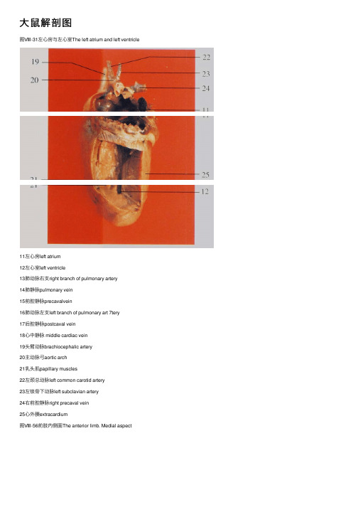
⼤⿏解剖图图Ⅷ-31左⼼房与左⼼室The left atrium and left ventricle11左⼼房left atrium12左⼼室left ventricle13肺动脉右⽀right branch of pulmonary artery14肺静脉pulmonary vein15前腔静脉precavalvein16肺动脉左⽀left branch of pulmonary art 7tery17后腔静脉postcaval vein18⼼中静脉 middle cardiac vein19头臂动脉brachiocephalic artery20主动脉⼸aortic arch21乳头肌papillary muscles22左颈总动脉left common carotid artery23左锁⾻下动脉left subclavian artery24右前腔静脉right precaval vein25⼼外膜extracardium图Ⅷ-56前肢内侧⾯The anterior limb. Medial aspect图Ⅷ-57后肢内侧⾯(1)The posterior limb. Medial aspect(1)1腋神经axillary nerve 2桡神经radial nerve3头静脉cephalic vein 4正中神经median nerve5正中动脉median artery 6肌⽀muscular branch7尺神经ulnar nerve 8臂动脉brachial artery9胸长神经long thoracic nerve 10胸外侧动脉external thoracic artery I1颈总动脉common carotid artery 12主动脉⼸aorta arch13左⼼⽿left auricle 14右⼼⽿right auricle15⾷管esophagus 16胸主动脉thoracic aorta17腹壁浅动脉superficial epigastric artery 18腹壁浅静脉superficial epigastric vein 19隐动脉saphenous artery 20隐⼤静脉great saphenous vein 图Ⅷ-52⽣殖器官(♂)(5)The genital organs(♂)(5)图Ⅷ-53腹主动脉及其分⽀The abdominal aorta and branches1 直肠 rectum2精囊seminal vesicle3 膀胱urinary bladder4前列腺prostate5精索spermatic cord6阴茎penis7 附睾epididymis8睾丸testis9膈diaphragm10后腔静脉postcaval vein11腹腔动脉celiac artery12肾动脉renal artery13右肾 right kidkey14前肠系膜动脉anterior mesenteric artery 15精索内动脉internal spermatic artery 16输尿管ureter17髂腰动、静脉iliolumbar artery and vein 18髂总动脉common iliac artery19贮精囊vesicula seminalis20隐动脉saphenous artery21⾷管esophagus22膈下动脉inferior phrenic artery23肾上腺adrenal glands24肾上腺上动脉superior adrenal artery25左肾left kidney26后肠系膜动脉posterior mesenteric artery27髂外动脉external iliac artery28股动脉femoral artery29腹壁阴部动脉⼲arterial epigastro-pudendal trunk 30输精管ductus deferens1直肠rectum 2膀胱urinary bladder3肾kidney 4脂肪fat5精囊seminal vesicle 6前列腺prostate7阴茎penis 8附睾 epididymis9精索spermatic cord图Ⅷ-46⽣殖器官(♀)(1)The genital organs(♀)(1)图Ⅷ-47⽣殖器官(♀)(2)The genital organs(♀)(2)1⼩肠small intestine 2输卵管uterine tube 3脂肪fat 4胃stomach 5⼦宫⾓horn of uterus 6直肠rectum7膀胱urinary bladder 8脾spleen9⼦宫体body of uterus1空肠jejunum2横结肠transverse colon3升结肠ascending colon4盲肠cecum5腹壁浅动脉superficial epigastric artery 6隐动脉saphenous artery 7隐⼤静脉great saphenous vein8肝liver9胃stomach10脾spleen11降结肠descending colon12 膀胱 urinary bladder13尾中动脉middle coccygeal artery14回肠ileum15盲肠尖apex of cecum16盲肠体body of cecum17盲肠基部base of cecum图Ⅷ-41腹腔器官(6)The organs in the abdominal cavity (6)图Ⅷ-42胃The stomach 图Ⅷ-43胃壁内⾯The wall of stomach. Inner surface1膈diaphragm2前腔静脉precaval vein3胸腺thymus4肺lung5⼼heart6肾上腺adrenal gland7肾kidney8肾门静脉renal portal vein9精索内静脉internal spermatic vein10髂腰静脉iliolumbar vein11膀胱urinary bladder12⼗⼆指肠duodenum13幽门pylorus14胃⼤弯greater curvature of stomach15⾷管esophagus16胃底fundus of stomach17胃⼩弯lesser curvature of stomach18胃体body of stomach19贲门pylorus20胃粘膜⽪区cutaneous part of gastric mucous 21胃粘膜腺区glandular part of gastric mucous1肝右中叶right middle lobe of l iver 2肝右外叶right lateral lobe of liver 3⼩肠small intestine 4⼤⽹膜greater omentum 5结肠colon 6胸腺thymus7肺lung 8⼼heart9 膈 diaphragm l0肝左中叶left middle lobe of liver 11肝左外叶left lateral lobe of liver 12胃stomach13脾spleen 14附睾epididymis15睾丸testis 16阴茎penis17股动、静脉femoral artery and vein5空肠jejunum 6剑突xiphoid process7肝左中叶left middle lobe of liver 8肝左外叶left lateral lobe of liver 9胃stomach 10脾spleen1l脂肪fat 12肝liver13胰pancreas 14结肠colon图Ⅷ-35胸主动脉The thoracic aorta1甲状腺thyroid gland 2迷⾛交感神经链vagosympathetic chain 3颈总动脉common carotid artery 4锁⾻下动脉subclavian artery 5⽆名动脉innominate artery 6前腔静脉precaval vein7后腔静脉postcaval vein 8 膈 diaphragm9颈动脉⼲carotid trunk 10臂神经丛brachial plexus11腋动脉axillary artery 12胸外侧动脉lateral thoracic artery13椎动脉vertebral artery 14肋颈动脉⼲costocervical trunk15主动脉⼸aorta arch 16肺动脉pulmonary artery17胸主动脉thoracic aorta 18肋间动脉intercostal artery19⾷管esophagus图Ⅷ-33颈总动脉The common carotid artery图Ⅷ-34主动脉⼸The aorta arch1⾆动脉lingual artery2甲状腺thyroid gland3迷⾛交感神经链vagosympathetic chain 4颈外静脉external jugular vein。
大鼠解剖方面的资料

1. 外观大鼠外观与小鼠相似,但个体较大。
一般成年大鼠体长不小于18-20cm。
尾上覆有短毛和环状角质鳞片,数量多于200片。
上下颌各有两个切齿和六个臼齿,共16颗牙齿。
齿式为(1003/1003)×2。
2. 大鼠骨骼约105-108块,大鼠的生长发育期长,长骨长期有骨骺存在,不骨化。
切齿终生不断生长,大鼠需不断啃咬磨牙以维持其长度恒定,故垫料中应有部分小木块供其啃咬。
3. 大鼠唾液腺包括腮腺、颌下腺和舌下腺。
分别位于下颌骨后缘至锁骨的腹外侧、下颌骨后缘和胸腔入口的腹侧、颌下腺口侧。
颈区肩胛部间沉积的脂肪组织呈腺体状,称为冬眠腺,在产热中起着重要作用。
4. 胃由前后两部分组成,前胃为无腺区,后胃为有腺区,前后两部分由一个界限嵴分开,食管通过界限嵴的一个褶进入胃小弯,此褶是大鼠不能呕吐的原因。
5. 肠道分为十二指肠、空肠、回肠、盲肠、结肠、直肠。
其中小肠最长,约114cm(102-126),盲肠较长,约6-8cm。
6. 肝脏呈紫红色,占体重的比例大,约为体重的1/25,由四叶组成(右侧叶、中叶、左叶和尾叶)。
肝脏的再生能力强,经部分肝切除术后仍可再生。
成年大鼠切除肝2/3,在一周内肝脏生长最快,三周内肝脏重量可恢复到接近正常。
大鼠无胆囊,各肝叶的胆管会合成胆总管,开口于十二指肠。
胰脏位于胃和十二指肠的弯曲处,呈淡粉色,形状不规则,似脂肪。
7. 心脏重量约占体重的1/30-1/20,由左心房、左心室、右心房、右心室组成。
左心室发出主动脉弓,由此分出无名动脉、左颈总动脉、左锁骨下动脉。
无名动脉又分出右颈总动脉和右锁骨下动脉。
主动脉弓到心脏背侧沿脊柱下行,形成背主动脉,背主动脉再分支到髂部和四肢。
8. 肺脏为海绵状,淡粉色,位于胸腔中部,分为左、右两部分。
左肺为一个大叶,右肺分为4叶(前叶、中叶、副叶、后叶)。
9. 肾脏呈暗红色、蚕豆状,位于腹腔背侧脊柱两侧。
每侧肾都和一条白色细长的输尿管相连,输尿管下接膀胱。
- 1、下载文档前请自行甄别文档内容的完整性,平台不提供额外的编辑、内容补充、找答案等附加服务。
- 2、"仅部分预览"的文档,不可在线预览部分如存在完整性等问题,可反馈申请退款(可完整预览的文档不适用该条件!)。
- 3、如文档侵犯您的权益,请联系客服反馈,我们会尽快为您处理(人工客服工作时间:9:00-18:30)。
2018年1月第28卷 第1期中国比较医学杂志
CHINESEJOURNALOFCOMPARATIVEMEDICINEJanuary,2018Vol.28 No.1
[基金项目]华中科技大学实验技术研究项目(华中科技大学实验室与设备管理处,设字[2013]13号)。[作者简介]王蕾(1996—),女,医学本科在读,研究方向:比较医学及实验动物疾病模型。E⁃mail:272492016@qq.com[通信作者]叶明霞(1971—),主管技师,医学学士,研究方向:动物实验及手术造模。E⁃mail:yemx@hust.edu.cn
研究报告
大鼠尾部血管的解剖结构与鼠尾的生理功能探讨王 蕾1,叶明霞2∗(1.华中科技大学同济医学院护理学院,武汉 430030;2.华中科技大学同济医学院实验动物学部,武汉 430030)
【摘要】 目的 通过对实验大鼠尾部血管的解剖研究,进一步明确其动静脉走行和分布,以加深对其生理功
能的理解,为动物实验技术的规范化和实验动物模型设计提供解剖学基础。方法 选用成年SD大鼠,采用红蓝两色颜料对尾部血管进行灌注后解剖、腹主动脉生理压力甲醛灌注固定后尾部组织切片、动脉显微血管造影,三种方法配合互相印证。结果 证实了大鼠尾部浅层3套纵向动静脉系统,明确了背侧的动静脉链状结构,填补了深层血管分布的空白。提出了动静脉血管分布不匹配特点及其生理基础,明确了尾部血管的双层笼状沟通结构。结论本研究准确地显露了大鼠尾部丰富的血管结构,揭示了鼠尾深层血管系统的存在,并提出双层框架血管结构的理念,为探讨其生理功能的相关研究,避免重大损伤、后肢缺血代偿反应等打下良好的基础。【关键词】 SD大鼠;尾部血管;造影;HE染色【中图分类号】R⁃33 【文献标识码】A 【文章编号】1671⁃7856(2018)01⁃0051⁃05doi:103969/j.issn.1671-7856201801009
Anatomyofcaudalvesselsoftheratandphysiologicalfunctionsofrattail
WANGLei1,YEMingxia2∗
(1.SchoolofNursing;2.FacultyofLaboratoryAnimals;TongjiMedicalCollege,HuazhongUniversityofScienceandTechnology,Wuhan430030,China)
【Abstract】 Objective Tofurtherdefinitethedistributionofcaudalarteriesandveinsofratbyanatomicaldissectionandtodeepentheunderstandingoftheirphysiologicalfunctions,andprovideabasisforstandardizationofanimalexperimentaltechniquesanddesignofanimalmodels.Methods EighteenSPFadultSDratswereusedinthisstudy.Severaltechniqueswereusedincombinationtostudytheanatomyandhistologyoftherattailbloodvessels:paraformaldehydeperfusionthroughtheabdominalaortawasperformedforrapidandthoroughfixation,blueandredpaintswereinjectedtovisualizethetailveinsandarteries,respectively,arterialmicroangiographywasperformedtoillustratethedistributionoftailarteries,andthemicroscopicstructureofarteriesandveinswasverifiedbyhistologicalexamination.Results Threelongitudinalsuperficialarterialandvenoussystemsofrattailwereconfirmedandadorsalarterialandvenouschainstructurewasdefined,whichdeepedourknowledgeaboutthedistributionofthedeepbloodvessels.Inaddition,thecaliberofarterieswasnotcorrespondingwiththatofveins,providingabasisoftheirphysiologicalfunctions.Abilayercageconnectingstructureoftherattailvasculaturewasforthefirsttimedefined.Conclusions Therichvascularstructureofrattailisdescribedindetailsinthisstudy.Theexistenceofbasalvascularsystemofrattailisclarified.Aconceptofbilayerframeworkoftherattailvasculatureisproposed,whichlaysagoodfoundationforrelatedresearchesoftheirphysiologicalfunctions,andprovidesagoodbasisforavoidingmajorinjuriesandcompensatoryresponsesofhindlimbischemiaduringanimalexperiments.【Keywords】 SDrats;tailvasculature;angiography;HEstaining 大鼠是医学研究中最常用的实验动物之一,尾部主要大血管分布表浅,尾侧静脉适于注射给药,尾正中动脉适于动脉采血、练习显微外科血管吻合技术等,实际应用相当普遍。许多动物模型也使用鼠尾。但是关于大鼠尾部血管的解剖学特点,国内外完善的资料较少,与人类的解剖学研究有相当大的距离,目前还存在不少空白之处。为了充分了解大鼠尾部血管的解剖分布,从而深入了解其生理功能,并为相关疾病模型造模设计提供解剖学依据,本课题组对18只大鼠采取不同的方法,对大鼠尾部血管做了详细的研究,报告如下。1 材料和方法11 实验动物成年SPF级SD大鼠18只,250~300g,雌雄各半,由华中科技大学同济医学院实验动物学部提供[SCXK(鄂)2016-0009],实验操作在华中科技大学同济医学院实验动物学部屏障动物实验设施进行[SYXK(鄂)2016-0057]。12 主要试剂与仪器美国FaxitronBioptics公司小动物X⁃光机LX60型,高敏感X⁃光底片,自行研制动脉造影剂,注射器泵,戊巴比妥钠,4%多聚甲醛,莱卡切片机,Olympus显微镜,佳能照相机,自制实体血管模型材料(乳胶漆加水粉颜料调配)。13 实验方法18只大鼠随机分为3组,一组麻醉后行红蓝两色颜料对尾部血管进行灌注后解剖观察,一组做尾部压力固定组织切片观察,一组做尾部及后肢动脉显微造影,三种方法配合互相印证。该实验研究的伦理审批编号为:[2013]伦审字(S856)号。131 尾部血管颜料灌注后解剖2%戊巴比妥钠按45mg/kg计算的用量,大鼠行腹腔注射麻醉,动静脉彩色颜料灌注,蓝色染料经尾侧静脉近尾根部,向远心方向行静脉注射1mL。结扎双侧髂总动脉和肠系膜下动脉后,于髂腰动脉起始部远端的腹主动脉,向远心方向注射红色染料05mL,结扎注射端口,然后行大体解剖。132 尾部压力固定病理组织切片2%戊巴比妥钠按45mg/kg计算用量,对大鼠行腹腔注射麻醉。下腹部正中纵向开腹,将腹腔内脏器推向一边,暴露腹主动脉下端,尾根部预留结扎线,结扎双侧髂总动脉和肠系膜下动脉。剪开后腔静脉。于髂腰动脉起始部远端的腹主动脉,向远心方向匀速灌注4%多聚甲醛固定液10mL。封闭后腔静脉切口后,再继续灌注固定液1mL。即刻结扎尾根部的预置线。处死大鼠。沿尾根部结扎线近端1mm处切下鼠尾,放入甲醛固定液中固定,48h后切除结扎部分。取材不同节段的尾椎骨,以整
节尾椎骨为单位,椎骨上端、中端、下端分别脱水、脱钙、石蜡包埋,予以标注,连续切片,HE染色。133 动脉造影
大鼠行腹腔注射麻醉,2%戊巴比妥钠按45mg/kg计算用量。沿腹正中线,自剑突下到包皮腺上缘
开腹。充分暴露腹主动脉和后腔静脉。结扎肠系膜下动静脉。于髂腰动脉起始部远端的腹主动脉,向远心方向抵达荐中动脉之前做动脉插管。剪开后腔静脉后,灌注肝素生理盐水2mL,再灌注特制动脉造影剂1mL。结扎后腔静脉切口后,再灌注造影剂05mL。断颈处死大鼠,置X⁃光机中静置15min,35kV
电压,曝光2min,X⁃光片冲洗后,扫描读片。2 结果
大鼠尾部血管丰富,供血来源于荐中动脉和臀上动脉两个动脉,形成尾椎节段性分布和纵向贯通分布相结合的特点,尾部浅层3套纵向动静脉系统,鼠尾背侧为不规则动静脉链状结构,深层血管与浅层血管双层笼状以每节脊椎为单位沟通,且呈动静脉血管径不匹配的特点。详述如下(图1~3)。21 纵向贯通大血管
尾正中动脉:一根,为荐中动脉的直接延续,断面位于6点钟位置。止于距尾端2mm处,其后分散成数支。纵向贯穿尾部全长。是尾部主要供血血管。尾正中静脉:一根,伴随同名动脉,但是明显小于动脉。肉眼观察时往往被忽视。汇入后腔静脉末端背侧。尾侧动脉:左右各一根。为臀上动脉的直接延续,断面位于2点钟和10点钟位置。明显小于同名静脉。很容易被遗漏。尾侧静脉:左右各一根。伴随同名动脉。表浅而粗大,常用于静脉穿刺。两侧血管大小不一定对称。有时可见此两静脉间有一交通支斜行于背面连通。汇入臀上静脉。22 纵向非贯通的血管
尾副动脉:有两支。起自荐中动脉,纵向伴行
25中国比较医学杂志2018年1月第28卷第1期 ChinJCompMed,January2018,Vol.28.No.1