一种新组织工程方法桥接修复缺损坐骨神经
人工组织神经移植物桥接修复缺损一月的大鼠坐骨神经
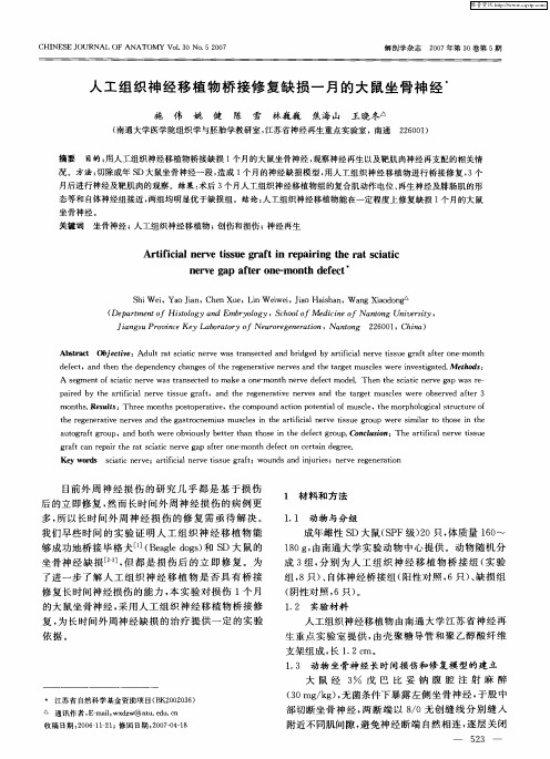
S h i We i ,Ya o J i a n,Ch e n Xu e ,Li n We i we i ,J i a o Ha i s h a n,W a n g Xi a o d o n
( D e p a r t m e n t o f Hi s t o l o g y a n d E mb r y o l o g y。S c h o o l o f Me d i c i n e o f Na n t o n g U n i v e r s i t y, J i a n g s u Pr o v i n c e Ke y L a b o r a t o r y o f Ne u r o r e g e n e r a t i o n, Na n t o n g 2 2 6 0 0 1 ,C h i n a )
A b s t r a c t O b j e c t i v e : Ad u l t r a t s c i a t i c n e r v e w a s t r a n s e c t e d a n d b r i d g e d b y a r t i f i c i a l n e r v e t i s s u e g r a f t a f t e r o n e - mo n t h
坐骨神经 。
关键词 坐骨神经 ;人工组织神经移植物 ; 创伤 和损伤 ; 神经再生
Ar t i f i c i a l n e r v e t i s s u e g r a f t i n r e pa i r i n g t h e r a t s c i a t i c n e r v e g a p a f t e r o n e - mo n t h d e f e c t
神经导管修复大鼠坐骨神经缺损实验研究
神经导管修复大鼠坐骨神经缺损实验研究(作者:___________单位: ___________邮编: ___________)作者:张凤久赵志英刘启黄丽华【摘要】目的:采用一种自行研制的具有良好生物相容性的壳聚糖构建人工神经来修复大鼠坐骨神经缺损,研究证实其修复效果。
方法:选用30只健康雄性Wistar大鼠,随机分为正常对照组(A组)、原位神经移植组(B组)、壳聚糖神经导管桥接组(C组)3组,分别切断坐骨神经后做相应处理,12周后进行神经电生理检测。
结果:C组12周后神经已经长过缺损段,神经传导功能恢复。
结论:这种套管能够有效的桥接10 mm长的大鼠坐骨神经缺损,可应用于周围神经缺损的治疗,同时可以作为进一步开发组织工程化的人工神经的良好载体。
【关键词】神经导管;周围神经;缺损;壳聚糖Abstract Objective: Using a self-developed chitosan with good biocompatibility to build artificial nerves to repair defects in rat’s sciatic nerve and a study was made to confirm the repair effect. Methods: 30 healthy male Wistar rats wererandomly divided into 3 groups (A: normal control group, B: in situ nerve graft group, C: Artificial neural bridge group). Sciatic nerve were cut off with the deal accordingly, 12 weeks later, the nerve electrophysiological testing was performed. Results:12 weeks later, the nerve has been a long paragraph defect, the function of nerve conduction recovered. Conclusion: This casing can effectively bridge the Sciatic Nerve defect of 10mm in rats and can be used in the treatment of peripheral nerve defects. At the same time it can serve as a good carrier for the further development of tissue-engineered artificial nerves.Key words Nerve conduit; Peripheral nerve; Defect; Chitosan周围神经损伤在临床中非常多见,在很多情况下,易导致长期且严重的功能障碍。
新型支架移植修复脊髓损伤

新型支架移植修复脊髓损伤中国北京积水潭医院袁宁博士所在团队设计了一种新型双层不等隙胶原膜,作为神经干细胞的支架,具有双层结构,内层为疏松层,外层为致密层。
移植时将大量的神经干细胞浸润于多孔隙的疏松层,这样能为目标脊髓移植位置提供大量的神经干细胞存在的三维定向。
移植于目标位置时,将疏松层放置于内部,而致密层放置于外侧,致密层具有较小的孔隙,从而有利于阻止周围瘢痕组织的进入,并减少疏松层中神经干细胞及其分化后生成的促神经生长因子的流失,在目标脊髓位置中形成有利于损伤脊髓修复的微环境。
将这种双层不等隙胶原膜作为神经干细胞支架用于修复大鼠脊髓不完全损伤,可使大鼠脊髓病理损伤明显减轻,运动功能得到有效的恢复。
相关文献发表于《中国神经再生研究(英文版)》杂志2014年5月第10期。
扫描电镜观察到神经干细胞在双层不等隙胶原膜上生长良好,且双层不等隙胶原膜结构未出现变化Article: " Neural stem cell transplantation in a double-layer collagen membrane with unequal pore sizes for spinal cord injury repair," by Ning Yuan1, Wei Tian1, Lei Sun2, Runying Yuan2, Jianfeng Tao2, Dafu Chen2 (1 Department of Spine, Beijing Jishuitan Hospital, Beijing, China; 2 Beijing Institute of Orthopedics and Traumatology, Beijing, China)Yuan N, Tian W, Sun L, Yuan RY, Tao JF, Chen DF. Neural stem cell transplantation in a double-layer collagen membrane with unequal pore sizes for spinal cord injury repair. Neural Regen Res. 2014;9(10):1014-1019.欲获更多资讯:Neural Regen ResUsing a novel scaffold to repair spinal cord injuryDr. Ning Yuan, Beijing Jishuitan Hospital, China and his colleagues, developed a novel neural stem cell scaffold that has two layers: the inner loose layer and the outer compact layer. The loose layer was infiltrated with a large amount of neural stem cells before it was transplanted in vivo. Thus a plenty of neural stem cells can be provided at the target spinal cord site. The loose layer was adhered to the injured side and the compact layer was placed against the lateral side. The compact layer has very small holes, so it can prevent ingrowth of adjacent scar tissue. It can also prevent the loss of inner neural stem cells and the neural growth factors secreted by the differentiated neural stem cells. Thus a good microenvironment forms to help spinal cord injury repair. Yuan Ning and colleagues found that transplantation of neural stem cells in a double-layer collagen membrane with unequal pore sizes is an effective therapeutic strategy to repair an injured spinal cord in rats. Related results were published in Neural Regeneration Research (Vol. 9, No. 10, 2014).Through transmission electron microscope, neural stem cells attached to the double-layer collagen membrane with unequalpore sizes and there was no structural change in the double-layer collagen membrane.Article: " Neural stem cell transplantation in a double-layer collagen membrane with unequal pore sizes for spinal cord injury repair," by Ning Yuan1, Wei Tian1, Lei Sun2, Runying Yuan2, Jianfeng Tao2, Dafu Chen2 (1 Department of Spine, Beijing Jishuitan Hospital, Beijing, China; 2 Beijing Institute of Orthopedics and Traumatology, Beijing, China)Yuan N, Tian W, Sun L, Yuan RY, Tao JF, Chen DF. Neural stem cell transplantation in a double-layer collagen membrane with unequal pore sizes for spinal cord injury repair. Neural Regen Res. 2014;9(10):1014-1019.。
组织工程修复周围神经缺损的研究概况[兼容模式]
![组织工程修复周围神经缺损的研究概况[兼容模式]](https://img.taocdn.com/s3/m/f04df0170812a21614791711cc7931b765ce7b03.png)
组织工程修复周围神经缺损的研究概况[兼容模式]组织工程修复周围神经缺损的研究概况陈琳敖强左焕琮清华大学玉泉医院神经外科周围神经缺损:临床常见病–创伤–理化因素–肿瘤治疗金标准神经移植治疗金标准:神经移植– Balane 1932年首次采用较大缺损间隙临床上目前仍多采用自体较大缺损间隙:临床上目前仍多采用自体神经移植修复自体神经移植缺点–供区神经的缺损–供区神经来源有限–与受区神经难以匹配–供失神经支供区失神经支配中空导管桥接缺损神经两断端:引导神经再生– 20世纪80年代初修复方法:–单一的显微外科–细胞移植技术–组织工程技术Elsevier?SD ? nerve and cell*?and?(transplant or?implant nerve?and?cell and (transplant* or implant* or?graft*?or?engraft*) ? Li i Limit?to?patient and?peripheral?nerve i d i h l ? 1400?papers p p一、导管桥接神经再生机制导管桥接神经再生机制神经修复导管引导神经再生的能力–“临界轴突延伸长度”( i i l axonelongation,“临界轴突延伸长度”(critical l i Lc)–轴突通过率达到50%时导管内神经断端间隙的最长长度硅胶管中神经缺损长度的微小增加会导致神经轴突通过率的急剧下降(Lundborg?1982)突通过率的急剧下降(Lundborg 1982) ?硅胶管内神经再生的特征曲线:在这个S型的特征曲线上,在拐点记录到 Lc ( Yannas?2005,2007)再生神经轴突通过导管比率的特征曲线中空硅胶管桥接大鼠和小鼠坐骨神经不同长度缺损神经缺损长度:横坐标神经缺损长度横坐标再生轴突通过率:纵坐标导管的相对性能–实验导管的特征曲线与硅胶管的标准曲线比较导管引导神经再生的能力–△L=实验导管Lc -硅胶管Lc△L–导管的可变参数决定–组成成分、导管结构、通透性等依据神经再生的基本理论–优化神经导管参数–构建理想新型导管–提高引导神经再生能力周围神经再生理论(1)神经营养论神经营养理论–神经远段雪旺细胞分泌营养因子诱导轴突再生–已经被许多实验所证实–难以解释以下实验现象:难解释实现象 ? 在神经缺损间隙有微小增加的情况下神经轴突通过率大幅度下降,因为距离微小的增加不会引起营养因子浓度的急剧下降,因而不足以解释神经轴突通过率的骤降 ? 导管促进神经再生的能力却由于加入有方向性基质材料而增加,这也是神经营养理论不能解释的周围神经再生理论(2)接触引导论接触引导理论–轴突的延伸需要接触合适的基质–有方向性的导管内基质构型:促进成纤维细胞和雪旺细胞增殖、迁移,引导轴突延伸和雪旺细胞增殖迁移引导轴突延伸周围神经再生理论(3)基膜管论基膜管理论–周围神经节段缺损后成纤维细胞首先增殖迁移到神经缺损间隙,形成纤维缆连接两神经断端–雪旺细胞随后沿着纤维缆形成柱状基膜管大雪旺细胞随后沿着纤维缆形成柱状基膜管,大约直径10-20μm –轴突延伸长入基膜管后形成髓鞘–利于雪旺细胞轴向迁移的导管构型,引导有髓神经纤维生长导管引导周围神经再生种机制导管引导周围神经再生3种机制:–相辅相成–不能孤立强调某一机制,忽视其他机制提高导管△L 提高导管△L:–必须综合考虑这三方面因素–构建具有诱导活性与引导结构的神经修复导管二、周围神经组织工程神经修复支架材料的基本要求(1)良好的生物相容性作为人工神经移植物植人体内,首先不能引起免疫排异反应,具有良好的细胞亲能引起免疫排异反应具有良好的细胞亲和性及组织相容性,不会产生炎症刺激及诱发炎症反应,无血液毒性(2)良好的生物可降解性良的生物降解性理想的神经修复材料应具可控的体内降解性能,材料的降解应与神经轴突再生相同步,降解太快会管道膨胀阻碍其再生,同步降解太快会管道膨胀阻碍其再生降解太慢则会导致神经卡压及慢性异物反应(3)良好的生物力学性能良的生物力学性能有适宜的强度和弹性。
壳聚糖导管桥接修复周围神经缺损新进展

壳聚糖导管桥接修复周围神经缺损新进展陈琳;张玉琪;敖强;左焕琮【摘要】周围神经缺损是临床常见的致残性疾病,虽然科技和手术技术的发展使疗效获得进步,但对于长距离组织缺损,治疗仍面临很大困难.多种材料制造的神经导管的使用均可以提高修复再生的长度,其中壳聚糖是一种可降解材料,生物相容性优良,已经较多用于导管支架的制备,而联合使用各种可促神经再生的物质,进一步提高了效果.本文就壳聚糖基导管桥接修复周围神经缺损的最新研究进展作一简述.【期刊名称】《神经损伤与功能重建》【年(卷),期】2017(012)003【总页数】3页(P238-239,246)【关键词】壳聚糖;导管;桥接;神经修复;周围神经缺损【作者】陈琳;张玉琪;敖强;左焕琮【作者单位】清华大学玉泉医院神经外科中心北京100040;清华大学玉泉医院神经外科中心北京100040;清华大学玉泉医院神经外科中心北京100040;清华大学玉泉医院神经外科中心北京100040【正文语种】中文【中图分类】R741;R741.05全球每年新增1 000~1 500 万创伤病例,其中周围神经损伤占1.5%~4.0%[1]。
随着显微外科设备和技术的发展,周围神经损伤修复的临床疗效不断提高,缺损间隙较短可以直接精细缝合或小间隙套管修复;但缺损距离较长时,则需采用神经组织移植或神经导管桥接。
周围神经导管材料包括生物材料和人工合成材料两大类,壳聚糖是一种自然生物可降解材料,具有良好的生物相容性。
近年来,有许多关于壳聚糖在神经导管中应用的研究报道。
前期的研究大多使用单一的壳聚糖导管修复神经缺损,现在多在壳聚糖导管中复合各种可促神经再生的物质,例如其他种类的构建材料、因子、药物以及种子细胞,来协同修复神经损伤,提高修复效能[2-11]。
壳聚糖又名脱乙酰几丁质、聚氨基葡萄糖、可溶性甲壳素,可在体内降解为单糖,是一种良好的修复材料,对神经细胞基本无毒害作用[12]。
优点:①降解速度可调节,通过原料的脱乙酰度、分子量以及交联等方法;②有良好成膜性,通过旋转蒸发法或冷冻干燥法,可加工成管;③来源丰富,良好生物相容性;④能促进内皮细胞生长,利于新血管发生;⑤抑制成纤维细胞的生长,减少瘢痕形成,保持很小的组织粘连;⑥为再生的轴突提供营养[13]。
成年大鼠骨髓间充质干细胞复合组织工程人工神经修复1cm坐骨神经缺损

11 实验动物 无特定病原体 (SPF) 级雄性 SD大鼠 6只,
体重 180~200g;SPF级雄性 SD大鼠 16只,体重 200~250g。全部实验动物均由广东医学院动物实 验中心提供。 12 大鼠骨髓间充质干细胞分离培养
成年 SD雄性大鼠 (体重 180~200g),颈椎 脱臼致死。75%乙醇浸泡 5min无菌条件下取出两 侧股骨,剪去股骨两端,用 5ml注射器抽取含 10% 胎 牛 血 清 (以 色 列 KibbutzBeitHaemek公 司 ), 2μmol/LL谷氨酰胺(美国 Gibco公司),10ng/ml 表皮生长因子 (epidermalgrowthfactor,EGF,美国 Peprotech公司) 和 1% 青霉素链霉素的杜氏培养 基 (DulbeccosmodifiedEaglesmedium,DMEM) / F12的 BMSC完全培养液反复冲洗骨髓腔。收集骨 髓细胞 悬 浮 液,接 种 在 25ml的 塑 料 培 养 瓶 中, 37℃、5% CO2和 95%湿度的培养箱培养,48h 后首次全量换液,换液前用 001mmol/L磷酸盐缓 冲液 (PBS) 轻 轻 冲 洗 未 贴 壁 的 细 胞。以 后 每 隔 2~3d换液 1次。待约 80% ~90%的 BMSC接近 融合时,吸出旧培养液,用 001mmol/LPBS冲洗 2次,加入 025% 胰蛋白酶及 002% 乙二胺四乙 酸 (ethylenediaminetetraaceticacid,EDTA) 混合 消化液,显微镜下观察细胞已全部变圆并开始从瓶 壁脱落时,用吸管轻轻吹打细胞,加入 BMSC完全 培养 液 终 止 消 化,收 集 细 胞 悬 液,200 ×g离 心 5min。弃上清液,沉淀的细胞用 001mmol/LPBS 冲洗 1次后,用含 BMSC完全培养液重悬悬浮细
骨髓间充质细胞构建组织工程神经修复坐骨神经缺损

骨髓间充质细胞构建组织工程神经修复坐骨神经缺损何红云;邓仪昊;佟晓杰;成家茂;杜赵康【期刊名称】《中国组织工程研究》【年(卷),期】2009(013)028【摘要】BACKGROUND: Schwann calls are seed cells for constructing tissue engineered peripheral nerves. But their in vitro isolation, culture and purification are difficult. Acellular nerve allografts (ANA) have a great effect on repairing peripheral nerve defects, and it can induce marrow stromal cells (MSCs) into Schwann-like calls. Accordingly, MSCs can be used as seed calls theoretically in constructing tissue engineered peripheral nerves, instead of Schwann cells OBJECTIVE: To observe the repairing effect of tissue engineered peripheral nerves constructed with MSCs on sciatic nerve defects and to assess the feasibility of repairing peripheral nerve defects with MSCs as seed calls. DESIGN, TIME AND SE'I-rlNG: A randomized control animal experiment was conducted in the laboratory of Basic Medicine Collage of Dali Collage from July to December in2008.MATERIALS: A total of 30 SD rats were divided randomly into 3 groups, with 10 in each group. In MSCs+ ANA group, end-to-end anastomosis were performed with 10/0 non traumatic suture to broken ends of rat sciatic nerves and tissue engineered nerves cultured using MSCs combined with ANA; In ANA group, ANA were bridged to broken ends of sciatic nerves; In METHODS: The 10ram sciatic nerve defects in ratswere repaired using MSCs-constructed tissue engineered peripheral nerves, whose repairing effects were evaluated at week 12 post transplant through sciatic function index (SFI), gastrocnemius wet weight recovery rate, S-100 immunohistochemical staining and electron microscope observation, etc. MAIN OUTCOME MEASURES: Morphological changes of calls were observed during the culture of compounds; SFI and gastrocnemius wet weight recovery rates were detected after transplantation; New myelinization, axon growth and nerve fiber distdbuUon were observed with toluidine blue staining; Schwann calls growth and nerve fiber regeneration were observed with transmission electron microscope combined with S-100 protein immunohistochemical stainingmethod.RESULTS: The detection results showed that SFI and gastrocnemius wet weight recovery rate were higher in the MSCs+ ANA group than in the ANA group (P < 0.05). Compared the ANA group, the MSCs+ ANA group had more S-100 proteins expressed in its compounds obviously, and the number, the diameter of its myelinated nerve fibers as well as its myelin sheath thickness were all greater than those of the ANA group (P < 0.05). The repairing effect of the MSCs+ ANA group was close to that of the autotransplantation group.CONCLUSION: MSCs-constructed tissue engineered nerves have a better effect on repairing peripheral nerve defects than ANA, and MSCs as seed cells have a high value in peripheral nerve tissue engineering.%背景:许旺细胞是周围神经组织工程的种子细胞,但体外分离、培养、纯化许旺细胞较困难.脱细胞同种异体神经移植物具有较强的修复外周神经缺损的能力,且可诱导骨髓间充质细胞分化为类许旺细胞,理论上骨髓间充质细胞可替代许旺细胞作为种子细胞应用于周围神经组织工程.目的:观察骨髓间充质细胞构建组织工程神经修复坐骨神经缺损的效果,评估骨髓间充质细胞作为种子细胞修复周围神经缺损的可行性.设计、时间及地点:随机对照动物实验,于2008-07/12在大理学院基础医学院实验室完成.材料:将30只SD大鼠按随机数字表法分为3组,每组10只.骨髓间充质细胞+异体移植组将骨髓间充质细胞复合脱细胞同种异体神经移植物培养的组织工程神经与两断端用10/0无创线端端吻合;异体移植组将脱细胞同种异体神经移植物桥接;自体移植组将切断的坐骨神经旋转180°端端吻合.方法:运用骨髓间充质细胞构建的组织工程神经修复大鼠10 mm坐骨神经缺损,移植后12周通过坐骨神经功能指数、腓肠肌湿质量恢复率、S-100免疫组织化学染色、电镜等方法观察移植物修复效果.主要观察指标:复合物培养时观察细胞形态的变化;移植后观察坐骨神经功能指数及腓肠肌湿质量恢复率;通过甲苯胺蓝染色观察新生髓鞘形成和轴突生长及神经纤维的分布情况,结合透射电镜及S-100蛋白免疫组织化学染色,观察许旺细胞生长和神经纤维再生情况.结果:坐骨神经功能指数及腓肠肌湿质量恢复率的检测结果显示骨髓间充质细胞+异体移植组优于异体移植组(P<0.05).骨髓间充质细胞+异体移植组复合物中S-100的表达明显高于异体移植组,有髓神经纤维数量、有髓纤维直径和髓鞘厚度均大于异体移植组(P< 0.05),修复效果接近自体移植组.结论:骨髓间充质细胞构建的组织工程神经修复周围神经缺损的效果优于单纯的脱细胞同种异体神经移植物,骨髓间充质细胞作为种子细胞在周围神经组织工程中具有较强的应用价值.【总页数】5页(P5562-5566)【作者】何红云;邓仪昊;佟晓杰;成家茂;杜赵康【作者单位】大理学院基础医学院人体解剖学教研室,云南省大理市,610000:;大理学院基础医学院人体解剖学教研室,云南省大理市,610000:;中国医科大学基础医学院人体解剖学教研室,辽宁省沈阳市,110001;大理学院基础医学院人体解剖学教研室,云南省大理市,610000:;大理学院基础医学院人体解剖学教研室,云南省大理市,610000:【正文语种】中文【中图分类】R318【相关文献】1.天然脱细胞神经支架复合骨髓间充质细胞修复坐骨神经缺损 [J], 何红云;邓仪昊;佟晓杰;贾桦;张旭2.脱细胞神经支架联合干细胞构建组织工程神经修复坐骨神经缺损的meta分析[J], 向飞帆;阳运康;谭小琦;魏代清;杨琨;孙远林;周举3.脱细胞神经支架联合干细胞构建组织工程神经修复坐骨神经缺损的meta分析[J], 向飞帆;阳运康;谭小琦;魏代清;杨琨;孙远林;周举4.无细胞神经支架复合骨髓间充质干细胞构建组织工程人工神经修复坐骨神经缺损[J], 张彩顺;吕刚5.无细胞神经移植物复合骨髓间充质干细胞构建组织工程人工神经修复坐骨神经缺损 [J], 张彩顺;吕刚;张基仁因版权原因,仅展示原文概要,查看原文内容请购买。
复合转化生长因子-β的组织工程化周围神经修复坐骨神经缺损

复合转化生长因子-β的组织工程化周围神经修复坐骨神经缺损张勇杰;金岩;聂鑫;王彦亮;刘鹏;沈楠;国生辉【期刊名称】《生物医学工程学杂志》【年(卷),期】2007(24)2【摘要】探讨了转化生长因子-β(Transforming growth factor-β,TGF-β)在周围神经缺损修复中的作用。
将50 ng·ml-1 TGF-β加入体外培养的雪旺细胞(Sehwann cells,SC)中,MTT和流式细胞仪观测到TGF-β能够明显促进SC增殖;ELISA方法检测到TGF-β组上清神经生长因子(NGF)含量高于对照组(P<0.05);将牛去细胞基质(Bovine acellular matrix,BAM)、SC、血清和培养基按一定的比例混合,注入聚乳酸羟基乙酸共聚物(PLGA)神经导管去修复15 mm坐骨神经缺损。
30只SD大鼠分为3组,实验组:PLGA导管+SC+TGF-β;空白组PLGA导管+SC;和自体神经移植组。
16周后通过电生理、透射电镜等检测方法显示各组坐骨神经均得到再生,修复效果实验组与自体神经移植组无显著性差异,均优于空白组。
TGF-β一方面可以明显促进SC的增殖,另一方面可以增强SC分泌NGF的功能。
因此周围神经修复过程中使用外源性的TGF-β对修复周围神经缺损有较好的疗效。
【总页数】5页(P394-398)【关键词】转化生长因子-β;组织工程;神经再生【作者】张勇杰;金岩;聂鑫;王彦亮;刘鹏;沈楠;国生辉【作者单位】第四军医大学口腔医学院组织工程实验中心组织病理教研室;第四军医大学口腔医学院颌面外科【正文语种】中文【中图分类】R587.2【相关文献】1.组织工程化神经修复周围神经缺损的研究进展 [J], 曹常松;张基仁2.自体骨髓间充质干细胞组织工程化神经修复坐骨神经缺损的研究 [J], 吴坚;刘炎;王洁;姚健;顾晓松3.应用组织工程化周围神经修复大鼠坐骨神经缺损的实验研究 [J], 崔树森;魏壮;尹维田;刘建国;甘志华;张文杰;徐莘香4.小肠黏膜下层复合雪旺细胞构建组织工程化人工神经修复周围神经缺损的研究[J], 苏琰;张长青;曾炳芳;张开刚;谢雪涛5.新型组织工程化神经导管修复大鼠周围神经缺损 [J], 张振辉;王庆德;梅伟;毛克政;姜文涛因版权原因,仅展示原文概要,查看原文内容请购买。
- 1、下载文档前请自行甄别文档内容的完整性,平台不提供额外的编辑、内容补充、找答案等附加服务。
- 2、"仅部分预览"的文档,不可在线预览部分如存在完整性等问题,可反馈申请退款(可完整预览的文档不适用该条件!)。
- 3、如文档侵犯您的权益,请联系客服反馈,我们会尽快为您处理(人工客服工作时间:9:00-18:30)。
一种新组织工程方法桥接修复缺损坐骨神经
化学脱细胞同种异体神经不但去除了许旺细胞、髓鞘及其崩解碎片等可引起排斥反应的物质,减轻了移植修复后的免疫排斥反应,同时也保留了为缺损神经修复再生过程中提供良好再生支架的神经基质管,具有与自体神经移植近似的引导神经再生的功能,为神经再生提供良好的局部微环境。
来自中国郑州大学国际教育学院的张雁儒团队,设想用大鼠双侧坐骨神经制备化学去细胞同种异体神经,并联合脑源性神经营养因子复合骨髓间充质干细胞桥接修复大鼠坐骨神经1 0mm缺损后发现,大鼠的坐骨神经指数、三头肌湿质量恢复率、髓鞘厚度、有髓神经纤维数量在3组中的变化为:脱细胞同种异体神经+脑源性神经营养因子+间充质干细胞组>脱细胞同种异体神经+间充质干细胞组>脱细胞同种异体神经组。
修复效果优于单独应用化学脱细胞同种异体神经桥接和脱细胞同种异体神经复合骨髓间充质干细胞移植。
文章发表在《中国神经再生研究(英文版)》杂志2014年10月第20期。
Article: " Combining acellular nerve allografts with brain-derived neurotrophic factor transfected bone marrow mesenchymal stem cells restores sciatic nerve injury better than either intervention alone," by Yanru Zhang1, 2, Hui Zhang3, Gechen Zhang3, Ka Ka1, Wenhua Huang2 (1 School of International Education, Zhengzhou University, Zhengzhou, Henan Province, China; 2 Institute of Clinical Anatomy, Southern Medical University, Guangzhou, Guangdong Province, China; 3 Department of Orthopedic Surgery, First Affiliated Hospital of Zhengzhou University, Zhengzhou, Henan Province, China)
Zhang YR, Zhang H, Zhang GC, Ka K, Huang WH. Combining acellular nerve allografts with brain-derived neurotrophic factor transfected bone marrow mesenchymal stem cells restores sciatic nerve injury better than either intervention alone. Neural Regen Res. 2014;9(20):1814-1819.
欲获更多资讯:Neural Regen Res/
A novel tissue engineering method for repair of sciatic nerve defects
Chemically extracted acellular nerve allografts can eliminate Schwann cells, myelin and disintegrating debris that can cause rejection, attenuate immune rejection after transplantation, and also help retain the nerve canal. These canals can provide a good scaffold for the regeneration of nerve defects, and have a similar effect to autologous nerve transplantation in guiding nerve regeneration, offering a local environment for nerve recanalization. Yanru Zhang at School of International Education, Zhengzhou University, China and his colleagues chemically extracted acellular nerve allografts from bilateral sciatic nerves, and repaired 10-mm sciatic nerve defects in rats using these grafts and brain-derived neurotrophic factor (BDNF) transfected bone marrow mesenchymal stem cells (BMSCs). Sciatic functional index, triceps wet weight recovery rate, myelin thickness, and number of myelinated nerve fibers were significantly changed in the acellular nerve allograft group, acellular nerve allograft + BMSCs group, and acellular nerve allograft + BDNF transfected BMSCs group. Variations were the largest in the acellular nerve allograft + BDNF transfected BMSCs, followed by acellular nerve allograft + BMSCs group, and lastly the acellular nerve allograft group. Experimental findings suggest that chemically extracted acellular nerve allograft combined with nerve factor and mesenchymal stem cells can promote the restoration of sciatic nerve defects. The repair effect of acellular nerve allograft + BDNF transfected BMSCs is superior to single application of acellular nerve allograft or acellular nerve allograft combined with BMSCs transplantation. This article has been published in Neural Regeneration Research (Vol. 9, No. 20, 2014).
Article: " Combining acellular nerve allografts with brain-derived neurotrophic factor transfected bone marrow mesenchymal stem cells restores sciatic nerve injury better than either intervention alone," by Yanru Zhang1, 2, Hui Zhang3, Gechen Zhang3, Ka Ka1, Wenhua Huang2 (1 School of International Education, Zhengzhou University, Zhengzhou, Henan Province, China; 2 Institute of Clinical Anatomy, Southern Medical University, Guangzhou, Guangdong Province, China; 3 Department of Orthopedic Surgery, First Affiliated Hospital of Zhengzhou University, Zhengzhou, Henan Province, China)
Zhang YR, Zhang H, Zhang GC, Ka K, Huang WH. Combining acellular nerve allografts with brain-derived neurotrophic factor transfected bone marrow mesenchymal stem cells restores sciatic nerve injury better than either intervention alone. Neural Regen Res. 2014;9(20):1814-1819.。