简单的显微镜+显示器的配置
A包荧光显微镜参数要求如下

A包(荧光显微镜)参数要求如下:主要技术参数:1、粗、中、细同轴3档调焦装置,带上限锁定及松紧调节功能。
2、10倍物镜具备17mm以上的工作距离。
3、与显微镜同品牌专业显微镜数码照相系统,像数:3M,2048×1536;CCD连RGB滤片,最高像素点700万。
曝光时间:0.1毫秒到2秒,30Bit RGB彩色深度,BINNING模块,彩色或黑白。
一般技术参数:1、新型无限远、完整修正光学系统。
模块化设计,可升级。
采用T型一体化机身设计,整机可进行明场及荧光观察。
三目镜筒,30度人工学观察设计角度,宽视野目镜:l0X/22mm2、7位及以上物镜转换。
底座带3位滤光片支架,配日光、灰色、减光滤光片。
3、电源:可在90—250V之间自动稳压内置电源,≥12V/l00W卤素灯照明,带外置灯箱。
4、荧光装置:≥5位荧光滤快转盘,l00W汞灯。
紫外荧光滤块激发波长:BP340—380nm,蓝色荧光滤块激发波长:BP450—490nm,绿色荧光滤块激发波长:BP515—560nm5、对焦钮:上下高度可调。
XY操纵杆可自行左右手拆换使用。
6、载物台:抗刮抗腐蚀,载物台中心陶瓷面可独立拆卸清洗。
配双玻片样品夹。
7、多功能聚光镜,带彩色条纹标记,颜色标记可与物镜颜色标记相匹配。
8、全中文界面图像处理软件:具测量等功能,能准确地进行周长、直径、面积等参数测量。
多图像画廊浏览,多参数图像信息记录,具备标注、剪头等功能有丰富的图文库,实现教学功能,具查询功能,能打印各种形式报告单。
9、物镜:5X高级平场消色差物镜,数值孔径NA≥0.12,WD≥12mm10X高级平场消色差物镜,NA≥0.25,WD≥17mm20X高级平场消色差物镜,NA≥0.40,WD≥1.15 mm40X高级平场消色差物镜,NA≥0.65,WD≥0.40mm100X高级平场消色差油镜,NA≥1.25,WD≥0.12mm配置要求:显微镜主机1台,品牌电脑(主流配置)1台B包(心电图机)技术参数要求如下:主要技术参数:1、同步15导联采集、存储、评估,有助于迅速识别右心室和后壁的心肌梗塞;2、机器内置预采集模式,即按下打印按键时,可出按键前10秒心电图;3、FDA认证的测量诊断功能,有年龄特异性分析;4、交流85—264V 50Hz,适应各种使用环境;5、可选择低频响应范围0.04Hz,0.08Hz,0.16Hz;一般技术参数:1、急性心肌缺血非时间依赖性预测评分;2、频率响应:0.03—150Hz(-3db);3、电池充足后可记录≥50人/次;4、灵敏度选择:5、10、20、10/5mm/mV;5、具有交流滤波、肌电滤波、漂移滤波功能;6、ECG输入方式:前置模数转换盒;7、采集盒可控制系统采集,打印等操作;8、输入阻抗≥10MΩ;9、具有抗除颤电击保护功能;10、抗极化电压≥±500mV;11、共模抑制比≥120dB;12、采样率:2000sample/Channel/s;13、显示器规格≥10.4英寸的液晶显示屏;14、显示12导联波形;显示菜单、心率、病人姓名、导联选择、走纸速度、增益、滤波器、时钟、导联脱落等;15、高级儿童心电分析模式;配置要求:1、热敏式点阵打印机;2、床旁控制心电图打印;3、走纸速度:5、12.5、25、50 mm/sec;4、分辨率:200dpi;5、A4格式折纸;6、连续记录通道数:3,6,12,15;7、内存及SD卡存储;8、LAN宽带接口;9、交流和内置可充电电池;10、14 根导联线;。
一、学生用体视显微镜(电脑型)技术参数(共40台)

一、学生用体视显微镜(电脑型)技术参数:(共40台)1.目镜类型放大倍数视场(mm)目镜10X φ202.物镜变倍范围 0.7X-4.5X3.显微镜为连续变倍,变倍比为6.5:14.双瞳距调节范围 54-76mm5.移动工作距离: 95mm6.总放大倍数: 7—90X7.观察头:三目观察45度倾斜,360度旋转8.载物台:圆形载物台直径:95mm9.照明光源:12V/10W上下卤素灯亮度可调或上卤下荧光灯放大倍数:10.可以直接电视机或电脑上观察实物图像系统组成: 1.立体显微镜 2.适配镜 3.摄像器(CCD)二、教师用体视显微镜(电脑型)技术参数:(1台)1.目镜类型放大倍数视场(mm)目镜10X φ202.物镜变倍范围 0.8X-5X3.显微镜为连续变倍,变倍比为6.5:14.双瞳距调节范围 55-75mm5.移动工作距离: 95mm6.总放大倍数: 7—360X(以17寸显示器,2X倍的大物镜为例)7.观察头:三目观察45度倾斜,360度旋转8.载物台:圆形载物台直径:95mm9.照明光源:12V/10W上下卤素灯亮度可调或上卤下荧光灯10.可以直接电视机或电脑上观察实物图像系统组成:1、立体显微镜 2、专用适配镜 3、摄像器(300万) 4、图像采集及处理软件 5、测量软件 6、数据线三、深色不透光窗帘窗户尺寸:长*高=4.5m*3m(相同尺寸,共4扇窗户)要求:左右拉,深色不透光布帘,四、空调(数量:2台)立体柜式,3匹,可以独立除湿。
经营管理模拟沙盘实训室建设参数1.1.1内饰物品清单及预算1.1.5配套软件1.1.6装修预算梁光平团队II期实验室建设——整改及布置家具采购梁光平团队II期实验室建设——实验设备采购明细紫外可见分光光度计参数: 1台主机参数:1 工作环境1.1使用温度范围:15°C to 35°C1.2 使用湿度范围:30% to 80%1.3 仪器尺寸:450Wx600Dx250H mm2 技术规格2.1 分光系统2.1.1 光学系统: 双光束2.1.2 分光器: 双单色器,象差校正型切尼尔一特纳装置2.1.3 设定波长范围:190~900nm▲2.1.4 测试波长范围: 185-1100nm(需更换PMT)▲2.1.5 衍射光栅刻线数:≥1800 lines/mm2.1.6 波长准确性: ±0.1nm(656.1nm)±0.3nm(全波段)▲2.1.7 波长重复精度: ±0.05nm▲2.1.8 波长扫描速度: 波长移动速度: ≥14000nm/min; 最大扫描速度:≥4500nm/min;2.1.9 波长设定: 扫描开始波长和扫描结束能够以1nm单位设置;其它为0.1nm单位2.1.10 光源切换波长: 和波长同步自动切换290.0 nm~370.0 nm▲2.1.11 谱带宽度: 0.1/ 0.2/ 0.5/ 1/ 2/ 5nm 6档或以上可调2.1.12 分辨率:≥0.1nm▲2.1.13 杂散光: KCI< 1%T (198nm)NaI < 0.00005%T (220nm)NaNO2< 0.00002%T (340nm)2.1.14 测光方式: 双光束测光方式2.1.15 测光类型: 吸光度(Abs),透射率(%),反射率,能量(E)▲2.1.16 测光范围: 吸光度:-8.5~8.5 Abs2.1.17 光度准确性±0.002Abs(0-0.5Abs)±0.003Abs(0.5-1Abs)±0.006Abs(1.0-2.0Abs)±0.3%T2.1.18光度重现性±0.001Abs(0.5Abs)±0.001Abs(1Abs)±0.003Abs(2Abs)±0.1%T▲2.1.19 噪音0.00005Abs RMS (500nm) 以下。
透射电子显微镜
透射电子显微镜1.工作条件:1.1电力供应:220V(±10%),50Hz,单相;380V(±10%),50Hz,三相1.2工作温度:15︒C-25︒C1.3工作湿度:< 60%1.4仪器运行的持久性:连续使用1.5独立地线:≤100欧姆2.设备用途和功能:用于金属材料、无机非金属材料、生物材料、化工材料、高分子材料等材料的微观精细结构、形貌观察,衍射花样分析,成分分析,高分辨成像等。
3. 技术规格:3.1 六硼化镧透射电镜基本单元3.1.1 电子枪:六硼化镧型*3.1.2 分辨率点分辨率:≤0.23nm线分辨率:≤0.14nm3.1.3 加速电压最高加速电压: 200kV*3.1.4 稳定度加速电压稳定性:≤2 ppm/min物镜电流稳定性:≤1 ppm/min*3.1.5 放大倍数50×—1,500,000×*3.1.6 物镜球差系数:≤1.0mm色差系数:≤1.4mm最小聚焦步长:≤1.5nm3.1.7束斑尺寸TEM模式:≥20nmEDS/NBD/CBD模式:≤1.0nm3.1.8相机长度: 80~2000mm3.1.9 样品移动:X: ≤2mm ;Y: ≤2mm;Z:≤0.4mm3.1.10 最大倾斜角:≥+35°3.1.11 X射线能谱分析固体角:≥0.13sr*3.1.12 取出角:≥25°*3.1.13 计算机控制系统操作系统:Windows XP及以上,控制系统采用分级分块方式控制,即使计算机死机,高压系统、真空系统、操作面板系统仍能正常工作。
实验状态(电子光学状态)记忆功能:可多用户储存各自的实验状态并随时恢复样品位置记忆功能:具备计算机工作站:i3及以上处理器,内存4GB以上,硬盘500GB,专业的图形处理用显示卡,DVD刻录功能,所有声音、网络以及输出功能俱全,19寸及以上专业图形液晶显示器3.1.14 真空系统: 自动控制样品室真空度:好于2.7 ×10⁻⁵Pa3.1.15 透射电镜具备自动断电、断水保护功能,具备自动诊断功能*3.1.16 透射电镜具备自动烘烤功能3.1.17 扫描透射附件明场分辨率:≤1.0nm暗场分辨率:≤1.0nm可采集明场像、暗场像和HAADF像3.2 能谱仪(EDS)的技术规格3.2.1 功能:该附件是透射电镜的必要附件,用于材料微区的定性、定量成份分析*3.2.2探测器:电制冷*3.2.3 探测器面积:≥60mm23.2.4 分辨率:≤133eV(Mn K 线)3.2.5 分辨元素范围:5B -U923.2.6 峰背比:≥18,000:13.2.7系统工作站:i3-2100处理器,内存4GB以上,硬盘2×250GB,专业的图形处理用显示卡,所有声音、网络以及输出功能俱全,22寸液晶显示器3.2.8 软件:导航分析器,全中文软件操作界面及中文实时帮助系统,实时帮助系统,信息管理系统,实验报告系统等3.3 数字化CCD相机技术规格3.3.1 功能:该附件是透射电镜的必要附件,用于透射电镜形貌像和电子衍射花样的数字化图像的记录,具有数字化图像处理的功能*3.3.2像素尺寸≥9 μm x 9 μm3.3.4视野范围41 mm x 41 mm3.3.5分辨率2048 x 2048 Pixel*3.3.6耦合方式2:1光纤耦合3.3.7动态范围≥14位3.3.8曝光时间 1 ms - 100 s3.3.9双制冷系统芯片温度15度在室温25度时3.3.10安装位置底部同轴3.3.10抗光晕指数100x3.3.11操作界面可选中文、英文等语言3.4 冷却循环水(原装进口,匹配电镜)3.4.1冷却能力:5230 W (4500 kca l/h)3.4.2控温精度:0.1︒C/h3.4.3流量:7.5 L/min3.4.4水温:15-20℃3.4.5水压:0.2 to 0.3 M Pa3.5稳压电源(原装进口,可以匹配电镜)3.5.1 输出电压: 单相 200 V AC3.5.2 输入电压波动范围: ±10%3.5.3 频率: 50/60 Hz4. 离子减薄仪4.1 离子枪:潘宁式离子枪,装载微小磁铁,聚焦离子束设计,无耗件。
显微镜光学配置图说明书

Supplementary MethodsOptical configuration:A diagram of the optical configuration used for the photobleaching experiments is shown insupplemental figure 1 below.Supplemental Figure 1. Diagram of the optical configuration of the side-port for photobleaching.e. he d or the YFP bleaching experiment shown in figure 1, the microscope filter cube used for bleaching rom 0DCSXscanning mirrorsView is from above, and the scanning mirrors are at the rear of a Nikon TE-300 inverted microscop The dichroic mirror near the tube lens is a long-pass extended reflection mirror (650 DCXRU). The light from an argon-ion laser (Coherent Sabre) was coupled into an optical fiber and connected to the side-port at the connector shown at the right side of the figure. To preview the fluorescence and select cells, an in-house developed fiber-optic coupled high-power blue light emitting diode (LED) source (patent pending) was connected in place of the laser coupling fiber. Depending on the experiment a band-pass filter was sometimes included before coupling the LED emission to the fiber. In some experiments an additional dichroic mirror, 440 nm long-pass (440DCLP), was included between t fiber-optic coupler and the divergence-correcting lens to allow the additional coupling of a liquid-fille light guide (Oriel) to permit the use of a xenon arc lamp (Cairn).F contained a 545nm long-pass dichroic (545DCLP) and a 540-600nm band-pass emission filter(HQ570/30). A portion of the YFP emission could be monitored visually while scattered light f the 514.5 nm laser excitation was blocked. For bleaching experiments using laser lines shorter than 514.5, a filter cube containing a 510 long pass dichroic and a 510-560 band-pass emission filter (510DCLP and HQ535/50 respectively) was used. The filter cube for the 2-photon excitationcontained a 700 nm short-pass, UV reflecting, dichroic and a 710 short pass emission filter (70and E710SP respectively).YFP photoconversion revisited: confirmation of the CFP-like speciesMichael T Kirber, Kai Chen & John F Keaney JrIn experiments to check if the fluorescent decay product was visible with arc lamp illumination, a filter experiments to check if bleaching using arc lamp illumination produced the fluorescent decay ht , and en rlabs. ther than observing that the fluorescent decay product of YFP could be excited using a configuration ell culture and plasmid transfection: cube containing a 405-445 band-pass excitation filter, a 455 long-pass, extended reflection dichroic, and a 460-500 nm band-pass emission filter (D425/40X, 455DCXRU, and D480/40M respectively) was used. Infrared light was removed from the arc lamp output using a short wavelength visible and UV reflecting cold mirror (Thorlabs).In product, the fiber coupler was removed from the side port and the output from the liquid-filled lig guide and a collimating lens were coupled directly in place of the fiber-optic coupler. The 440DCLP dichroic was left in place. The filter cube used for bleaching contained, a 460-500nm band passemission filter inserted in reverse direction in the excitation position, a 510 nm long-pass dichroic a 510-560 nm band-pass emission filter ( HQ480/40M, 510DCLP, and HQ535/50M respectively) Bleaching YFP about 50% took 2 hours and when the 440DCLP mirror was removed and the CFP filter cube (D425/40X, 455DCXRU, and D480/40M) selected, the fluorescent decay product was se with the arc lamp illumination. All filters and dichroic mirrors were purchased from ChromaTechnology. Optical fiber, lenses, and optomechanical components were purchased from ThoO appropriate for exciting CFP using single photon (arc lamp) or 2-photon excitation, we did not try to determine the excitation spectrum of this byproduct. Such experiments might well be worthwhile but we are not presently set up to carry them out.Cvitrogen) and transfection with overexpression plasmids was ) was age acquisition and display:COS-7 cells were cultured in DMEM (In carried out using Fugene 6 (Roche) in cells at 70% confluency according to the manufacturer’sinstructions. PTP1B trapping mutant expression vector pcDNA6.2/YFP-PTP1B (D181A/Q262A constructed by using PCR subcloning technique with pcDNA6.2/N-YFP vector (Invitrogen) and pJ3H-PTP1B (D181A/Q262A)(kindly provided by Dr. Zhong-Yin Zhang, Indiana University). Cells were grown on glass cover slides and fixed with 4% paraformaldehyde.Imas designed and built in-house in collaboration with the laboratory of inal irror d between the number of counted photons and the brightness of the display.The 2-photon microscope used w Dr. Peter So at the Massachusetts Institute of Technology. The optical microscope portion of the system was a Nikon TE300 and the objective used was a 100X, 1.3 NA, oil-immersion type. Orig images obtained with 2-photon excitation (800 or 916 nm excitation) were 384x384 pixels with each pixel imaging 130 by 130 nm in the sample, which is beyond the resolution capability of the system. The dwell time at each pixel was 2 ms. Images shown in figure 1 are cropped from the originals and are 280 by 280 pixels. The fluorescence at each channel was measured with photon counting photomultiplier tubes (Hamamatsu R4700P-01 for the green channel and R4700P for the bluechannel). The green channel was separated from the red using an extended reflection dichroic m 565 nm long-pass (565DCXR) and the blue and green channels were separated using a 500nm long-pass extended reflection dichroic mirror (500DCXR). The pass-bands of the filters are given in the body of the text. The number of counts at each location in the sample was stored as a 16 bit unsigne integer. Images were imported into ImageJ as raw data. The colors for images in figure 1a-f were chosen to approximate the actual color of the fluorescence. The scale bars indicate the relationshipColocalization:In quantitatively assessing the degree of colocalization of two distinctly fluorescently labeled (or the different colored images each as a sample of a e or expressed) compounds in a cell, it is useful to treat random process which accurately represents it statistically (ergodicity). The normalized covarianc correlation coefficient between 2 sets of data, which in this case are images E and F which are N i x N j in size and contain elements e ij and f ij respectively, can be written as}Where E{ } denotes the “expected value” and E ¯ is the arithmetic mean of the pixel values in image E nd F ¯ is the mean of image F. This calculation has been applied to quantitatively assess colocalization E ()()ij ij e E f F ρ−−a (Vereb et al .). Because expectation is a linear operator, this expression can be rearranged so that the contribution at each pixel pair to the correlation coefficient is clear.ρ==We can then display an image G* with pixel values g*ij , the sum of all the pixels being the correlationoefficient. c*ij ij ij e f E F N N g −=or simplicity we normalize this so that we display images where the mean of all of the pixels, G ¯, is e correlation coefficient.F thij g =ll of the values can be easily calculated using ImageJ (Wayne Rasband, NIH).this manner we can easily compare different size sets of images and not lose the quantitative ove comparing the degree of colocalization under different experimental conditions some caution is oth ference:atko J, Vamosi G, Ibrahim SM, Magyar E, Varga S, Szollosi J, Jenei A, Gaspar R Jr, AIn information. Areas where the normalized covariance image is positive indicate colocalization ab that predicted by random uniform distributions of the 2 fluorescent species. Additionally, there are generally some regions where the pixels have a negative value. These regions correspond to areas where the colocalization of the 2 labeled compounds is less than would be predicted by a uniform random distribution.In needed. For example the regions for which the computation is performed should be similar under b sets of conditions (i.e. ratio of area of background to area of cell interior).re Vereb G, M Waldmann TA, Damjanovich S. Proc Natl Acad Sci U S A. 2000 May 23;97(11):6013-8.。
B203LED双目生物显微镜
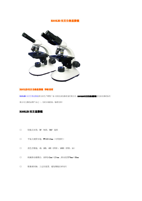
河南兄弟仪器设备有限公司依靠自身强大的社会资源、品牌优势、经营优势,以优于市场的 B203LED
双目生物显微镜 的价格向广大新老顾客朋友提供价格实惠、品质优良的该仪器设备;河南兄弟仪器设备公 司拥有最齐全的 B203LED 双目生物显微镜的型号,欢迎新老顾客朋友订购。河南兄弟仪器设备公司承诺以 最新 B203LED 双目生物显微镜的报价、最完善的服务回报顾客朋友。兄弟仪器设备公司希望早日与您结下 兄弟情谊!
○
○
○
LEDR
○
○
○
○
○
LED
○
○
○
○
○
TR-1
注:“●”为必备件,“○”为选购件
B 系列生物显微镜选购件
目镜 物镜 反光镜 移动尺 C 型接口 数码相机接口 数码摄像头
5X,WF16X,10X 测微目镜(0.1mm),10X 指针目镜,10X/20 20X,60X(弹簧) 凹凸双面镜 移动范围:30×50mm 0.5X,1 X NIKON,OLYMPUS 等数码相机专用 DV130(130万像素),DV200 (200万像素)
转换 四孔
器 三孔
●
固定 压簧片
●
载物 移动尺 ○
平台 台
机械移动平台
聚光 单片式
●
镜 阿贝式 N.A.1.25
照明 220V/20W 白炽灯,亮度可调
●
组 6V/20W 卤素灯,亮度可调
●
● ●● ●● ●● WF10
●
●
●
●
●
●
●
●
●
●
●
●
B-A4 B-A10 B-A40
○
●
○
●
电子显微镜技术参数

电子显微镜技术参数一、电镜主机技术要求:1.总则:1.1提供相应货物的技术规格文件,在应答的品目标题下,标明货物的型号、商标名称及生产厂家。
1.2货物的制造和检验,必须是按照现行的中国国家标准,或通用国际标准。
1.3仪器设备如需特殊工作条件(如:水、电源、磁场强度、特殊温度、湿度、振动强度等),应在相关文件中加以说明。
2环境条件:除该品目在技术要求中另有说明外,所有仪器、设备和装置,均应适合以下条件:a)电源:220V(±10%),50Hz/60Hz;b)工作环境温度:15~23度c)工作环境湿度:<60%RHd)运行持久性:连续使用e)安装条件:地线接地电阻小于100欧姆3.技术要求:3.1分辨率:0.2nm3.2加速电压:20-120KV(以100V为步长调节)3.3放大倍数:高反差模式:X200~X200,000高分辨模式:X4,000~X600,000低倍模式:X50~X1,0003.4图像旋转:最大范围X1,000~X40,000,旋转角度:±90度(15度/步)3.5衍射长度:高反差方式 0.2~8.0m高分辨方式 0.2~2.0 m3.6电子枪:钨灯丝,具有电流自动控制,灯丝计时,气压式自动升枪等功能3.7使用高灵敏度的荧光屏CMOS相机取代了传统的荧光屏,将TEM操作统一于显示器上。
配置双CCD,使图像显示与操作一体化3.8样品位移:X/Y ±1mm(CPU控制马达驱动),Z±0.3mm,样品台倾斜角:±30度。
可显示样品位置、倾角等。
3.9透镜系统:高倍观测时8级透镜3.10照明透镜级数:2级聚光镜3.11成像系统:CPU控制的6级透镜系统,物镜、中间镜和投影镜均为两级3.12不更换硬件的前提下,可在同一台仪器上实现高分辨和高反差模式的自由切换3.13 高反差模式焦长:≥6.1mm,高分辨模式焦长:≤3.1mm3.14物镜活动光阑:4孔光阑(10-20-50-80微米)3.15 可以实现8K x 8K像素快速自动拼图(4张x4张拼图仅需4分钟)3.16电镜控制界面与相机图像一体化,配置1600万像素直插相机3.17真空系统:真空逻辑由测量值控制配有皮拉尼规,用于测量低真空度潘宁规,用于测量高真空度3.18不使用扩散泵,标准配置分子泵 1台,转速不低于250L/s,旋转泵 1台,转速不低于135L/min。
采购显微镜技术指标要求

申购工业显微镜技术参数要求
一、设备特性与部门需求
1. 多点测量:可按其所需设置每片样品上的采样点数——少许几点适用于快速检测,多点可用于详细分析。
2. 大面积成像:可自动按次序对多个视场进行成像,然后由软件拼接成一张大面积的样品表面三维图像。
3. 自动形貌特征测量:可设置操作配方对样品的各种指定形貌特征如尺寸、间距、高度甚至整片样品的弧度等进行自动测量。
4.部门需求:可测量线切割使用钢线的直径,颗粒高度等;可测量制绒后硅片表面制绒绒面分布效果;
二、标准系统配置
1.显微镜系统
光源:高亮度白光LED
物镜:5x,10x,20x,50x,100x
耦合透镜:0.5x
自动载物台:200毫米x200毫米XY驱动范围
Z轴:30毫米纵向驱动范围
数码相机: 1024x768像素,1/3 英寸CCD
2.计算机控制系统
处理器:英特尔®酷睿2 双核(最低2.5GHz)
随机存储器3GB
硬盘驱动器:320GB
显示器:22英寸宽屏液晶显示器(1680x1050像素)
3. 三维测量软件
实时视频成像
快速数据采集(每处<1分钟)
三维表面浏览:
倾斜,旋转,缩放,过滤•
体积计算
二维表面分析:
特性尺寸,直径,面积
图案间距,标准偏差
表面粗糙度
区域平均台阶高度
轮廓测量
台阶高度与粗糙度
多横截面分析
平均值测量标尺
倾斜度与表面波度补偿
阵列式采样序列
自动数据与图像输出用于生产线SPC。
显微镜技术参数

显微镜技术参数
1.放大率:电动变倍,缩放比例1:6,通过手柄或脚控调节
2.调焦系统:内部,电动,连续可调,通过手柄或脚控触发,自动聚焦
3.工作范围:200-415mm,或增强至219-530mm
4.主双目镜筒:折叠式双目镜筒,0-180°上下倾角可调镜筒,左右360°旋转
可调,广角目镜12.5X,放大倍数1.9X~18.2X
5.助手镜:倾角可调,直视/0-45°/0-180°
6.目镜PD屈光补偿:+5D—-8D
7.照明:冷光源光纤照明,氙灯用于主照明和备用照明,备用氙灯位于灯箱内,
带一键快速切换功能,可通过手柄调节亮度,光斑直径大小可调,范围
11-165mm
8.手柄功能:变焦、变倍、电磁锁开关、亮度调节、数码照相机快门控制
9.控制单元:液晶显示面板,可控参数,可自定义手柄按键功能设定,内置1CCD
摄像视频参数设定,故障诊断码提示
10.额定电压:220~240V±10%
11.额定频率:50Hz-60Hz
12.功耗:115V,Max10A 230V,Max8A
13.电流断路器:自动,短路自动保护装置
14.支架:升降支柱的落地式支架,承重19kg,全电磁锁控制
15.外置视频组件:三晶片高清摄像系统、标清视频记录器、照相系统适配器、
高清平板显示器(1080p)。