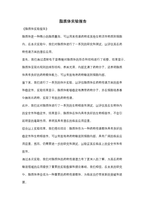
透射电镜观察脂质体
脂质体及其在化妆品体系中的表征

脂质体的水合粒径表征,应根据具体的样品体系选择合适的表示方法
.
41
包封率
.
42
方法
原理
特点与适用范围
缺点
HPLC法
参照磷脂含量测定
适用范围广,
/
可同时用于磷脂含量和包封率测定
透析法
利用半透膜分子大小差别,通过及时更换 简单,稳定性好,可处理大量样品,
外相达到分离的目的
适合小粒径,稳定性好的脂质体
Eric Betzig
Stefan W. Hell 2014诺贝尔化学奖
W. E. Moerner
STORM 技术的开创者庄小威
.
35
低分辨荧光显微镜和超高分辨荧光显微镜图片的对比
优点:突破光学极限的分辨率: 1nm 左右, 具有专属性,可作为通用的方法
局限性:外部资源少
北大工学院席鹏课题组 上海交大李小卫课题组
耗时较长,所耗介质较多
微柱离心法
凝胶柱层析 法
利用游离药物与含药脂质体的重力差异进 行分离,计算包封率
利用脂质体与游离药物分子质量和粒径大 小的差异进行分离
常规SEM
1. 将含有脂质体的样品滴在干净的硅片上面 2. 晾干或者烘干 3. 对其进行喷金处理(增强样品的导电性)
缺点局限性 1.脂质体干燥过程中会发生结构变化。 2. 对小颗粒分辨率不高。100nm左右。
1.只能看表面结构,不能区分脂质体和其它纳米颗粒 2.脂质体不导电,需要喷金处理。
AFM
1.把样品稀释为浓度1Wt%左右 2. 样品滴在云母片上 3. 放置待溶剂挥发干后进行测试
二、对于一些特例。比如低倍的Cyro-TEM或者切片法观测不到脂质体,不确 定是否稳定存在于体系中,可以尝试使用荧光法。该方法对样品纯度没有 要求。如果在共聚焦显微镜不能看到完整的脂质体形状,说明脂质体极大 可能已经被破坏,那么这样的脂质体在体系中发挥不了增溶的作用,已经 没有太大的实用价值。
脂质体实验报告
脂质体实验报告
《脂质体实验报告》
脂质体是一种微小的脂质囊泡,可以用来传递药物或其他生物活性物质到细胞内。
在本次实验中,我们对脂质体进行了一系列的研究和测试,以评估其在药
物传递方面的潜在应用。
首先,我们通过透射电子显微镜对脂质体的形态和结构进行了观察。
结果显示,脂质体呈现出规则的球形结构,表面光滑,内部充满了药物分子。
这表明脂质
体具有良好的药物载体能力,可以有效地将药物输送到细胞内部。
接下来,我们进行了一系列的体外实验,以评估脂质体在药物传递方面的效率
和稳定性。
实验结果显示,脂质体能够稳定地携带药物分子,并在细胞培养基
中释放出药物,实现了有效的药物传递。
此外,我们还对脂质体进行了一系列的生物相容性测试,以评估其在生物体内
的安全性和稳定性。
结果显示,脂质体在体内具有良好的生物相容性,不会引
起明显的毒副作用,表明其具有潜在的临床应用前景。
综合以上实验结果,我们得出结论:脂质体作为一种药物传递载体具有良好的
稳定性和生物相容性,可以有效地将药物输送到细胞内部,具有广阔的临床应
用前景。
然而,仍需要进一步的研究和测试,以验证其在临床上的安全性和有
效性。
通过本次实验,我们对脂质体的药物传递潜力有了更深入的了解,为其在药物
输送领域的应用提供了重要的实验数据和理论基础。
我们相信,在未来的研究中,脂质体将会成为一种重要的药物传递载体,为临床治疗带来新的突破和进展。
空白温敏脂质体电镜形态-概述说明以及解释
空白温敏脂质体电镜形态-概述说明以及解释1.引言1.1 概述概述温敏脂质体是一种具有特殊响应性的纳米载体,具有很大应用潜力。
温敏脂质体的响应性主要通过温度的变化来实现,当环境温度超过一定阈值时,脂质体会发生相变,从而改变其结构和特性。
这种特殊的温敏性使得温敏脂质体在生物医药领域中具有广泛的应用前景。
温敏脂质体的制备方法主要包括薄膜法、乳化法、逆乳化法、脂质薄膜分散法等。
这些方法能够控制脂质体的大小、形状和结构,从而影响其特性和应用。
此外,温敏脂质体中可加入多种功能性物质,如药物、基因、蛋白质等,以实现其在药物传递、基因治疗、肿瘤治疗等方面的应用。
温敏脂质体的电镜形态是观察和分析其微观形貌的重要手段。
电镜技术可以提供高分辨率的图像,帮助我们了解温敏脂质体的结构、形状、大小等特征。
目前,常用的电镜技术包括透射电镜(TEM)和扫描电镜(SEM)。
通过这些技术,我们可以观察到温敏脂质体的核心-壳结构、孔洞形貌、表面形态等信息,为进一步研究和应用温敏脂质体提供了重要依据。
本文旨在综述温敏脂质体的电镜形态,并阐述其在生物医药领域中的应用前景和研究进展。
通过对温敏脂质体电镜形态的研究和思考,可以为深入理解其结构特性以及优化制备方法提供有益的启示。
同时,对于温敏脂质体的电镜形态进行深入探究,有助于揭示其与药物释放、生物相容性等关键性质之间的关联。
最终,我们期望本文能够为温敏脂质体的研究和应用提供有益的参考和指导。
1.2文章结构1.2 文章结构本文主要分为引言、正文和结论三个部分。
其中,引言部分包括概述、文章结构和目的三个小节。
正文部分主要涵盖温敏脂质体的定义和特点、温敏脂质体的制备方法以及温敏脂质体的电镜形态三个小节。
结论部分包括温敏脂质体的应用前景、温敏脂质体的研究进展以及对温敏脂质体电镜形态的思考三个小节。
在引言部分的概述部分,对温敏脂质体的基本概念和背景进行简要介绍,针对温敏脂质体在生物医学领域的重要性进行说明。
脂质体的质量评价

1、形态、粒径及其分布脂质体的形态为封闭的多层囊状或多层圆球。
其粒径大小可用显微镜法测定,小于 2μm 时须用扫描电镜或透射电镜。
也可用电感应法 ( 如 Coulter 计数器 ) 、光感应法 ( 如粒度分布光度测定仪 ) 、激光散射法等测定脂质体的粒径及其分布。
2、包封率的测定包封于脂质体内的药物与体系中总药量之比称为包封率。
根据计算单位不同可分为质量包封率和体积包封率。
可采用葡聚糖凝胶法、超速离心法或透析法等进行分离,然后用适当方法进行含量测定,再按式 (16-1) 计算包封率。
包封率 = (16-1)影响包封率的因素有:(1) 类脂质材料的比例:处方中增加胆固醇含量时可提高水溶性药物的载药量;(2) 脂质体电荷的影响:当相同电荷的药物包封于脂质体双层膜中,同电相斥致使双层膜之间的距离增大,可包封更多亲水性药物;(3) 脂质体粒径大小的影响:当类脂质的量不变,类脂质双分子层的空间体积愈大,所载药物量就愈多,但是粒径的大小应控制在适当范围内,以确保药物的靶向特性。
(4) 药物溶解度的影响:极性药物在水中溶解度愈大,在脂质体水层中的浓度愈高。
水层空间愈大,能包封极性药物愈多。
通常多室脂质体的体积包封率远比单室的大。
非极性药物的脂溶性愈大,体积包封率愈高,水溶性与脂溶性均小的体积包封率也低。
3、脂质体的稳定性由于脂质体膜有一定的通透性,放置一定时间后包封的药物可渗漏到膜外,导致包封率下降,故渗漏率是衡量脂质体稳定性的重要指标。
渗漏率计算:将脂质体储存在特定介质中,一定温度放置,于不同时间用透析或离心等方法分离,测定介质中的药量,根据放置前后介质中药量差值占体系药物总量的百分率即为样品的渗漏率。
分别测定储存前脂质体中包封的药量和储存一定时间后介质中的药量,利用渗漏率指标可比较不同工艺、不同配方的脂质体包封药物的稳定性,在制剂处方工艺筛选中有较为广泛的应用价值。
通常在膜材中加一定量的胆固醇以加固脂质双分子层膜,降低膜流动,可减小渗漏率。
脂质体制备实验报告
脂质体制备实验报告1. 引言脂质体是一种由磷脂、胆固醇等组分构成的微小球形结构,广泛应用于药物传递、基因治疗等领域。
本实验旨在通过简单的实验步骤,了解脂质体的制备方法及其特性。
2. 实验材料•卵磷脂(L-α-磷脂酰胆碱)•胆固醇•氯仿•甲醇•磷酸盐缓冲液(pH 7.4)3. 实验步骤步骤一:制备脂质体的脂质溶液1.取适量的卵磷脂和胆固醇,按磷脂和胆固醇的摩尔比例混合(通常为10:1)。
2.将混合的脂质溶液置于干燥密闭容器中,加入适量的氯仿。
3.使用超声波仪器对溶液进行均匀混合,直到形成乳白色的透明溶液。
步骤二:制备脂质体悬浮液1.取适量的脂质溶液,将其加入磷酸盐缓冲液(pH 7.4)中。
2.使用超声波仪器对溶液进行均匀混合,直到脂质体悬浮分散均匀。
步骤三:脂质体特性分析1.利用动态光散射仪(DLS)测定脂质体的平均粒径和粒径分布。
2.利用透射电子显微镜(TEM)观察脂质体的形貌。
4. 实验结果与讨论经过实验制备得到的脂质体悬浮液呈乳白色,具有较好的分散性。
通过DLS测定,发现脂质体的平均粒径约为100 nm,粒径分布较窄。
透射电子显微镜观察结果显示,脂质体呈现球形结构,表面光滑。
这些结果表明,本实验制备的脂质体具有良好的稳定性和合适的粒径。
脂质体的制备方法简单、成本较低,适用于大规模制备。
脂质体具有良好的生物相容性,可被细胞摄取,并能够在细胞内释放药物。
因此,脂质体在生物医学领域具有广阔的应用前景,例如用于药物传递、基因治疗等方面。
5. 结论本实验通过简单的步骤制备了脂质体,并对其进行了特性分析。
实验结果表明,制备的脂质体具有较小的粒径和良好的稳定性,适用于药物传递等应用。
本实验为脂质体制备提供了一个简单可行的方法,为进一步研究和应用脂质体奠定了基础。
6. 参考文献[1] Torchilin, V. P. (2005). Recent advances with liposomes as pharmaceutical carriers. Nature Reviews Drug Discovery, 4(2), 145-160.[2] Allen, T. M., & Cullis, P. R. (2004). Drug delivery systems: entering the mainstream. Science, 303(5665), 1818-1822.。
积雪苷柔性纳米脂质体的制备与表征分析
积雪苷柔性纳米脂质体的制备与表征分析作者:任艳贺兴冬尚北城鲍秀坤王艳芳马吉胜来源:《中国中药杂志》2013年第19期[摘要]积雪苷是中药积雪草的有效成分,临床主要用于促进手术创面愈合和瘢痕修复,治疗效果明显。
然而其透皮吸收差和作用时间短限制了药物的推广应用。
本实验采用逆相-挤出-冻干法制备积雪苷柔性脂质体冻干制剂,测定了其电镜结构、粒径、Zeta电位、包封率、载药量、稳定性和药物释放特征,并采用智能透皮吸收仪测定柔性脂质体膏体的体外透皮作用。
积雪苷柔性脂质体为白色球状,pH 7.03,粒径70.14 nm,Zeta电位-36.5 mV,3批样品的平均包封率为31.43%,3批冻干样品每1 mg含积雪苷的平均值为0.134 mg,显示积雪苷柔性脂质体具有良好的理化性质和药学特征,并且提高了药物的透皮性能。
[关键词]柔性纳米脂质体;积雪苷;制备;表征[摘要]积雪苷是中药积雪草的有效成分,临床主要用于促进手术创面愈合和瘢痕修复,治疗效果明显。
然而其透皮吸收差和作用时间短限制了药物的推广应用。
本实验采用逆相-挤出-冻干法制备积雪苷柔性脂质体冻干制剂,测定了其电镜结构、粒径、Zeta电位、包封率、载药量、稳定性和药物释放特征,并采用智能透皮吸收仪测定柔性脂质体膏体的体外透皮作用。
积雪苷柔性脂质体为白色球状,pH 7.03,粒径70.14 nm,Zeta电位-36.5 mV,3批样品的平均包封率为31.43%,3批冻干样品每1 mg含积雪苷的平均值为0.134 mg,显示积雪苷柔性脂质体具有良好的理化性质和药学特征,并且提高了药物的透皮性能。
[关键词]柔性纳米脂质体;积雪苷;制备;表征积雪苷是从伞形科植物积雪草Centella asiatica中提取出的一种白色针状结晶,有清热解毒和促进伤口愈合作用,临床上使用的剂型有片剂和软膏,主要用于治疗手术创伤、烧伤、外伤、瘢痕修复等[1-3]。
通常情况下,活性物相对分子质量越小,越容易透过皮肤,一旦相对分子质量大于600,活性物就很难被皮肤吸收了。
《紫苏叶花色苷脂质体制备及其功能特性研究》范文
《紫苏叶花色苷脂质体制备及其功能特性研究》篇一一、引言紫苏作为一种重要的中药材,具有丰富的药用价值和营养价值。
近年来,紫苏叶中的花色苷成分因其抗氧化、抗炎、抗癌等生物活性而备受关注。
为了更好地利用紫苏叶中的花色苷,本篇论文旨在研究紫苏叶花色苷脂质体的制备工艺及其功能特性。
二、材料与方法1. 材料(1)紫苏叶:选择优质紫苏叶作为原料。
(2)其他辅助材料:如磷脂、胆固醇等。
2. 方法(1)紫苏叶花色苷的提取与纯化。
(2)脂质体的制备:采用旋转蒸发法、薄膜分散法等方法制备紫苏叶花色苷脂质体。
(3)脂质体的表征:通过透射电镜、粒度分析仪等手段对制备的脂质体进行表征。
(4)功能特性的研究:通过体外实验和动物实验等方法,研究紫苏叶花色苷脂质体的抗氧化、抗炎、抗癌等生物活性。
三、紫苏叶花色苷脂质体的制备1. 紫苏叶花色苷的提取与纯化采用适当的提取方法从紫苏叶中提取花色苷,通过柱层析、高效液相色谱等方法进行纯化。
2. 脂质体的制备以磷脂和胆固醇为主要成分,采用旋转蒸发法或薄膜分散法制备紫苏叶花色苷脂质体。
具体步骤如下:(1)将磷脂和胆固醇溶于有机溶剂中,形成均匀的薄膜。
(2)将纯化的紫苏叶花色苷加入薄膜中,充分混合。
(3)采用旋转蒸发法或薄膜分散法去除有机溶剂,形成脂质体。
3. 脂质体的表征通过透射电镜观察脂质体的形态,粒度分析仪测定脂质体的粒径及分布。
四、紫苏叶花色苷脂质体的功能特性研究1. 抗氧化活性研究通过DPPH自由基清除实验、ABTS自由基清除实验等方法,评价紫苏叶花色苷脂质体的抗氧化活性。
2. 抗炎活性研究通过细胞实验和动物实验,观察紫苏叶花色苷脂质体对炎症的抑制作用。
3. 抗癌活性研究通过细胞实验和动物实验,研究紫苏叶花色苷脂质体对癌细胞的抑制作用及对肿瘤生长的影响。
五、结果与讨论1. 制备结果透射电镜观察显示,制备的紫苏叶花色苷脂质体形态规整,粒径分布均匀。
粒度分析结果显示,脂质体的粒径在适宜范围内。
6_种阿达帕林脂质体的制备及透皮性能比较研究
广东药科大学学报Journal of Guangdong Pharmaceutical University Nov,2023,39(6)6种阿达帕林脂质体的制备及透皮性能比较研究任书杉,付琳,李东泽,王超,赵雅欣,张向宇(佳木斯大学药学院,黑龙江佳木斯154007)摘要:目的制备6种不同的阿达帕林脂质体,研究其粒径、皮肤透过率和皮内滞留量。
方法以阿达帕林原料药为对照,选用薄膜水化法制备载阿达帕林的脂质体、2种温敏脂质体、柔性脂质体、弹性纳米脂质体和可变形脂质体,分别测定包封率、粒径。
并以大鼠腹部皮肤为实验模型,进行透皮扩散试验,考察不同脂质体的皮肤透过效果。
结果6种脂质体的粒径在79~303n m范围内,其中温敏脂质体与弹性纳米脂质体的粒径较小,粒径分布范围较窄。
6种脂质体的包封率在58.38%~85.25%范围内,其中温敏脂质体的包封率最高,为80.15%。
该剂型在12h 累积渗透量为0.7088mg/cm2,高于载阿达帕林的脂质体、柔性脂质体、弹性纳米脂质体和可变形脂质体。
体外经皮渗透试验结果表明阿达帕林脂质体的渗透具有缓慢和持续的特征,符合一级动力学模型。
结论阿达帕林脂质体均在不同程度上增加了阿达帕林原料药的皮肤透过量及皮内滞留量,在37℃时,温敏脂质体达到了相变温度,透皮吸收量及皮内滞留量最高。
关键词:阿达帕林;温敏脂质体;体外经皮渗透试验;皮内滞留中图分类号:R943;TQ460.1文献标识码:A文章编号:2096-3653(2023)06-0085-07DOI:10.16809/ki.2096-3653.2023070403Study on the preparation and transdermal permeation of six adapalene liposomesREN Shushan,FU Lin,LI Dongze,WANG Chao,ZHAO Yaxin,ZHANG Xiangyu*(College of Pharmacy of Jiamusi University,Jiamusi154007,China)*Corresponding author Email:*********************Abstract:Objective Six different adapalene liposomes were prepared to study their particle size,skin permeability,and intradermal retention.Methods Adapalene API was used as the control,and the film hydration method was chosen to prepare adapalene-loaded liposomes,two kinds of temperature-sensitive liposomes,flexible liposomes,elastic nano-liposomes,and deformable liposomes,and the encapsulation rate and the particle size were determined respectively.The transdermal diffusion test was also performed by rat abdominal skin as an experimental model to investigate the skin permeation effect of different liposomes.Results The particle size of six types of liposomes was in the range of79-303nm,among which the temperature-sensitive liposomes and elastic nanoliposomes had smaller particle sizes and narrower particle size distribution.The encapsulation rates of six types of liposomes were in the range of58.38%-85.25%,among which the temperature-sensitive liposomes had the highest encapsulation rate of80.15%.The cumulative penetration of this dosage form at12h was0.7088mg/cm2,which was higher than that of adapalene-loaded liposomes,flexible liposomes,elasticnanoliposomes,and deformable liposomes.The results of the in vitro transdermal permeation assay showed that the permeation of adapalene liposomes was characterized by slow and sustained permeation,which was by the first-order kinetic model.Conclusion Adapalene liposomes all increased the dermal permeation and intradermal retention of adapalene API to different degrees,and the temperature-sensitive liposomes reached the phase transition temperature at37℃,with the highest transdermal absorption and intradermal retention.Key words:adapalene;temperature-sensitive liposomes;in vitro percutaneous permeation experiments;intradermal retention收稿日期:2023-07-04基金项目:黑龙江省属本科高校基本科研业务费科研项目(2022-KYYWF-0609)作者简介:任书杉(1997-),女,2020级硕士研究生,Email:******************通信作者:张向宇(1980-),男,副教授,博士,功能性靶向缓释给药系统研究,Email:*********************。
- 1、下载文档前请自行甄别文档内容的完整性,平台不提供额外的编辑、内容补充、找答案等附加服务。
- 2、"仅部分预览"的文档,不可在线预览部分如存在完整性等问题,可反馈申请退款(可完整预览的文档不适用该条件!)。
- 3、如文档侵犯您的权益,请联系客服反馈,我们会尽快为您处理(人工客服工作时间:9:00-18:30)。
Energy-filtered cryotransmission electron microscopy of liposomes prepared from human stratum corneum lipidsM. C.SCHMIDTGEN,*M.DRECHSLER,†SCH‡&R.SCHUBERT**Department of Pharmaceutical Technology,Albert-Ludwigs-University,Hermann-Herder-Str .9,D-79104Freiburg,Germany†Cardiological Research Laboratory of the Charite´,Humboldt-University,Ziegelstr .5-9,D-10117Berlin,Germany‡Institute of Physiological Chemistry,Martin-Luther-University,Hollystr .1,D-06114Halle,GermanyKey words.Ceramides,cryotransmission electron microscopy (cryo-TEM),human stratum corneum lipids (hSCLs),liposomes,membranes.SummaryWe used cryo-TEM to examine the morphology of vesicles formed from lipids of the human stratum corneum (hSC).Human stratum corneum lipid liposomes (hSCLLs)were prepared in buffer at various pH values,using different preparation methods (film method,extrusion,ultrasonica-tion,detergent dialysis).The morphology of hSCLLs at pH 7·4differed markedly from that of liposomes formed by phospholipids,showing folds,stacks and membrane thick-ening.At pH 5·0,corresponding to natural conditions at the skin surface,membrane structures are essentially the same as those prepared at pH 7·4.Sharp edges in hSCLLs,branching membranes and stable membrane stacks were explained by the presence of ceramides,the major components and structural elements of human stratum corneum lipids (hSCLs).Thickened areas in the membranes may be caused by the local accumulation of triacylglycerols and cholesterol esters in the hydrophobic interior of the bilayer.1.IntroductionThe human stratum corneum (hSC)represents the main permeation barrier between the human body and its environment.This uppermost layer of the epidermis is continuously exposed to external stresses such as radiation of varying wavelengths,changing temperatures and moisture.It also experiences attack from pathogens and xenobiotics.Its main biological functions therefore are to guarantee impermeability to particles such as viruses,bacteria,etc.,or aggressive substances and to form aprotective layer against mechanical stresses.Furthermore,in order to prevent excessive water loss,it must also reduce water permeation to a tolerable limit.To meet these demands,human stratum corneum lipids (hSCLs)must have special properties (Elias,1981).The formation of lipid lamellae,which originate from the fusion of flattened vesicles,the so-called lamellar discs,is one of the main factors capacitating the barrier function (Landmann,1986).The lipid composition of the hSCLs mainly differs from that of common biomembranes in its content of various ceramides,cholesterol esters,fatty acids and its lack of phospholipids.The small polar headgroups (hydroxy groups)of the components and the lack of phospholipids result in very low lipid hydration.Furthermore,owing to the infrequency of double bonds,hSCLs form gel state structures with short distances between neighbouring molecules.Ceramide 1,a lipid with one prolonged lipophilic chain,is able to connect adjacent monolayers and narrows the space between lipid bilayers (Wertz &Downing,1982;Wertz et al.,1987).The other ceramides are also of mixed chain length,which,as suggested by Swartzendruber et al.(1989),results in interdigitated chain packing.As a consequence of these unique features,hSCLs in situ seem to form tight multilamellar structures composed of several connected monolayers,which significantly reduce the diffusion of water,as well as of hydrophilic or hydrophobic compounds.In the topical administration of drugs,this barrier must be overcome in order to provide a sufficient drug dose at the target tissue.In order to develop improved methods of topical drug administration,the properties of hSCLs are of specific interest.One of the first approaches involved the examination of model lipid liposomes,the so-called stratum corneum lipid liposomes (SCLLs)made from commercialJournal of Microscopy,Vol.191,Pt 2,August 1998,pp.177–186.Received 18October 1996;accepted 15January 1998177᭧1998The Royal Microscopical SocietyDedicated to Emeritus Professor Dr Kurt-Heinz Bauer.Correspondence to:M.C.Schmidtgen.lipids(Gray&White,1979;Wertz et al.,1986).Owing to the apparent absence of ceramide1,the lamellae produced were probably non-interdigitating.Liposomes formed from hSCLs should therefore be an improved model for the in situ aggregation behaviour of these lipids.Skin lipid structures have been investigated by small angle X-ray scattering(Bouwstra et al.,1995),wide angle X-ray scattering,electron spin resonance(Subczynski et al., 1994;Alonso et al.,1995)and nuclear magnetic resonance spectroscopy(Jendrasiak et al.,1990;Fujisawa&Komoda, 1993).Transmission electron microscopy provides direct information on structures down to nanometre size. Conventional preparation methods for electron microscopy, which involve chemical treatment forfixation,dehydration and plastic embedding of the samples,are often connected with artefacts(morphological changes due to chemical fixation and lipid extraction due to dehydration).Cryo-TEM combines physicalfixation and the potential to view,in situ, bulk ultrastructures of thin samples(Dubochet&McDowall, 1981;Lepault,1985;Schro¨der,1991).Related artefacts such as dehydration,owing to the draining of thinfilms during preparation,can be avoided or employed when necessary(Frederik et al.,1989).The ultrastructure of the human stratum corneum has been investigated mainly by means of negative staining (Swartzendruber et al.,1989),freeze fracturing,freeze etching(Bodde´et al.,1990;Holman et al.,1990;Alonso et al.,1995;Hofland et al.,1995),and freeze substitution (Aoki et al.,1994).As yet the structures of hSCLLs have only been studied using freeze-fracture techniques(Lasch et al.,1994).In this study we have examined frozen–hydrated dispersions of hSCLLs using cryo-TEM,thereby providing the potential to view different morphologies, which arise under various conditions,without the common artefacts caused by conventional electron-microscopical preparation procedures.2.Materials and methodsLipidshSCLs were prepared using a modified Bligh-Dyer extraction from chemically untreated sole scrapings(Lasch et al., 1994).Total hSCLs are composed as follows(wt%):sterols 43·5,sterol esters9·4,cholesterol sulphate2·1,ceramides 20·3,free fatty acids20·2,triglycerides4·6(Zellmer&Lasch, 1997).Phospholipids were not detectable.A ceramide-enriched fraction was prepared from total hSCLs by silica column chromatography.Activated Silicagel60was used as the stationary phase.The mobile phase consisted of chloro-form/methanol/glacial acetic acid(190:9:1,v/v).The composition of this fraction was ceramides68wt%,sterols and sterol esters26wt%and4·5wt%,respectively,with triacylglycerols making up the remainder.Sodium cholate was purchased from Sigma Chemical Co. (St.Louis,MO,U.S.A.)and octylglucoside from Calbiochem, Behring Diagnostics(La Jolla,CA,U.S.A.).Preparation of liposomesForfilm method liposomes,a lipidfilm was prepared by dissolving the lipids in a mixture of dichloromethane and methanol(2:1,v/v),and by subsequent rotary evaporation of the solution in a round-bottomedflask.Glass beads (2mm)and either phosphate buffer or distilled water were then added andflask was shaken for a few minutes. When required,these lipid suspensions were sized by extrusion through polycarbonate membranes,using a LiposoFast extruder(Avestin,Ottawa,Canada)and passing them through membranes with pore sizes of0·2m m and subsequently of0·08m m(Nuclepore,Tu¨bingen,Germany). The device consists of two syringes connected with the membrane in a suitable holder.During extrusion,the lipid suspension is forced from one syringe through the membrane holder into the other syringe.To ensure that the suspended lipid has passed through the membrane, extruded lipids have to be taken from the acceptor syringe. Therefore,an odd number of extrusion steps is required.We performed the extrusion51times with each membrane filter to ensure complete sizing of the lipid suspension.To decrease the rigidity of the lipid aggregates,ceramide-enriched liposomes were extruded at55ЊC,thus minimizing clogging of the lipids on the polycarbonate membrane. Ultrasonication offilm method liposomes was performed by continuous tip sonication(Branson Co.,Danbury, Canada)at a power of50W without cooling for10min. After that time the lipid suspension had the same temperature as used for the extrusion to achieve a similar mechanism of formation of lipid aggregates.For partial solubilization and reconstitution of hSCLs, lipids and cholate(at a molar ratio of0·77),or lipids and octylglucoside(at a molar ratio of0·35)were mixed and dissolved in a mixture of dichloromethane and methanol (2:1,v/v)in a round-bottomedflask.Solvent was removed under reduced pressure.After adding glass beads and phosphate buffer pH7·4and then shaking the mixture, mixed micelles,co-existing with dispersed lipids,were formed.This suspension was dialysed against buffer for 24h in a MiniLipoprep TM dialyser(Dianorm,Munich, Germany)using a highly permeable dialysis membrane (cut-off10kDa;Diachema AG,Langnau,Switzerland).In previous studies it was shown that vesicles prepared by this fast and controlled dialysis are essentially free of octylgluco-side or cholate,i.e.the amount of detergent was found to be less than1mol%(Schubert et al.,1986).Phosphate buffer for hSCLLs consisted of10mmol NaH2PO4and150mmol NaCl and was adjusted with NaOH or HCl,respectively,to the required pH of7·4or5·0.178M. C.SCHMIDTGEN ET AL.᭧1998The Royal Microscopical Society,Journal of Microscopy,191,177–186᭧1998The Royal Microscopical Society ,Journal of Microscopy ,191,177–186Fig.1.Liposomes from human stratum cor-neum lipids prepared by the film method in phosphate buffer pH 7·4:e,edge of holey carbon film;A,oligovesicular vesicle;B,membrane aggregate;a,membrane folds;b,free membrane edges;c,protruding membranesheet.Fig.2.Liposomes from human stratum corneum lipids prepared by detergent dialysis with octylglucoside at pH 7·4:a,bilamellar vesicle;b,thickening of membrane;i,ice crystals as artefacts.CRYO-TEM OF LIPOSOMES179For the preparation of extruded ceramide liposomes only distilled water was used as a suspending medium.The use of a high ionic strength buffer media had to be avoided,because it resulted in large aggregates,which could be only partially sized by extrusion.Cryotransmission electron microscopyGrids for specimen preparation were prepared according to Fukami &Adachi (1965).Copper grids (200mesh,Science Services,Munich,Germany)were coated with a holey film prepared from Triafol-BN-foil (Merck KGaA,Darmstadt,Germany)and subsequently vapour-deposited with carbon.Finally ,the Triafol film was removed by washing the grid with ethylacetate to obtain a pure holey carbon film.The size of the holes,varying between 2and 12m m,depended on the conditions during preparation (i.e.temperature and humidity of the environment).A drop of the sample was put on the untreated coated grid.Most of the liquid was removed with blotting paper leaving a thin film stretched over the holes.The best hole coverage yield was achieved using a hydrophobic film surface.The specimens were instantly shock-frozen by plunging them into liquid ethane,cooled to 90K by liquid nitrogen in a temperature-controlled freezing unit (Zeiss,Oberkochen,Germany).The temperature was monitored and kept constant at 90K in this cryo-preparation box during all the sample preparation steps.After freezing the specimens,the remaining ethane was removed using blotting paper.The specimens were inserted into a cryo-transfer holder (Zeiss,Oberkochen,Germany)and trans-ferred to a Zeiss CEM 902,equipped with a cryo-stage.Examinations were carried out at a constant temperature of 90K.The TEM was operated at an accelerating voltage of 80kV and a beam current of 12m A,with a condensor diaphragm of 100m m and an objective entrance apertureofFig.3.Liposomes from human stratum cor-neum lipids made by detergent dialysis with octylglucoside at pH 7·4:a,swollen membrane region;b,bug-likevesicle.Fig. 4.Schematic model of a membrane pocket formed by human stratum corneum lipids filled with cholesterol esters and tri-acylglycerols.180M. C.SCHMIDTGEN ET AL.᭧1998The Royal Microscopical Society ,Journal of Microscopy ,191,177–18617mrad (i.e.with a diameter of 90m m).The focal length of the objective lens was 2·6mm.Zero-loss filtered images (D E ¼0eV)were taken under low-dose conditions,ing the minimal dose focusing device.The cumulative specimendose was calculated according to Drechsler &Cantow (1991)as Ϸ0·7ke nm ¹2for a primary microscopical magnification of 20000and 1·4ke nm ¹2for a magnifica-tion of 30000.Representative micrographs of the different liposome preparations are shown below.3.Results and discussionAccording to Abraham et al.(1988),SCLLs are generally stable at neutral pH.Representative micrographs of similar hSCLLs made by the film method are shown in Fig.1.The dark lines (e)next to the liposomes in the micrographs indicate the edge of the holey carbon film.A characteristic feature of this preparation at pH 7·4was the presence of neat,round vesicles.In particular,vesicle A shows some irregularities in its membrane.In this case it is not clear whether the smaller vesicles are entrapped,or attached above or below the larger vesicle.In the complex membrane aggregate (B),probable features such as membrane folds (a)and free membrane edges (b)were common structures.᭧1998The Royal Microscopical Society ,Journal of Microscopy ,191,177–186Fig.5.Schematic model of the contact area between vesicle wall and partition wall in a bug-like vesicle formed by human stratum corneumlipids.Fig.6.Liposomes from human stratum corneum lipids prepared by detergent dialysis with sodium cholate at pH 7·4:a,cauliflower-like struc-ture formed by aggregated vesicles;b,stacks of membranes;c,broadened membranes.CRYO-TEM OF LIPOSOMES181Ceramides appear to have a stabilizing effect on free membrane edges (see also the suggested structure of an edge in Fig.11),similar to detergents such as octylgluco-side,which stabilize free edges in opened vesicles during the solubilization of egg lecithin membranes (Vinson et al.,1989).Branching membranes,which can also be observed in native hSC,exist in (c),where a membrane sheet protrudes from an open vesicular structure.This may also be explained by the presence of ceramides,a major compound and structural element of hSCLs,which may act as a source of branching.In Fig.2,vesicles prepared from hSCLs by detergent dialysis with octylglucoside (OG)at pH 7·4are shown.The dark,and sometimes aggregated particles (i)are ice crystals,resulting from water contamination during sample pre-paration and subsequent transfer to the electron micro-scope.Such artefacts are also visible in Figs.7–10.This preparation using OG resulted predominantly in rather homogeneous larger unilamellar vesicles,which were essentially free of detergent after dialysis.In this specimen the vesicles are embedded in a frozen water film with a thickness of Ϸrger vesicles form a thickening in the film,which is visible by a continuous darkening to the middle of the vesicles,resulting from an increasedscatteringFig.7.Liposomes from human stratum cor-neum lipids prepared by ultrasonication at pH 5·0:a,stacks of membrane sheets;b,membrane folds;c,twisted membrane disc;i,icecrystals.Fig.8.Liposomes from human stratum cor-neum lipids prepared by ultrasonication at pH 5·0:a,membrane folds;i,ice crystals.182M. C.SCHMIDTGEN ET AL.᭧1998The Royal Microscopical Society ,Journal of Microscopy ,191,177–186of electrons.Vesicle a is probably bilamellar,i.e.a smaller vesicle is trapped in the larger one.A position of the smaller vesicle on the outside of the larger one would result in a more pronounced shadowing owing to a thicker waterfilm, as explained above.Vesicle b shows a thickening of the well-reproduced bilayer.One explanation may be the branching of one bilayer into two,which remain in close contact.The brightness of both bilayer projections is almost the same as in the other parts of the vesicle.This points to real membrane branching rather than to a double projection of the membrane caused by different local orientation within one bilayer.Thickened areas in the membranes may also be caused by local separation of long-chain ceramides from ceramides of normal size,the major bilayer-forming component.An even more pronounced example of mem-brane thickening is shown in Fig. 3.The edges of the ‘swollen’membrane region(a)show only a blurred contour with no resolved membrane.This very thick area may be explained by the presence of non-polar triacylglycerols and cholesterol esters,whichfill the hydrophobic space between two monolayers(see Fig.4).The smaller vesicle has a bug-like shape,with two aqueous compartments separated by one joint membrane.The sharp angles between the vesicle wall and the partition wall indicate the presence of exceptional lipids,which stabilize these structures.As depicted in Fig.5,such membrane branching can be induced by the presence of ceramides.Figure6shows hSCL vesicles prepared by detergent dialysis with sodium cholate at pH7·4.This was the most remarkable structure found.On both the right and the left, small vesicles are entrapped in a large membrane system. The central membrane structure encloses an aggregate of small vesicles that stick together like foam bubbles,forming a cauliflower-like structure(a).The area in which the large membrane structures(a)make contact shows stacks of membranes in very close proximity,resembling structures observed in native hSC samples(Landmann,1986).Again some membranes appear widened(c)owing to the apparent stacking of two bilayers.These structures cannot be formed in phosholipid preparations.In human skin there exists a pH gradient from pH5at the top to pH7at deeper layers(O¨hman&Vahlquist,1994). Lipid association in situ may vary owing to different proton concentrations.We therefore prepared hSCLLs by ultra-sonication at pH5·0and pH7·4,to see if there was much difference in morphology.Figure7presents a micrograph of the structures found in a sample prepared at pH5·rge membrane sheets were stacked(a)and folded(b).Some smaller andflattened,similarly folded vesicles formed twisted membrane discs(c).These structures are similar to those in the stratum granulosum(the layer immediately below the stratum corneum),in whichflat membrane discs are formed.These membrane disks then fuse at the edges to form a system of multiple double layers that lie on top of each other‘leaf on leaf’,which thus form the lipid barrier between the corneocytes(Landmann et al.,1984).Figure8shows similar structures at pH5·0.Again,well-defined folds(a)are predominant.In the area in which the membrane sheets protrude from the underlying membrane, no ultrastructural detail could be resolved.Figure9is a micrograph of vesicles prepared by ultrasonication at a pH of7·4.They also show folds(a), which are in part tightly stacked(b),in addition to membrane undulations,which resemble ripple structures (c).Based on the evaluation of additional numerous᭧1998The Royal Microscopical Society,Journal of Microscopy,191,177–186Fig.9.Liposomes from human stratum cor-neum lipids prepared by ultrasonication atpH7·4:a,membrane folds;b,stacked mem-brane folds;c,membrane undulations;i,icecrystals.CRYO-TEM OF LIPOSOMES183micrographs not shown,it may be concluded that the pH gradient between 5·0and 7·4has only a negligible influence on the structure of the lipid layers in the stratum corneum.However,as pointed out in Materials and Methods,an influence of the ion strength on the aggregation behaviour of skin ceramides is obvious.As ceramides are predominant and structuring compo-nents of hSCLs,we studied the structures formed by enriched human stratum corneum ceramides to establish their influence on hSCL in vitro structures.A typical sample of these vesicles,produced by extrusion in distilled water,is shown in Fig.10(A)and 10(B).Drop-like vesicles with sharp tips or edges (a),hemispheres with one flattened part of the membrane (b),and bug-like liposomes with two hemispheres (c)are the most prominent features aside from the spherical vesicles.The mechanism controlling edge formation in mem-branes is related to the presence of structuring molecules.As ceramides are the main component in this preparation (68wt%),it suggests that these molecules are involved in non-vesicular membrane structures.The molecular model portrayed in Fig.11proposes the molecular arrangement.In this model,acylceramide (ceramide 1)is an integral component.It has two hydrophilic centres and,through folding,is able to protect the membrane’s hydrophobic core from its aqueous surrounding.We found that the method of preparation was very important for the appearance of the vesicles.AfterFig.10.Vesicles prepared from enriched human stratum corneum ceramides by extrusion:a,drop-like vesicle;b,vesicles with flattened side;c,bug-like vesicle;i,icecrystals.184M. C.SCHMIDTGEN ET AL.᭧1998The Royal Microscopical Society ,Journal of Microscopy ,191,177–186destroying the in situ structures by extraction of the lipids with organic solvent,the dried lipids are arranged differently by using mechanical forces or detergents.This finally results in a variety of ultrastructures of lipids,most of which are also seen in the stratum corneum.On the one hand,by ultrasonication a higher mechanical energy is exerted on the membranes than by a mild hand shaking when using the film method.This results in folds with uncommon features.Notably ,sonicated hSCLs seemed to preserve these structures for several weeks (results not shown).On the other hand,when using detergents such as sodium cholate or octylglucoside,only parts of the skin lipids are extracted into mixed micelles and reorganize to membrane structures with a particular lipid composition after complete detergent removal.In conclusion,when aqueous dispersions of isolated skin lipids are used as models for the study of the ultrastructure of stratum corneum lipids,of their permeability properties,or of their specific interaction with drugs or excipients,the interpretation of the data should take into account the method of hSCLL preparation.However,all hSCLL prepara-tions showed membrane structures remarkably different from phosphatidylcholine vesicle preparations.From our studies it is obvious that ceramides are responsible for membrane branching,as found in intact stratum corneum,whereas very high local ceramide concentration results in the formation of free membrane edges.It could also be shown that cholesterol esters or triglycerides in the stratum corneum can be accumulated in the hydrophobic interior of a ceramide-rich membrane.The influence of cholesterol sulphate and fatty acids upon the ultrastructure of the skin lipid lamellae has to be elucidated in additional studies.AcknowledgmentsThis project was supported by the Deutsche Forschungsge-meinschaft and Woelm Pharma GmbH.We gratefully acknowledge the help of M.Brandl and N.Skalko for the final corrections.ReferencesAbraham,W .,Wertz,P .W .&Downing, D.T.(1988)Effect of epidermal acylglucosylceramides ana acylceramides on the morophology of liposomes prepared from stratum corneum lipids.Biochim.Biophys.Acta ,939,403–408.Alonso,A.,Meirelles,N.C.&Tabak,M.(1995)Stratum Corneum intercellular lipid as compared to erythrocyte ghosts:an ESR study of thermotropic behaviour and nitroxide reduction.Physiol.Chem.Phys.Med.27,63–72.Aoki,N.,Ito,M.,Ejiri,S.&Ozawa,H.(1994)Ultrastructure of human skin by a rapid freezing technique:structural preserva-tion and antigenicity .J.Invest.Dermatol .102,354–361.Bodde´,H.E.,Holman,B.,Spies,F .,Weerheim,A.,Kempenaar,J.,Mommaas,M.&Ponec,M.(1990)Freeze–fracture electron microscopy of in vitro reconstructed human epidermis.J.Invest.Dermatol.95,108–116.Bouwstra,J.A.,Gooris,G.S.,van der Spek,J.A.&Bras,W .(1995)Structural investigations of human stratum corneum by small-angle X-ray scattering.J.Invest.Dermatol .97,1005–1012.Drechsler,M.&Cantow,H.-J.(1991)EELS data acquisition,processing and display for the Zeiss CEM 902based on Lotus 1-2-3®:application examples from a biological system and inorganic transition metal compounds.J.Microsc .162,61–76.Dubochet,J.&McDowall,A.W .(1981)Vitrification of pure water for electron microscopy .J.Microsc.124,3–4.Elias,P .M.(1981)Lipids and the epidermal permeability barrier.Arch.Dermatol.Res .270,95–117.Frederik,P .M.,Stuart,M.C.A.,Bomans,P .H.&Busing,W .M.(1989)Phosholipid,nature’s own slide and coverslip for cryo-electron microscopy .J.Microsc .153,81–92.Fukami,A.&Adachi,K.(1965)A new method of preparation of a self-perforated micro plastic grid and its application.J.Electron Microsc .14,112–118.Fujisawa,S.&Komoda,Y .(1993)Further NMR-spectroscopic studies of interaction of phospholipid liposomes with methacry-loyloxydecyl dihydrogen phosphate (MDP)in dental adhesives.Dent.Mater .J .12,69–74.Gray ,G.M.&White,R.J.(1979)Epidermal lipid liposomes:a novel non-phospholipid membrane system.Biochem..Soc.Trans .7,1129–1131.Hofland,H.E.J.,Bouwstra,J.A.,Bodde´,H.E.,Spies,F .&Junginger,H.E.(1995)Interactions between liposomes and human stratum corneum in vitro :freeze fracture electron microscopical visuali-zation and small angle X-ray scattering studies.Br .J.Dermatol .132,853–866.Holman,B.P .,Spies,F .&Bodde´,H.E.(1990)An optimized freeze-fracture replication procedure for human skin.J.Invest.Dermatol .94,332–335.Jendrasiak,G.L.,McIntosh,T.J.,Ribeiro,A.&Porter,R.S.(1990)Amiodarone–liposome interaction:a multinuclear NMR and X-ray diffraction study .Biochim.Biophys.Acta ,1024,19–31.᭧1998The Royal Microscopical Society ,Journal of Microscopy ,191,177–186Fig.11.Schematic model of an edge or a tip in membranes formed by human stratum corneum lipids.CRYO-TEM OF LIPOSOMES185Landmann,L.(1986)Epidermal permeability barrier:transforma-tion of lamellar granule disks into intercellular sheets by a membrane fusion process.J.Invest.Dermatol.87,202–209. Landmann,L.,Wertz,P.W.&Downing,D.T.(1984)Acylglucosyl-ceramide causes stacking andflattening of liposomes:an analogy for assembly of the epidermal barrier.Biochim.Biophys. Acta,778,412–418.Lasch,J.,Schmitt,U.,Sternberg,B.&Schubert,R.(1994)Human stratum corneum lipid-based liposomes(hSCLLs).J.Liposome Res.4,93–106.Lepault,J.(1985)Cryo-electron microscopy of helical particles TMV and T4polyheads.J.Microsc.140,73–80.O¨hman,H.&Vahlquist,A.(1994)In vivo studies concerning a pH gradient in human stratum corneum and upper epidermis.Acta Derm.Venereol.74,375–379.Schro¨der,R.R.(1991)Zero-loss energy-filtered imaging of frozen-hydrated proteins:model calculations and implications for future developments.J.Microsc.166,389–400.Schubert,R.,Beyer,K.,Wolburg,H.&Schmidt,K.H.(1986) Structural changes in membranes of large unilamellar vesicles after binding of sodium cholate.Biochemistry,25, 5263–5269.Subczynski,W.K.,Wisniewska,A.,Yin,J.J.,Hyde,J.S.&Kusumi,A.(1994)Hydrophobic barriers of lipid bilayer membranes formed by reduction of water penetration by alkyl chain unsaturation and cholesterol.Biochemistry,33,7670–7681. Swartzendruber, D.C.,Wertz,P.W.,Kitko, D.J.,Madison,K.C.& Downing,D.T.(1989)Molecular models of the intercellular lipid lamellae in mammalian stratum corneum J.Invest.Dermatol.92, 251–257.Vinson,P.K.,Talmon,Y.&Walter, A.(1989)Vesicle–micelle transition of phosphatidylcholine and octylglucoside elucidated by cryo-transmission electron microscopy.Biophys.J.56,669–681. Wertz,P.W.,Abraham,W.,Landmann,L.&Downing,D.T.(1986) Preparation of liposomes made from stratum corneum lipids. J.Invest.Dermatol.87,582–584.Wertz,P.W.&Downing,D.T.(1982)Glycolipids in mammalian epidermis:structure and function in the water barrier.Science, 217,1261–1262.Wertz,P.W.,Swartzendruber,D.C.,Abraham,W.,Madison,K.C.& Downing, D.T.(1987)Essential fatty acids and epidermal integrity.Arch.Dermatol.123,1381–1384.Zellmer,S.&Lasch,J.(1997)Individual variation of human plantar stratum corneum lipids,determined by automated multiple development of high-performance thin-layer chromatography plates.J.Chromatogr.B,691,321–329.186M. C.SCHMIDTGEN ET AL.᭧1998The Royal Microscopical Society,Journal of Microscopy,191,177–186。