肺癌淋巴结图谱(2009年最新)
肺癌淋巴结图谱(2009年最新)
图11
• 5区,主动脉弓下淋巴结,位于纵隔胸膜内, 区 主动脉弓下淋巴结,位于纵隔胸膜内, 主动脉弓下或主肺动脉窗淋巴结位于动脉 韧带或主动脉、左肺动脉外侧, 韧带或主动脉、左肺动脉外侧,并且接近 左肺动脉第一分支。 左肺动脉第一分支。 6区.主动脉旁淋巴结,位于升主动脉前方或 主动脉旁淋巴结, 区 主动脉旁淋巴结 侧面,主动脉弓上下缘之间。 侧面,主动脉弓上下缘之间。
特定区域淋巴结图解如下: 特定区域淋巴结图解如下
图2
• 1区,锁骨上淋巴结:包括下颈部、锁骨上、 区 锁骨上淋巴结 包括下颈部、锁骨上、 锁骨上淋巴结: 胸锁颈静脉切迹区域。上界: 胸锁颈静脉切迹区域。上界:环状软骨下 下界:锁骨与胸骨柄上缘。 缘,下界:锁骨与胸骨柄上缘。气管中线 的分界线。 是1R与1L的分界线。 与 的分界线
图9
• 肺动脉主干上方层面显示下部气管旁多发 淋巴结,此外还有第三、五区淋巴结。 淋巴结,此外还有第三、五区淋巴结。
图10
• 气管下部隆突上层面,气管左侧4L淋巴结, 气管下部隆突上层面,气管左侧 淋巴结 淋巴结, 位于肺动脉主干与降主动脉之间, 位于肺动脉主干与降主动脉之间,由于是 位于动脉韧带内侧, 位于动脉韧带内侧,不算主肺动脉窗内淋 巴结,肺动脉干外侧的属于第五区淋巴结。 巴结,肺动脉干外侧的属于第五区淋巴结。
图4
• 3区.血管前与椎前淋巴结,第三区淋巴结不 区 血管前与椎前淋巴结 血管前与椎前淋巴结, 像第二区淋巴结那样靠近气管, 像第二区淋巴结那样靠近气管,它们位于 血管前或食管后椎体前。 血管前或食管后椎体前。纵隔镜对第三区 淋巴结的发现无帮助, 淋巴结的发现无帮助,食管超声可以发现 3P淋巴结。 淋巴结。 淋巴结
肺癌淋巴结图谱
(Lung cancer-lymph node map-update) )
肺癌淋巴结分区定义(附CT影像图)
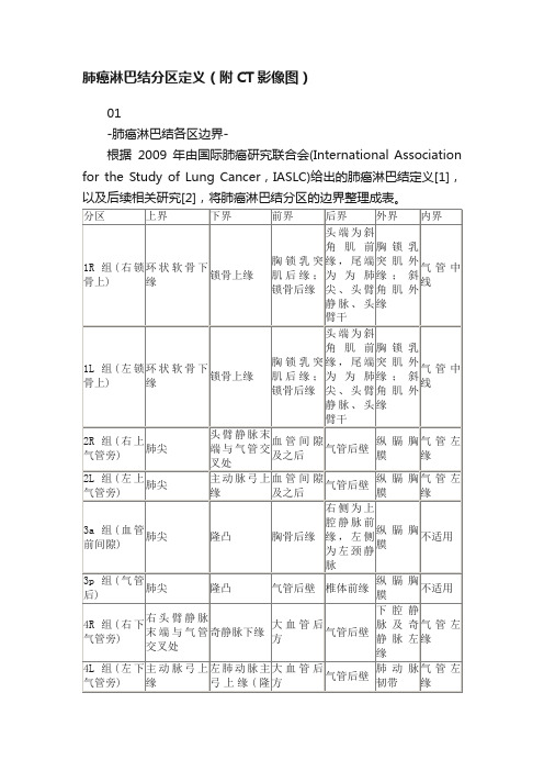
肺癌淋巴结分区定义(附CT影像图)01-肺癌淋巴结各区边界-根据2009年由国际肺癌研究联合会(International Association for the Study of Lung Cancer,IASLC)给出的肺癌淋巴结定义[1],以及后续相关研究[2],将肺癌淋巴结分区的边界整理成表。
来源于《胸部肿瘤放疗规范和靶区定义》2009 IASLC map-AJCC来源于Rusch VW,Asamura H,Watanabe H,et al. The IASLC Lung Cancer Staging Project:A Proposal for a New International Lymph Node Map in the Forthcoming Seventh Edition of the TNM Classification for Lung Cancer[J]. J Thorac Oncol,2009,4(5):568-577.FIGURE:The International Association for the Study of Lung Cancer (IASLC) lymph node map, including the proposed grouping of lymph node stations into “zones” for the purposes of prognostic analyses.FIGURE A–F: Illustrations of how the International Association for the Study of Lung Cancer (IASLC) lymph node map can be applied to clinical staging by computed tomography scan in axial (A–C), coronal (D), and sagittal (E, F) views. The border between the right and left paratracheal region is shown in A and B. Ao, aorta; AV, azygos vein; Br, bronchus; IA, innominate artery; IV, innominate vein; LA, ligamentum arteriosum; LIV, left innominate vein; LSA, left subclavian artery; PA, pulmonary artery; PV, pulmonary vein; RIV, right innominate vein; SVC, superior vena cava.02-肺癌淋巴结各区CT影像勾画-图1 环状软骨下缘水平(1组上界)图2 第2胸椎上缘水平(1组)此处第1肋骨连线的前方为1组。
肺癌淋巴结图谱

10区:肺门淋巴结,包括临近主支气管与肺门血管淋巴结。在右 侧自奇静脉下缘至叶间区域,左侧自肺动脉上缘至叶间区域。
特定区域淋巴结图解如下:
图2
1区,锁骨上淋巴结:包括下颈部、锁骨上、胸锁颈静脉切迹区域。
上界:环状软骨下缘,下界:锁骨与胸骨柄上缘。气管中线是1R 与1L的分界线。
图3
2R区. 右上气管旁淋巴结:2R淋巴结延伸至气管左侧旁。上界:
图16
10区.肺门淋巴结,肺门淋巴结临近肺叶淋巴结及纵隔胸膜反折,
在右侧临近中间段支气管。10-14区淋巴结不位于纵隔内,因此均 为N1期淋巴结。
下面再完整的了解一下淋巴结在CT图片上的准确位置:
纵隔镜可以检查到的淋巴结:
食管超声可以发现的淋巴结:
图5
3A区,血管前间隙淋巴结,右侧气管旁淋巴结,即4R淋巴结。
图6
4R区.右侧下部气管旁。上界:无名静脉足侧与气管交界区,下界:
奇静脉。4R淋巴结可以延伸至气管左侧。
图7
4R区,气管旁淋巴结,主动脉弓外侧淋巴结,即第六区淋巴结。
图8
4L区.左侧下部气管旁。4L淋巴结位于下部气管左侧缘,水平上界
为主动脉弓上缘,在左上叶支气管上缘延伸至左侧主支气管。包 括位于动脉韧带内侧气管旁淋巴结。5区(主肺动脉窗)淋巴结位 于动脉韧带侧面。
图9
肺动脉主干上方层面显示下部气管旁多发淋巴结,此外还有第三、
五区淋巴结。
图10
气管下部隆突上层面,气管左侧4L淋巴结,位于肺动脉主干与降
主动脉之间,由于是位于动脉韧带内侧,不算主肺动脉窗内淋巴 结,肺动脉干外侧的属于第五区淋巴结。
图13
8区.食管旁淋巴结 位于隆突下延伸至横膈。
肺癌淋巴结图谱

图15
• 9区.肺韧带淋巴结,肺韧带淋巴结位于肺韧 带内,包括下肺静脉后壁及下方淋巴结。 肺韧带是纵隔胸膜在肺门部反折向下延伸 所致。
图16
• 10区.肺门淋巴结,肺门淋巴结临近肺叶淋 巴结及纵隔胸膜反折,在右侧临近中间段 支气管。10-14区淋巴结不位于纵隔内,因 此均为N1期淋巴结。
下面再完整的了解一下淋巴结在CT图片上 的准确位置:
图9
• 肺动脉主干上方层面显示下部气管旁多发 淋巴结,此外还有第三、五区淋巴结。
图10
• 气管下部隆突上层面,气管左侧4L淋巴结, 位于肺动脉主干与降主动脉之间,由于是 位于动脉韧带内侧,不算主肺动脉窗内淋 巴结,肺动脉干外侧的属于第五区淋巴结。
图11
• 5区,主动脉弓下淋巴结,位于纵隔胸膜内, 主动脉弓下或主肺动脉窗淋巴结位于动脉 韧带或主动脉、左肺动脉外侧,并且接近 左肺动脉第一分支。 6区.主动脉旁淋巴结,位于升主动脉前方 或侧面,主动脉弓上下缘之间。
• 10-14区:肺门、肺叶及其主要分支淋巴结, 属于N1淋巴结。 10区:肺门淋巴结,包括临近主支气管与 肺门血管淋巴结。在右侧自奇静脉下缘至 叶间区域,左侧自肺动脉上缘至叶间区域。
特定区域淋巴结图解如下:
图2
• 1区,锁骨上淋巴结:包括下颈部、锁骨上、 胸锁颈静脉切迹区域。上界:环状软骨下 缘,下界:锁骨与胸骨柄上缘。气管中线 是1R与1L的分界线。
图3
• 2R区. 右上气管旁淋巴结:2R淋巴结延伸 至气管左侧旁。上界:胸骨柄上缘,下界: 无名静脉与气管交汇处。 2L区.左上气管旁。上界:胸骨柄上缘,下 界:主动脉弓上缘。如图2所示气管前第二 区淋巴结,血管前3A淋巴结。
图4
• 3区.血管前与椎前淋巴结,第三区淋巴结 不像第二区淋巴结那样靠近气管,它们位 于血管前或食管后椎体前。纵隔镜对第三 区淋巴结的发现无帮助,食管超声可以发 现3P淋巴结。
图谱:十分钟搞懂肺癌淋巴结分区
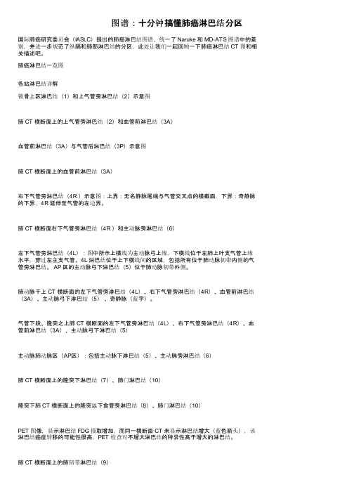
图谱:十分钟搞懂肺癌淋巴结分区国际肺癌研究委员会(IASLC)提出的肺癌淋巴结图谱,统一了 Naruke 和 MD-ATS 图谱中的差别,并进一步规范了纵膈和肺部淋巴结的分区,此处让我们一起回顾一下肺癌淋巴结 CT 图和相关描述吧。
肺癌淋巴结一览图各站淋巴结详解锁骨上区淋巴结(1)和上气管旁淋巴结(2)示意图肺 CT 横断面上的上气管旁淋巴结(2)和血管前淋巴结(3A)血管前淋巴结(3A)与气管后淋巴结(3P)示意图肺 CT 横断面上的血管前淋巴结(3A)右下气管旁淋巴结(4R )示意图:上界:无名静脉尾端与气管交叉点的横截面,下界:奇静脉的下界,4R 延伸至气管的左边界。
肺 CT 横断面右下气管旁淋巴结(4R )和主动脉旁淋巴结(6)左下气管旁淋巴结(4L):图中所示上横线为主动脉弓上缘,下横线位于左肺上叶支气管上缘水平,穿过左主支气管。
4L 淋巴结位于上下横线间的区域,包括所有位于肺动脉韧带内侧的气管旁淋巴结。
AP 区的主动脉弓下淋巴结(5)位于肺动脉韧带外侧。
肺动脉干上 CT 横断面的左下气管旁淋巴结(4L)、右下气管旁淋巴结(4R)、血管前淋巴结(3A)、主动脉弓下淋巴结(5)、奇静脉(蓝字)。
气管下段、隆突之上肺 CT 横断面的左下气管旁淋巴结(4L)、右下气管旁淋巴结(4R)、血管前淋巴结(3A)、主动脉弓下淋巴结(5)主动脉肺动脉区(AP区):包括主动脉下淋巴结(5)、主动脉旁淋巴结(6)肺 CT 横断面上的隆突下淋巴结(7)、肺门淋巴结(10)隆突下肺 CT 横断面上的隆突以下食管旁淋巴结(8)、肺门淋巴结(10)PET 图像,显示淋巴结 FDG 摄取增加,而同一横断面 CT 未显示淋巴结增大(蓝色箭头),该淋巴结癌症转移的可能性很高,PET 检查对不增大淋巴结的特异性高于增大的淋巴结。
肺 CT 横断面上的肺韧带淋巴结(9)肺 CT 横断面上的隆突下淋巴结(7)、肺门淋巴结(10)淋巴结活检:纵膈镜、超声内镜下细针抽吸活检术(EUS)传统纵膈镜:如下各组淋巴结可以通过颈部纵膈镜进行活检:上气管旁淋巴结(2L、2R)、下气管旁淋巴结(4L、4R)、隆突下淋巴结(7)。
肺癌淋巴结图谱

6区:主动脉旁淋巴结
位于升主动脉前方或侧面,主动脉弓 上下缘之间。 上界:主动脉弓上缘 下界:隆突下层面 前界:在左头臂静脉后缘和胸骨 后方; 后界:在主动脉弓和升主动脉前 1/2和肺动脉 干前缘; 双侧界:位于纵隔胸膜内
7区:隆突下淋巴结
位于气管隆突下,与肺内下叶支 气管、动脉无关。 上界:隆突下层面 下界:隆突下(约3cm) 右侧:向下延伸至中间段支气管 左侧:延伸至下叶上界。 前界:左右主支气管前壁水平线 或右肺动脉后缘; 后界:于椎体前缘;
1区:锁骨上淋巴结:
位于三角形的锁骨上窝内(后外缘: 锁骨内2/3,左外上:胸锁乳突肌下 缘,右外上:肩胛舌骨肌。) 包括下颈部、锁骨上、胸锁颈静脉 切迹区域。 上界:环状软骨下缘, 下界:锁骨与胸骨柄上缘。 气管中线是1R与1L的分界线。
2区:上气管旁淋巴结:
2R区右上气管旁淋巴结。 上界:胸骨柄上缘, 下界:无名静脉与气管交汇处。 2L区左上气管旁淋巴结。 上界:胸骨柄上缘, 下界:主动脉弓上缘。
肺癌淋巴结图谱
2009年(IASLC)国际肺癌研究协会
提出新的肺癌淋巴结分布图,目的是折NSRUKE
与美国胸科学会在淋巴结分区上的差异,并且改
良了每一区淋巴结的解剖界限的定义。
1区:锁骨上淋巴结 2区:上气管旁淋巴结 3A:血管前淋巴结 3P:椎前淋巴结 4区:下气管旁淋巴结 5区:主动脉下淋巴结 6区:主动脉旁淋巴结 7区:隆突下淋巴结 8区:隆突下食管旁淋巴结 9区:肺韧带淋巴结位于肺韧带区 N1淋巴结 10区:肺门淋巴结 11区:叶间淋巴结,12 区:叶淋巴结 13 区:段淋巴结,14区:亚段淋巴结
4R区.右侧下部气管旁淋巴结 上界:无名静脉足侧与气管交界区 下界:奇静脉。4R淋巴结可以延伸 至气管左侧。
肺癌纵膈肺门淋巴结分区及CT图像

5-6区:主动脉淋巴结
5区:主动脉下淋巴结。 这些淋巴结不是位于主动脉与肺动脉主干之间,而是位于 主肺动脉窗肺动脉韧带外侧。
6区:主动脉旁淋巴结。
位于升主动脉与主动脉弓前方与外侧。
7-9区:下纵隔淋巴结
7区:隆突下淋巴结。 8区:隆突以下食管旁淋巴结。
9区:肺韧带淋巴结位于肺韧带区。
• 10-14区:肺门、肺叶及其主要分支淋巴结。 • 10区:肺门淋巴结,包括临近主支气管与肺门血管淋巴结。 在右侧自奇静脉下缘至叶间区域,左侧自肺动脉上缘至叶 间区域。
淋巴结特定分区及图像
10区.肺门淋巴结
肺门淋巴结临近肺叶淋巴结及纵隔胸 膜反折。 右侧临近中间段支气管,自奇静脉下 缘至叶间区域。 左侧自肺动脉上缘至叶间区域。
淋巴结特定分区及图像
11区:叶支气管开口之间 12区:紧邻叶支气管淋巴结。 13区:段支气管周围淋巴结。 14区:紧邻亚段支气管淋巴结
淋巴结特定分区及图像
动脉。
纵膈血管CT图像
• 胸锁关节层面(平扫)
纵膈血管CT图像
• 胸锁关节层面(增强)
纵膈血管CT图像
• 胸骨柄层面
• 该层面相当主动脉弓上水平。气管前方较粗的血管断面为无名动脉 (头臂干,头臂动脉),气管左侧为左颈总动脉,其左后方为锁骨下 动脉。无名动脉与左颈总动脉的前外方分别为右侧及左侧头臂静脉。 (5条血管断面)
淋巴结特定分区及图像
8区:食管旁淋巴结
位于隆突下延伸至横膈。 淋巴结位于食道两侧, 邻近食道壁,不包括隆 突下淋巴结。
淋巴结特定分区及图像
8区:食管旁淋巴结
位于隆突下延伸至横膈。 淋巴结位于食道两侧, 邻近食道壁,不包括隆 突下淋巴结。
淋巴结特定分区及图像
肺癌胸腹部淋巴结分组及图片

The Radiology AssistantLung cancer - Lymph Node Map - Updateby Robin SmithuisRadiology department of the Rijnland Hospital in Leiderdorp, the NetherlandsIASLC lymph node map 2009Specific Lymph Node StationsAxial CT of Lymph NodesMediastinoscopy and EUSConventional mediastinoscopyExtended mediastinoscopyEUS-FNAThis is an update of the 2007 article, which used theMountain-Dresler regional lymph node classificationfor lung cancer staging (MD-ATS maps)(1).In 2009 a new Lung cancer lymph node map wasproposed by the International Association for theStudy of Lung Cancer (IASLC) in order to reconcile thedifferences between the Naruke and the MD-ATSmaps and refine the definitions of the anatomicboundaries of each of the lymph node stations (2).In this article we provide illustrations and CT-imagesfor a better understanding of this IASLC lymph nodemap.Publicationdate:8-6-2010IASLC lymph node map 2009Regional lymph node classification for lung cancer stagingadapted from the American Thoracic Society mappingschemeSupraclavicular nodes1. Low cervical, supraclavicular and sternal notchnodesFrom the lower margin of the cricoid to the claviclesand the upper border of the manubrium.The midline of the trachea serves as border between1R and 1L.Superior Mediastinal Nodes 2-42R. Upper Paratracheal2R nodes extend to the left lateral border of thetrachea.From upper border of manubrium to the intersectionof caudal margin of innominate (left brachiocephalic)vein with the trachea.2L. Upper ParatrachealFrom the upper border of manubrium to the superiorborder of aortic arch.2L nodes are located to the left of the left lateralborder of the trachea.3A. Pre-vascularThese nodes are not adjacent to the trachea like thenodes in station 2, but they are anterior to thevessels.3P. Pre-vertebralNodes not adjacent to the trachea like the nodes instation 2, but behind the esophagus, which isprevertebral.4R. Lower ParatrachealFrom the intersection of the caudal margin ofinnominate (left brachiocephalic) vein with the tracheato the lower border of the azygos vein.4R nodes extend from the right to the left lateralborder of the trachea.4L. Lower ParatrachealFrom the upper margin of the aortic arch to the upperrim of the left main pulmonary artery.Aortic Nodes 5-65. SubaorticThese nodes are located in the AP window lateral tothe ligamentum arteriosum.These nodes are not located between the aorta andthe pulmonary trunk but lateral to these vessels.6. Para-aorticThese are ascending aorta or phrenic nodes lyinganterior and lateral to the ascending aorta and theaortic arch.Inferior Mediastinal Nodes 7-97. Subcarinal8. ParaesophagealNodes below carina.9. Pulmonary LigamentNodes lying within the pulmonary ligaments.Hilar, Lobar and (sub)segmental Nodes 10-14These are all N1-nodes.10. Hilar nodesThese include nodes adjacent to the main stembronchus and hilar vessels.On the right they extend from the lower rim of theazygos vein to the interlobar region.On the left from the upper rim of the pulmonaryartery to the interlobar region.Specific Lymph Node Stations1. Supraclavicular zone nodes 1. Supraclavicular zone nodesThese include low cervical, supraclavicular and sternal notch nodes.Upper border: lower margin of cricoid.Lower border: clavicles and upper border of manubrium.The midline of the trachea serves as border between1R and 1L.2R. Right Upper Paratracheal2R nodes extend to the left lateral border of the trachea.Upper border: upper border of manubrium.Lower border: intersection of caudal margin of innominate (left brachiocephalic) vein with the trachea.2L. Left Upper ParatrachealUpper border: upper border of manubrium.Lower border: superior border of aortic arch.On the left a station 2 node in front of the trachea, i.e.a 2R-node.There is also a small prevascular node, i.e. a station3A node.3. Prevascular and Prevertabral nodesStation 3 nodes are not adjacent to the trachea like station 2 nodes.They are either:3A anterior to the vessels or3B behind the esophagus, which lies prevertebrally. Station 3 nodes are not accessible with mediastinoscopy.3P nodes can be accessible with endoscopicultrasound (EUS).3A and 3P nodesOn the left a 3A node in the prevascular space.Notice also lower paratracheal nodes on the right, i.e. 4R nodes.4R. Lower Paratracheal nodes4R. Right Lower ParatrachealUpper border : intersection of caudal margin of innominate (left brachiocephalic) vein with the trachea.Lower border :lower border of azygos vein.4R nodes extend to the left lateral border of thetrachea.On the left we see 4R paratracheal nodes.In addition there is an aortic node lateral to the aortic arch, i.e. station 6 node.4L. Lower paratracheal nodes4L. Left Lower Paratracheal4L nodes are lower paratracheal nodes that arelocated to the left of the left tracheal border , between a horizontal line drawn tangentially to the uppermargin of the aortic arch and a line extending across the left main bronchus at the level of the upper margin of the left upper lobe bronchus.These include paratracheal nodes that are located medially to the ligamentum arteriosum.Station 5 (AP-window) nodes are located laterally to the ligamentum arteriosum.On the left an image just above the level of the pulmonary trunk demonstrating lower paratracheal nodes on the left and on the right.In addition there are also station 3 and 5 nodes.On the left an image at the level of the lower trachea just above the carina.To the left of the trachea 4L nodes.Notice that these 4L nodes are between the pulmonary trunk and the aorta, but are not located in the AP-window, because they lie medially to the ligamentum arteriosum.The node lateral to the pulmonary trunk is a station 5 node.5. Subaortic nodesSubaortic or aorto-pulmonary window nodes are lateral to the ligamentum arteriosum or the aorta or left pulmonary artery and proximal to the first branch of the left pulmonary artery and lie within the mediastinal pleural envelope.6. Para-aortic nodesPara-aortic (ascending aorta or phrenic) nodes are located anteriorly and laterally to the ascending aorta and the aortic arch from the upper margin to the lower margin of the aortic arch.7. Subcarinal nodesThese nodes are located caudally to the carina of the trachea, but are not associated with the lower lobe bronchi or arteries within the lung.On the right they extend caudally to the lower border of the bronchus intermedius.On the left they extend caudally to the upper border of the lower lobe bronchus.On the left a station 7 subcarinal node to the right of the esophagus.8 Paraesophageal nodesThese nodes are below the carinal nodes and extend caudally to the diafragm.On the left an image below the carina.To the right of the esophagus a station 8 node..On the left a PET image demonstrating FDG uptake ina station 8 node.On the corresponding CT image the node is notenlarged (blue arrow).The probability that this is a lymph node metastasis isextremely high since the specificity of PET inunenlarged nodes is higher than in enlarged nodes.9. Pulmonary ligament nodesPulmonary ligament nodes are lying within thepulmonary ligament, including those in the posteriorwall and lower part of the inferior pulmonary vein.The pulmonary ligament is the inferior extension ofthe mediastinal pleural reflections that surround thehila.10 Hilar nodesHilar nodes are proximal lobar nodes, distal to themediastinal pleural reflection and nodes adjacent tothe intermediate bronchus on the right.Nodes in station 10 - 14 are all N1-nodes, since theyare not located in the mediastinum.Axial CT of Lymph NodesScroll through the images on the left.1.Sternal notch nodes are just seen at this level andabove this level2.Upper Paratracheal: below clavicles and on the rightabove the intersection of caudal margin ofinnominate (left brachiocephalic) vein with thetrachea and on the left above the aortic arch.3.Pre-vascular and Retrotracheal : anterior to thevessels (3A) or prevertebral (3P)4.Lower Paratracheal : below upper margin of aorticarch down to level of main bronchus5.Subaortic (A-P window): nodes lateral toligamentum arteriosum or lateral to aorta or leftpulmonary arteryView more images: 1/86.Para-aortic: nodes lying anterior and lateral to the ascending aorta and the aortic arch beneath the upper margin of the aortic arch 7.Subcarinal8.Paraesophageal (below carina)9.Pulmonary Ligament: nodes lying within the pulmonary ligament.10.-14: nodes are all N1 nodesMediastinoscopy and EUSConventional mediastinoscopyThe following nodal stations can be biopsied by cervical mediastinoscopy: the left and right upper paratracheal nodes (station 2L and 2R), left and right lower paratracheal nodes (station 4L and 4R) and the subcarinal nodes (station 7).Station 1 nodes are located above the suprasternal notch and are not routinely accessed by cervicalmediastinoscopy.Extended mediastinoscopyLeft upper lobe tumors may metastasize to the subaortic lymph nodes (station 5) and paraaortic nodes (station 6).These nodes can not be biopsied through routine cervical mediastinoscopy.Extended mediastinoscopy is an alternative for the anterior-second interspace mediastinotomy which is more commonly used for exploration of mediastinal nodal stations.This procedure is far less easy and therefore less routinely performed than conventional mediastinoscopy.EUS-FNAEndoscopic Ultrasound with Fine Needle Aspiration can be performed of all the mediastinal nodes that that can be assessed from the oesophagus.In addition the left adrenal gland and the left liver lobe can be visualized.EUS particularly provides access to nodes in the lower mediastinum (station 7,8 and 9)References1.Regional lymph node classification for lung cancer stagingby CF Mountain and CM DreslerChest, Vol 111, 1718-17232.The IASLC Lung Cancer Staging Project: A Proposal for a New International Lymph Node Map in the ForthcomingSeventh Edition of the TNM Classification for Lung Cancerby Valerie Rusch et alJournal of Thoracic Oncology: May 2009 - Volume 4 - Issue 5 - pp 568-5773.Conventional mediastinoscopyby Paul De Leyn and Toni Lerut.in the Multimedia Manual of Cardiothoracic Surgery4.Mediastinal Staging of Non Small-Cell Lung Cancerby Christian Lloyd, MD, and Gerard A.Silvestri, MD, FCCP Christian Lloyd, MD, and Gerard A.Silvestri, MD, FCCP Cancer Control, July/August 2001,Vol.8, No.4 Cancer Control 3115.State of the art lecture: EUS and EBUS in pulmonary medicineby J. T. Annema, and K. F. RabeEndoscopy 2006; 38: 118-1226.Imaging of the Patient with Non Small Cell Lung Cancer, What the Clinician Wants to Knowby Reginald F. Munden, MD, DMD, Stephen S. Swisher, MD, Craig W. Stevens, MD, PhD and David J. Stewart, MDRadiology 2005; 237:803-818。
- 1、下载文档前请自行甄别文档内容的完整性,平台不提供额外的编辑、内容补充、找答案等附加服务。
- 2、"仅部分预览"的文档,不可在线预览部分如存在完整性等问题,可反馈申请退款(可完整预览的文档不适用该条件!)。
- 3、如文档侵犯您的权益,请联系客服反馈,我们会尽快为您处理(人工客服工作时间:9:00-18:30)。
(Lung cancer-lymph node map-update)
齐新刚
• 2009年,International Association for the Study of Lung Cancer(IASLC)国际肺癌研 究协会提出新的肺癌淋巴结分布图,目的 是折中NSRUKE与美国胸科学会在淋巴结分 区上的差异,并且改良了每一区淋巴结的 解剖界限的定义。本章节,为了更好理解 国际肺癌研究协会淋巴结分布图,我们提 供注释与CT图像。
图3
• 2R区. 右上气管旁淋巴结:2R淋巴结延伸至 气管左侧旁。上界:胸骨柄上缘,下界: 无名静脉与气管交汇处。 2L区.左上气管旁。上界:胸骨柄上缘,下 界:主动脉弓上缘。如图2所示气管前第二 区淋巴结,血管前3A淋巴结。
图4
• 3区.血管前与椎前淋巴结,第三区淋巴结不 像第二区淋巴结那样靠近气管,它们位于 血管前或食管后椎体前。纵隔镜对第三区 淋巴结的发现无帮助,食管超声可以发现 3P淋巴结。
图12
• 7区.隆突下淋巴结,位于气管隆突下,与肺 内下叶支气管、动脉无关。在右侧向下延 伸至中间段支气管,左侧延伸至下叶上界。 图示第七区淋巴结位于食管右侧。
图13
• 8区.食管旁淋巴结 位于隆突下延伸至横膈。
图14
• PET显示第八区淋巴结FDG摄取,对应的CT 图像淋巴结未见增大(蓝箭),由于PET对 非增大淋巴结转移的摄取特异性高于增大 淋巴结。
• 纵隔镜可以检查到的淋巴结:
• 食管超声可以发现的淋巴结:
图15
• 9区.肺韧带淋巴结,肺韧带淋巴结位于肺韧 带内,包括下肺静脉后壁及下方淋巴结。 肺韧带是纵隔胸膜在肺门部反折向下延伸 所致。
图16
• 10区.肺门淋巴结,肺门淋巴结临近肺叶淋 巴结及纵隔胸膜反折,在右侧临近中间段 支气管。10-14区淋巴结不位于纵隔内,因 此均为N1期淋巴结。
下面再完整的了解一下淋巴结在CT图片上的 准确位置:
肺癌淋巴结图谱
1区:锁骨上淋巴结。
下颈部、锁骨上与胸骨颈静脉切迹淋巴结, 自环状软骨下缘至锁骨、胸骨柄上缘。气 管中线是1L与1R的分界线。
2-4区:上纵隔淋巴结。
2R.上气管旁2R淋巴结向气管左外侧缘延伸。自胸 骨柄上界至无名静脉足侧与气管交汇处。 2L.上气管旁,胸骨柄上缘至主动脉弓上缘。2L淋巴 结位于气管左侧缘的左侧。 3A.血管前,这些淋巴结同2区淋巴结一样不靠近气 管,位于血管前方。 3P.椎前淋巴结,位于食管之后椎体之前。 4R.下气管旁,自无名静脉与气管交界区至奇静脉下 界。4R淋巴结自右侧至气管左侧缘。 4L.下气管旁,自及其主要分支淋巴结, 属于N1淋巴结。 10区:肺门淋巴结,包括临近主支气管与 肺门血管淋巴结。在右侧自奇静脉下缘至 叶间区域,左侧自肺动脉上缘至叶间区域。
特定区域淋巴结图解如下:
图2
• 1区,锁骨上淋巴结:包括下颈部、锁骨上、 胸锁颈静脉切迹区域。上界:环状软骨下 缘,下界:锁骨与胸骨柄上缘。气管中线 是1R与1L的分界线。
图5
• 3A区,血管前间隙淋巴结,右侧气管旁淋 巴结,即4R淋巴结。
图6
• 4R区.右侧下部气管旁。上界:无名静脉足 侧与气管交界区,下界:奇静脉。4R淋巴 结可以延伸至气管左侧。
图7
• 4R区,气管旁淋巴结,主动脉弓外侧淋巴 结,即第六区淋巴结。
图8
• 4L区.左侧下部气管旁。4L淋巴结位于下部 气管左侧缘,水平上界为主动脉弓上缘, 在左上叶支气管上缘延伸至左侧主支气管。 包括位于动脉韧带内侧气管旁淋巴结。5区 (主肺动脉窗)淋巴结位于动脉韧带侧面。
5-6区:主动脉淋巴结。
5区:主动脉下淋巴结。 这些淋巴结不是位于主动脉与肺动脉主干 之间,而是位于主肺动脉窗肺动脉韧带外 侧。 6区:主动脉旁淋巴结。 位于升主动脉与主动脉弓前方与外侧。
7-9区:下纵隔淋巴结。
7区:隆突下淋巴结。 8区:隆突以下食管旁淋巴结。 9区:肺韧带淋巴结位于肺韧带区。
图9
• 肺动脉主干上方层面显示下部气管旁多发 淋巴结,此外还有第三、五区淋巴结。
图10
• 气管下部隆突上层面,气管左侧4L淋巴结, 位于肺动脉主干与降主动脉之间,由于是 位于动脉韧带内侧,不算主肺动脉窗内淋 巴结,肺动脉干外侧的属于第五区淋巴结。
图11
• 5区,主动脉弓下淋巴结,位于纵隔胸膜内, 主动脉弓下或主肺动脉窗淋巴结位于动脉 韧带或主动脉、左肺动脉外侧,并且接近 左肺动脉第一分支。 6区.主动脉旁淋巴结,位于升主动脉前方或 侧面,主动脉弓上下缘之间。