S4800扫描电镜(SEM)操作手册课件
S4800扫描电镜(SEM)操作手册

观测条件的选择
2、工作距离(WD)的设定 WD(working distance)是指物镜下端面到 焦点面之间的距离。 WD越小,图像的分辨率越高,而景深越浅。 做能谱时,WD必须设定为15mm。
观测条件的选择
3、探测器的选择 S-4800配备有上下两个二次电子探测器, 在WD较小时(<5),推荐使用上探测器, 在WD较大时(>10),推荐使用下探测器。 如果选择MIX,则同时使用两个探测器, 在整个WD的可变范围内,信号量不会发 生极端的变化。
2、加载样品
3)将样品架插入样品室
插入样品之前需要确认: (a)样品台的位置处于交换位置,并且没有处于锁定状态(Lock开关的灯 不亮) (b)加速电压处于OFF状态
(1)按交换室操作部分的AIR键,使交换室放气; (2)峰鸣器响后(对应键的灯不闪),将交换室打 开; (3)轻轻推入交换棒,此时保证交换棒后端的旋钮 处于UNLOCK状态; (4)用一只手拿着交换棒的旋钮,另一只手将样品 插入交换棒中;
观测条件的选择
1、加速电压的选择 从电子光学的角度讲,随着加速电压的升 高,电镜的分辨率也提高,但是电压越高,电 子束在样品中扩散范围就越大,同时放电和辐 照损伤现象也越严重。因此要根据样品的种类 及所要得到的信息,适当地选择加速电压。对 于导电性好的样品,可以选择较高的加速电压; 相反,对于导电性较差的样品,则应选择较低 的加速电压。
样品台旋转Rotation——R (360°连续)
样品台
样品台以通过其表面中心的法线为轴做平面内 旋转,是靠马达驱动的(自动)。
样品台旋转Rotation——R
Eucentric:当选中时,表示样品台旋转后, 观察区域仍然保持在当前位置。 Abs:绝对角度 正值0~360° Rel:相对角度(始终以当前位置为0) 可以输入正值0~360 °(顺时针) 负值0~-360 °(逆时针)
SEM_hitachi s4800

Loading Procedure for Hitachi 48001)login the computer with your active directory passwd, SEM program will startautomatically, just leave the blank when prompt for S4800 password2)Turn on IR lamp and cameraSample loading1)Mount your sample to sample holder and check height (make sure it is lower2)Check the computer monitor to make sure the stage is at EXC (exchangeposition), use mouse to select EXC to move the stage to exchange position,and make a note in the log sheet.3)Press AIR button to vent the load-lock4)After AIR button stop flashing (indicating vent complete), open the load-lockdoor (Do Not use the loading stick to open the door to avoid bending)5) Push the loading stick out a little bit and mount your sample holder to theloading stick. (Do Not touch the loading stick to avoid removing the grease on it)6) Pull back the loading stick all the way out to avoid it get suck in when start pumping later.7) Close the load-lock door (Do Not use the loading stick to close the door to avoid bending)8) Press EVAC to pump the load-lock9) After EVAC button stop flashing (indicating loadlock pressure is lowenough), press OPEN to open the gate valve10) After OPEN (take a few second, be patient ) button stop flashing (indicating gate valve is fully open) push the loading stick in. After the sample holder engaged to the stage , unlock the loading stick and pull back the loading stick all the way out .11) Press CLOSE to close the gate valve while keeping another hand to hold the loading stick to avoid it from sucking in.Sample imaging2. click “HOME” position to move the sample to the center1 inch3.4.When asked to flash, please use intensity 1, HV must be off to flash the tip.5.Turn on the HV by clicking “ON”6.Click “H/L” to go to LM (low magnification) to find the area you need to look at7.Go back to High Mag after locating the area you need to look at8.Adjust “focus”, “astigmatism”, “brightness/contrast”, “Mag”1Magnification Astigmatism FocusBrightness/ContrastBeam shift9. can be used for automatic brightness contrast control.10.choose “Slow3” icon for slow scan image capture11.capture.12.PCI software can be use for image analysis.Sample unloading1)Turn off High voltage from the computer2)Move the stage back to EXC (exchange position) by using mouse to selectEXC3)press OPEN to open the gate valve4)After OPEN button stop flashing (indicating gate valve is fully open) push theloading stick in. After the sample holder engaged to the loading stick, lock theloading stick to sample holder and pull back the loading stick all the way out.5)Press CLOSE to close the gate valve.6)After CLOSE button stop flashing (indicating gate valve is fully close), pressAIR button to vent the load-lock.7)After AIR button stop flashing (indicating vent complete), open the load-lockdoor (Do Not use the loading stick to open the door to avoid bending)8)Remove the sample holder from the loading stick9)Close the load-lock door (Do Not use the loading stick to close the door toavoid bending)10)Press EVAC to pump the load-lockAccess SEM image over network1.Right click “My computer”2.Select “Map Network Drive”3.Choose any unused Drive letter4.Enter \\\image for folder5.when enter your login name, you may need to put uofi\ before your netid6.uncheck Reconnect at logon7.Click “Finish”。
SEM原理及操作课件--

用真空主要原因:
电子束系统中的灯丝在普通大气中会迅速氧化而失 效,所以在使用SEM时需要用真空,或以纯氮气或 惰性气体充满整个真空柱。为了增大电子的平均自 由程,从而使得用于成像的电子更多。
b.电子束系统
电子束系统由电子枪和电磁透镜两部分组成,主 要用于产生一束能量分布极窄的、电子能量确定的电 子束用以扫描成像。
样品放好后,在软件界面上选择合适的高压和发射电流, 然后点击”ON”加高压,如图所示。
在软件面板中的Stage 部分,选择合适的样 品台大小,如图所示
在加高压时会弹出样品台大小、高度确认对话 框,如图5所示。高度前面已确认是8mm标准高 度,如果样品台大小(平面尺寸)和对话框中数 值不相符,则要点击cancel。
3.样品的制备及装入
样品的制备
样品制备简单,对样品要求较低,只要能放进样品室,都可进行 观察,高度保持 在 2cm 以内,直径在 4cm 以内。但对样 品的性质有特殊要求 a.化学上和物理上稳定的干燥固体,表面清洁,在真空中及 在 电子束轰击下不挥发或变形,无放射性和腐蚀性。 b.样品必须导电,非导电样品,可在表面喷镀金膜。 c.磁性样品须退磁,工作距离(WD)要大于8.0mm。
二次电子是指被入射电子轰击出来的核外电子。 携 带有样品的表面形貌信息,通常来自样品表面550 nm的 区域,能量为0-50 eV。 由于它发自试样表面层,入射电子还没有较多次 散 射,因此产生二次电子的面积与入射电子的照 射面积没多大区别。所以二次电子的分辨率较高, 一般可达到50-100 Å。 扫描电子显微镜的分辨率通常就是二次电子分辨 率。二次电子产额随原于序数的变化不明显,它 主要取 决于表面形貌。
背散射电子BSE
背散射电子是指被固体样品中的原子核反弹回来的 一 部分入射电子。包括弹性背散射电子和非弹性背 散射电子。从数量上看,弹性背散射电子远比非弹 性背散射电子所占 的份额多。背散射电子能量在数 千到数万电子伏。 背散射电子的产生范围在1000 Å到1 μm深,由于背 散射电子的产额随原子序数的增加而增加,所以, 利用背散射电子作为成像信号不仅能分析形貌特征, 也可用来显示原子序数衬度,定性地进行成分分析。
S4800扫描电镜(SEM)操作手册

• 如您的样品带有磁性或操作过程中遇到意外情况, 请垂询技术员彭开武/郭延军
( Tel: 82545516 )。
启动操作程序PC-SEM 加载样品
加高压及条件设定 调节电子光学系统
观察样品 记录图像 图像处理 SEM数据管理器 取出样品 刻录光盘 结束操作
采用慢扫描的方式能够提高图像的信噪比,从而提高图像的 质量,因此通常采用慢扫描方式记录图像。但是扫描速度越慢, 样品的放电现象越显著。所以,如果样品放电现象比较严重, 不能采用慢扫描方式记录图像,这时可以采用多次快速扫描叠 加的方式记录图像,同样可以提高信噪比。但是对于漂移明显 的样品无法采用这种技术。对于既放电又漂移的样品,采用单 次快速扫描的方式反而可以获得最高质量的图像。
倾转注意:
a)千万不要超过允许的倾转范围,为了安全起见, 不要倾转到极限,应留有一定的余量(至少1 °)
b)如果进行了倾转操作,当结束操作后使样品台回 到交换位置时,一定要先把T调回到0,然后再调Z 到8mm。
We are at your service!
可编辑
3)将样品架插入样品室
插入样品之前需要确认:
(a)样品台的位置处于交换位置,并且没有处于锁定状态(Lock开关的灯不 亮)
(b)加速电压处于OFF状态
(1)按交换室操作部分的AIR键,使交换室放气;
(2)峰鸣器响后(对应键的灯不闪),将交换室打 开;
(3)轻轻推入交换棒,此时保证交换棒后端的旋钮 处于UNLOCK状态;
样品台
样品台以通过其表面中心的法线为轴做平面内 旋转,是靠马达驱动的(自动)。
Eucentric:当选中时,表示样品台旋转后,观察区域仍然保 持在当前位置。
S-4800扫描电镜操作步骤
扫描电镜S-4800操作规程一、日常开机打开Display开关,电脑自动开机进入s-4800用户界面,PC_SEM程序自运行,点击确认进入软件界面。
二、装样品1.将样品台装在样品座上,根据标尺调整高度及确认样品位置后旋紧。
2.按下AIR键,当AIR灯变绿时拉开样品交换室,水平向前推出交换杆,把样品座插在交换杆上,逆时针旋转交换杆(即按照杆上的标示转至LOCK)锁定样品座后,将交换杆水平向后拉回原处。
3.关闭交换室,按下EV AC键,当EV AC绿灯亮时,按OPEN键至绿灯亮样品室阀门自动打开。
4.水平插入交换杆,直至样品座被卡紧为止,顺时针旋转交换杆(即按照杆上的标示转至UNLOCK)后水平向后拉回原处,点CLOSE键至绿灯亮样品室阀门自动关闭。
三、图像观察1.加高压点击屏幕左上方的高压控制窗口,弹出HV Control对话窗。
选择合适的观察电压和电流,点击ON,弹出提示样品高度的对话框,点击确定出现HV ON 提示条,待图像出现后,关闭HV Control对话窗。
2.在低倍、TV模式下,找到所要观察的样品,点击H/L按钮切换到高倍模式,通过调节样品位置,找到所要观察的视场。
3.聚焦、消像散选好视场后,放大到合适的倍数聚焦消像散。
先调节聚焦粗调和细调旋钮,使图像达到最佳状态,若图像有拉长现象,则需进行消像散。
调节STIGMATOR/ALIGNMENT X使图像在水平方向的拉长消失,再调节STIGMATOR/ALIGNMENT Y使图像在垂直方向的拉长消失。
4.图像采集及保存用A.B.C.键或BRIGHTNISS/CONTRAST旋钮自动或手动调节图像的对比度和亮度,扫描速度变为慢扫,点击抓拍按钮进行采集。
采集后暂时存放在窗口下侧,选中要保存的图像,点击Save,弹出Image Save对话框,输入文件名,选好存储位置保存即可。
5.对中调整改变加速电压和电流时,或图像在高倍聚焦发生漂移时,需要进行对中调整,方法如下:(1)选取样品上一个具有明显特征的位置放在视场中心。
S4800扫描电镜操作说明书
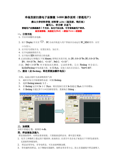
冷场发射扫描电子显微镜S4800操作说明(普通用户)燕山大学材料学院材料管A104(场发射,钨灯丝)编写人:李月晴吕益飞普通用户在熟练操作1个月后,如无不良记录,可申请高级用户培训。
高倍调清晰:局部放大(Red) →聚焦Focus→消像散一、日常开机1,开启冷却循环水电源。
2,按下Display开关至,PC自动开机进入用户界面并自动运行PC_SEM程序,以空口令登入。
3,打开信号采集开关,位置打到1,为打开。
4,打开电源插排的开关。
5,打开装有EDS软件的主机电源。
6,记录仪器运行参数(右下角Mainte),即钨灯丝真空度。
如:IP1:0.0×10⁻⁸Pa;IP2:0.0×10⁻⁸Pa;IP3:9.6×10⁻⁷Pa。
PeG-1,<1×10⁻³;PeG-2,<1×10+2。
注意:PeG≤1×10⁻³Pa时才能加高压测量。
记录的参数:①点Flashing时会显示:In2(Ie)Flashing时电流最大值,如32.9μA;②加上高压后会显示,V ext=3.4kV。
二、轰击(点flashing,即在阴极加额外电压)目的:高温去除针尖表面吸附的气体1,最好在每天开始观察样品前一时做flashing;2,选择flashing intensity为2 ;3,若flashing运行时Ie小于20µA,则反复执行直至Ie值超过20µA且不再增加。
4,若flashing后超过8个小时仍继续使用,重新执行flshing 。
三、加液氮容积不要超过1L,能维持4~6h。
四、样品制备及装入样品制备简单,对样品要求较低,只要能放进样品室,都可进行观察。
1,化学上和物理上稳定的干燥固体,表面清洁,在真空中及在电子束轰击下不挥发或变形,无放射性和腐蚀性。
2,样品必须导电,非导电样品,可在表面喷镀金膜。
3,带有磁性的样品,由于物镜有强磁性,制样必须非常小心,防止在强磁场中样品被吸入物镜或分散在样品室中,工作距离(WD) 要大于8.0mm。
S-4800日立扫描电子显微镜(SEM)简易操作指南(通用版)
S-4800日立扫描电子显微镜(SEM)简易操作指南一、开机前准备1.1制备样品(带口罩与手套进行)SEM样品制备相对简单,原则上只要能放入样品室的样品,都可进行观察。
但需注意以下事项:a)样品在物理上和化学上必须要保持稳定,在真空中和电子束轰击下不挥发或变形,没有腐蚀性和放射性。
(通常是干燥固体。
)b)由于光源是电子,样品必须导电,非导电样品可喷镀金膜。
金膜在一定程度上会影响样品原有形貌。
(若样品本身导电,衬底不导电,如蓝宝石上的ZnO,只需用导电胶把样品表面连到样品台。
)c)由于物镜有强磁性,带有磁性的样品制样必须非常小心,防止在强磁场中样品被吸入物镜或分散在样品室中。
通常磁性样品必须退磁,且工作距离(WD)须大于8mm。
具体操作过程:(1) 按待测样品数量选择样品台,(支持直径d=5mm,15mm,1 inch,2 inch等规格,若要观测截面可选择带角度的样品台)。
(2) 剪一小段导电胶,粘到样品台上。
若样品为粉末,则把粉末撒到导电胶上,用吸耳球或高压氮气吹扫掉导电胶上未粘紧的粉末;若是块状样品,则把样品牢牢粘到导电胶上,用手轻轻推,样品不会左右晃动。
(为观测时方便定位,将样品排列成行(或列),并在行下方(或列左侧)标上数字编号。
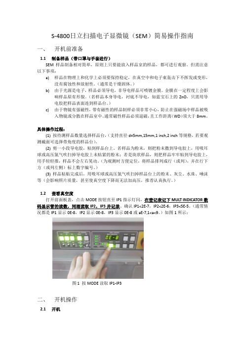
)(3) 样品粘贴完成后,用吸耳球或高压氮气吹扫掉样品台上的粉末、灰尘、水珠、唾沫等(会影响照片质量,甚至使真空度下降而无法加高压,推荐认真执行。
)1.2查看真空度打开前面板盖,点击MODE按钮直至IP1指示灯闪,在登记表记下MULT INDICATOR数码显示管的读数,同理读取IP2,IP3并记录。
确认IP1<2E-7,IP2<2E-6,IP3<5E-5。
(通常情况都是IP1显示0E-8,IP2显示0E-8,IP3显示0E-8或aE-7,1<a<9。
)如图1所示:图1 按MODE读取IP1-IP3二、开机操作2.1开机a)开启冷却循环水电源,循环水温度显示应在15-20℃,水位应浸没金属线圈。
高分辨扫描电镜 日立S-4800操作步骤

P r o c e d u r e f o r FIRST USER ofFIRST USER of day1.Turn on CPU power (inside lower cabinet)2.Turn on Computer Monitor3.Login to Windows (no password)4.Login to S-4800 software by clicking icon PC-SEM to open control panel (nopassword)5.Click box (Vacc, Ie) upper left of Toolbar (if dialog box w/FLASHING button isnot open)6.Click Flashing.7.“Flashing Execute ok?” Click Execute (Intensity 2 is normally ok).8.Repeat steps 5 & 6; Ie will transiently register current – should be between 20 and40 µA.9.Check Evacuation Control Panel for ion pump readings (IP1, 2, 3). Values are in10-3 Pa (For Lab Manager Only)ALL USERSU s e r S e t-u p f o r ALL1.For sample exchange, be sure that the sample stage settings – Rotation (R) = 0.0,Tilt (T) = 0.0, X = 25.0, Y = 25.0 and Z = 8.0 are all set to these positions asindicated on the front of the microscope.2.Click AIR button on right side of microscope – after a few seconds, air isadmitted to the Sample Exchange Chamber (SEC). There is a beep when the SEC is at atmospheric pressure.3.Pull open and turn black knob CW to UNLOCK position and remove sampleholder.4.While wearing gloves and at workbench, not on stage, install new sample onsample holder and adjust the height using the fixture console. See pictures taped to wall for details. This will give a working distance of 8 mm when the sample is inserted into the sample stage.5.Insert sample holder aligning to dual bayonet connectors in the SEC; rotate blackknob CCW to LOCK position. Then pull SEC and rod out to guide endpoint.(Check that rod is pulled out to endpoint). CLOSE SEC door.6.Press EVAC button – air is pumped out of the SEC. A beep sounds when vacuumis attained.7.Press OPEN to open the chamber door – a beep sounds when the chamber door isopen. Visually examine to check that stage is in chamber.8.Push rod all the way in until the sample holder fully engages the sample stage;rotate black knob CW to UNLOCK position.9.Grasp black knob and pull back to end of guides. Then make sure that the sampleholder remains on the stage.10.Press CLOSE to close the chamber door. Beep sounds when the door is closedand the vacuum is ready.11.To turn HV ON. Click ON (left of gray box on Toolbar) – for normal operation,V acc=15 kV and I e=10 µA. I e will drop during the first ~30 minutes of usage due to formation of a monolayer of absorbed gas on the tungsten emitter tip. To set Accl Voltage (VAcc) click large black arrow and set KV at .5 to 30. The lower the KV the more surface detail imaged and greater the KV will increase imaging the interior structure/views. Periodically click SET (right side of Toolbar) to re-set the emission to ~10 µA (this will increase V ext).12.Scan rate is selectable on the toolbar – TV1, Slow1 and Slow3.13.H/L button on toolbar selects low magnification (30 X – 2 KX) or highmagnification (800X – 500KX) modes. The magnification can be adjusted with the knob on the control box.14.ABC button on toolbar is Auto Brightness / Contrast. These can also be adjustedusing the Brightness and Contrast knobs on the control box.15.At the beginning of a session, it is important to check the alignment of theelectron beam. Click H/L and set magnification at ~5000X. While increasing magnification Beep will occur at 2000X. The beep is a reminder to click on H/L to reset mag to high. Click ALIGN on the toolbar. A menu box will appear.Click on BEAM ALIGN and center the beam using X and Y knobs(stigma/alignment) on the control box. Click STIGIMATION X, and stopmovement of image using X and Y knobs on control box. Click STIGIMATION Y and stop image movement using X and Y knobs. Click APERTURE ALIGN and stop movement of image using X and Y knobs on control box.Click OFF in ALIGN menu box.16.Adjust coarse focus until WD is ~ 5mm. It may be required to click on “resetfocus condition” in popup dialogue box.17.To obtain the best image resolution, carefully adjust coarse, fine focus, x-stigmation and y-stigmation KNOBS at a high magnification. Adjust brightness and contrast, and observe image in Slow2 mode (button on toolbar). N.B., set Working Distance (WD) only in Hi Mag mode to ~ 5mm by using the Z adjust knob18.Set magnification at ~ 1000X. Then lower Z by turning Z knob on scope.19.Find field of interest by moving X/Y stage. Set to required magnification.20. Before Image captur, be sure to set slow scan mode for best image quality.21.To capture an image, click the 1280 button on the toolbar to the right of the H/Lbutton. This will capture a 1280 x 960 image. The resolution can be changed using the pull-down arrow.22.When the image is captured, it is displayed in the lower left, highlighted inyellow.23.Click the PCI button in the lower left to export the image to QuartzPCI imageanalysis software.24.In QuartzPCI, click File – can SAVE or EXPORT image (e.g. to a JPEG file).Clicking SAVE will actually create 3 files of which one is PCI format. EXPORT, which creates 1 file, is more efficient choice.25.Back in S-4800 software, click RUN on toolbar to resume scanning.S h u t D o w n P r o c e d u r e1.Turn off beam2.Return stage to proper settings.3.Press OPEN4.Insert rod at unlocked position5.Turn rod to locked position6.Pull rod and sample out to endpoint.7.Press CLOSE8.Press AIR, wait for beep9.Slide open SEC10.Turn black knob to UNLOCK and remove sample11.If done, close SEC and press evacuate to pump down chamber12.Turn off monitorCommon Abbreviations and DefinitionsVext (Extracting voltage) Voltage that is applied between the cathode and first anode.Electrons are emitted from the cathode.Vacc (Accelerating voltage) electrons are accelerated by an accelerating voltage.Flashing- cleansing of the cathode (FE tip) by turning on the flashing power supply in order to remove absorbed gas on the surface of the cathode. This is to be done,before usage, first thing in the morning or the evening.Ie (emission current) is generally set to 10 μA for normal operation.HV (high voltage) Applies HV to the electron gun and controls the extracting voltage to obtain the emission current.Beam monitor (adjustment of the reference voltage) is provided to reduce tip noise, which is a low frequency noise caused by fluctuations of the emission current.Dividing the image signal by a reference signal that is proportional to probe current can stabilize it. It is recommended to keep the beam monitor on for normaloperations.Beam indicator (box with cross hairs) to reset image shift to the center, click theindicator areaUnder signal selection box, L.A is low angle. When BSE is selected, the amount of BSE signal is controlled by BSE ratio selection box. Low angle BSE will be detected with L.A0 to L.A100. With the larger number, the amount of SE is suppressed and results in a BSE richer image.HR is high resolution mode where the short working distance range is limited and it is easier to use at a longer working distance (>5mm).UHR is ultrahigh resolution mode where the full working distance range is availableUnder scan size…Small screen mode has faster frame speeds and in some cases results in better image quality.Dynamic focus allows you to focus the beam for the entire field of view.Condenser lens 1 setting to lower values (1-16 range) results in weaker excitation and larger Probe Current and larger “spot size” and vice versa. Recommended value is 5. Unclicking box will turn off lens and this condition is typically used for mechanical alignments.Focus depth is recommended to be set at 1.0 or larger to increase focus depth.Degauss button should be clicked after changing focus widely or before making the electron optical axis alignment (You can do this by hitting the F2 hotkey)To select many images, press and hold down the control keyLayout button opens the Report Generation window for printing the imageSC stands for the specimen chamberApt heat should be set at auto. If the objective aperture lens is contaminated, charging will degrade image quality and the image will drift because of micro discharge. Such problems are noticeable at low accelerating voltages. The aperture is heated to about 150C to remove contaminants to one tenth or less of what it would be at room temperature.The display switch is on when the line is depressed (a 1 or switch is up is on)For use at high magnifications or low accelerating voltages, the use of the anti-contamination trap is recommended to prevent specimen contamination by hydrocarbon build-up. Fill the trap with liquid nitrogen (LN2). The Dewar is usable for about 5 hours at room temperature. ** Before introducing air into the chamber, wait for a few hours until the LN2 Dewar has completely emptied so that the trap will not frost up and deteriorate the vacuum. The air introduction value does not have a protection link with the cold trap.WD (working distance) is the distance between the bottom face of the objective lens and the surface of the specimen. At a shorter working distance, higher resolution is obtainable. At a longer WD, a larger tilt angle and a greater depth of focus are obtainable. To change the WD, move the Z to a lower #, which moves the stage up. Short-Cut KeysCtrl O Open SEM Data ManagerCtrl P PrintCtrl C Copy ImageCtrl L Open Captured Image windowF1 Help can be openedF2 Activates Degauss functionF5 Runs or stops scanning alternatelyF4 Changes alignment mode to the next stepShift F4 Changes alignment mode to previous stepNotes for AdministratorPassword is “hitachi”To preset magnifications, open the Image Tab of the Setup dialog window and input desired values in the three Preset Magnification boxes. A PM mark is indicated in the Magnification indicator area when the preset magnification is set.Setting up logins: Option menu, login setting (must be logged in as S-4800) to create or change login names and passwords for each user. Can also change password setting under Option menu.TroubleshootingWhen operating at short working distances and experiencing uneven brightness at low magnifications, turn specimen bias voltage off.Magnetic samples such as iron can cause astigmatism correction to be difficult.The use of double-sided adhesive tape may cause specimen drift. Use the least amount possible to minimize out gassing.Perform beam and aperture alignment when you change HV value, accelerating voltage, probe current mode, or setting of Cond Lens1. For all alignments, either drag the mouse in the grid area of the Alignment dialog window or adjust theSigma/alignment X and Y knobs on the control panel. Set the magnification at 5, 000X for aperture alignment.The lowest magnification that can be obtained at a WD of 25mm or more is 20X.To maximize brightness and contrast, start B/C monitor mode by double clicking the MontiF button or operate menu, BC monitor. When the maximum and minimum values of the waveform are adjusted to fit within the upper and lower reference lines, appropriate brightness and contrast will be obtained. To terminate this mode, click the cancel button in the BC monitor mode message or one of the scanning speed buttons.To maximize focus, set magnification to 1,000X and start the focus monitor by clicking the Monitor button (MontiF) on the control panel and focus the image so that the waveform shows sharp peaks. To close the focus monitor, click the cancel button in the focus monitor mode or click one of the scanning speed buttons.Auto focus or auto stigma functions will not work correctly with little or no surface detail on the specimen or when the specimen is charging. These functions should also be performed at magnifications higher than 5,000X.For high magnification work, staging locking is recommended for better mechanical stability. The Z and T axes are locked or released by the Lock button on the specimen stage. ** If the Z or T axis is moved while the stage is locked, the stage mechanism may be damaged. X, Y, and R axes are free and movable when the stage is locked.Objective lens apertures are 50 μm for settings 2 or 3. The electron optical column of the HRSEM is designed to achieve highest resolution with a 50 μm aperture. Suggestions on getting better image quality:1.Higher spatial resolution can be obtained at higher accelerating voltages2.For uncoated, insulator specimens, accelerating voltages less than 1kV arerecommended for minimizing charging. In some cases, high accelerating voltages (20kV or higher) may produce a better image.3.Influence of contamination at lower voltages is more pronounced4.Disturbances by leakage magnetic field (wobbling or distortion of the image) aregreater at low accelerating voltages.5.Generally a soft-tone image is obtained at low accelerating voltages because moreSEs are detected than BSEs.6.Ion Pump readings should be this or better:IP1: 2 x 10^-7 PaIP2: 2 x 10^-6IP3: 5 x 10^-5SC: 2 x 10^-3Stereo ImagingPg 3-165BSE ImagingA mixed signal is available. When +BSE is selected, the amount of BSE signal is controlled by the BSE ratio selection box. Low angle BSE will be detected with LA0 to LA100. With a larger number, the amount of SE is suppressed and this will result in a BSE richer image. HA results in a high angle BSE image.Refer to sections 3.5.1 SE Detector and 3.5.1.2 Signal control。
- 1、下载文档前请自行甄别文档内容的完整性,平台不提供额外的编辑、内容补充、找答案等附加服务。
- 2、"仅部分预览"的文档,不可在线预览部分如存在完整性等问题,可反馈申请退款(可完整预览的文档不适用该条件!)。
- 3、如文档侵犯您的权益,请联系客服反馈,我们会尽快为您处理(人工客服工作时间:9:00-18:30)。
来,这样会造成机械故障。
学习交流PPT
10
2、加载样品
3)将样品架插入样品室
插入样品之前需要确认:
(a)样品台的位置处于交换位置,并且没有处于锁定状态(Lock开关的灯不 亮)
记录图像
图像处理
SEM数据管理器
取出样品
刻录光盘
结束操作
学习交流PPT
7
1、启动操作程序PC-SEM
• 打开显示器Display的开关; • 系统启动,要求输入系统的用户名和密码; • 核实用户名和密码后,电镜操作程序PC-
SEM自动启动,要求输入程序的用户名和密 码; • 核实用户名和密码后,电镜操作程序打开。
(b)加速电压处于OFF状态
(1)按交换室操作部分的AIR键,使交换室放气;
(2)峰鸣器响后(对应键的灯不闪),将交换室打 开;
(3)轻轻推入交换棒,此时保证交换棒后端的旋钮 处于UNLOCK状态;
(4)用一只手拿着交换棒的旋钮,另一只手将样品 插入交换棒中;
学习交流PPT
11
2、加载样品
(5)按逆时针方向旋转交换棒的旋钮到LOCK状态, 然后将交换棒完全拉出;
学习交流PPT
8
2、加载样品
1)将样品装在样品托(specimen stub)上
(1)根据样品大小选择合适尺寸的样品托 (15mm,1inch,1.5inch,2inch);
(2)用碳导电胶带或银导电胶将样品粘在样 品托上。
注意:
(a)对于不导电或导电性不好的样品,需要进行喷金等的导电处 理;
(b)当在较高倍率下观察时(大于等于10万倍),建议使用银导 电胶,可以防止样品漂移;用了导电胶的样品需要用台灯烤干 或用吹风机吹干后再插入样品室。
开始自动加电压
学习交流PPT
13
3、加高压及条件设定
4)条件设定 (1)选择图形信号及探测器类型:SE,BSE (2)设定工作距离WD (3)设定高度Z Z与WD密切相关,它们设定的先后顺序并不
重要,最终目的是在Z与WD的允许范围内使 图像聚焦清楚。为安全起见,本机高度Z应 在3mm以上(范围3-30)。
(6)关闭交换室,按EVAC键,交换室抽真空;
(7)峰鸣器响后,按OPEN键,打开样品室与交换 室之间的门(气阀);
(8)峰鸣器响后,一边探视里面,一边将样品架 充分地插入样品台的槽中(交换棒推到底);
(9)按顺时针方向旋转交换棒至UNLOCK位置,卸 下样品,然后将交换棒完全拉出;
(10)按CLOSE键,气阀关闭。
Hitachi S-4800 型扫描电镜 简易操作指南
国家纳米科学中心纳米检测实验室
学习流PPT
1
注意!
• 本文件的目的在于帮助用户记忆培训的内容, 不能代替培训。有意自己操作扫描电镜的用 户请到现场参加培训。
• 为了把此文件的篇幅限制在一个合理程度, 文件内容难于面面俱到。
• 欢迎各位对本文件的内容提出宝贵意见!
学习交流PPT
14
4、调节电子光学系统
加高压后即可寻找感兴趣的区域观察图像了。为了获得高质 量的图像,通常要进行电子光学系统的调节,也称为合轴 或对中(Alignment)。
(1)点击ALIGN键,出现合轴画面 (2)主要的合轴有: a.电子束合轴:Beam Align 目标:把光圈调到中心 b.物镜光栏合轴:Aperture Align 目标:把图像晃动量调到最小 c.象差校正合轴:Stigma Align X,Y 目标:把图像晃动量调到最小 (3)操作:调节操作面板的STIGMA/ALIGNMENT X 和Y
学习交流PPT
2
S-4800 扫描电镜外观
学习交流PPT
3
S-4800 扫描电镜剖面图
学习交流PPT
4
S-4800主要技术参数
学习交流PPT
5
扫描电镜使用时的安全注意 事项
• 扫描电镜及其附属设备中有高压电、低温、高温 、高压气流等危险因素,因此不正确的使用有可 能造成人身伤亡。请您正确操作仪器,不要打开 仪器的面板或试图接触培训过程中未允许您操作 的部分,即使您对自己的操作很有信心。未获得 授权的用户请勿操作电镜。
(4)校正象散使用操作面板上的 STIGMA/ALIGNMENT (不在Align窗口时)
学习交流PPT
16
6、记录图像
1)捕捉并保存
可以在任一种扫描模式(TV,FAST,SLOW)下 记录图像,每一种模式又包括几种条件,可以 在拍照前具体设定。设定好后,先点击所需要 的扫描模式,然后按CAPTURE键。捕捉只是指将 图像暂时保存在Captured Image 中,选中需要保 存的图像,然后点击SAVE按钮,会出现一个对 话框,可以设定图片被保存的位置及文件名称、 类型等信息。可以选中所有的图片进行全部保 存。
• 请勿用扫描电镜观察磁性样品,磁性样品有可能 给电镜造成严重伤害。
• 如您的样品带有磁性或操作过程中遇到意外情况 ,请垂询技术员彭开武/郭延军
( Tel: 82545516 )。
学习交流PPT
6
S-4800扫描电镜的基本操作 过程
启动操作程序PC-SEM
加载样品
加高压及条件设定
调节电子光学系统
观察样品
学习交流PPT
9
2、加载样品
2)将样品托装在样品架(specimen holder) 上
(1)把样品托安装在样品架的顶端; (2)调整样品的高度,使得样品的上表面与
样品高度计的下面尽量相平。
注意: (a)取放样品操作时必须带手套以减少污染; (b)必须使用样品高度计调节样品的高度,使WD与Z尽量保持一
学习交流PPT
12
3、加高压及条件设定
1)设定样品尺寸 点击STAGE设定键,在SPECIMEN窗口画面,点击
SET键,设定样品的尺寸。 2)设定加速高压和发射电流 点击加速电压显示部分,设定所需要的加速电压和
发射电流。发射电流一般设为10A 3)加高压 点击ON键,出现确认样品尺寸对话框,确定后,
学习交流PPT
15
5、观察样品
(1)移动样品可以通过操作面板上的滚动球来实 现,在低倍下寻找感兴趣的区域,然后到合适的 放大倍数观察和记录图像
(2)亮度和对比度调节使用操作面板上的BRIGHT 和CONTRAST旋钮,或点击ABCC进行自动调整
(3)聚焦样品使用操作面板上的FOCUS,包括粗 调COARSE和微调FINE