视网膜疾病
什么是视网膜色素变性会带来哪些症状

什么是视网膜色素变性会带来哪些症状在我们的眼睛这个精妙的器官中,视网膜扮演着至关重要的角色。
然而,有一种疾病会悄悄地侵袭视网膜,给我们的视力带来严重的影响,那就是视网膜色素变性。
视网膜色素变性,这是一种遗传性的眼部疾病,它会导致视网膜中的感光细胞逐渐受损和死亡。
简单来说,视网膜就像是我们眼睛内的“底片”,而感光细胞则是这张“底片”上的关键元素。
当这些感光细胞出现问题时,我们的视力也就会随之出现障碍。
那么,视网膜色素变性会带来哪些具体的症状呢?夜盲往往是视网膜色素变性最早出现的症状之一。
在昏暗的环境中,比如夜晚或者光线较暗的地方,患者会发现自己的视力明显下降,难以看清周围的物体。
这是因为视网膜中的视杆细胞,它们对于光线较弱的环境特别敏感,而在视网膜色素变性中,视杆细胞往往是最先受到损害的。
所以,当夜幕降临,其他人能够正常行走和活动时,患者可能会感到步履维艰,甚至需要依靠他人的帮助。
随着病情的进展,患者的视野会逐渐缩小。
想象一下,我们的视野原本就像一个宽阔的全景画面,但由于视网膜色素变性,这个画面好像被一只无形的手从四周向中心挤压。
患者可能会发现自己只能看到正前方的一小部分物体,而周围的景象则变得模糊不清,甚至完全看不见。
这种视野的缩小会严重影响日常生活,比如过马路时无法看到两侧来车,在房间里容易碰撞到家具等等。
视力下降也是不可避免的症状之一。
一开始,可能只是轻微的视力模糊,但随着时间的推移,视力会越来越差,最终可能导致失明。
这对于患者的生活和工作无疑是巨大的打击,读书、看报、看电视等日常活动都会变得异常困难。
除了上述这些主要的症状,视网膜色素变性还可能引起其他一些问题。
例如,患者对颜色的感知能力可能会下降,原本鲜艳的色彩在他们眼中变得暗淡无光。
还有,眼睛对强光的敏感度可能会增加,在阳光明媚的日子里,患者可能会感到眼睛刺痛、不适。
视网膜色素变性的症状通常是逐渐加重的,而且这个过程可能非常缓慢,以至于患者在早期很难察觉到明显的变化。
眼睛视网膜脱落常见疾病症状和治疗方法
眼睛视网膜脱落常见疾病症状和治疗方法视网膜是位于眼睛内部的感光层,负责将光线转化为神经信号并传递至大脑。
然而,由于各种原因,视网膜有可能脱落,导致视力受损甚至完全丧失。
视网膜脱落是一种常见的眼科疾病,本文将介绍其常见症状和治疗方法。
一、视网膜脱落的症状视网膜脱落通常表现为以下几种常见症状:1. 视力下降:视网膜脱落会导致视力急剧下降,患者可能感到模糊或失明的区域。
2. 闪光和飞蚊症:有些患者在视网膜脱落前会出现闪光或飞蚊症的症状,即眼前出现闪烁的亮点或浮动的黑点。
3. 突发黑暗影子:患者可能感到视野中出现不明原因的黑暗影子或幕状遮挡物。
4. 外观变化:有些患者可能会感到眼球异常移动或位置改变,甚至可能出现眼球震颤。
5. 视野缺失:在视网膜脱落的严重情况下,患者可能会出现视野缺失或扭曲,无法清晰看到某些区域。
二、视网膜脱落的治疗方法视网膜脱落是一种严重的眼科疾病,如果不及时治疗,可能导致永久的视力损害。
下面是常见的视网膜脱落治疗方法:1. 激光治疗:对于视网膜脱落的早期病情,激光治疗可以有效地将视网膜重新固定到眼球壁上。
这种治疗方法需要专业医师进行操作,通过激光的作用将视网膜与眼球壁进行粘合。
2. 冷冻治疗:冷冻治疗是一种非侵入性治疗方法,通过低温冷冻的作用固定视网膜。
这种方法适用于早期视网膜脱落,可以帮助恢复视力和预防病情进一步发展。
3. 玻璃体手术:对于较为严重或复杂的视网膜脱落,可能需要进行玻璃体手术。
这种手术通过切除玻璃体并修复视网膜,以恢复视力和稳定眼球内部结构。
4. 植入眼球填充物:在一些特殊情况下,医生可能会推荐植入眼球填充物。
这种方法通过注射填充物填充眼球,以恢复眼球形状和视网膜位置。
5. 视网膜游离手术:当视网膜脱落较为严重或治疗方法无效时,可能需要进行视网膜游离手术。
这种手术通过将视网膜切除、植入硅胶气囊并加压,以使其重新固定。
三、预防视网膜脱落的方法除了治疗方法,正确的预防措施也可以帮助降低视网膜脱落的风险。
视网膜血管疾病
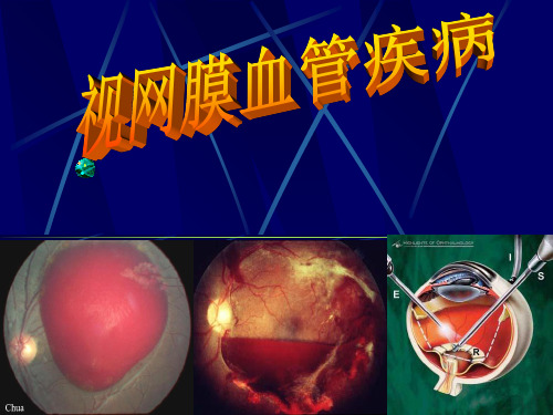
视网膜中央静脉阻塞
视网膜新生血管增生
虹膜新生血管
严重并发症——新vein occlusion
治疗 保护或改善视功能 防止或减少并发症 1治疗原发病如高血压或糖尿病; 2尿激酶溶栓 激素 3激光光凝大面积无灌注区 新生血管者 黄斑囊样水肿; 4手术治疗 视神经鞘切开术 视网膜动脉鞘切开
病因:不明;炎症 内分泌失调 视网膜小血管床先天性发育异常 视网膜血管为主要病变所在;血管失代偿血浆渗漏
Coats Disease
好发于男性儿童或青年;多单眼; 临床表现:早期因可无症状;波及黄斑视力减退; 儿童猫眼征或斜视时就诊 眼底所见:玻璃体清;眼底白色或黄白色不规则类脂样渗出;深层出血或胆固醇样结晶;异常血管;黄斑水肿和渗出;视网膜脱离 并发症:白内障;新生血管性青光眼;虹睫炎及眼球萎缩
三 视网膜静脉周围炎 Eales disease
发病年龄多2030岁;男性多;双眼;先后 临床表现:视力突然严重减退;一眼有症状;另眼亦可发现眼底病变 眼底所见:1 玻璃体出血;混浊;2 视网膜血管改变;主要位于周边部;小静脉扩张;迂曲;伴有白鞘;周围出血;渗出;视网膜大静脉分支有时亦受累 并发症:增殖性视网膜玻璃体病变;继发性视网膜脱离; 新生血管性青光眼与并发性白内障
临床表现 症状——主干阻塞时视功能明显下降 分枝阻塞时相应视野缺损 程度及速度均较中央动脉阻塞为轻; 检查——视盘水肿;以视盘为中心的大量浓密的放射状出血;棉绒斑;静脉怒张;时隐时现; 黄斑囊样水肿; FFA ——视网膜静脉充盈时间延长;视乳头边界糊;静脉迂曲扩张;渗漏及血管壁染;黄斑可有囊样水肿 缺血型:毛细血管无灌注区;微血管瘤;动静脉短路;视网膜新生血管; 视网膜电流图 b波低于正常60%;易发生新生血管性青光眼
视网膜疾病如何保护视力

视网膜疾病如何保护视力视网膜是人眼中负责感光的部分,它对于保持良好的视力非常重要。
然而,随着生活方式的变化,视网膜疾病的发病率也在增加。
那么,我们该如何保护视力,预防视网膜疾病呢?本文将从日常生活中的注意事项、健康饮食以及定期眼科检查三个方面来探讨视网膜疾病的保护方法。
一、日常生活中的注意事项1.避免长时间用眼: 长时间用眼容易导致视网膜疲劳,增加患视网膜疾病的风险。
因此,我们应该避免连续长时间盯着电脑、手机或电视屏幕。
每隔一段时间,可以合理安排休息,进行远离屏幕的适当眼保健操。
2.佩戴护眼设备: 在进行长时间用眼时,可以佩戴护目镜或者挂护目组合。
这样,可以有效减少电脑辐射和光线的刺激,降低对视网膜的损伤。
3.防范眼部伤害: 注意避免眼部受伤,避免颠簸剧烈运动、高风险活动以及使用尖锐物品等。
4.室内光线良好:保持室内光线明亮,避免太过昏暗的环境,这样可以有效减少眼睛对暗光的适应,减轻对视网膜的压力。
二、健康饮食对视网膜疾病的保护1.摄入富含维生素A的食物: 维生素A与视网膜健康密切相关,因此,摄入富含维生素A的食物,如胡萝卜、菠菜、柿子椒等,有助于预防视网膜疾病并保护视力。
2.增加摄入抗氧化剂:抗氧化剂可以有效中和体内自由基,减轻视网膜的氧化压力。
食物中的一些抗氧化剂,如维生素C、维生素E、花青素等,可以通过食用番茄、柑橘、蓝莓等水果蔬菜来摄入。
3.适量摄入ω-3脂肪酸: 研究表明,适量摄入富含ω-3脂肪酸的食物,如鱼类、亚麻籽油等,有助于维持视网膜的正常功能。
4.控制甜食摄入: 大量摄入高糖食物会导致血糖升高,损害视网膜血管,增加患眼底病变的风险。
因此,我们应该控制甜食的摄入,保持血糖平稳。
三、定期眼科检查定期眼科检查是预防和及早发现视网膜疾病的关键。
眼科医生能够通过检查眼底,观察视网膜的情况,及时发现异常,给予干预治疗。
建议人们每年至少定期进行一次全面的眼科检查,以保证视力的健康。
总结起来,保护视力、预防视网膜疾病需要我们在日常生活中注意眼部卫生,减少眼睛的疲劳,佩戴护眼设备,防范眼部伤害。
视网膜疾病的专业讲解PPT课件

目录 导言 视网膜疾病概述 青光眼 糖尿病视网膜病变 视网膜脱离 结论
导言
导言
视网膜疾病是一类常见的眼科疾病 ,严重影响视力和生活质量。 本课程将带您深入了解视网膜疾病 的类型、病因、症状和治疗方法。
视网膜疾病概 述
视网膜疾病概述
视网膜疾病指的是影响视网膜结构和功 能的各种疾病。 视网膜疾病可分为青光眼、糖尿病视网 膜病变、视网膜脱离等多个类型。
糖尿病视网膜病变
糖尿病视网膜病变的治疗方法包括控制 血糖、激光治疗和手术治疗等。
视网膜脱离
视网膜脱离
视网膜脱离是指视网膜与眼底 脱离的状况。 症状包括眼前闪光、黑点和视 野缺损等。
视网膜脱离
视网膜脱离的治疗方法包括激光治疗和 手术治疗等。
结论
结论
视网膜疾病是常见的眼科疾病,需 要及时诊断和治疗。 通过本课程的学习,您对视网膜疾 病的类型、病因、症状和治疗方法 有了更深入的了解。
青光眼
青光眼
青光眼是一种常见的视网膜疾 病,主要由眼内压增高引起。 症状包括视力模糊、眼部疼痛 和视野缺损等。
青光眼
青光眼的治疗方法包括药物治疗、手术 治疗和激光治疗等。
糖尿病视网膜 病变
糖尿病视网膜病变
糖尿病视网膜病变是糖尿病患者常 见的并发症之一。 症状包括视力模糊、视野缺损和眼 底出血等。
视网膜疾患的健康宣教

04
康复训练:进行视力训练,提高视力和视觉功能
视网膜疾患的护理
饮食护理
均衡饮食:保证营养均衡,多吃蔬菜水果
01
01
02
03
04
避免刺激性食物:避免辛辣、油腻、高糖食物
补充维生素:补充维生素A、C、E等对视网膜有益的营养素
控制体重:保持正常体重,避免肥胖对视网膜造成压力
02
03
04
心理护理
01
保持乐观积极的心态,避免焦虑和抑郁
定期进行眼部检查
定期检查的重要性:及时发现视网眼部检查
检查项目:包括视力检查、眼底检查、眼压检查等
检查注意事项:保持良好的生活习惯,避免过度用眼,保持良好的心理状态,积极配合医生进行检查。
避免过度用眼
控制使用电子产品的时间,避免长时间使用电脑、手机等设备。
抗血小板药物:如阿司匹林、氯吡格雷等,用于治疗糖尿病视网膜病变等疾病。
抗氧化剂:如叶黄素、玉米黄质等,用于治疗老年性黄斑变性(AMD)等疾病。
激素类药物:如地塞米松、曲安奈德等,用于治疗葡萄膜炎等疾病。
抗感染药物:如万古霉素、头孢曲松等,用于治疗感染性眼内炎等疾病。
其他药物:如钙通道阻滞剂、前列腺素类似物等,用于治疗青光眼等疾病。
手术治疗
手术类型:视网膜剥离术、玻璃体切割术、激光治疗等
手术目的:修复视网膜损伤,改善视力
手术风险:手术并发症、术后感染等
术后护理:定期复查、保持眼部卫生、避免剧烈运动等
4
康复治疗
01
药物治疗:使用抗炎、抗血管生成等药物,控制病情发展
02
激光治疗:通过激光照射,破坏病变组织,促进视网膜修复
03
手术治疗:针对不同病情,进行玻璃体切割、视网膜剥离等手术
视网膜病

第一节概述视网膜(retina)为眼球后部最内层组织,结构精细复杂,其前界为锯齿缘,后界止于视神经头。
视网膜由神经感觉层与色素上皮层组成。
神经感觉层有三级神经元:视网膜光感受器(视锥细胞和视杆细胞)、双极细胞和神经节细胞,神经节细胞的轴突构成神经纤维层,汇集组成视神经,是形成各种视功能的基础。
神经感觉层除神经元和神经胶质细胞外,还包含有视网膜血管系统。
一、视网膜解剖结构特点1.视网膜由神经外胚叶发育而成,胚胎早期神经外胚叶形成视杯,视杯的内层和外层分别发育分化形成视网膜感觉层(神经上皮层)和视网膜色素上皮(RPE)层。
神经上皮层和RPE层间粘合不紧密,有潜在的间隙,是两层易发生分离(视网膜脱离)的组织学基础。
2.RPE有复杂的生物学功能,为感觉层视网膜的外层细胞提供营养、吞噬和消化光感受器细胞外节盘膜,维持新陈代谢等重要功能。
RPE与脉络膜最内层的玻璃膜(Bruch膜)粘连极紧密,并与脉络膜毛细血管层共同组成一个统一的功能单位,即RPE-玻璃膜-脉络膜毛细血管复合体,对维持光感受器微环境有重要作用。
很多眼底病如年龄相关性黄斑变性、视网膜色素变性、各种脉络膜视网膜病变等与该复合体的损害有关。
3.视网膜的供养来自两个血管系统,内核层以内的视网膜由视网膜血管系统供应,其余外层视网膜由脉络膜血管系统供养。
黄斑中心凹无视网膜毛细血管,其营养来自脉络膜血管。
4.正常视网膜有两种血-视网膜屏障(blood-retinal barrier, BRB)使其保持干燥而透明,即视网膜内屏障和外屏障。
视网膜毛细血管内皮细胞间的闭合小带(zonula occludens)和壁内周细胞形成视网膜内屏障;RPE和其间的闭合小带构成了视网膜外屏障。
上述任一种屏障受到破坏,血浆等成分必将渗入神经上皮层,引起视网膜神经上皮层水肿或脱离。
5.视网膜通过视神经与大脑相通,视网膜的内面与玻璃体连附,外面则与脉络膜紧邻。
因此,玻璃体病变、脉络膜、神经系统和全身性疾患(通过血管和血循环)均可累及视网膜。
视网膜病人的护理

根据病因和发病机制,视网膜疾病可分为多种类型,如视网膜脱离、视网膜血 管病变、黄斑病变等。
发病原因及危险因素
发病原因
视网膜疾病的发病原因多种多样,包括遗传、环境、生活习 惯等多种因素。例如,高度近视、眼部外伤、眼部炎症等都 可能导致视网膜疾病的发生。
危险因素
年龄、性别、遗传、环境等因素也可能增加患视网膜疾病的 风险。例如,老年人、女性、有家族遗传史的人群可能更容 易患视网膜疾病。
等,提高其生活质量。
社交能力提升策略
心理疏导
对视网膜病患者进行心理疏导,帮助其克服自卑、焦虑等心理障 碍,增强社交信心。
社交技能培训
通过专门的社交技能培训课程,教授视网膜病患者如何与他人交流 、表达情感等,提高其社交能力。
参加社交活动
鼓励视网膜病患者参加各种社交活动,如社区活动、志愿者活动等 ,增加其与他人交流的机会,提高社交能力。
应对焦虑、抑郁等情绪问题
识别情绪问题
密切关注病人的情绪变化,及时发现焦虑、抑郁等情绪问题,为病 人提供必要的心理支持和干预。
心理干预措施
针对不同情绪问题,采取相应的心理干预措施,如认知行为疗法、 放松训练等,帮助病人缓解情绪压力,改善心理状态。
寻求专业帮助
对于严重情绪问题,及时引导病人寻求专业心理咨询或治疗,以便得 到更专业的帮助和支持。
如出现视力下降、眼前黑 影飘动等症状,应及时就 医检查。
03
视网膜病人日常护理措施
饮食调整与营养补充
饮食清淡
保持饮食清淡,避免过多摄入辛 辣、油腻食物,以免加重眼部负
担。
增加营养摄入
多食用富含维生素A、C、E的食物 ,如胡萝卜、菠菜、西兰花等,有 助于视网膜健康。
- 1、下载文档前请自行甄别文档内容的完整性,平台不提供额外的编辑、内容补充、找答案等附加服务。
- 2、"仅部分预览"的文档,不可在线预览部分如存在完整性等问题,可反馈申请退款(可完整预览的文档不适用该条件!)。
- 3、如文档侵犯您的权益,请联系客服反馈,我们会尽快为您处理(人工客服工作时间:9:00-18:30)。
Central Retinal Artery Occlusion (#1,3), Angiogram Comment: The late angiogram shows very slow perfusion of the retinal vessels as indicated by the so called box-car formation in the vessels
体 征
1、瞳孔直接光反射消失,间接光反射存在。 2、视乳头变白,边缘模糊,压迫眼球在视乳头上 不能压出动静脉搏动。 3、视网膜动脉显著变细 呈线状,或伴有白线。有 的动脉距视乳头不远即消失。静脉也变细,血 柱常间断成节状或念珠状。
4、视网膜呈急性贫血状。于眼底后极部呈乳白色半透 明混浊水肿区。
5、黄斑部因无视网膜内层,部分脉络膜色泽经透明的 视网膜外层透露而出,与其周围水肿的灰白色内层 相对比,呈鲜红色,即所谓“樱桃红点”。此为本病 的典型现象。
7、如果有视网膜睫状动脉存在时,由此血管供养的视网 膜可不坏死,该处仍保存正常色泽和机能,呈典型红 色舌状区。视野检查时,可发现一个小的中央岛状区, 因而得以保持中央视力。 8、中央动脉阻塞时,很少伴有视网膜出血。如果有出血, 多系合并有小静脉血栓。
Central Retinal Artery Occlusion (#1,1), with Cilioretinal Artery Comment: Faded whitening (a sign of a little older event) of the retina and cherry-red spot. Only the papillo-macular bundle looks normal. Vision is 0.01.
系视网膜中央动脉的主干或其分支阻塞,使 其所供血的区域发生急性缺血,致使该区营 养切断而引起极度的机能障碍。本病发病急, 多见于单眼。
病 因
筛板水平的粥样硬化所致 系统性疾病:偏头痛、外伤、凝血障 碍、炎症或感染性疾病 视网膜脱离手术或眶内手术
临床表现
症状 一只眼突然发生无痛性完全失明。 有的病人在发作前有阵发性黑朦。
视网膜中央动脉阻塞 视乳头退色,边缘模糊,颞侧缘可见小出血灶;动脉 细,有的走行不明;后极视网膜混浊肿胀,黄斑中心“樱桃红”。
视网膜中央动脉阻塞
后极视网膜肿胀显著,动脉细,பைடு நூலகம்斑樱桃红明显。
Central Retinal Artery Occlusion Edema makes the retina look whitish exept for the fovea where it is so thin that one can see the perfused underlying tissues (cherry red spot)
视网膜疾病
视网膜疾病
先天性 血管性 黄斑部 退行变性 全身病相关性 视网膜脱离 视网膜肿瘤
视网膜动脉阻塞
(Retinal artery occlusion)
分类
一.视网膜中央动脉阻塞 二.视网膜分支动脉阻塞 三.视网膜毛细血管前小动脉阻塞 四.眼缺血综合症
视网膜中央动脉阻塞
(central retinal artery occlusion)
Central Retinal Artery Occlusion (#1,2) with Cilioretinal Artery. Angiogram. Comment: While all other vessels are not perfused the cilio-retinal artery with its veins is open
Central Retinal Artery Occlusion (#4), Total Optic Atrophy Comment: After complete occlusion of the central artery ascending total optic atrophy and obliteration of vessels.
formation, as a sign of very slow perfusion.
Central Retinal Artery Occlusion (#1,1) (CRAO), Few Days Old
Regressing edema of the retina, disappearing red spot in fovea and narrow arteries. The disc not yet atrophic
6、数周后,视网膜水肿可逐渐消散,眼底恢复红色,但视 网膜完全萎缩,视神经纤维变性。视神经乳头因缺乏营 养而萎缩,呈苍白色,边缘整齐。血管呈白线状。视力 永久不能恢复。
Central Retinal Artery Occlusion
After a while the retinal edema turns into atrophy and the retina becomes transparent again. In some of the vessels one sees an interrupted blood column, so called box-car
Central Retinal Artery Occlusion
Atrophic retinal periphery where the retina has become transparent again with ensheathed and/or occluded vessels
Central Retinal Artery Occlusion (#1,2), (CRAO), Old, Angiogram Comment: Retinal vessels are only filling near the disc while there is a good choroideal flush.