Specificity characteristics of monoclonal antibodies to wheat grain storage proteins
Development and Production of Commercial Therapeutic Monoclonal
Current Pharmaceutical Biotechnology, 2008, 9, 447-467 4471389-2010/08 $55.00+.00© 2008 Bentham Science Publishers Ltd.Development and Production of Commercial Therapeutic Monoclonal An-tibodies in Mammalian Cell Expression Systems: An Overview of the Cur-rent Upstream TechnologiesMichel Chartrain * and Lily Chu *Department of Bioprocess R&D, Merck Research Laboratories, P.O. Box 2000, Rahway NJ, 07065, USAAbstract: This article provides an overview of the upstream technologies used in the industrial production of therapeutic monoclonal antibodies (mAbs) based on the cultivation of mammalian cells. More specifically, in a first section, after a short discussion of relevant biochemical characteristics of antibodies, we review the cell lines currently employed in commercial production and the methods of constructing and isolating production clones. This is followed with a review of the most current methods of commercial scale production and their associated technologies. Selected references and short discussions pertaining to emerging and relevant technologies have been embedded throughout the text in order to give a sense of the overall direction the field is taking.INTRODUCTIONTherapeutic monoclonal antibodies (mAbs) are as of to-day a well accepted class of therapeutics especially in the fields of oncology, immunology, and organ transplant, where the use of these targeted biologics has profoundly revolution-ized treatments paradigms. This is especially true in the treatment of various cancers where mAbs have proven to carry fewer side effects than the traditional cytotoxic drugs and have resulted in improved patient quality of life [1-3]. As of today, there are twenty two therapeutic mAbs, or fragments, currently registered for marketing in the US (see table 1) with a market size over $ 17B in 2007 and an ex-pected cumulative annual rate of growth of 14 % [4, 5] . This success is likely to carry for many years since it has recently been reported that the number of mAbs that entered clinical studies tripled over the past decade [4].Commercial production of currently registered mAbs relies on the cultivation of mammalian cells that have been genetically engineered to over-produce the mAb of interest. One exception is the use of the bacterium Escherichia coli in the production of fragments. The production of mAbs at industrial scale is a multifaceted endeavor that encompasses many technically complex and lengthy steps. Briefly, a com-mercial mAb production process starts with the generation of a mAb via immunization of an animal or via molecular biol-ogy methods, the identification and optimization of the cod-ing DNA sequence and the construction and identification of a highly producing and stable clone. These steps are fol-lowed with the development of a well designed cultivation process that encompasses the full control and scale up of associated operations that will support early clinical evalua-tions. Typical cycle times for these activities usually range between 16-24 months [6]. In this article, we will provide an overview of the state of the current technologies that lead to*Address correspondence to these authors at the Department of Bioprocess R&D, Merck Research Laboratories, P.O. Box 2000, Rahway NJ, 07065, USA; E-mails: michel_chartrain@; lily_chu@ achieving high efficiency in these process steps and will briefly discuss potential technological improvements which are likely to benefit the current technology over the next few years.1. OVERVIEW OF RELEVANT BIOCHEMICAL FEATURES AND PROPERTIES OF MONOCLONAL ANTIBODIESThis section will familiarize the reader with the basic concepts that are later discussed in the following sections. 1.1. The Concept of Therapeutic AntibodyVia interaction with specialized components of the im-mune system, the in vivo role of antibodies is to clear the host from invading pathogens and from any non-self mole-cules these micro-organisms may release (toxins for exam-ple). Antibodies present exquisite specificity for their target (antigens), with the ability to recognize and bind exclusively to a small region (epitope) of a given antigen. When binding to an antigen, the complex formed allows for rapid recogni-tion and clearance by specialized components and cells of the immune system. Natural killer cells NK or NKC recog-nize antibody-target cell complexes and trigger the lysis and destruction of the invading cell in a process known as anti-body dependent cell-mediated cytotoxicity (ADCC). Another facet of the immune response involves the complement, a multi-protein complex that sequentially bind to the antibody-target complex leading to either its recognition and en-gulfment by macrophages in a process known as opsoniza-tion, or to the lysis of the target cell in a process known as complement-dependent cell cytotoxicity (CDC). This very succinct and simplified overview of the immune response can be supplemented by consulting the following references for more extensive discussions [7, 8].These properties have made antibodies a very attractive choice for novel therapeutic approaches to diseases where externally exposed membrane-bound or circulating proteins could be specifically targeted for a specific action. The over-448 Current Pharmaceutical Biotechnology, 2008, Vol. 9, No. 6 Chartrain and Chuall expectation is that specific interactions between the mAb and its specific circulating or membrane-bound protein target will either result in the blockage of key signals of the disease pathology, will achieve the specific delivery of conjugated small molecules such as cytotoxins or radionucleides to tar-get cells, or will lead to the destruction of the target cell via ADCC or CDC. Several therapeutic mAbs whose mode of action fits one or several of these desired traits are currently in clinical use. A few are highlighted here.For example, the mAbs cetuximab (Erbitux®) and trasta-zumab (Herceptin®) interfere with the over-expressed mem-brane-bound growth factor receptors EGFR and Her2, both known to be implicated in signaling for proliferation and survival of cancer cells. By preventing ligand binding, cetuximab prevents cancer cells from proliferating. Although the exact mode of action is still unclear, trastazumab is be-lieved to act via interactions with the effectors of the im-mune system. The association of the mAb with a membrane protein triggers various mechanisms that can lead to the de-struction of the targeted cell by triggering apoptosis (a pre-programmed self destruction pathway) or cell lysis via CDC or ADCC. Both these two mAbs are two highly successful clinical examples of interference with a membrane-bound receptor. Based on a different approach, rather than directly interfering with a receptor, the mAbs bevacizumab (Avastin®) and adalimumab (Humira®) respectively block tumor angiogenesis and the signals leading to rheumatoid arthritis by respectively sequestering the circulating ligand, VEGF and TNF . The concept of delivering a toxic payload to specific cells targeted for destruction is exemplified by three mAbs that are currently in use in the treatment of can-cers. Mylotarg® (gemtuzumab) is conjugated to ozogamy-cin, a derivative of the antitumor-antibiotic calicheamycin and targets CD33, a protein often over-expressed on cancer-ous myeloid cells. Bexxar® (tositumomab) conjugated with the radionucleide 131I, and Zevalin® (ibritumomab) conju-gated with either 111In or 90Y both target CD20, an antigen expressed on the surface of myeloid cells. Palivizumab (Synagis®) which is directed at the envelope trans-membrane F (fusion) protein of the respiratory syncytial vi-rus (RSV is a very serious pediatric infection) is the only mAb currently in use for the treatment of infectious disease.Fragments of mAbs that are made up of the domain of the mAb that confers affinity to its target but that do not in-clude the domain of the molecule that interacts with the ef-fectors of the immune system are also used. In addition to not having interactions with the effectors of the immune sys-tem, fragments are rapidly cleared from the body, a second feature that is useful when seeking short term therapeutic action. Three fragments are currently registered with the FDA. Reo-pro® (abciximab) is used to control platelet ag-gregation during percutaneous coronary intervention. Lucen-tis® (ranibizumab) which is directly delivered to the eye has been very successfully used in the management of age re-lated wet macular degeneration. Very recently, the approval of Cimzia® (certolizumab pegol), a pegylated fragment di-rected at TNF , marked the introduction of fragments de-signed for extended in vivo life time through conjugation with the polymer PEG. These few selected descriptions ex-emplify how mAbs have been successfully used in the treat-ment of complex and serious diseases. The list of the 22 cur-rently US approved mAbs, their protein target and their clinical applications can be found in Table 1. Additional general information can be found in the following publica-tions [9-16].1.2. Biochemical Overview of Therapeutic AntibodiesAs of today all therapeutic mAbs are of the Immuno-globulin G (IgG) sub-class. These molecules are made up of two heavy chains and two light chains that are held together and folded via intra and inter-chain disulfide bounds. The average molecular weight of each sub-chain is about 25Kd (or about 220 amino acids) for the light chain and 50 Kd (about 450 amino acids) for the heavy chain. The heavy chain is made up of 3 constant and one variable domains, while the light chain is comprised of one constant and one variable domains. Specificity of the antibody molecule is dictated by the amino acid sequence at specialized sites, called complementarity determining regions (CDR) present in the variable domains of both light and heavy chains. It is these loop-forming hyper-variable regions of the molecule that interact and form a tight association with the intended antigen target. All IgG sub-class antibodies are based on this general structure and mostly differ in the sequence of the constant regions of the heavy chains, the part of the molecule that interacts with other components of the immune system via ADCC and/or CDC. The IgG1 sub-class molecules pre-sent the strongest association with the receptors of the effec-tor components of the immune system [17]. IgG antibodies have an additional layer of complexity through a conserved N-linked glycosylation site at ASN-297 on each of the heavy chains [18]. These short glycans are of bi-antennary structure and can be variable in many ways that include; the terminal sugar composition (presence or absence of galactose, cap-ping with a sialic acid) and the presence or absence of bisect-ing fucose and N-acetyl glucose amine residues. This high degree of variability and relative abundance of each glycan species results in the high likelihood that glycans of a differ-ent structure are attached to each of the heavy chains of a given mAb molecule. The presence and composition of these glycans has been found to greatly affect the activity of the antibodies in terms of their interactions with the immune system effector functions [19]. For example, glycans devoid of the bisecting fucose will confer the mAb a stronger inter-action between the antibody and the NKC, resulting in higher ADCC mediated immune response [20-26]. Similar increases in ADCC function have also been noticed when the bisecting N-acetyl glucose amine is present, although this effect seems to be more antibody dependent [27-29]. How-ever while all ADCC activity is abolished by the complete removal of the glycans, partial removal of the terminal sialic acid and galactose have no gross apparent impact on ADCC [30]. Although they represent a small part of the antibody molecule, glycans, because they are assembled post transla-tionally are more likely to exhibit higher diversity and to thereby influence the overall activity of the mAb. Fig. (1) presents a composite pictorial of an antibody with a compos-ite of the glycan chain presented in detail.In addition, it is also important to note that not only are antibody molecules highly complex in their primary structure and heterogeneity; they also have complex secondary and tertiary structures that greatly influence their activity. ThisDevelopment and Production of Commercial Therapeutic Monoclonal Antibodies Current Pharmaceutical Biotechnology, 2008, Vol. 9, No. 6 449short overview of the biochemistry of IgG antibodies high-lights the fact that synthesis of biologically active mAbs is a highly complex and well orchestrated biochemical process that encompasses folding, trafficking, post-translational modifications, and effective secretion. The choices of a pro-ducing platform (host cell) and of the physico-chemical con-ditions that will support mAb synthesis have been shown to greatly affect the abundance and quality of the N-linked gly-cans, molecule folding, and the yield of the mAb of interest. Since the advent of mAb production in the late 1970s, production platforms have relied on mammalian cells. As discussed above, monoclonal antibodies are highly complex molecules that require post translational modifications such as inter and intra-chain disulfide bond formations, and the addition of N-linked glycans at specific sites. Until recent metabolic engineering advances, only mammalian cells which possess the intrinsic required machinery could pro-duce mAbs with the desired folding and post-translational modifications, and were therefore the de facto production platforms for mAbs. Although considerable progress has recently been (and continues to be) made in the use of other biological production platforms [31-33], to our knowledge, as of today only mammalian cells are used for the production of commercial and late stage clinical mAb supplies. Judging by the pace and the significance of the discoveries made in the use of yeasts, it is however very likely that during the next decade, these non-mammalian expression systems will be in use for the production of clinical or commercial mAbs.2. GENERATION AND MOLECULAR BIOLOGY BASED MANIPULATIONS OF MABS. 2.1. General PrinciplesTraditionally, the generation of most therapeutic antibod-ies relies on the immunization of mice or other mammals with the desired antigen target. Upon repeated injections, the animals develop a strong response to the antigen and will present a large number of cells secreting antibodies against the injected antigen. It is important to understand that multi-ple antibodies directed at different epitopes of the antigen are secreted by a mixed population of B cells with each cell only secreting one specific antibody [34]. Therefore if one secret-ing B cell was to be isolated and expanded (cloned), only one type of antibody molecule (monoclonal or mAb) would be secreted. Unfortunately, secreting B cells can only repli-cate a limited number of times, therefore rendering produc-tion of mAbs by their cultivation all but impossible. In vitro production of mAbs was only a concept until the ground-breaking hybridoma technology was developed in the 1970s by Kohler and Milstein [35-37]. Antibody secreting cells originating from the spleen of an animal immunized with the antigen target of choice are fused with immortalized (i.e. cells that will divide forever when cultivated in permissive conditions) non-antibody secreting cells. To facilitate the isolation of the desired fused clones, the immortalized non-secreting cells are engineered to be deficient in a key meta-bolic pathway. For example, deficiencies in the hypoxan-Fig. (1). Schematic representation of an IgG mAb.The molecule is symmetric and can engage through its binding regions with two antigens at once. The presence of the hinge region allows for flexibility and facilitates this process.The insert provides details of the glycan structure. Symbols key is as follows: Fucose (FUC ), N acetyl glucose amine (NAGA ) galactose (GAL ), mannose (MAN ), N-acetylneuramic acid or sialic acid (NANA).450 Current Pharmaceutical Biotechnology, 2008, Vol. 9, No. 6 Chartrain and Chuthine guanidine phosphorybosyl transferase (HGPRT), an enzyme that holds a key role in the synthesis of guanidine tri-phosphate (GTP) in the "nucleotide synthesis salvage pathway" have been extensively used. These cells are still able to synthesize their nucleotides via the main (or de novo) pathway, however, when cultivated in the presence of aminopterin, an inhibitor of dehydrofolate reductase (DHFR) an enzyme that produces tetrahydrofolate, a key co-factor in the synthesis of thymidine tri-phosphate (TTP) from dUMP in the main nucleotide synthesis pathway, these cells can not grow since each of the pathways leading to the production of nucleotides are non-functional under these conditions. Since they have a functioning HPRT enzyme, B cells originating from the spleen are able to survive in the presence of aminopterin. However as somatic cells, they only can repli-cate a few times before dying. Only fusion clones that have inherited the ability to replicate indefinitely from the non-secreting cells and the functional HPRT activity from the B-cells will grow for extensive number of generations in the presence of aminopterin. When coupled with the isolation of colonies originating from one fused cell, this technology rendered possible the production of mAbs. Clones that pro-duce the mAbs with affinity to the desired target can be iden-tified using classical affinity biochemistry methods. The de-velopment of this technology in the late 1970s was hailed as a major breakthrough and opened the door to generation of therapeutic monoclonal antibodies and is now a well estab-lished protocol [38-40]. Fig. (2) represents an overview of the process of mAb production by generating hybridomas.Although the first recombinant mAbs were produced using this technology, the mAb hybridoma production plat-form presents some serious drawbacks. Since the coding region of the mAb directly originates from the immunized animal without any subsequent genetic manipulations, the mAb produced is of mouse origin in its amino acid sequence, and upon repeated injections rapidly triggers an immune response from the human recipient (referred to as Human Anti Mouse Antibody or HAMA). Upon these repeated infu-sions, the therapeutic mAb is rapidly cleared from the circu-lation and therefore rendered therapeutically ineffective. About 80% of the patients develop an immune response to the first commercial therapeutic mAb, OKT3, a murine anti-body used in the treatment of organ transplant rejection [41]. Further improvements were obviously needed in order to make monoclonal antibodies a therapeutic reality, and con-sequently there are only two murine therapeutic monoclonal antibodies approved at this time (Table 1).2.2. Sequence HumanizationFollowing these first clinical attempts to use mAbs of murine structure, molecular biology manipulations greatly improved the immunogenic profiles of the subsequent mAbs tested in humans by reducing the amount of amino acid se-quences of mouse origin while retaining the appropriate af-finity for the intended target. The most common approach starts with the generation of mouse hybridomas as described above. Once clones are generated, they are screened for their ability to produce a mAb with the desired affinity for the target antigen, employing biochemical screening methods [42-47]. The selected clone is further cultivated and the mAb genetic coding sequences are isolated and modified to yield antibodies that are more and more human-like in their struc-ture. Briefly these technologies have evolved over the past decade, from first the construction of chimeric antibodies, made up of the constant regions of human antibodies at-tached to the variable region of mouse origin (responsible for the binding to the target), to techniques of generating "hu-manized" antibodies by "grafting" and "veneering" only the essential mouse amino acid residues needed for affinityFig. (2). Overview of the various steps leading to the production of mAbs via the generation of hybridomas.Development and Production of Commercial Therapeutic Monoclonal Antibodies Current Pharmaceutical Biotechnology, 2008, Vol. 9, No. 6 451 Table 1. Therapeutic Monoclonal Antibodies and Antibody Fragments on the US MarketName Genericname Target IndicationRituxan®Rituximab CD20, a protein expressed on B cells but not on stem cell and differentiatedantibody secreted cells. Can trigger cell death via ADCC, CDC and by initiatingapoptosis.Non Hodgkin Lymphoma of B-CellsZevalin®IbritumomabConjugated with eitherIndium 111 or yttrium 90. Amurine antibody precursorto rituximab CD20. Same target as rituximab, often used in conjunction with rituximab Non HodgkinLymphoma of B-CellsBexxar®Tositumomab, a conjugatewith I131CD20. Specifically delivers a radionucleide to target cells. ADCC, CDC, andinduction of apotosis via ionizing radiation.Non HodgkinLymphoma of B-CellsHerceptin®Trastazumab Her2, a tyrosine kinase receptor over-expressed on the surface of cancer cells. Byblocking the most upstream signal, it reduces cell proliferation and survival. Canalso trigger cell death via ADCC and CDC.Cancer (breast)Mylotarg®Gemtuzumab ozogamicinA conjugate with the cyto-toxic ozogamycin,CD33, a protein often over-expressed on cancerous myeloid cells but not onpluripotent haematopoetic stem cells. Specifically delivers a cytotoxic drug.Cancer (Acute mye-loid leukemia)Campath®Alemtuzumab CD52, a protein expressed on the surface of differentiating lymphocytes. Ellicitscells death via ADCC, CDC, and the triggering of apoptosis.Cancer (Chronic myeloid leukemia)Erbitux®Cetuximab EGFr, a tyrosine kinase receptor over-expressed on the surface of cancer cells.By blocking the most upstream signal, it reduces cell proliferation and survival.Can also trigger cell death via ADCC and CDC.Cancer (Colon)Vectibix®Panitimumab EGFr Cancer (colon) Avastin®Bevacizumab VEGF, a circulating protein that triggers vascularization, a needed step for tumordevelopmentCancer (Colon)Orthoclone® OKT 3Mururomab CD3,a protein associated with T cell receptors. Steric inhibition by OKT3 in-duces pan-T cell depletionKidney transplantrejection preventionZenapax®Daclizumab CD25, an IL2 receptor present on the surface of activated but not resting T-lymphocytes, thereby blocking activation of cytotoxic lymphocytes involved intransplant rejectionKidney transplant rejection preventionSimulect®Basiliximab CD25Kidney transplantrejection prevention Remicade®Infliximab TNF , a proinflamotory cytokine involved in various inflammatory chronic diseases Crohn’s disease, RA Humira®Adalimumab TNF RAXolair®Omalizumab IgE, prevent the binding of this proinflamatory signaling immunoglubuling withits receptor Allergy relatedasthmaRaptiva®Efalizumab CD11a, a subunit of the T cell integrin LFA-1, a ICAM receptor involved in T-cell activation and migration, a hallmark of psoriasis.PsoriasisTysabri®Natalizumab 4 subunit of VLA-4 integrin of activated T cells, preventing interaction with itsbrain endothelium counter receptor VCAM-1,thereby preventing T-cells fromcrossing the blood brain barrier.Multiple sclerosisSynagis®Palivizumab F protein (fusion transmembrane protein) of the Respiratory Synsytial Virus.Binding to the F protein elicits recruitment of the immune system effector com-ponents and triggers virus clearance.RSV infectionsSoliris®Eculizumab Terminal complement protein C5. Prevents complement mediated lysis of redblood cells deficient in the regulatory proteins CD55 andCD59, which makes the cells sensitive tocomplement dependent destruction.Paroxysmal Nocturnal Haemoglo-binuria452 Current Pharmaceutical Biotechnology, 2008, Vol. 9, No. 6 Chartrain and Chu (Table 1) contd....Name Genericname Target IndicationReo-Pro®Abciximab(a Fab fragment)GPIIb/IIIa receptor complex of platelets. Binding to these integrins inhibits plate-let aggregation and prevent blood clots formation.Adjunct in coronaryinterventionsLucentis®Ranibizumab(a Fab fragment)VEGF. Binding with the VEGF ligand prevents interaction with its receptorsVEGFR1 and VEGFR2 on the surface of endothelial cells. When locally in-jected, It reducesendothelial cell proliferation, vascular leakageand the formation of new blood vessels on the central part of the retina.Wet maculardegenerescenceCimzia®Certolizumab pegol(A Fab fragment conju-gated with poly-ethylene-glycol)TNF- .Crohn's diseaseto the antigen target to a human antibody framework. Finally the production of fully "human" antibodies through the use of transgenic mice, which expresses human genes for IgGs, has been implemented to recently yield its first commercial-ized mAb, Vectibix® (panitumumab), an IgG2 molecule [48].All these approaches which rely on the immunization of animals require the construction of hybridomas and present limitations in some situations such as when generating a mAb directed against a toxin or a highly conserved antigen across species. To overcome these limitations, the generation of mAbs by recombinant technology was developed. Post isolation and amplification of the variable HC and LC ge-netic coding sequences from B lymphocytes by PCR, it relies on the construction of highly diverse libraries that have the potential to generate up to 1010 different mAbs via recombi-nation. Most screening protocols use phage display technol-ogy, an approach that by fusing the Fab region coding genes with the gene of a phage coat protein results in linking geno-type to phenotype. Briefly, the phage which displays the resulting Fab on its coat can be isolated by affinity technolo-gies. After amplification in a bacterium, the genome of the isolated phage contains the matching DNA sequence which can be isolated and further manipulated as desired such as increasing affinity for the antigen target [42-47, 49]. Other methods such as yeast and bacterial displays have also been developed and may present a simpler and faster approach to the development of synthetic mAbs [42-47]. As of today, the use of phage display technology has been successfully im-plemented in the generation of fully human antibodies, with Humira® an antibody directed at the human protein TNF , being the first to reach commercialization.Development of the production of chimeric mAbs fol-lowed by the refinements described here were the needed boosts that by greatly reducing or eliminating patient im-mune response to the therapeutic mAb allowed repeated in-fusions and propelled therapeutic mAbs to widespread and successful clinical uses.2.3. Construction of Genetic VectorsThe construction of producing clones follows the overall paradigm of classical molecular biology developed for mi-crobial systems. After selection of a hybridoma clone secret-ing the desired antibody (selection based on affinity for its target), the DNA sequence coding for the IgG is obtained and engineered to reduce immunogenicity as described above. Further refinements aimed at increasing the affinity of the mAb to its target antigen using phage display technology can take place at that point during the development cycle of a mAb [49]. This is sometimes a required step since affinity of the original murine mAb for its target typically decreases during the humanization process. The finalized mAb coding sequences are then inserted on classical genetic vectors (plasmids) that contain a selectable genetic marker and that are mass produced using microbial technology. The entire coding mAb sequence can be enclosed into a single plasmid, or two plasmids each containing the genes for the heavy chain (HC) and light chain (LC) respectively can be con-structed [50, 51]. The purified plasmid(s) is/are then trans-fected into non-antibody secreting immortalized mammalian cells, where upon insertion of one or multiple copies into one or several chromosomes, the foreign gene is transcribed to eventually yield secretion of a fully functional mAb. Various transfection approaches that all rely on the temporal destabi-lization of the membrane integrity, thus allowing the plasmid to gain access to the cytoplasm, have been used with success [52]. Vector design uses the same promotors for both heavy and light chains and in theory can support similar production of HC and LC, although more LC may be required since HC lower transcription efficiency and lower transcript stability has also been observed. Studies seem to indicate that excess LC synthesis may lead to better stabilization of the HC and to higher overall mAb secretion [53]. To the best of our knowledge, tunable expression systems that favor the tran-scription and translation of LC or that offer the possibility of controlling the onset of synthesis via induction have not yet been commercially implemented although the technology does exist [54].Since transfection and integration of genetic material into the chromosome of mammalian cells are low efficiency processes, in order to facilitate selection of cells that are likely to have integrated the foreign DNA and to secrete the desired mAb, selection protocols are routinely used. These rely on the principle that when the genes of the mAb and of a resistance/selectable marker are co-localized on the vector they will co-integrate and co-express with a high probability. Usually resistance to an antibiotic or the ability to grow in a。
生物化学及分子生物学(人卫第九版)-16基因表达调控说课讲解
色氨酸操纵子的结构及其关闭机制
A.前导序列的结构特征;B.在Trp低浓度时,核糖体停滞在序列1上,2/3发卡结构形成,转录继续进行; C.在Trp高浓度时,3/4发卡结构和多聚U序列使得转录提前终止
3.转录衰减的机制 ①色氨酸的浓度较低时,前导肽的翻译因色氨酸量的不足而停滞在第10/11的色氨酸密码子 部位,核糖体结合在序列1上,因此前导mRNA倾向于形成2/3发夹结构,转录继续进行; ②色氨酸的浓度较高时,前导肽的翻译顺利完成,核糖体可以前进到序列2,因此发夹结构 在序列3和序列4形成,连同其下游的多聚U使得转录中途终止,表现出转录的衰减。
3.真核生物编码蛋白质的基因是不连续的,转录后需要剪接去除内含子,这就增加了基因表 达调控的层次。
4.原核生物的基因编码序列在操纵子中,多顺反子mRNA使得几个功能相关的基因自然协调 控制;而真核生物则是一个结构基因转录生成一条mRNA,即mRNA是单顺反子 (monocistron),许多功能相关的蛋白、即使是一种蛋白的不同亚基也将涉及多个基因的 协调表达。
1.原核生物大多数基因表达调控是通过操纵子机制实现的
2.操纵子(operon):由结构基因、调控序列和调节基因组成 ①结构基因:包括数个功能上有关联的基因,它们串联排列,共同构成编码区。这些结 构基因共用一个启动子和一个转录终止信号序列,因此转录合成时仅产生一条mRNA长 链,为几种不同的蛋白质编码。这样的mRNA分子携带了几个多肽链的编码信息,被称 为多顺反子(polycistron)mRNA。
5种E.coli 启动子的共有序列
b. 操纵元件:是一段能被特异的阻遏蛋白识别和结合的DNA序列。 ③调节基因(regulatory gene):编码能够与操纵序列结合的阻遏蛋白
Monoclonal Antibodies
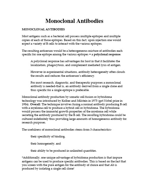
Monoclonal Antibodies MONOCLONAL ANTIBODIESMost antigens such as a bacterial cell possess multiple epitopes and multiple copies of each of those epitopes. Based on this fact, upon injection one would expect a variety of B cells to interact with the various epitopes.The resulting antiserum would be a heterogeneous mixture of antibodies each specific for one epitope among the various epitopes = a polyclonal response.A polyclonal response has advantages for host in that it facilitates thelocalization, phagocytosis, and complement mediated lysis of antigen.However in experimental situations, antibody heterogeneity often cloudsthe results and reduces the antiserum’s efficiency.For most research, diagnostic, and therapeutic purposes a monoclonalantibody is needed-that is, an antibody derived from a single clone andthus specific for a single epitope is preferable.Monoclonal antibody production by somatic cell fusion or hybridoma technology was introduced by Kolher and Milstein in 1975 (got Nobel prize in 1984). Overall: The technique involves fusing a normal antibody producing B cell with a myeloma cell to produce a hybrid cell or hybridoma. The hybridoma would possess the immortal growth properties of the myeloma cell while secreting the antibody produced by the B cell. The resulting hybridoma could be cultured indefinitely thus providing large amounts of homogeneous antibody for research purposes.The usefulness of monoclonal antibodies stems from 3 characteristics- their specificity of binding,their homogeneity, andtheir ability to be produced in unlimited quantities.*Additionally, one unique advantage of hybridoma production is that impure antigens can be used to produce specific antibodies. This is based on the fact that you screen with the pure antigen for the antibody of choice and that Ab is produced by isolating a single cell clone!MONOCLONAL PRODUCTIONFirst mice are injected with an Ag+adjuvant mixture- mice generallyreceive a primary injection followed by two boosts (14 to 28 days later).Why?After the final boost the mice are euthanized 3 to 5 days later and thespleen is harvested for cell fusion.A single cell suspension from the spleen is made and mixed with themyeloma partner. A fusing agent such as polyethylene glycol is thenadded. PEG fuses the plasma membranes of adjacent cells forming asingle cell with two or more nuclei.This heterokaryon retains these nuclei until the nuclearmembranes dissolve prior to mitosis. During mitosis andfurther rounds of division, the individual chromosomes aresegregated into daughter cells. Often chromosomes are lost.Even in the most efficient hybridoma fusions, only about1%of the starting cells are fused, and only about 1 in 105 formviable hybrids. This leaves a large number of unfused cellsstill in the culture. The cells from the immunized animal donot continue to grow in tissue culture because they areterminally differentiated cells and thus only capable oflimited growth, and so do not confuse further work.However, the myeloma cells are well adapted to tissueculture and must be killed. This is usually done by drugselection.DRUG SELECTIONCells have two pathways for the synthesis of nucleotides, the de novo and salvage pathways. Since cells in tissue culture can survive by using either of these pathways, mutations in the enzymes responsible for nucleotide synthesis have become common and readily manipulated targets for mutagenesis of mammalian cells. The most common target is the enzyme hypoxanthine-guanine phosphoribosyl transferase (HGPRT).HPGRT performs one of the essential steps in the salvage pathwayMutations in the HPGRT gene can be selected by growing cells in thepresence of purine analogs such as 8-azoguanine (8-AG). HPGRT willrecognize 8-AG as a substrate and convert it to the monophosphatenucleotide. The 8-AG-containing nucleotide is then processed further and incorporated into DNA and RNA, where it is toxic substitution. Therefore, cells with a functional HPGRT enzyme grown in the presence of 8-AG will die.However, because the HPGRT enzyme is part of a nonessential pathway(the de novo pathway is still available) cells harboring a mutant HPGRTgene can continue to grow.Therefore, selection with 8-AG will kill cells with a wild-type HPGRT but will not affect cells with a mutant HPGRT. The normal rate of mutagenesis is sufficient for treating cells with 8-AG and the surviving cells areHPGRT deficient.SP2/0 myeloma cell line from the ATCC (american type culture collection) The myeloma cell line does not synthesize or secrete any immunoglobulin chains, and is HGPRT-.HAT Selection for HybridomaDrugs like aminopterin block de novo synthesis of nucleotides. Bothpurine and pyrimidine synthesis are inhibited so both need to beproduced by the salvage pathway and precursors for these pathways(such as hypoxanthine and thymidine) must be included in the selectionmedia.Myeloma fusion partners are deficient in the enzyme required for thesalvage pathway of nucleotide synthesis. These cells will die in HATcontaining medium because aminopterin blocks normal nucleotidesynthesis and the enzyme deficiency blocks utilization of the salvagepathway.If the myeloma and B cell fuse, the resulting hybridoma will liveindefinitely in culture because the normal cell supplies the missingenzyme and the myeloma immortalizes the cell line. Unfused normal cells will only survive in tissue culture for approximately 1 week before theydie off.After 7-10 days, most of the wells contain dead cells but a few containclusters of viable cells - each cluster represents a clonal expansion of ahybridoma!At this point it is necessary to screen your fusion. We use the ELISA(enzyme linked immunosorbent assay) method. Screening at this timepoint is important because non-Ab producing hybrids will overgrow theslower growing Ab producers!ELISACoat 96 well plates with antigen at 1 ug/ml in carbonate buffer pH 9.6(add 100ul per well).Incubate overnight at 4o C. (Polystyrene binds protein noncovalently).Remove the contents of the wells and block plate with 3% NFDM in PBSto block nonspecific antibody binding. Incubate. (Blocking must beperformed to prevent nonspecific binding of Igs to any sites on the plate).Wash plate with PBS-tween.Add 100ul of (antibody containing) supernatant from Fusion into Elisaplates.Incubate.Wash plate and add 100ul of anti-Ig labeled with horseradish peroxidase.Incubate then wash plate.Add 100ul of substrate solution to each well. Incubate.Evaluate wells for positives- If an Ag+Ab+enzyme labeled Ab + enzymesubstrate is in the well there will be a color reaction!Positive wells contain hybrids making specific Ab to your test Ag!Once clones have been identified, the positive wells need to be expandedand cloned by limiting dilution.You can’t be sure that the positive well arose from only one hybrid cloneso the cells have to undergo limiting dilution- you dilute out the cells inone well into a series of wells until only one cell is in the well to insureclonality.After each cloning the cells are again screened by ELISA to insure Abs are still being produced.Because hybridoma cells have a very low plating effiency, single cellcloning is normally done in the presence of feeder cells. We use a singlecell suspension of spleen cells-these cells produce growth factors and also provide cell contact for your hybridoma.We usually do limiting dilution 3 consecutive times to one clone to assure clonality. ELISA screening is performed each time.At this point, once you have a clone the cells can be expanded forantibody production and experimental use!Cells can be frozen in a DMSO-FCS mixture and stored in liquid nitrogenfor years. * Cells should be frozen down at each step of the cloningprocedure in case of contamination. If you do get contamination-you don’t have to start all over- you simply retrieve the cell line from the previouscloning and start at that point!CLINICAL USE- Wagner et al. Hybridoma 16(1):33-40 1997Generated an IgG1 murine monoclonal Ab -anti-idiotypic Ab (Ab2) designated ACA125 which mimics an epitope on the tumor associated antigen designated CA125. This antigen is expressed by most malignant ovarian tumors.Used ACA125 Ab2 for vaccine therapy in 16 patients with advancedepithelial ovarian cancer. Patients with CA125 positive tumors areimmunologically tolerant to CA125 -in other words they don’t produce an immune response to it.Tolerance - a state in which lymphocytes are present that wouldnormally respond but are somehow prevented from doing so.It affects both the T and B lymphocytes.When the antigen is cleared, and with production of newlymphocytes - you eventually regain response ability- so toleranceis a temporary state.Certain conditions favor tolerance induction:state of the lymphoid system- its easier to induce withimmature immune systems or immune systems damaged byirradiation, drugs, ectphysiochemical properties of the Ag-size (small, soluble Ags areharder to capture from circulation by the phagocytic system,chemical nature (d-amino acids are toleragens whereas l-amino acids are immunogens) , epitope density on molecule(too many induces tolerance)route of Ag administration- determines accessibility of Ag toMOsAg dose- very low and very high doses induce tolerance.Does the tumor display a very low or very high epitope density toinduce tolerance? Does it flood the system with soluble Ag?The patients received 3 to 19 injections of 2mg/inj of the Ab2.Patients showed development of specific humoral and cellular immuneresponses to an otherwise nonimmunogenic tumor antigen. Theydeveloped Ab3 (anti-anti-id) and had an increase in IFNg production.(*Remember an increase in IFNg results in an increase of TNF).9 patients produced the Ab3 with few side effects and displayed a medianprogression free survival for 11 +/-5.6 (5.4-16.6) months compared to 8+/-4.2 (3.8-12.2) months in the Ab3 negative group.Human HybridomasWhen mouse monoclonals are introduced into humans they are recognized as foreign and evoke an Ab response. The induced human anti-mouse antibodies quickly reduce the effectiveness of the mouse monoclonal by clearing it from the bloodstream. Also, circulating immune complexes of mouse + human Abs can cause allergic reactions or you can get a buildup of complexes on the basement filter membranes of the kidney- glomerulonephritis. One area of development over the last 5 years has been the development of systems for the production of human hybridomas. Human monoclonal antibodies can be used for clinical applications.The progress has been slow because of the lack of a suitable myelomapartner but several line have been isolated and are now in use.The use of EBV transformed Ab secreting cells has solved some of theproblems but these transformants seldom secrete large amounts of Abs.This has been overcome in some cases by fusing the EBV transformed cell with a mouse myeloma cell line to increase Ab secretion- aheterohybridoma.Also there is the problem of obtaining Ag primed B cells in humans-can’t take their spleens! So human hybridomas have to be prepared from B cells in human peripheral blood. Lymphocytes only make up about 20-30% ofyour WBC population and most (~90%) of the lymphocytes in circulation are T cells.Plus human volunteers can not be just immunized with a range ofantigens- have tried in vitro Ag priming of the B cells but conditions arenot optimal so only see low affinity IgM production.ImmunotoxinsTumor specific monoclonal antibodies with lethal toxins coupled to it are potentially valuable therapeutic reagents. Toxins such as ricin and shigella are so toxic that one molecule can kill a cell. These toxins are composed of two chains- one is the toxin chain and the other is a binding chain that interacts with receptors on the cell surface. Without this binding chain the toxin is harmless since it cannot enter the cell. In theory: the monoclonal Ab with the attached toxin chain will bind specifically to a tumor cell where it will be endocytosed and the toxin will cause death by inhibiting protein synthesis.。
单克隆抗体制备的基本流程和应用

英文回答:The basic process for the preparation of monoclonic resistance consists of the selection of suitable antigens and the immunisation of animals to induce the production of specific antibodies。
The nucleic acid is then extracted from the spleen cells of immunosuppressants and, when integrated with tumour cells, it is formed into a hybrid tumour cell。
A hybrid cell is screened to obtain a single cloned hybrid cell strain of high—yield antibodies。
These monoclonal antibodies are strictly identified to ensure their purity and specificity。
This preparation process aims to ensure the quality and stability of monoclon antibodies, in line with the country ' s approach to scientific and technological development, and to promote innovative development in the field of medicine and the flourishing of health。
药物分析专业英语词汇

药物分析专业英语词汇AAbbe refractometer 阿贝折射仪absorbance 吸收度absorbance ratio 吸收度比值absorption 吸收absorption curve 吸收曲线absorption spectrum 吸收光谱absorptivity 吸收系数accuracy 准确度acid-dye colorimetry 酸性染料比色法acidimetry 酸量法acid-insoluble ash 酸不溶性灰分acidity 酸度activity 活度additive 添加剂additivity 加和性adjusted retention time 调整保存时间adsorbent 吸附剂adsorption 吸附affinity chromatography 亲和色谱法aliquot 〔一〕份alkalinity 碱度alumina 氧化铝ambient temperature 室温ammonium thiocyanate 硫氰酸铵analytical quality control〔AQC〕分析质量控制anhydrous substance 枯燥品anionic surfactant titration 阴离子外表活性剂滴定法antibiotics-microbial test 抗生素微生物检定法antio*idant 抗氧剂appendi* 附录application of sample 点样area normalization method 面积归一化法argentimetry 银量法arsenic 砷arsenic stain 砷斑ascending development 上行展开ash-free filter paper 无灰滤纸〔定量滤纸〕assay 含量测定assay tolerance 含量限度atmospheric pressureionization(API) 大气压离子化attenuation 衰减Bback e*traction 反萃取back titration 回滴法bacterial endoto*ins test 细菌毒素检查法band absorption 谱带吸收baseline correction 基线校正baseline drift 基线漂移batch, lot 批batch(lot) number 批号Benttendorff method 白田道夫〔检砷〕法between day (day to day, inter-day) precision 日间精细度between run (inter-run) precision 批间精细度biotransformation 生物转化bioavailability test 生物利用度试验bioequivalence test 生物等效试验biopharmaceutical analysis 体药物分析,生物药物分析blank test 空白试验boiling range 沸程British Pharmacopeia (BP) 英国药典bromate titration 溴酸盐滴定法bromimetry 溴量法bromocresol green 溴甲酚绿bromocresol purple 溴甲酚紫bromophenol blue 溴酚蓝bromothymol blue 溴麝香草酚蓝bulk drug, pharmaceutical product 原料药buret 滴定管by-product 副产物Ccalibration curve 校正曲线calomel electrode 甘汞电极calorimetry 量热分析capacity factor 容量因子capillary zone electrophoresis (CZE) 毛细管区带电泳capillary gas chromatography 毛细管气相色谱法carrier gas 载气cation-e*change resin 阳离子交换树脂ceri(o)metry 铈量法characteristics, description 性状check valve 单向阀chemical shift 化学位移chelate pound 鳌合物chemically bonded phase 化学键合相chemical equivalent 化学当量Chinese Pharmacopeia (ChP) 中国药典Chinese material medicine 中成药Chinese materia medica 中药学Chinese materia medica preparation 中药制剂Chinese Pharmaceutical Association (CPA) 中国药学会chiral 手性的chiral stationary phase (CSP) 手性固定相chiral separation 手性别离chirality 手性chiral carbon atom 手性碳原子chromatogram 色谱图chromatography 色谱法chromatographic column 色谱柱chromatographic condition 色谱条件chromatographic data processor 色谱数据处理机chromatographic work station 色谱工作站clarity 澄清度clathrate, inclusion pound 包合物clearance 去除率clinical pharmacy 临床药学coefficient of distribution 分配系数coefficient of variation 变异系数color change interval 〔指示剂〕变色围color reaction 显色反响colorimetric analysis 比色分析colorimetry 比色法column capacity 柱容量column dead volume 柱死体积column efficiency 柱效column interstitial volume 柱隙体积column outlet pressure 柱出口压column temperature 柱温column pressure 柱压column volume 柱体积column overload 柱超载column switching 柱切换mittee of drug evaluation 药品审评委员会parative test 比拟试验pleteness of solution 溶液的澄清度pound medicines 复方药puter-aided pharmaceutical analysis 计算机辅助药物分析concentration-time curve 浓度-时间曲线confidence interval 置信区间confidence level 置信水平confidence limit 置信限congealing point 凝点congo red 刚果红〔指示剂〕content uniformity 装量差异controlled trial 对照试验correlation coefficient 相关系数contrast test 对照试验counter ion 反离子〔平衡离子〕cresol red 甲酚红〔指示剂〕crucible 坩埚crude drug 生药crystal violet 结晶紫〔指示剂〕cuvette, cell 比色池cyanide 氰化物cyclode*trin 环糊精cylinder, graduate cylinder, measuring cylinder 量筒cylinder-plate assay 管碟测定法Ddaughter ion 〔质谱〕子离子dead space 死体积dead-stop titration 永停滴定法dead time 死时间decolorization 脱色deposition point 分解点deflection 偏差deflection point 拐点degassing 脱气deionized water 去离子水deliquescence 潮解depressor substances test 降压物质检查法derivative spectrophotometry 导数分光光度法derivatization 衍生化descending development 下行展开desiccant 枯燥剂detection 检查detector 检测器developer, developing reagent 展开剂developing chamber 展开室deviation 偏差de*trose 右旋糖,葡萄糖diastereoisomer 非对映异构体diazotization 重氮化2,6-dichlorindophenol titration 2,6-二氯靛酚滴定法differential scanning calorimetry (DSC) 差示扫描热量法differential spectrophotometry 差示分光光度法differential thermal analysis (DTA) 差示热分析differentiating solvent 区分性溶剂diffusion 扩散digestion 消化diphastic titration 双相滴定disintegration test 崩解试验dispersion 分散度dissolubility 溶解度dissolution test 溶出度检查distilling range 馏程distribution chromatography 分配色谱distribution coefficient 分配系数dose 剂量drug control institutions 药检机构drug quality control 药品质量控制drug release 药物释放度drug standard 药品标准drying to constant weight 枯燥至恒重dual wavelength spectrophotometry 双波长分光光度法duplicate test 重复试验Eeffective constituent 有效成分effective plate number 有效板数efficiency of column 柱效electron capture detector 电子捕获检测器electron impact ionization 电子轰击离子化electrophoresis 电泳electrospray interface 电喷雾接口electromigration injection 电迁移进样elimination 消除eluate 洗脱液elution 洗脱emission spectrochemical analysis 发射光谱分析enantiomer 对映体end absorption 末端吸收end point correction 终点校正endogenous substances 源性物质enzyme immunoassay(EIA) 酶免疫分析enzyme drug 酶类药物enzyme induction 酶诱导enzyme inhibition 酶抑制eosin sodium 曙红钠〔指示剂〕epimer 差向异构体equilibrium constant 平衡常数equivalence point 等当点error in volumetric analysis 容量分析误差e*citation spectrum 激发光谱e*clusion chromatography 排阻色谱法e*piration date 失效期e*ternal standard method 外标法e*tract 提取物e*traction gravimetry 提取重量法e*traction titration 提取容量法e*trapolated method 外插法,外推法Ffactor 系数,因数,因子feature 特征Fehling‘s reaction 费林反响field disorption ionization 场解吸离子化field ionization 场致离子化filter 过滤,滤光片filtration 过滤fineness of the particles 颗粒细度flame ionization detector(FID) 火焰离子化检测器flame emission spectrum 火焰发射光谱flask 烧瓶flow cell 流通池flow injection analysis 流动注射分析flow rate 流速fluorescamine 荧胺fluorescence immunoassay(FIA) 荧光免疫分析fluorescence polarization immunoassay(FPIA) 荧光偏振免疫分析fluorescent agent 荧光剂fluorescence spectrophotometry 荧光分光光度法fluorescence detection 荧光检测器fluorimetyr 荧光分析法foreign odor 异臭foreign pigment 有色杂质formulary 处方集fraction 馏分freezing test 结冻试验funnel 漏斗fused peaks, overlapped peaks 重叠峰fused silica 熔融石英Ggas chromatography(GC) 气相色谱法gas-liquid chromatography(GLC) 气液色谱法gas purifier 气体净化器gel filtration chromatography 凝胶过滤色谱法gel permeation chromatography 凝胶渗透色谱法general identification test 一般鉴别试验general notices 〔药典〕凡例general requirements 〔药典〕通则good clinical practices(GCP) 药品临床管理规good laboratory practices(GLP) 药品实验室管理规good manufacturing practices(GMP) 药品生产质量管理规good supply practices(GSP) 药品供给管理规gradient elution 梯度洗脱grating 光栅gravimetric method 重量法Gutzeit test 古蔡〔检砷〕法Hhalf peak width 半峰宽[halide] disk method, wafer method, pellet method 压片法head-space concentrating injector 顶空浓缩进样器heavy metal 重金属heat conductivity 热导率height equivalent to a theoretical plate 理论塔板高度height of an effective plate 有效塔板高度high-performance liquid chromatography (HPLC) 高效液相色谱法high-performance thin-layer chromatography (HPTLC) 高效薄层色谱法hydrate 水合物hydrolysis 水解hydrophilicity 亲水性hydrophobicity 疏水性hydroscopic 吸湿的hydro*yl value 羟值hyperchromic effect 浓色效应hypochromic effect 淡色效应Iidentification 鉴别ignition to constant weight 灼烧至恒重immobile phase 固定相immunoassay 免疫测定impurity 杂质inactivation 失活inde* 索引indicator 指示剂indicator electrode 指示电极inhibitor 抑制剂injecting septum 进样隔膜胶垫injection valve 进样阀instrumental analysis 仪器分析insulin assay 胰岛素生物检定法integrator 积分仪intercept 截距interface 接口interference filter 干预滤光片intermediate 中间体internal standard substance 标物质international unit(IU) 国际单位in vitro 体外in vivo 体iodide 碘化物iodoform reaction 碘仿反响iodometry 碘量法ion-e*change cellulose 离子交换纤维素ion pair chromatography 离子对色谱ion suppression 离子抑制ionic strength 离子强度ion-pairing agent 离子对试剂ionization 电离,离子化ionization region 离子化区irreversible indicator 不可逆指示剂irreversible potential 不可逆电位isoabsorptive point 等吸收点isocratic elution 等溶剂组成洗脱isoelectric point 等电点isoosmotic solution 等渗溶液isotherm 等温线KKarl Fischer titration 卡尔·费歇尔滴定kinematic viscosity 运动黏度Kjeldahl method for nitrogen 凯氏定氮法Kober reagent 科伯试剂Kovats retention inde* 科瓦茨保存指数Llabelled amount 标示量leading peak 前延峰least square method 最小二乘法leveling effect 均化效应licensed pharmacist 执业药师limit control 限量控制limit of detection(LOD) 检测限limit of quantitation(LOQ) 定量限limit test 〔杂质〕限度〔或限量〕试验limutus amebocyte lysate(LAL) 鲎试验linearity and range 线性及围linearity scanning 线性扫描liquid chromatograph/mass spectrometer (LC/MS) 液质联用仪litmus paper 石蕊试纸loss on drying 枯燥失重low pressure gradient pump 低压梯度泵luminescence 发光lyophilization 冷冻枯燥Mmain constituent 主成分make-up gas 尾吹气maltol reaction 麦牙酚试验Marquis test 马奎斯试验mass analyzer detector 质量分析检测器mass spectrometric analysis 质谱分析mass spectrum 质谱图mean deviation 平均偏差measuring flask, volumetric flask 量瓶measuring pipet(te) 刻度吸量管medicinal herb 草药melting point 熔点melting range 熔距metabolite 代物metastable ion 亚稳离子methyl orange 甲基橙methyl red 甲基红micellar chromatography 胶束色谱法micellar electrokinetic capillary chromatography(MECC, MEKC) 胶束电动毛细管色谱法micelle 胶束microanalysis 微量分析microcrystal 微晶microdialysis 微透析micropacked column 微型填充柱microsome 微粒体microsyringe 微量注射器migration time 迁移时间millipore filtration 微孔过滤minimum fill 最低装量mobile phase 流动相modifier 改性剂,调节剂molecular formula 分子式monitor 检测,监测monochromator 单色器monographs 正文mortar 研钵moving belt interface 传送带接口multidimensional detection 多维检测multiple linear regression 多元线性回归multivariate calibration 多元校正Nnatural product 天然产物Nessler glasses(tube) 奈斯勒比色管Nessler‘s reagent 碱性碘化汞钾试液neutralization 中和nitrogen content 总氮量nonaqueous acid-base titration 非水酸碱滴定nonprescription drug, over the counter drugs (OTC drugs) 非处方药nonproprietary name, generic name 非专有名nonspecific impurity 一般杂质non-volatile matter 不挥发物normal phase 正相normalization 归一化法notice 凡例nujol mull method 石蜡糊法Ooctadecylsilane chemically bonded silica 十八烷基硅烷键合硅胶octylsilane 辛〔烷〕基硅烷odorless 无臭official name 法定名official specifications 法定标准official test 法定试验on-column detector 柱上检测器on-column injection 柱头进样on-line degasser 在线脱气设备on the dried basis 按枯燥品计opalescence 乳浊open tubular column 开管色谱柱optical activity 光学活性optical isomerism 旋光异构optical purity 光学纯度optimization function 优化函数organic volatile impurities 有机挥发性杂质orthogonal function spectrophotometry 正交函数分光光度法orthogonal test 正交试验orthophenanthroline 邻二氮菲outlier 可疑数据,逸出值overtones 倍频峰,泛频峰o*idation-reduction titration 氧化复原滴定o*ygen flask bustion 氧瓶燃烧Ppacked column 填充柱packing material 色谱柱填料palladium ion colorimetry 钯离子比色法parallel analysis 平行分析parent ion 母离子particulate matter 不溶性微粒partition coefficient 分配系数parts per million (ppm) 百万分之几pattern recognition 模式识别peak symmetry 峰不对称性peak valley 峰谷peak width at half height 半峰宽percent transmittance 透光百分率pH indicator absorbance ratio method pH指示剂吸光度比值法pharmaceutical analysis 药物分析pharmacopeia 药典pharmacy 药学phenolphthalein 酚酞photodiode array detector(DAD) 光电二极管阵列检测器photometer 光度计pipeclay triangle 泥三角pipet(te) 吸移管,精细量取planar chromatography 平板色谱法plate storage rack 薄层板贮箱polarimeter 旋光计polarimetry 旋光测定法polarity 极性polyacrylamide gel 聚丙酰胺凝胶polyde*tran gel 葡聚糖凝胶polystyrene gel 聚苯乙烯凝胶polystyrene film 聚苯乙烯薄膜porous polymer beads 高分子多孔小球post-column derivatization 柱后衍生化potentiometer 电位计potentiometric titration 电位滴定法precipitation form 沉淀形式precision 精细度pre-column derivatization 柱前衍生化preparation 制剂prescription drug 处方药pretreatment 预处理primary standard 基准物质principal ponent analysis 主成分分析programmed temperature gas chromatography 程序升温气相色谱法prototype drug 原型药物provisions for new drug approval 新药审批方法purification 纯化purity 纯度pyrogen 热原pyometric method 比重瓶法Qquality control(QC) 质量控制quality evaluation 质量评价quality standard 质量标准quantitative determination 定量测定quantitative analysis 定量分析quasi-molecular ion 准分子离子Rracemization 消旋化radioimmunoassay 放射免疫分析法random sampling 随机抽样rational use of drug 合理用药readily carbonizable substance 易炭化物reagent sprayer 试剂喷雾器recovery 回收率reference electrode 参比电极refractive inde* 折光指数related substance 有关物质relative density 相对密度relative intensity 相对强度repeatability 重复性replicate determination 平行测定reproducibility 重现性residual basic hydrolysis method 剩余碱水解法residual liquid junction potential 剩余液接电位residual titration 剩余滴定residue on ignition 炽灼残渣resolution 分辨率,别离度response time 响应时间retention 保存reversed phase chromatography 反相色谱法reverse osmosis 反渗透rider peak 驼峰rinse 清洗,淋洗robustness 可靠性,稳定性routine analysis 常规分析round 修约〔数字〕ruggedness 耐用性Ssafety 平安性Sakaguchi test 坂口试验salt bridge 盐桥salting out 盐析sample applicator 点样器sample application 点样sample on-line pretreatment 试样在线预处理sampling 取样saponification value 皂化值saturated calomel electrode(SCE) 饱和甘汞电极selectivity 选择性separatory funnel 分液漏斗shoulder peak 肩峰signal to noise ratio 信噪比significant difference 显著性差异significant figure 有效数字significant level 显著性水平significant testing 显著性检验silanophilic interaction 亲硅羟基作用silica gel 硅胶silver chloride electrode 氯化银电极similarity 相似性simultaneous equations method 解线性方程组法size e*clusion chromatography(SEC) 空间排阻色谱法sodium dodecylsulfate, SDS 十二烷基硫酸钠sodium he*anesulfonate 己烷磺酸钠sodium taurocholate 牛璜胆酸钠sodium tetraphenylborate 四苯硼钠sodium thiosulphate 硫代硫酸钠solid-phase e*traction 固相萃取solubility 溶解度solvent front 溶剂前沿solvophobic interaction 疏溶剂作用specific absorbance 吸收系数specification 规格specificity 专属性specific rotation 比旋度specific weight 比重spiked 参加标准的split injection 分流进样splitless injection 无分流进样spray reagent 〔平板色谱中的〕显色剂spreader 铺板机stability 稳定性standard color solution 标准比色液standard deviation 标准差standardization 标定standard operating procedure(SOP) 标准操作规程standard substance 标准品stationary phase coating 固定相涂布starch indicator 淀粉指示剂statistical error 统计误差sterility test 无菌试验stirring bar 搅拌棒stock solution 储藏液stoichiometric point 化学计量点storage 贮藏stray light 杂散光substituent 取代基substrate 底物sulfate 硫酸盐sulphated ash 硫酸盐灰分supercritical fluid chromatography(SFC) 超临界流体色谱法support 载体〔担体〕suspension 悬浊液swelling degree 膨胀度symmetry factor 对称因子syringe pump 注射泵systematic error 系统误差system model 系统模型system suitability 系统适用性Ttablet 片剂tailing factor 拖尾因子tailing peak 拖尾峰tailing-suppressing reagent 扫尾剂test of hypothesis 假设检验test solution(TS) 试液tetrazolium colorimetry 四氮唑比色法therapeutic drug monitoring(TDM) 治疗药物监测thermal analysis 热分析法thermal conductivity detector 热导检测器thermocouple detector 热电偶检测器thermogravimetric analysis(TGA) 热重分析法thermospray interface 热喷雾接口The United States Pharmacopoeia(USP) 美国药典The Pharmacopoeia of Japan(JP) 日本药局方thin layer chromatography(TLC) 薄层色谱法thiochrome reaction 硫色素反响three-dimensional chromatogram 三维色谱图thymol 百里酚〔麝香草酚〕〔指示剂〕thymolphthalein 百里酚酞〔麝香草酚酞〕〔指示剂〕thymolsulfonphthalein ( thymol blue) 百里酚蓝〔麝香草酚蓝〕〔指示剂〕titer, titre 滴定度time-resolved fluoroimmunoassay 时间分辨荧光免疫法titrant 滴定剂titration error 滴定误差titrimetric analysis 滴定分析法tolerance 容许限toluene distillation method 甲苯蒸馏法toluidine blue 甲苯胺蓝〔指示剂〕total ash 总灰分total quality control(TQC) 全面质量控制traditional drugs 传统药traditional Chinese medicine 中药transfer pipet 移液管turbidance 混浊turbidimetric assay 浊度测定法turbidimetry 比浊法turbidity 浊度Uultracentrifugation 超速离心ultrasonic mi*er 超生混合器ultraviolet irradiation 紫外线照射undue to*icity 异常毒性uniform design 均匀设计uniformity of dosage units 含量均匀度uniformity of volume 装量均匀性〔装量差异〕uniformity of weight 重量均匀性〔片重差异〕Vvalidity 可靠性variance 方差versus …对…,…与…的关系曲线viscosity 粘度volatile oil determination apparatus 挥发油测定器volatilization 挥发法volumetric analysis 容量分析volumetric solution(VS) 滴定液vorte* mi*er 涡旋混合器Wwatch glass 外表皿wave length 波长wave number 波数weighing bottle 称量瓶weighing form 称量形式weights 砝码well-closed container 密闭容器**ylene cyanol blue FF 二甲苯蓝FF 〔指示剂〕*ylenol orange 二甲酚橙〔指示剂〕Zzigzag scanning 锯齿扫描zone electrophoresis 区带电泳zwitterions 两性离子zymolysis 酶解作用。
名词释义(生物化学)
名词释义:仅限公办临床医学、眼科、口腔、麻醉、影像等专业(100学时教学平台)(参考人卫第八版:p.504-p.517)cDNA文库(cDNA library)、DNA印迹(DNA blotting)、DNA变性(DNA denature)、DNA聚合酶(DNA polymerase)、DNA损伤(DNA demage)、DNA重组(DNA recombination)、G蛋白偶联受体(G protein-coupled receptor)、Klenow片段(Kleow fragment)、Km值(Km value)、P/O 比值(P/O ratio)、RNA干扰(RNA interference, iRNA)、RNA印迹(RNA blotting)、RNA剪接(RNA splicing)、SD序列(SD sequence)、SH2结合域(SH2 domain)、Warburg效应(Warburg effect)、γ-谷氨酰基循环(γ-glutamyl cycle)、癌基因(oncogene)、氨基酸代谢库(amino acid metabolic acid)、氨基酰-tRNA合成酶(aminiacyl-tRNA synthetase)、巴斯德效应(Pasteur effect)、半保留复制(semiconservative replication)、必需脂肪酸(essential fatty acid)、标志补救(marker rescue)、表型克隆(phenotype cloning)、别构调节(allosteric regulation)、不对称转录(asymmetric transcription)、层析(chromatography)、长链非编码RNA(long noncoding RNA, LncRNA)、短链非编码RNA(Small noncoding RNA, SncRNA)、超家族基因(superfamily gene)、沉默子(silencer)、初级胆汁酸(primary bile acid)、次级胆汁酸(secondary bile acid)、错配修复(mismatch repair)、代谢组学(metabonomics)、单体酶(monomeric enzyme)、单顺反子(monocistron)、胆固醇逆向转运(reverse cholesterol transport, RCT)、胆汁酸的肠肝循环(enterohepatic circulation of bile acid)、蛋白激酶(protein kinase)、蛋白质变性(protein denaturation)、蛋白质的靶向输送(protein targeting)、蛋白质的腐败作用(putrefaction)、蛋白质的互补作用(complementary action)、蛋白质的等电点(protein isoelectric point, PI)、蛋白质相互作用结构域(protein interaction domain)、蛋白质组学(proteomics)、氮平衡(nitrogen balance)、低密度脂蛋白受体(LDL receptor)、底物水平磷酸化(substrate-level phosphorylation)、第二信使(second messenger)、电泳(electrophoresis)、端粒及端粒酶(telomere and telomerase)、断裂基因(spilt gene)、多基因家族(multigene family)、多聚核糖体(polyribosome)、多顺反子(polycitron)、翻译后加工(post-translational processing)、反式作用因子(trans-acting factor)、反义RNA(antisense RNA)、泛素(ubiquitin)、非蛋白氮(non protein nitrogen)、分子伴侣(molecular chaperon)、分子病(molecular disease)、干扰小RNA(small interfering RNA, siRNA)、感受态细胞(competent cells)、冈崎片段(Okazaki fragment)、功能克隆(functional cloning)、共有序列(consensus sequence)、管家基因(house-keeping gene)、核苷酸的从头合成(de no synthesis)、核酸分子杂交(nucleic acid hybridization)、核糖体循环(ribosomal cycle)、核糖核酸酶保护实验(ribonuclease protein assay, RPA)、核小RNA(small nuclear RNA, snRNA)、基因捕获(gene trapping)、基因打靶(gene targeting)、基因定位(gene location)、基因定位的连锁分析(linkage analysis)、基因剔除(gene knock out)、基因芯片(gene chip)、基因诊断(gene diagnosis)、基因治疗(gene therapy)、基因组DNA文库(genomic DNA library)、基因组学(genome)、激素敏感性脂肪酶(hormone sensitive lipase, HSL)、剪接体(spliceosome)、碱基堆积力(base stacking interaction)、结构域(domain)结合胆红素(conjugated bilirubin)、解偶联剂(uncoupler)、聚合酶链反应(polymerase chain reaction, PCR)、开放阅读框(open reading frame, ORF)、可诱导基因(inducible gene)、空间特异性(spatial specificity)、框移突变(frame shift mutation)、磷酸戊糖途径(pentose phosphate pathway)、磷脂酶A2(phospholipase A2, PLA2)、卵磷脂:胆固醇酯酰转移酶(lecithin: cholesterol acyl trans)、酶的共价修饰(covalent modification)、酶的活性中心(active center)、酶的竞争性抑制(competitive inhibition)、酶原(zymogen)、密码子(codon)、免疫印迹(immunoblot)、模体(motif)、内含子(intron)、逆转录PCR(reverse transcription PCR, RT-PCR)、鸟苷酸结合蛋白(guanine nucleotide binding protein, G protein)、柠檬酸循环(citric acid cycle)、启动子(promoter)、切除修复(excision repair)、乳糜微粒(chylomicron, CM)、乳酸循环(Cori cycle)、乳糖操纵子(lac operon)、生物转化(biotransformation)、受体(receptor)、实时PCR(real-time PCR)、顺式作用元件(cis-acting element)、探针(probe)、糖的无氧氧化(anaerobic oxidation)、糖的有氧氧化(aerobic oxidation)、糖酵解(glycolysis)、糖生物学(glycobiology)、糖异生(gluconeogenesis)、糖原(glycogen)、同工酶(isoenzyme)、酮体(ketone body)、退火(annealing)、外显子(exon)、微RNA(microRNA, miRNA)、微量元素(microelement)、维生素(vitamin)、未结合胆红素(unconjugated bilirubin)、细胞色素(cytochrome)、细胞色素P450单加氧酶(cytochrome P450 monooxygenase)、细胞外基质(extracellular matrix)、纤连蛋白(fibronection)、限速步骤(rate-limiting step)、限制性核酸内切酶(restriction endonuclease, RE)、协调表达(coordinate expression)、锌指结构(zinc finger)、信号序列(signal sequence)、信号转导(signal transduction)、血脂(plasma lipids)、亚基(subunit)、氧化呼吸链(oxidative respiration)、氧化磷酸化(oxidative phosphorylation)、一碳单位(one carbon unit)、引发体(primosome)、营养必需氨基酸(nutritionally essential amino acid)、诱导多能干细胞(induced pluripotent stem cell, iPS)、载体(vector)、载脂蛋白(apolipoprotein)、增强子(enhancer)、增色效应(hyperchromic effect)、脂蛋白(lipoprotein)、脂蛋白脂肪酶(lipoprotein lipase, LPL)、脂肪动员(fat mobilization)、脂组学(lipidomics)、肿瘤抑制基因(tumor suppressor gene)、重组DNA技术(recombinant DNA technology)、转氨基作用(transamination)、转基因动物(transgenic animal)、转基因技术(transgenic technology)、转录(transcription)、转录后加工(post-transcriptional processing)、转录泡(transcription bubble)、转录因子(transcription factor)、转录组学(transcriptomics)、组成性基因表达(constitutive gene expression)、阻遏蛋白(repressor)。
roche免疫产品原料简介
- use identical immunoreactive amino acid sequences in the framework regions of the interference eliminating reagent as in the test antibodies.
肿瘤:
项 抗体 目
抗体
批号
CEA MAB<CEA>TU2 IgG 11 353 713 MAB<CEA>M-TU3 IgG 103 10 777 498 103
AFP MAB<AFP>M-LJ738 IgG MAB<AFP>M-TU11 IgG
11 492 101 103 11 492 080 103
英文名称
批号
Streptavidin,recombinant
11 520 679 103
D-Biotin-N-hydroxysuccinimide 10 734 250
ester
103
D-Biotinoyl-ε-aminocaproic acid-N-hydroxysuccinimide ester
11 003 933 103
Streptavidin Magnetic Particles 11 636 502 103
备注 冻干 白色干粉,短链
白色干粉,长链
悬浊液 磁珠直径2um左右
干扰消除蛋白
非特异性干扰
非特异性干扰消除蛋白
BSA V
Cat. No. 10738328
有机化学专业英语词汇
有机化学专业英语词汇(精)acetal 醛缩醇-ene 烯acetal- 乙酰epi-表acid 酸epoxy-环氧-al 醛-ester 酯alcohol 醇-ether 醚-aldehyde 醛ethoxy-乙氧基ethyl乙基alkali-碱allyl烯丙基[propenyl(丙烯基)]alkoxy-烷氧基fluoro-氟代-amide酰胺form 仿amino-氨基的-glycol 二醇-amidine 月米-amine 胺-ane 烷hemi-半anhydride 酐hendeca-十^一anilino-本胺基hepta-七aquo-含水的heptadeca-十七-ase 酶hexa-六-ate含氧酸的盐、酯hexadeca-十六-atriyne 三炔-hydrin 醇azo-偶氮hydro-氢或水hydroxyl 羟基benzene 苯hypo-低级的,次bi-在盐类前表示酸式盐hyper-高级的,高bis-双-borane 硼烷-ic酸的,高价金属bromo-澳-ide无氧酸的盐,酰替胺,酐butyl 」基-il偶酰-imine亚胺-carbinol 甲醇iodine 碘carbonyl 羰基iodo-碘代-carboxylic acid 羧酸iso-异,等,同centi- 10-2-ite亚酸盐chloro-氯代cis-顺式keto-酮ketone 酮condensed缩合的、冷凝的cyclo-环-lactone 内酯deca-十deci 10-1mega- 106-dine 啶meta-间,偏dodeca-十二methoxy-甲氧基methyl甲基micro- 10-6milli- 10-3mono- ( mon-) 一,单nano- 10-9 nitro-硝基nitroso-亚硝基nona-九nonadeca-十九octa-八octadeca-十八-oic酸的-ol醇-one 酮ortho-邻,正,原-ous亚酸的,低价金属oxa-氧杂-oxide氧化合物-oxime月亏oxo- 酮oxy-氧化-oyl 酰para-对位,仲penta-五pentadeca-十五per-高,过petro-石油phenol苯酚phenyl苯基pico- 10-12 poly-聚,多quadri-四quinque-五semi-半septi-七sesqui 一个半sulfa-磺胺sym-对称syn-顺式,同,共ter-三tetra-四tetradeca-十四tetrakis-四个thio-硫代trans-反式,超,跨thio-硫代tri-三trideca-十三tris-三个undeca-十一uni-单,一unsym-不对称的,偏位-yl基-ylene撑(二价基,价在不同原子上) -yne 炔organic compounds 有机化合物compounds of carbon 碳化合物hydrocarbons and their derivatives 碳氢化合物及其衍生物organic chemistry 有机化学structure of molecule 分子结构chemical bond 化学键covalent bond 共价键hybrid orbital 杂化轨道bond length 键长bond angle 键角bond energy 键能polarity 极性dissociation energy 离解能constitution 构造contiguration 构型conformation 构象stereochemistry 立体化学tetrahedral正四面体cis-顺trans-反isomerism同分异构现象isomer异构体stereoisomer立体异构constitutional isomer 构造异构structural formula 结构式octet八隅体perspective 透视式eclipsed conformation 重叠式构象staggered conformation 交叉式构象newman projection 纽曼投影式functional group 官能团chain compoud链状化合物carbocyclic compound 碳环化合物heterocyclic compound 杂环化合物dipole-dipole interactions 偶极-偶极相互作用van der Waals forces 范德华力hydrogen bonding 氢键dipole moment 偶极矩electronegativity 电负性physical property 物理性质melting point 熔点boiling point 沸点reaction mechanism 反应机理homolysis 均裂free redical 自由基heterolysis 异裂ionic type 离子型electrophilic reagent 亲电试剂electrophilic reaction 亲电反应nucleophilic reagent 亲核试剂nucleophilic reaction 亲核反应英文名汉文名Angular methyl group 角甲基Alkylidene group 亚烷基Allyl group 烯丙基Allylic烯丙型[的]Phenyl group 苯基Aryl group 芳基Benzyl group 苄基Benzylic 苄型[的]Activating group 活化基团Chromophore 生色团Auxochrome 助色团Magnetically anisotropic group 磁各向异性基团Smally ring 小环Common ring 普通环Medium rimg 中环Large ring 大环Bridged-ring system 桥环体系Spiro compound螺环化合物Helical molecule螺旋型分子Octahedral compound八面体化合物Conjugation 共轭Conjugated-system 共轭体系Acyl cation酰[基]正离子Benzylic cation 苄[基]正离子Arenirm ion 芳[基]正离子Ketyl radical羰自由基Radical ion自由基离子Radical cation自由基正离子Radical anion自由基负离子Isomerism异构[现象] Aci form 酸式Fluxional structure 循变结构Stereochemistry 立体化学Optical activity 光学活性Dextro isomer 右旋异构体Laevo isomer左旋异构体Tetrahedral configuration 四面体构型Stereoisomerism 立体异构[现象]Asymmetric atom不对称原子Asymmetric carbon 不对称碳Pseudoasymmetric carbon 假不对称碳Phantom atom 虚拟原子Homotopic 等位[的] Heterotopic 异位[的] Enantiotopic对映异位[的]Diastereotopic非对映异位[的]Configuration 构型Absolute configuration 绝对构型Chirality 手性Chiral手性[的]Chiral center手性中心Chiral molecule 手性分子Achiral非手性[的]Fischer projection费歇尔投影式Neoman projection 纽曼投影式D-L system of nomenclature D-L 命名体系R-S syytem of nomenclature R-S 命名体系Cahn-Ingold-Prelon sequence 顺序规则Symmetry factor 对称因素Plane of symmetry 对称面Mirror symmetry 镜面对称Enantiomer对映[异构]体Diastereomer非对映[异构]体Epimer差向异构体Anomer端基[差向]异构体Erythro configuration 赤型构型Erythro isomer赤型异构体Threo configuration 苏型构型Threo isomer苏型异构体Trigonal carbon 三角型碳Cis-trans isomerism 顺反异构 E isomer E异构体Z isomer Z异构体Endo isomer内型异构体Exo isomer外型异构体Prochirality 前手性Pro-R group 前R 基团Pro-S proup 前S 基团Re face Re 面Si face Si 面Racemic mixture外消旋混合物Racemic compound夕卜消旋化合物Racemic solid solution 外消旋固体溶液Meso compound内消旋化合物Quasi recemate准外消旋体Conformation 构象Conformational 构象分析Torsion angle 扭转角Rotamer旋转异构体Anti conformation 反式构象Bisecting conformation 等分构象Anti periplanar conformation 反叠构象Synperiplanar conformation 顺叠构象Synclinal conformation 反错构象Synclinal conformation 顺错构象Eclipsed conformation 重叠构象Gauche conformation, skew con-formation邻位交叉构象Staggered conformation 对位交叉构象Steric effect空间效应Steric hindrance 位阻Atropismer阻转异构体Puckered ring 折叠环Conformational inversion 构象反转Chair conformation 椅型构象Boat conformation 船型构象Twist conformation 扭型构象Skew boat conformation 扭船型构象Half-chair conformation 半椅型构象Pseudorotation 假旋转Envelope conformation 信封[型]构象Axial bond 直[立]键Equatorial bond 平[伏]键Cisoid conformation 顺向构象Transoid conformation 反向构象Retention of configuration 构型保持Regioselectivity 区域选择性Regiospecificity 区域专一性Stereocelectivity 立体选择性Stereospecificty 立体专一性Conformer构象异构体Conformational effect 构象效应Cram,s rube克拉姆规则Prelog,rule普雷洛格规则Stereochemical orientation 立体[化学]取向Conformational transmission 构象传递Homolog同系物Ipso position 本位Ortho position 邻位Meta position 间位Para position 对位Amphi position 远位Peri position 近位Trigonal hybridization 三角杂化Molecular orbiral method 分子轨道法Valence bond method 价键法Delocalezed bond 离域键Cross conjugation 交叉共轭Vinylog插烯物Mesomeric effect 中介效应Resonance 共振Resonance effect 共振效应Hyperconjugation 超共轭Isovalent hyperconjugation 等价超共轭No-bond resonance 无键共振Aromaticity 芳香性Aromatic sexter 芳香六隅 Huckel’ru咻克尔规则Paramagnetic ring current 顺磁环电流Diamagnetic ring cruuent 抗磁环电流Homoaromaticity 同芳香性Antiaromaticity 反芳香性Alternant hydrocarbon 交替烃Non-alternant hydrocarbon 非交替烷Pericyclic reaction 周环反应Electrocyclic rearrangement 电环[化]重排Conrotatory 顺旋Disroatatory 对旋Cycloaddition 环力口成Symmetry forbidden-reaction 对称禁阻反应Synfacial reaction 同面反应Antarafacial reaction 异面反应Mobius system默比乌斯体系Leois structure路易斯结构Coordinate-covalent bond 配位共价键Banana bond 香蕉键Pauling electronegativity scale 鲍林电负性标度Polarizability 可极化性Inductive effect 诱导效应Field effect 场效应Electrical effect 电场效应tautomerism互变异构Tautomerization互变异构化Keto-enol tautomerism 酮-烯醇互变异构Phenol-keto tautomerism 酚-酮互变异构Imine-enamine atutomerism 亚胺-烯胺互变异构Ring-chain tautomerism 环-链互变异构Valence tautomerism 价互变异构Ambident 两可[的]Solvent effect 溶剂效应Acid-base catalyxed reaction 酸性溶剂Basic solvent碱性溶剂Dielectric constant 介电常数Solvated electron 溶剂化电子Acid-base catalyzed reaction 酸碱催化反应Conjugate base 共轭酸Conjugate base 共轭碱Therm odynamic acidity 热力学酸度Kinetic acidity动力学酸度Electron donof-acceptor complex,EDAcomplex 电子给[体]受体络合物Host主体Guest客体Primary isotope effect 一级同位素效应Secondary isotope effect 二级同位数效应Inverse isotope effect 逆同位素效应Kinetic control动力学控制Thermodynamic control 热力学控制Substrate 底物Intermediate 中间体Reactive intermediate 活泼中间体Microscopic reversibility 微观可逆性Hammond postulate 哈蒙德假说Linear free energy 线性自由能Non-bonded interaction 非键相互作用Torsional effect 扭转效应Pitzer strain皮策张力Restricted rotation 阻碍旋转Eclipsing effect 重叠效应Eclipsing strain 重叠张力Small-angle strain 小角张力Large angle strain 大角张力Transannular interaction 跨环相互作用Transannular strain 跨环张力I strain内张力F strain前张力B strain后张力Anomeric effect端基异构效应Walden inversion瓦尔登反转Racemization外消旋化Isoinversion 等反转Isoracemization 等消旋Homochiral纯手性[的] Mechanism 机理Unimolecular nucleophilic 单分子亲核取代Bimolecular nucleophilic sub-stitution 双分子亲核取代Bimolecular nucleophilic substi-tution(with allylic rearrange-ment) 双分子亲核取代(含烯丙型重排) Internal nucleophilic substiru-tion 分子内亲核取代Aromatic nucleophilic substitu-tion 芳香亲核取代Unimolecular electrophilic sub-stitution 单分子亲电取代Bimolecular electrophilic substi-tution 双分子亲电取代Nucleophile-assisted unimolecu-larelectrophilic substitution 亲核体协助单分子亲电取代Unimolecular elimination 单分子消除Bimolecular elimination 双分子消除Unimolecular elimination through theconjugate base单分子共轭碱消除Bimolecular elimination through theconjugate base双分子共轭碱消除Bimolecular elimination with for-mation ofa carbonyl group双分子羰基形成消除Unimolecular acid-catalyzed acyl-oxygencleavage单分子酸催化酰氧断裂Bimolecular base-catalyzed acyl-oxygencleavage双分子碱催化酰氧断裂Unimolecular acid-catalyzed alkyl-oxygencleavage 单分子酸催化烷氧断裂Bimllecular base-catalyzed al- kyl-oxygencleavage双分子碱催化烷氧断裂n-allyl complex mechanism 兀烯丙型络合机理Borderline mechanism 边理机理Homolysis 均裂Heterolysis 异裂Heterolytic michanism 异裂机理Counrer[gegen]ion 反荷离子Ion pair离子对Carbocation碳正离子Nonclassical carbocation 非经典碳正离子Carbanion碳负离子Masked carbanion掩蔽碳负离子Carbenoid卡宾体Carbene 卡宾Nitrene 氮宾Carbine 碳炔Electrophilic addition 亲电加成Electrophile 亲电体Diaxial addition双直键加成Markovnikov,s rube马尔科夫尼科规则Anti-Markovnikov addition 反马氏力口成Michael addition迈克尔加成Substitution 取代Electrophilic substitution 亲电取代Addition-elimination mechanism 加成消除机理Electrophilic aromatic substitu-tion 亲电芳香取代Electron transfer 电子转移Electron-donating group 给电子基团Electron-Withdrawing group 吸电子基团Deactivating group 钝化基团Orinentation 取向Ortho-para directing group 邻对位定位基Meta directing group 间位定位基Ortho effect邻位效应Partial rate factor 分速度系数Nucleophilic reaction 亲核反应Internal return 内返Nucleophilicity 亲核体Nucleophilicity 亲核性a-effect a-效应Backside attack 背面进攻Inversion 反转Umbrella effect 伞效应Push-pull effect 推拉效应Leaving group 离去基团Electrofuge 离电体Nucleofuge 离核体Phase-transfer catalysis 相转移催化Neighboring group participation 邻基基参与Neighboring proupassistance,anchimeric assistance 邻助作用Neighboring group effect 邻基效应Apofacial reaction 反面反应Briddgehead displacement 桥头取代Aryl action芳正离子Benzyne 苯炔Zaitsev rule札依采夫规则Anti-Zaitsev orientation 反札依采夫定向Hofmann,s rule霍夫曼规则Bredt rule布雷特规则Initiation 引发Anionic cleavage负离子裂解Partial bond fixation键[的]部分固定化02. 3有机化学反应Alkylation 烷基化C- alkylation C-烷基化O- alkylation O-烷基化N-alkylation N-烷基化Silylation硅烷[基]化Exhaustive methylation 彻底甲基化Seco alkylation断裂烷基化Demethylation 脱甲基化Ethylation 乙基化Arylation芳基化Acylation 酰化Formylation 甲酰化Carbalkoxylation 烷氧羰基化Carboamidation 氨羰基化Carboxylation 较基化Amination 氨基化Bisamination 双氨基化Cine substitution 移位取代Transamination 氨基交换Hydroxylation羟基化acyloxyation 酰氧基化Decarboxylative nitration 脱竣卤化Allylic halogenation 烯丙型卤化Dehalogenation 脱卤Nitration 硝化Decarboxylative nitration 脱竣硝化Nitrosation 亚硝化Sulfonation 磺化Chlorosulfonation 氯磺酰化Desulfonation 脱磺酸基Sulfenylation 亚磺酰化Sulfonylation 磺酰化Chlorosulfenation 氯亚磺酰化Chlorocarbonylation 氯羰基化Diazotization 重氮化Diazo transfer重氮基转移Coupling reaction 偶联反应Diazonium coupling 重氮偶联Cross-coupling reaction 交叉偶联反应1,4-addition 1, 4-加成Conjugate addition 共轭力口成Dimerization 二聚Trimefization 三聚Additive dimerization 加成二聚sulfurization 硫化Selenylation 硒化Hydroboration 硼氢化Oxyamination 羟氨基化Insertion 插入carbonylation 较基化Hydroformylation加氢甲酰基化Hydroacylation 加氢酰化Oxo process羰基合成Decarbonylation 脱羰Hydrocarboxylation 氢较基化Homologization 同系化Cyanoethylation 氰乙基化Decyanoethylation 脱氧乙基Ring clsure 环合Diene synthesis 双烯合成Dienophile亲双烯体Endo addition内型加成Exo addition夕卜型力口成Diels-Alder reaction第尔斯-尔德反应Retro Diels-Alder reaction 逆第尔斯- 阿尔德反应Ene synthesis单烯合成Anionic cycloaddition 负离子环力口成Dipolar addition 偶极加成-elimination -消除-elimination -消除-elimination -消除-elimination -消除Dehydrohalogenation 脱卤化氢Deamination 脱氨基Pyrolytic elimination 热解消除Elimination-addition 消除-力口成Decarboxylation 脱羧Decarboxamidation脱酰胺Decyanation 脱氧基Alkylolysis,alkyl cleavage 烷基裂解Acylolysis,acyl cleavage 酰基裂解Flash pyrolysis 闪热裂Fragmentation 碎裂Chiletropic reaction 螯键反应Chelation 螯环化Esterification 酯化Transesterification 酯交换Saponification 皂化Alcoholysis 醇解Ethanolysis 乙醇解Cyanomethylation 氟甲基化Aminomethylation 氨甲基化Hydroxymethylation 羟甲基化Hydroxyalkylation 羟烷基化Cholromethylation 氯甲基化Haloalkylation 卤烷基化Transacetalation 缩醛交换Enolization 烯醇化Haloform reaction 卤仿反应Condensation 缩合Aldol condensation 羟醛缩合Cross aldol condensation 交叉羟醛缩合Retrograde aldol condensation 逆羟醛缩合Acyloin condensation 偶姻缩合Cyclization 环化Annulation,annelation 增环反应Spiroannulation 螺增环Autoxidation 自氧化Allylic hydroperoxylation 烯丙型氢过氧化Epoxidation 环氧化Oxonolysis 臭氧解Electrochemical oxidation 电化学氧化Oxidative decarboxylation 氧化脱竣Aromatization 芳构化Catalytic hydrogenation 催化氢化Heterogeneous hydrogenation 多相氢化Homogeneous hydrogenation 均相氢化Catalytic dehydrogenation 催化脱氢Transfer hydrogenation 转移氢化Hydrogenolysis 氢解Dissolving metal reduction 溶解金属还原Single electron transfer 单电子转移Bimolecular reduction 双分子还原Electrochemical reduction 电化学还原Reductive alkylation 还原烷基化Reductive acylation 还原酰化Reductive dimerization 还原二聚Deoxygenation 脱氧Desulfurization 脱硫Deselenization 脱石西Mitallation 金属化Lithiation 锂化Hydrometallation 氢金属化Mercuration 汞化Oxymercuration 羟汞化Aminomercuration 氨汞化Abstraction 夺取[反应] Internal abstraction 内夺取[反应] Rearrangement 重排Prototropic rearrangement 质了转移重排Double bond migration 双键移位Allylic migration 烯丙型重排Allylic migration 烯丙型迁移Ring contraction 环缩小[反应] Ring expansion,ring enlargement 扩环[反应]-ketol rearrangement -酮醇重排Pinacol rearrangement 频哪醇重排Retropinacol rearrangement 逆频哪醇重排Semipinacol rearrangement 半频哪醇重排Benzilic rearrangement 二苯乙醇酸重排Acyl rearrangement 酰基重排Migratory aptitude 迁移倾向Transannular insertion 跨环插入Transannular rearrangement 跨环重排Migration 迁移Prototropy质子转移Cationotropic rearrangement 正离子转移重排Anionotropy负离子转移Anionotropic rearrangement 负离子转移重排Sigmatropic rearrangement -迁移重排Homosigmatropic rearrangement 同迁移重排Electrophilic rearrangement 亲电重排Photosensitization 光敏化Forbidden transition 禁阻跃迁photooxidation 光氧化Photoisomerization 光异构化Photochemical rearrangement 光化学重排2. 4有机化合物类名Aliphatic compound脂肪族化合物Hpdrocarbon碳氢化合物Alkane 烷Wax蜡Paraffin wax 石蜡Alkene 烯Alkyen 炔Acetylide炔化物Active hydrogen compounds 活泼氢化合物Carbon acid 碳氢酸Super acid 超酸Diene双烯Triene三烯Allene 丙二烯Ccumulene累积多烯Enyne烯炔Diyne 二炔Alkyl halide 卤代烷Alcohol 醇Homoallylic alcohol 高烯丙醇Ether 醚Epoxide 环氧化物Cellosolve 溶纤剂Crown ether 冠醚Netro compound硝基化合物Amine 胺Quaternaryammonium com-pound 季铵化合物Amine oxide 氧化胺Diazoalkane 重氮烷Mercaptan 硫醇Sulfonic acid 磺酸Sulfoxide 亚飒Sulfone 飒Aldehyde 醛Detone 酮Aldehyde hydrate 醛水合物Ketone hydrate 酮水合物Hemiacetal 半缩醛Acetal缩醛Ketal 缩酮Dithiane 二口塞烷Aminal 缩醛胺imine 亚胺Aldimine 醛亚胺Oxime月亏Aldimine 醛肟Oxime 亚硝基化合物aldoxime 硝酮Hydrazone 腙Azine 嗪Semicarbazone 缩氯基脲Cyanohydrin 羟月青Pinacol频哪醇Enol烯醇Enol ether烯醇醚Enol ester烯醇酯Enamine 烯胺Ynamine 炔胺Mannich base曼尼希碱Carboxylic acid 较酸Ester 酯orthoester 原酸酯Acyl halide 酰卤Acyl fluoride 酰氟Acyl chloride 酰氯Acyl rtomide 酰澳Acyl iodide 酰碘Carbobenzoxy chloride 苄氧甲酰氯Acyl tosylate酰基对甲苯磺酸酐Ketene乙烯酮Peracid 过酸Perester过酸酯Acyl peroxide酰基过氧化物Nitrile 月青Nitrile oxide氧化月青Isonitrile 异月青Amide酰胺Imide二酰亚胺N-bromo compound N-澳化物Hydrazide 酰肼Acyl azide酰叠氮Amidine 月米Keto ester酮酸酯Acyl cyanide 酰月青Carbon suboxide 二氧化三碳Glycidic acid环氧丙酸Carbammic acid 氨基甲酸Carbamate氨基甲酸酯Urea 脲Cyanamide氨月青Carbodiimide 碳二亚胺Allophanate脲基甲酸酯Thioester硫代酸酯Thiol acid硫羰酸Lactone 内酯Lactol内半缩醛Macrolide大环内酯Amino acid氨基酸Zwitterions两性离子Inner salt 内盐Betaine甜菜碱Lactam内酰胺Hydantion乙内酰脲Peptide 肽Glycol 二醇Aldol 羟醛Acyloin 偶姻Carbohydrate碳水化合物Aldose醛糖Ketose酮糖Furanose呋喃糖Pyranose毗喃糖Glycoside 糖昔Glucoside葡[萄]糖昔Aglycon 苷元Saccharide 糖类Oligosaccharide 寡糖Polysaccharide 多糖Alditol 糖醇Osazone 月杀Alicyclic compound 脂环化合物Cycloalkene 环烷Spirane 环烯Cage compound 螺烷Propellane笼型化合物Rotazane螺桨烷Catenane 轮烷Rused ring 索烃。
分子生物学第一篇基因表达调控和蛋白质修饰
分子生物学第一篇: 基因表达调控和蛋白质修饰基因组(Genome): 生物个体所携带遗传性物质的总量。
即细胞中的DNA总量,或病毒的DNA或RNA量“C值悖论”(C-value paradox): C值:一种生物细胞中特异不变的DNA总量(单倍体基因组)。
物种的C值和它进化的复杂性之间没有严格的对应关系,这种现象称为C值悖论。
基因表达(Gene expression): 在一定调控机制下基因经过激活、转录、翻译、等过程产生具有生物学功能分子从而赋予细胞一定功能或表型,即基因的转录和翻译的过程。
基因表达调控(Regulation of gen expression): 细胞或生物体接受环境信号刺激或适应环境营养状况变化在基因表达水平上作出应答的分子机制。
这包括对表达基因种类和数量上的调调控。
基础基因表达(basic gene expression):又称持续性/组成型基因表达(constitutive gene expression): 不易受环境变化而改变的基因表达。
这其中包括一类“管家基因(housekeeping genes)”, 这类基因产物是细胞生存活动所必需的,在个体各生长阶段都表达。
可调节基因表达(regulated gene expression):易受环境变化而改变的基因表达。
对环境应答时被增强表达的过程称为诱导(induction), 被激活的基因称为可诱导基因(inducible genes);对环境应答时被抑制表达的过程称为阻遏repression),被抑制的基因称为可阻遏基因(repressible genes)基因表达规律:组织特异性(tissue specificity) 时间特异性(temporal specificity)基因表达调节的生物学意义:(一) 适应环境,维持生长和增殖(二) 维持个体发育与分化.真核细胞的结构特性:1、庞大基因组,结构复杂,大量重复序列,基因组大部分是非蛋白质编码的序列,基因内部常被内含子(intron)隔开2、结构基因转录产物是一条单顺反子(monocistron) mRNA,基本上没有操纵元件的结构,而且真核细胞的许多活性蛋白是由相同和不同的多肽链形成的亚基构成的,涉及到多个基因的协调表达。
Heterogeneity of Monoclonal Antibodies Revealed by Charge-Sensitive methods
Current Pharmaceutical Biotechnology, 2008, 9, 468-481 4681389-2010/08 $55.00+.00© 2008 Bentham Science Publishers Ltd.Heterogeneity of Monoclonal Antibodies Revealed by Charge-Sensitive MethodsJ. Vlasak * and R. IonescuBiologics and Vaccines PR&D, MRL, Merck & Co. Inc., West Point, PA, USAAbstract: The expanding field of monoclonal antibody-based pharmaceuticals has triggered increased interest in analyti-cal characterization of these large proteins and in understanding of their heterogeneity and degradation pathways. As a re-sult, a large number of enzymatic modifications as well as chemical and physical degradations have been reported in monoclonal antibodies in recent years. Most heterogeneity is related to changes in the surface charge of the antibody, ei-ther directly, as a change in the number of charged residues, or indirectly as a chemical or physical alteration that changes surface-charge distribution. This review presents an overview of the sources of charge-related heterogeneity in mono-clonal antibodies and the methods used for their detection. A detailed section is dedicated to deamidation of asparagine and isomerization of aspartic acid residues, two ubiquitous degradation pathways detected in antibodies and other proteins as well. Finally, kinetic modeling of the accumulation of antibody variants is presented as a tool to determine the expected fraction of molecules that have undergone one or more degradation reactions.Keywords: Monoclonal antibody, charge heterogeneity, degradation, chromatography, deamidation, isomerization 1. INTRODUCTIONMonoclonal antibodies possess many qualities such as high target-selectivity, the ability to initiate immune recogni-tion of the target, and long circulation half lives that make them attractive molecules for therapeutic development. The initial drawbacks with antibodies derived from immunized mice were overcome by humanization and, more recently, by the generation of fully human antibodies [1]. Together with improvements in antibody production [2] these advances are driving the increasing number of monoclonal antibodies en-tering pharmaceutical pipelines.Monoclonal antibodies are large proteins and their devel-opment for therapeutic use in humans poses many challenges throughout the entire process [3]. Analytical characterization and understanding of degradation pathways play important roles in ensuring product integrity and manufacturing consis-tency. During manufacture, different types of microhetero-geneity can be introduced either due to enzymatic processing or due to spontaneous degradation. These variants need to be characterized and further degradation during storage of the purified antibody needs to be minimized by a suitable formu-lation. Monoclonal antibodies, like other proteins, are subject to many posttranslational modifications and degradations, including modifications of the termini, glycosylation, deami-dation, isomerization, oxidation, fragmentation, aggregation, etc. [4-8]. Almost all of these changes can alter surface-charge properties of the antibody either directly by changing the number of charged groups or indirectly by introducing structural alterations. Even a small perturbation of the struc-ture may change the local distribution of charged residues or modify their pKa, thus changing the overall surface-charge*Address correspondence to this author at the WP78A-31, Merck & Co., Inc., West Point, PA 19486 USA; Tel: 215-652-2585; E-mail: josef_vlasak@ distribution of the antibody. Here, we review enzymatic and spontaneous processes that introduce charge-related hetero-geneity into the antibody molecule. We focus on examples where the heterogeneity was resolved in full-length antibod-ies by methods sensitive to charge properties of proteins be-cause this is the most common approach used in analytical characterization. Analysis of full-length antibodies allows quick assessment of the heterogeneity and is amenable to a high-throughput use. Detection of heterogeneity in the full-length antibody usually provides the trigger for further inves-tigation by more complex approaches such as peptide map-ping.2. STRUCTURE OF HUMAN IgGHuman IgG antibodies consist of two heavy chains and two light chains joined via disulfide bridges and non-covalent interactions. There are four isotypes of human IgG (1 through 4), depending on the gene for the heavy chain constant region. The light chain constant domain is coded by two genes ( or ). Consequently, each IgG isotype can be either or , for instance IgG1 . Currently, only IgG1 and IgG2 have significant pharmaceutical applications.The heavy chain and the light chain consist of four and two immunoglobulin domains, respectively. Each immuno-globulin domain consists of two anti-parallel -sheets and the structure is further strengthened by a disulfide bond, Fig. (1). The immunoglobulin domain is found in hundreds of proteins, owing to the high stability of the -sheet core and, at the same time, to the enormous flexibility to accommodate loops of different lengths and sequences. Antigens are rec-ognized through the N-terminal domain of the heavy chain and the light chain, referred to as variable domains (V H and V L , respectively). Three loops in each variable domain, called complementarity determining regions (CDRs), usually make the majority of the contact with the antigen. The flexi-Heterogeneity of Monoclonal Antibodies Revealed Current Pharmaceutical Biotechnology, 2008, Vol. 9, No. 6 469bility and solvent exposure of CDRs make them prone to various chemical degradations. The length of variable do-mains can differ slightly between antibodies, changing the numbering of homologous amino acids in the constant do-mains. The first fully sequenced antibody (Eu myeloma pro-tein) contained a heavy chain with 447 amino acids (includ-ing the C-terminal lysine) and a light chain with 214 amino acids. This antibody is often referred for amino acid number-ing in the constant regions (Eu numbering) [9].In the folded state the antibody molecule can be divided into two Fab fragments and one Fc fragment, connected by a stretch of the heavy chain called the hinge region, Fig. (1). The core of the hinge region is coded as a separate exon [10], a reminder of the evolutionary assembly of functional parts of the antibody. The hinge region is not structured and al-lows a high degree of movement for the three fragments [11]. Each of the two heavy chains of human IgG antibodies is glycosylated on a conserved Asparagine 297 and the whole antibody molecule therefore contains two glycan chains.An important part of antibody function is linking anti-gens with appropriate clearance mechanisms. This is achieved by highly specific interaction with the antigen (via the Fab fragments) and with various effector molecules (via the Fc fragment). Receptor activation upon Fc fragment binding may trigger a variety of functions including anti-body-dependent-cellular-cytotoxicity (ADCC), complement-dependent-cytotoxicity (CDC), phagocytosis, oxidative burst, release of inflammatory mediators, etc. [12]. The in-teraction of the Fc fragment with receptors recognizing the IgG Fc (Fc R) was studied in detail [13], and high resolution mapping of residues involved in the binding with Fc R and with the neonatal Fc receptor FcRn is available [14].3. METHODS FOR THE DETECTION OF CHARGE-RELATED HETEROGENEITYCharge-related heterogeneity in monoclonal antibodies is usually examined by ion-exchange HPLC or isoelectric fo-cusing (IEF), but other chromatographic and electrophoretic techniques can be employed as well [15].In ion-exchange HPLC, electrostatic interactions between the ionic groups of the stationary phase and ionic groups on the antibody surface form the basis of the separation. The sample is loaded on the column under pH conditions where the net charge of the antibody is the opposite of the charge of the stationary phase. The antibody is subsequently eluted by salt gradient, pH gradient, or by a combination of both. The pH gradient can be generated either by titration of a weak ion exchanger with an amphoteric buffer (so called chromatofo-cusing) [16] or by pre-column mixing of the ampholytes without the involvement of column resin in gradient forma-tion [17]. Since most human IgGs have a basic isoelectric point, cation-exchange HPLC (CEX) is typically used. Vari-ants that elute earlier than the main peak are referred to as acidic because they are less positively charged and therefore bind less tightly to the column. Variants that elute later than the main peak are referred to as basic. In CEX, the net sur-face-charge, its distribution, and the overall geometry of the protein play a role in the separation. For instance, deamida-tion events in different parts of the molecule can be resolvedFig. (1). The structure of an IgG monoclonal antibody. A space-fill model of a murine IgG1 (pdb file 1IGY) [112] is shown on the left. The two heavy chains are shown in blue and green, the two light chains are in aqua and dark green. Carbohydrates covalently attached to the two Asn 297 (one in each heavy chain) are shown in red. The Fab and Fc fragments and the hinge region in one of the two heavy chains are indicated. V H – variable heavy chain domain, C H – constant heavy chain domain (1 through 3), V L – variable light chain domain, C L – con-stant light chain domain. A ribbon diagram of V L domain structure is shown on the right. The loops containing CDRs are indicated in orange. The sulfur atoms of cysteine residues engaged in the disulfide bridge are shown as yellow spheres. The figure was prepared with programMOLMOL [113].470 Current Pharmaceutical Biotechnology, 2008, Vol. 9, No. 6 Vlasak and Ionescuby CEX [18]. Table 1 summarizes different modifications that were detected in antibodies by CEX. As seen in Table 1, ProPac WCX-10 column (Dionex Corp.), based on the po-lymeric tentacle resin [19], has become the gold standard for antibody analysis.In IEF, antibody variants are separated according to their isoelectric point (pI). A mixture of ampholytes is used to establish a pH gradient in a gel or a capillary. An electric field then drives migration of the antibody to a pH zone which is equal to its pI. Since the separation is based on the pI, variants with different surface-charge distribution that can be resolved by CEX may merge together during IEF separation [18, 20, 21]. Compared to CEX, collection of fractions from IEF is difficult and IEF is generally used for monitoring of variants but not for their characterization.Chromatographic techniques that separate based on hy-drophobicity, such as reversed-phase HPLC and hydrophobic interaction HPLC (HIC) can also provide information re-garding variations in surface-charge properties of antibodies. Reversed-phase HPLC is particularly attractive because it can be used in-line with mass spectrometry, eliminating the need for peak purification. Recent improvements in the use of reversed-phase HPLC for antibody analysis have demon-strated that many common charge-related modifications can be resolved. Higher resolution and recovery required many improvements, namely proper column selection, higher col-umn temperature, and a selection of suitable mobile phases [22, 23]. More recently, Ren et al. showed that reversed-phase HPLC columns with diphenyl stationary phase canTable 1. Modifications Detected by CEX in the Full-Length Antibody. LC – light chain, HC – heavy chain. The ProPac WCX-10 is manufactured by Dionex Corp. 1 – The elution is indicated relative to the unmodified antibody, for some variants, like those re-lated to C-terminal lysine cleavage or to pyroglutamate formation the main population may carry the modification. 2 - Formationof an amide bond between the carboxyl of maleuric acid and the N-terminal primary amine.Modification ColumnusedElution1 Explanation Ref.Lack of or abnormal glycosylation ProPac WCX-10 Basic Conformational [49] Sialylation of glycans ProPac WCX-10 Acidic New COOH [40-42]C-terminal lysine cleavage ProPac WCX-10PL-SCX (Polymer laboratories)Mono S HR5/5 (Pharmacia) Acidic LossofNH2 [19;40-42;53;54][55][18;44]C-terminal amidation ProPac WCX-10 Basic Loss of COOH [56]N-terminal glutamine cyclization PL-SCX (Polymer laboratories)ProPac WCX-10 Acidic LossofNH2 [55][41]Maleuric acid adduct ProPac WCX-10 Acidic 2Loss of NH2 [42] Thiosulfate adduct BakerBond WP Carboxy sulfon (J. T. Baker) Acidic New acidic group [71]Citrate or succinate adduct Not specified Acidic New COOH [72]Oxidation, HC M252 & M428 ProPac WCX-10 Basic Conformational [54] Cysteinylation, HC C104 ProPac WCX-10 Acidic Conformational [88]Disulfide related ProPac WCX-10ProPac WCX-10 Basic3Conformational [31][32]Deamidation, HC N55 ProPac WCX-10 Acidic New COOH [111]Deamidation, HC N55 ProPac WCX-10 Acidic New COOH [102]Deamidation, HC N55, N384,N389ProPac WCX-10 Acidic New COOH [41] Deamidation, LC N30, HC N55 BakerBond CSX Acidic New COOH [20] Deamidation, LC N161, HC N141 Mono S HR5/5 (Pharmacia) Acidic New COOH [18] Succinimide, HC D102 BakerBond CSX Basic Loss of COOH [20] Succinimide, LC D30 ProPac WCX-10 Basic Loss of COOH [32] Isomerization, HC D102 BakerBond CSX (J. T. Baker) Basic Conformational [20] Several peaks of comparable intensityHeterogeneity of Monoclonal Antibodies Revealed Current Pharmaceutical Biotechnology, 2008, Vol. 9, No. 6 471provide superior resolution compared to the conventional alkyl-based columns [24, 25].Due to the large size of antibodies, good chroma-tographic resolution can be difficult to achieve. Generation of Fab and Fc fragments by papain digestion has been used to improve the separation. Analysis of individual fragments also allows determination of which part of the antibody is involved in the heterogeneity. For instance, only a single peak was detected by reversed-phase HPLC in the full-length antibody, whereas heterogeneity due to heavy chain C-terminal lysine cleavage and cysteinylation of an unpaired cysteine in heavy chain CDR3 was resolved in Fc and Fab fragments, respectively [26]. Alternatively, the full-length antibody sample can present a complex chromatogram with overlapping peaks as a result of combinatorial mixing of modifications in the Fc and Fab fragments (see section 12 on modeling of the accumulation of antibody variants). The complexity can be significantly reduced by analyzing indi-vidual fragments separately [27]. It is important to keep in mind, however, that papain cleavage can occur in other parts of the antibody than the hinge region, which can undesirably complicate the analysis [28]. Alternatively, generation of Fab and Fc fragments has been successfully achieved by limited proteolysis with endoproteinase Lys-C [25, 26, 29].Identification of the nature of charge-related heterogene-ity is usually a challenging task and, to obtain an unambigu-ous assignment, orthogonal methods need to be employed. Determination of the mass of the antibody variant by mass spectrometry (MS) plays a key role in the process [30]. En-zymatic digestion of the antibody followed by analysis of resulting peptide fragments by liquid chromatography-mass spectrometry (LC-MS), referred to as peptide mapping, usu-ally provides enough information to identify the modifica-tion. The precise position within the peptide can be achieved by tandem MS. Typically, charge variants resolved in full-length antibodies or in Fab and Fc fragments are purified and then subjected to peptide mapping. This is a preferred ap-proach because chromatographic analysis of full-length anti-bodies can resolve variants that might elude detection by peptide mapping. For instance, heterogeneity related to disul-fide bridges [5, 23, 31] or a labile intermediate such as suc-cinimide [32] would be eliminated during peptide-mapping procedure unless special measures were taken. In addition, due to the complexity of the peptide map, low amounts of modified peptides may go unnoticed [33]. Furthermore, pep-tide mapping requires time-consuming sample preparation and data interpretation can be complicated. It is therefore more practical for identification of peaks resolved in the full-length antibody than for a routine analysis. Nevertheless, peptide mapping can be successfully used directly without prior separation of antibody variants [34-36]. Also, not every modification will be resolved in the full-length antibody and peptide mapping may be the only tool to reveal such modifi-cations.There is another aspect that underscores the utility of chromatographic separation of full-length antibodies. Sur-face-surface interactions between the protein and the station-ary phase can be dominated by certain areas on the protein, particularly those well exposed [37]. It is therefore possible that exposed CDR loops will have a significant impact on the chromatographic behavior, and, consequently, degradations within CDRs may be easier to detect than changes elsewhere within the molecule. For instance, deamidation of asparagine and isomerization of aspartate detected in full-length mono-clonal antibodies or their Fab and Fc fragments by chroma-tographic methods are almost entirely found in CDRs (Table 1 and 2). Although it is likely that the relative flexibility and solvent exposure of these loops compared to the rest of the molecule accelerate degradations in these regions, selective detection by chromatography may have some contribution to the frequent detection of these degradations in the CDRs. Thus, chromatographic analysis of full-length antibodies may selectively detect biologically relevant degradations that affect antigen binding properties of the antibody.4. HETEROGENEITY DUE TO N-LINKED GLYCO-SYLATIONGlycosylation is inherently heterogeneous because, un-like for protein synthesis on ribosomes, there is no rigorous quality-control mechanism for glycan processing. N-linked glycosylation starts co-translationally by the addition of a 14-carbohydrate block, containing mostly mannoses, on the nascent polypeptide [38]. The accessibility of the carbohy-drate chain in the folded protein and the repertoire of glycan-processing enzymes in the expressing cell dictate its further fate. High-mannose carbohydrates remain mostly unchanged, while complex carbohydrates undergo many steps of trim-ming and addition as the protein travels through the Endo-plasmic Reticulum and Golgi. Natural human IgG and re-combinant antibodies produced in the Chinese Hamster Ovary (CHO) cell line contain mainly complex, fucosylated, bi-antenary glycans varying in the number of terminal galac-tose residues [5, 39]. Individual moieties of the glycan chain are neutral carbohydrates, with the exception of sialic acid which can be added to the terminal galactose residues. In antibodies expressed in humans and in the CHO cell lines the level of sialylation is usually very low.The glycosylation profile has been highly scrutinized because it can significantly influence binding of the Fc frag-ment to receptors that mediate ADCC and CDC. Further-more, the glycosylation profile is prone to changes depend-ing on the cell-line clone and on fermentation conditions [39]. Careful monitoring of glycosylation profile is therefore important to ensure product quality and to demonstrate manufacturing consistency.Different levels of sialylation are a common source of charge-related heterogenenity (due to the presence of the carboxyl group within sialic acid) and have been detected by CEX [40-42] and IEF [43]. Sialic acid can be removed by an enzyme sialidase, which can be used to quickly determine the level of sialylation [40, 42, 43]. Alternatively, the entire glycan chain can be removed by Peptide N-Glycosidase F (PNGase F) [40, 41, 43, 44]. PNGase F treatment leaves an aspartate residue at position 297, resulting in two changes in the chromatogram – 1) the entire charge profile shifts to-wards the acidic side because of the new aspartate residue and 2) acidic peaks caused by sialylation are eliminated from the chromatogram. The role of sialylation of Fc N-linked carbohydrates in the ability of the antibody to mediate ADCC or CDC has not received much attention. Recent evi-472 Current Pharmaceutical Biotechnology, 2008, Vol. 9, No. 6 Vlasak and Ionescudence suggests, however, that a higher level of sialylation is associated with lower affinity to Fc RIIIa and a reduction in ADCC activity [45, 46].The conformation of the Fc fragment is influenced by glycosylation [47, 48] and it is therefore plausible that dif-ferent glycan variants could be resolved by surface-charge sensitive methods. This was recently demonstrated for anti-body variants that were not glycosylated or carried abnor-mally short glycans; both eluted basic by CEX compared to antibodies with the typical glycan profile [49].In addition to glycosylation of the conserved Asn 297 in the Fc fragment, N-linked glycosylation can occur in the variable domains and it is usually associated with higher levels of sialylation [39, 50]. Furthermore, O-linked glycosy-lation commonly found in human IgA1 antibodies has been recently described in a light-chain variable domain of human IgG2 [51]. Thus, glycosylation associated with variable re-gions of the antibody can further contribute to charge-related heterogeneity.5. HETEROGENEITY DUE TO C-TERMINAL LYS-INE CLEAVAGEAll human IgGs contain a lysine residue at the C-terminus of the heavy chain. Lysine and arginine residues at the C-terminus of proteins are usually removed by cellular enzymes called carboxypeptidases [52]. Typically, the re-moval is incomplete, resulting in two, so called lysine vari-ants, with lysine on either one or both heavy chains. In some antibodies the main population could still contain lysine on both heavy chains, because of a lower level of carboxypepti-dases in the producing cell line [19, 26].Lysine variants can be identified by a variety of methods, including CEX [19, 40-42, 44, 53-55] and IEF [40-44] where antibodies with the C-terminal lysines elute as basic peaks due to the presence of the lysine -amine. C-terminal lysine cleavage can be also detected by reversed-phase HPLC [22, 26] and by HIC [27] due to the increased hydrophobicity of antibodies lacking one or two lysine residues. Removal of C-terminal lysines with carboxypeptidase B (CPB) can be used as a quick method to determine the amount of lysine variants and to identify their peaks in the chromatogram [22, 27, 40, 42, 44]. The presence of lysine variants complicates analyti-cal characterization because every species is accompanied by its own lysine variants. Consequently, the number of peaks increases three fold and can lead to peak overlaps. Treatment with CPB prior to analysis can significantly simplify the chromatograms [18].Lysine variants are not expected to affect efficacy or safety. In fact, similar processing occurs in natural human antibodies [52]. In addition, lysine removal was found to have no effect on CDC [44]. Lysine variants, however, need to be monitored to ensure consistency of the manufacturing process and can serve as a surrogate marker of product qual-ity.6. OTHER EXAMPLES OF HETEROGENEITY DUE TO INCOMPLETE ENZYMATIC PROCESSINGA small amount of antibody missing C-terminal glycine and lysine and having the terminal proline -amidated was found to co-elute with a lysine variant in a basic CEX peak [56]. The mechanism of formation has not been identified but the authors speculated that due to the presence of a con-sensus sequence it is possible that the reaction had occurred enzymatically. In another instance, incomplete removal of the leader sequence was also detected by CEX [41].7. CYCLIZATION OF N-TERMINAL GLUTAMINE OR GLUTAMIC ACIDN-terminal glutamine (Q) or glutamic acid (E) can un-dergo cyclization forming a pyroglutamate (pE), Fig. (2). During cyclization of glutamine the N-terminal primary amine (positively charged at a neutral pH) is converted to a neutral amide, resulting in a change of the net charge of the antibody. The reaction is accompanied by a loss of ammonia (17 Da). Consequently, Q cyclization can be detected as acidic variants by CEX [41, 55] or as late-eluting peaks by reversed-phase HPLC due to the increased hydrophobicity after the loss of the N-terminal amine [57]. Cyclization of glutamic acid occurs via the carboxyl group of the side chain and the N-terminal amine, forming a neutral amide and re-leasing water (18 Da). The net charge remains the same be-cause one acidic and one basic group condense in the reac-tion, however, the loss of two charged residues increases the hydrophobicity of the molecule, allowing detection by re-versed-phase HPLC [58].Almost all human germline genes for the heavy chain contain Q or E at the N-terminus and a significant portion of light chain genes contain either E (for locus) or Q (for locus) (/igblast/showGermline. cgi). Pyroglutamate formation therefore occurs in virtually all antibodies and a different level of completeness of the process is a source of common heterogeneity. Purified anti-bodies generally contain the majority of N-terminal Q con-verted to pE [22, 41, 53, 55, 59]. In contrast, N-terminal E is typically found predominantly in the native form [36, 58-60].+H3NGlutamine (Q)HN+H3NGlutamate (E)pyroglutamate (pE)3Fig. (2). The formation of pyroglutamate from N-terminal glutamine or glutamic acid. The net charge changes only after cyclization of glu-tamine.Heterogeneity of Monoclonal Antibodies Revealed Current Pharmaceutical Biotechnology, 2008, Vol. 9, No. 6 473For instance, in an antibody with Q at the N-terminus of the heavy chain and E at the terminus of the light chain, 99% of Q and 2% of E were converted to pE in the antibody after manufacturing [59].The mechanism of pE formation in antibodies is not fully understood. Cyclization can occur spontaneously or it can be aided by an enzyme glutaminyl cyclase [61, 62]. Glutaminyl cyclase has been implicated in Q and E cyclization for vari-ous peptide hormones and proteins and the enzymatic activ-ity has been detected in different tissues [61-66]. Spontane-ous cyclization of Q [41, 55, 67] and E [36, 58, 60] in puri-fied antibodies stored at elevated temperatures has also been reported. It remains unclear whether glutaminyl cyclase is active in the CHO cell line, which is most commonly used for antibody production; however, rates of spontaneous cy-clization indicate that the reaction is likely non-enzymatic. For instance, when a synthetic peptide with the same se-quence as the N-terminus of a monoclonal antibody was in-cubated under conditions present in bioreactors, the extent of N-terminal Q cyclization was similar to that detected in the antibody [67].So far, no evidence implicating pE formation with changes in the binding affinity or safety of antibodies has been presented; however, similarly to lysine variants, moni-toring of pE can help to assess manufacturing consistency. 8. FORMATION OF COVALENT ADDUCTSChemicals present in culture media or in the formulation can react with antibodies and form various types of covalent adducts. Such reactions can alter the surface charge of the antibody in several ways. 1) A charged group on the anti-body may be involved in the reaction and thus lost in the process. 2) The small molecule reacting with the antibody could itself be charged. 3) Structural perturbation could po-tentially occur as well.Non-enzymatic addition of sugars (glycation) is com-monly observed in monoclonal antibodies [5, 33, 60, 68, 69]. Glycation occurs via the Maillard reaction between the car-bonyl group of a reducing sugar, such as glucose or lactose, and the -amine of a lysine residue or the primary amine of the N-terminus. Antibodies can be glycated during manufac-turing in glucose-rich culture media [5, 33] or during storage of the formulated material if a sugar is present in the formu-lation [33, 60, 68, 69]. The rate of the reaction at 4 ºC, how-ever, is very slow and the level of glycation even after pro-longed storage can be negligible [68, 69]. Glycation also occurs in human plasma and the level is dependent on glu-cose concentration [70]. In addition to mass spectrometry, glycation can be detected by boronate chromatography [5, 33, 68] or by methods that monitor the net charge of the molecule as a loss of positively charged primary amines. For instance, Andya et al. [68] observed an acidic shift of IEF bands in spray-dried samples incubated for 9 months at 30 ºC and the shift was more pronounced in samples with higher lactose content. Glycation events are usually distrib-uted over many lysine residues, although the degree of sol-vent exposure and the local microenvironment may enhance or reduce glycation at some sites [33, 69, 70]. In principle, glycation of a particular lysine residue in the antigen binding site of the antibody could significantly reduce binding affin-ity. However, the fraction of antibodies glycated at a given lysine residue is expected to be small due to the distribution of glycation events among many lysine residues and due to the overall low level of glycation found in antibodies. Con-sequently, it is not expected that glycation could have a sig-nificant adverse effect on antibody function [5].Several reports described other types of covalent modifi-cation in antibodies as a result of reaction with compounds present during manufacturing or in formulations. In one case, a maleuric acid adduct was found in a monoclonal antibody produced in transgenic goats [42]. The modification occurred during incubation of the antibody in goat milk and was de-tected by CEX as multiple acidic variants. The adduct was localized to the N-terminus of either heavy or light chains. In another case, a new acidic peak, presumably a thiosulfate adduct, was observed by CEX in an antibody stored in a formulation containing sodium thiosulfate [71]. Finally, in-cubation of monoclonal antibodies in citrate and succinate buffers (pH 5) at elevated temperatures resulted in the forma-tion of acidic CEX peaks, not present in other formulations. Mass determination of the acidic species indicated a reaction between the antibody and citrate or succinate but the exact chemistry was not determined [72].9. OXIDATIONOxidation is commonly observed in proteins, affecting several amino acids, mainly methionine, cysteine, histidine, and tryptophan [6]. In human IgG1 monoclonal antibodies, oxidation of methionine (M) residues to methionine sulfox-ide was repeatedly reported for two methionines located in the Fc fragment – M252 (Eu numbering) in the C H2 domain and M428 (Eu numbering) in the C H3 domain [5]. M252 and M428 oxidation can be induced by incubation with oxidizing agents like tert-butylhydroperoxide (t BHP) [29, 54, 73] or H2O2 [74], by ultraviolet light irradiation [71], and is also seen in formulated antibodies following incubation at eleva-ted temperatures [54, 71]. Methionine sulfoxide is more po-lar, bulkier and less flexible than methionine and its presence may affect local conformation, allowing detection by various chromatographic methods. Fc fragments oxidized at M252 and M428 were detected as early-eluting peaks by HIC, due to the lower hydrophobicity [5, 71, 73]. Similarly, oxidation of M252 was detected as an early-eluting shoulder on the main peak of the heavy chain or the Fc fragment by re-versed-phase HPLC [59]. Methionine oxidation of a human IgG2 monoclonal antibody was detected by reversed-phase HPLC with diphenyl chemistry of the stationary phase [25]. In addition, Chumsae et al. [54] reported that full-length antibodies oxidized at both sites (M252 and M428) eluted as basic shoulders on the intact-antibody peak by CEX.In the 3D-structure of a monoclonal antibody, the two methionine residues prone to oxidation are located at the interface between the C H2 and C H3 domains, in a close prox-imity to the binding site for FcRn. Although the two residues were not found critical for FcRn binding [14, 75], the impact of oxidation of these two methionines on the interaction with FcRn has not been determined.Oxidation of tryptophan residues located in CDRs was reported for several antibodies [76-78]. In the case of trypto-phan residue 103 in heavy chain CDR3 of human IgG2 the。
- 1、下载文档前请自行甄别文档内容的完整性,平台不提供额外的编辑、内容补充、找答案等附加服务。
- 2、"仅部分预览"的文档,不可在线预览部分如存在完整性等问题,可反馈申请退款(可完整预览的文档不适用该条件!)。
- 3、如文档侵犯您的权益,请联系客服反馈,我们会尽快为您处理(人工客服工作时间:9:00-18:30)。
(Received 10 June 1986)
Key words: Wheat; Gluten protein; Monoclonal antibody; Structural homology
A variety of monoclonal antibodies was prepared to wheat (Triticum aestivum L.) gluten proteins, and their antigenic specificities were assessed using non-denaturing polyacrylamide gradient and SDS-polyacrylamide gel electrophoresis and immunoblotting techniques. While most anti-gliadin monoclonal antibodies bound to all gliadin protein bands separated by one-dimensional electrophoresis, several antibodies binding to small groups of gliadin proteins were identified. At high concentrations, these 'specific' antibodies bound to an increasing number of gliadins; this is likely due to the very high sequence homologies between groups of gliadin proteins. Several monoclonal antibodies with specificities for glutenin proteins were produced. These antibodies bound to all major ghitenin subunits, although at low antibody concentrations some bound selectively to a single subunit. Other anti-glutenin antibodies bound to minor glutenin subunits and a variety of gliadin proteins. Several bound to gamma gliadins and high-molecular-weight glutenins, suggesting that these groups of proteins bear considerable homology. The results are reviewed in relation to known information on gluten protein structure, derived from DNA-sequencing studies.
Bio~imica et Biophvsica Acta 874 (1986) 245-254
245
Elsevier BBA 32682
Specificity characteristics of m o n o c l o n a l antibodies to wheat grain storage proteins
J o h n H. S k e r r i t t a a n d P. A n n e U n d e r w o o d b
" C S I R O Wheat Research Unit and h Dit#sion of Molecular Biology, North Rvde, N.S.W. (Australia)
Introduction
The major storage proteins of the wheat-grain endosperm, termed gliadins and glutenins, constitute over 80% of the total grain protein and are responsible for the unique theological properties of wheaten flour doughs [1-4]. The gliadins are monomeric proteins (M r 30000-80000) with unusually high glutamine and proline contents and low content of charged groups. They are poorly soluble in neutral buffers but dissolve readily in aqueous alcoholic, alkaline or acidic solutions. On the basis of electrophoretic mobility at acid pH they have been classified into a, fl, 3' and ¢0 classes; as a result of gene duplication and diDr. J.H. Skerritt, CSIRO Wheat Research Unit, PO Box 7, North Ryde, NSW 2113, Australia.
gence, over 50 discrete gliadin components exist in each variety [1-5]. Glutenins exist as polymeric moieties (subunit M r 11500-150000) associated by hydrophobic and hydrogen bonding and intermolecular sulfhydryl bonds [1-5]. In wheat, three homoeologous chromosomes are thought to bear structural genes for gluenins, giving rise to several dozen discrete polypeptides. The complexity of gluten, with its large number of components, all rather low in aqueous solubility, and bearing high degrees of amino acid sequence homology to one another, has complicated analysis of the structures and functions of the various protein components. Recent advances in understanding have come from application of DNA cloning and sequencing methods to genes for cereal seed storage proteins (see Refs. 6 and 7 for reviews). Immunochemical methods have also provided valuable information on homologies be-
Materials and Methods
Preparation of endosperm protein fractions Bread wheat (Triticum aestioum, cv. Timgalen)
was milled (Buhler mill, straight run) to produce white flour (75.5% extraction rate, 13.7% protein). Salt-soluble proteins were extracted by two 1 h extractions with 20 vol. 10% (w/v) NaC1 at 20°C and crude gliadin was prepared by extracting the saline-soluble residue twice with 70% (v/v) ethanol (20 vol., 2 x 1 h, 20°C). A glutenin fraction was extracted by treatment of this residue with 50% propanol/2% mercaptoethanol/1% acetic acid (10 vol., 16 h, 20°C). This glutenin fraction was chosen over SDS-extracted glutenin for screening of antibodies in dot-immunoassays since the detergent has been shown to markedly reduce glutenin antigenicity [14,15]. These extracts were dialyzed against distilled water (or 50 mM acetic acid, in the case of glutenin) then freeze-dried. Albumins and globulins were prepared from the dialyzed and lyophilized 10% (w/v) NaC1 extract as follows. Contaminating gliadin was removed by 1 h shaking in 0.5 ml/mg 70% (v/v) ethanol; the undissolved material was dissolved in 10% (w/v) NaCI and dialyzed against water. The precipitate forming upon dialysis is considered to contain globulins and the supernatant, albumins. Gliadin was repurified by dissolving the lyophilized ethanol extract in 0.5 ml/mg 70% ethanol, and precipitating gliadin by addition of 2 vol.