
肝脏体积
肝脏体积(liver volume,LV)测量不仅可以定量评价肝脏大小,还能间接反映肝功能情况,具有广泛而重要的临床应用价值,在评估肝硬化肝功能储备、肝脏肿瘤手术方式选择和预后评价、肝移植中都有重要意义。
1 肝脏体积测量方法常用的肝脏体积测量方法包括水测法、B超、SPECT、CT和MRI。
水测法被认为能够测量肝脏的实际体积,B超、SPECT、CT、MRI测量肝脏体积原理基本相同,又各有其优缺点,以下分别做简单介绍。
1.1 水测法将离体肝脏室温下放入盛满水的容器,放入标本后使容器内水外溢,收集全部溢出的水并测量其体积,即为待测肝脏体积。
该方法只能用于离体肝脏测量,不能用于术前活体肝体积评估。
Heinemann等[1]观察到在死亡当时至死后检查这段时间中LV无明显变化,但由于死亡原因不同,如循环血容量减少或心源性休克,及其他情况导致肝淤血,死后LV可能会比真实情况减少或增多[2]。
又由于肝脏的比重近似于水的比重[3,4],有学者在原位肝移植后,取下的肝脏在去除附着的韧带、胆囊、门静脉结构和其他组织,但不去除移出肝中的血液(因为活体肝含有大量血液)后立即称取肝脏重量,以此作为肝脏的实测体积,认为这种方法避免了经过福尔马林固定后因组织萎缩造成的人为误差[5]。
1.2 B超测量肝脏体积利用超声探头平行或垂直于人体纵轴得到肝脏纵切面或横截面,由计算机分别测出这些平行切面的面积,将所测面积与平行面积体间的距离进行积分,即可得到肝脏体积值。
V an Thiel等[3]发现超声测量肝脏体积值接近水测法体积值,而且比CT更准确。
Hatsuno等[6]比较超声和CT测量左外叶体积,发现没有明显差异,但超声测量值略低于CT测量值。
超声测量肝脏体积优势在于安全、无射线辐射或无需使用造影剂、可重复、价格较低,但超声检查易受操作者经验及腹腔内肠道气体、钙化等因素干扰,这些直接影响到断层的截取、肝脏边界的确定,使最后得出的结果存在误差。
三维超声体积计算在心血管、妇产科、空腔脏器(如胆囊、膀胱)及实质脏器(如前列腺、肾、脾)应用较多,应用于肝脏体积还有待进一步研究。
1.3 SPECT测量肝脏体积SPECT断层显像时,探头围绕人体长轴旋转360°,采集不同方向上的核素分布图像而形成原始的三维立体图像,再经计算机进行图像重建,可获得脏器各截面的核素分布图像,并可重建脏器的三维立体图像。
肝实质内的Kupffer细胞为单核巨噬细胞的主要成分之一。
经静脉注射99mTc-胶体显像剂后,正常时90%的胶体能被肝内Kupffer细胞吞噬,其余10%由其他脏器的单核巨噬细胞摄取,如脾、骨髓等。
当肝脏发生弥漫性或占位性病变或肝硬化时,病变部位的Kupffer细胞功能受损或数量减少,摄取放射性胶体的功能减低或受损,在显像图上表现为放射性稀疏区或缺损区。
因而SPECT测量肝脏体积同时,还能了解肝内病变存在情况及严重程度。
许敏等[7]用SPECT测量幼猪肝体积,与水测法肝体积进行比较,得出两者间差异无显著性。
Sugahara等[8]用SPECT评价1例急性肝衰病人,结果显示SPECT能准确测量功能性肝脏体积,并能反映病人的病情变化。
Sahoh等[9]研究同样认为SPECT能准确测量肝脏体积和功能性肝脏体积,同时结合循环中放射性胶体清除率,可用来预测肝切除术后肝衰发生可能性的大小。
1.4 CT测量肝脏体积1979年Heymsfield等[10]报道了应用CT测量尸体肝体积,测量值与水测法测得肝实际体积值相差<5%,1981年Moss等[11]也以CT测量肝体积,并与水测法测得肝体积进行比较,证实了前者的结论。
CT测量肝脏体积方法:扫描范围为从膈顶至肝脏下缘,包括全肝,层厚为0.5或1cm。
完成扫描后,勾画出每一层面肝脏的边界,由计算机计算出每一层面的面积,再将扫描层厚乘以各层面面积,即得到肝脏体积,并以下腔静脉和胆囊(即Cantlie线)、肝中静脉为界,分别测出左、右叶体积,以镰状韧带、门静脉左支矢状部为界测出左内、外叶体积,以肝右静脉为界测出右前、后叶体积。
通过类似的方法,还可测得肝内肿瘤的体积,帮助外科确定手术范围。
许多不同研究认为,CT测量体积与肝脏实际体积相差不大[10,12,13]。
Leelaudomlipi等[14]用CT测量了155例肝脏体积,并分别计算每一叶的体积,左外叶、左内叶、尾叶、右前叶、右后叶分别占总体积比为17%、14%、2%、37%、30%。
通常CT测量值高于肝脏实测体积,原因主要有:影像上未勾画出肝中静脉的周围行程;手术切除是靠近肝中静脉的一个较厚的层面,而描绘时通常是在肝中静脉的左侧;CT测量的是充盈的肝脏,而术后测量的是无血的肝脏[15]。
研究指出CT体积计算误差包括:部分容积效应、呼吸动度和观察者间变异[10,16]。
非螺旋式CT要得到准确连续的图像必须要有平稳的呼吸,呼吸的改变,特别是有大量腹水的病人可导致许多解剖影像的缺失而致诊断不准确。
1994年以来,螺旋CT扫描和三维重建技术的应用改善了测量的准确度,特别是多层螺旋CT,数据采集和成像处理速度更快,层厚更薄,由呼吸动度产生的伪影更少,令三维重建图像更准确,Hiroshige等[17]研究显示多层螺旋CT 3D成像测量肝脏体积错误率低于普通CT,因而更加准确,然而也有些研究表明,螺旋CT结合三维重建测量肝脏体积准确率提高<5%[18~20]。
另外,CT计算肝脏体积的观察者间差异在1.2%~4%之间[13,20]。
1.5 MRI测量肝脏体积MRI测量肝脏体积的方法类似于CT。
与CT相比,MRI对软组织分辨率更高,且没有X射线辐射伤害,易于为病人接受,但MRI为多参数成像,部分经验不足的医生不能完全理解其提供的大量信息;其次费用较高,一时难以普及。
Caldwell等[21]采用MRI冠状及轴位T1加权相计算出肝脏体积与水测法测出体积的相关系数达到0.90,认为MRI能够准确测量活体肝脏体积。
最近,Zhang等[22]从18层MR图像中重建出肝脏三维图像,并去除了肝脏周围器官,如胃、主动脉、脾、胰、肾等,采用表面再现或容积再现技术从不同视角展示,得到的肝脏体积与水测法相比存在8.7%的误差,肝硬化病人肝体积为(931±30)cm3,正常肝脏为(1070±412)cm3,肝硬化肝左叶体积为(238±53)cm3,超过正常人(176±69)cm3。
2 肝脏体积测量公式2.1 国外肝脏体积测量公式研究Urata K等[23]通过CT测量96例(65例儿童,31例青春期或成年人)正常肝脏,发现肝体积(LV)与体重(BW)之比随着年龄增长而降低,大约到16岁时达到稳定,并估计其肝体积与体表面积(BSA)之比,通过回归得到标准肝体积与体表面积公式:LV(ml)=706.2×BSA(m2)+2.4。
这个公式在美国、欧洲及肝移植工作团体中广泛使用,但德国学者Heinemann[1]通过1332例尸检测量肝脏体积与用日本公式计算值相比,发现计算值低于德国人实际肝体积,并进一步提出德国人群肝体积计算公式:LV(ml)=1072.8×BSA(m2)-345.7。
此后,美国学者V authey等[24]设计了一个多中心的研究来评价LV与BSA和BW 的关系,在美国和欧洲4个研究中心共292例未患有肝胆系统疾病,并排除了可能影响胆道系统疾病(如胰腺肿瘤)和与弥漫性肝脏疾病相关疾病(如淋巴瘤)的个体,进行CT检查,得到相应回归方程:LV(cm3)=-794.41+1267.28×BSA(m2)或LV=191.80+18.51×BW(kg)。
韩国学者Y u等[2]通过652例尸检发现日本公式同样低估了韩国人群肝脏体积,而德国公式则会产生随机误差,通过研究LV与BW、BSA及身高(BH)关系,提出相应的计算公式:LV(ml)=21.585×BW(kg)0.732+BH(cm)0.225。
2.2 国内肝脏体积测量公式研究国外人种的肝脏体积计算公式并不能完全适合中国人。
黎一鸣等[25]对280例健康志愿者CT测量肝脏体积,并分析其与体重、身高、体表面积、年龄、性别的关系,结果发现,CT测量值成年人在各年龄组间无差异,而与身高、体重、体表面积之间存在正相关关系,相关系数分别为0.79、0.90、0.96。
由于体表面积与代谢情况密切相关,同时包含了身高和体重这两个要素,其决定系数(r2)达到0.92,能够平衡身高、体重不同带来的偏差,因此提出成人标准肝体积公式:LV(cm3)=613×BSA(m2)+162.8。
王茂春等[26]对46例肝硬化原位肝移植病人和30例正常对照者行CT三维重建测量肝脏体积,其中46例肝硬化病人CT测量体积与水测法测量结果具有一致性,二者之间呈正相关,同时30例正常对照者肝脏体积与身高、体重、体表面积均呈正相关关系,依据体表面积提出LV(cm3)=570.1×BSA(m2)+168.8。
从以上研究看来,肝脏体积随人种而有所不同,随身高、体重、体表面积增加,肝脏体积也增大,因此,肝脏体积与身高、体重、体表面积有明显相关性,其中以体表面积与肝体积相关性更易应用于临床。
3 肝脏体积的临床意义3.1 肝脏体积与肝硬化肝功能储备肝硬化共同的影像表现有:边缘不整、形态不规则、方叶和右叶萎缩、尾叶和左外叶增大、纤维化和再生结节等。
肝脏体积大小可反映肝脏实质细胞容量的变化,间接反映肝脏血流灌注和代谢能力,在国外已被视为与Child分级同等重要的肝脏储备功能指标[5,20]。
Caldwell等[21]用MRI测量肝硬化病人肝脏体积,结果显示Child-Pugh A、B级病人肝脏体积1002~2470ml,平均(1986±568)ml,C级病人肝脏体积540~1889ml,平均(1433±379)ml,明显较前者缩小。
国内也有研究表明,病肝体积变化与肝功能Child分级有一定的对应关系,肝功能越差,病肝体积就越小。
但同一肝功能Child级别患者的肝脏体积变化情况并不一致,且在各级别间有交错现象[27]。
在基本病理状态下,肝脏体积越大,其储备功能就越好,对手术耐受性也越强,因此可用来指导选择合适的手术方式。
国内对门静脉高压病人术后肝性脑病的发生研究显示,肝脏体积<750cm3者门腔搭桥分流术后脑病发生率是体积>750cm3者的4.5倍,因此肝脏体积过小的病人不宜行分流手术[28]。
肝硬化病人肝细胞癌发病率高于正常人。
虽然对失代偿或晚期肝硬化缺乏有效的治疗措施,但对与病毒性肝炎有关的早期肝硬化,干预性治疗是有帮助的,因此,早期发现肝硬化能帮助选择适当的治疗措施。
标准肝体积计算公式

标准肝体积计算公式肝体积计算是临床上常见的一项重要指标,它对于评估肝脏健康状况、诊断肝脏疾病、制定治疗方案等方面具有重要意义。
在临床实践中,我们需要准确地计算患者的肝体积,以便更好地进行诊断和治疗。
本文将介绍标准的肝体积计算公式,帮助大家更好地理解和应用这一重要的临床指标。
肝体积计算的标准公式如下:肝体积(ml)= 0.6 ×身高(cm)+ 0.5 ×体重(kg)2.4。
其中,肝体积以毫升(ml)为单位,身高以厘米(cm)为单位,体重以千克(kg)为单位。
这个公式是根据大量的临床数据和统计分析得出的,具有较高的准确性和可靠性。
下面我们将详细解释这个公式的计算原理和临床应用。
首先,我们来解释各个参数的含义。
身高是指患者的身体高度,通常以厘米为单位。
体重是指患者的体重,通常以千克为单位。
这两个参数是计算肝体积的基本数据,需要准确测量并记录。
在实际应用中,我们可以使用体重仪和身高尺等工具来进行测量,确保数据的准确性和可靠性。
接下来,我们来解释公式中的系数和常数。
0.6和0.5是两个系数,它们代表了身高和体重对肝体积的影响程度。
在这个公式中,身高的影响系数略大于体重,这是因为肝脏是一个相对较大的器官,身高对其体积的影响更为显著。
-2.4是一个常数,它代表了肝脏的基础体积,这是根据大量的临床数据和统计分析得出的结果。
在实际应用中,我们可以根据患者的身高和体重,利用这个公式来计算其肝体积。
例如,一个身高170厘米、体重70千克的患者,其肝体积计算结果为:肝体积(ml)= 0.6 × 170 + 0.5 × 70 2.4 = 102 + 35 2.4 = 134.6。
这个计算结果告诉我们,这个患者的肝体积约为134.6毫升。
通过这样的计算,我们可以快速、准确地得出患者的肝体积,为临床诊断和治疗提供重要参考。
需要注意的是,肝体积的计算结果可能会受到一些因素的影响,例如年龄、性别、肝脏疾病等。
标准肝体积公式评估-中国人正常肝脏体积预测公式的评估
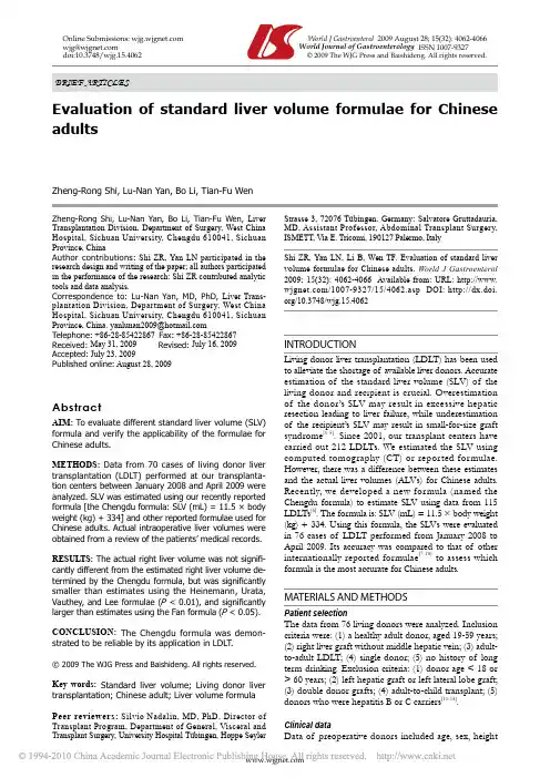
Zheng-Rong Shi, Lu-Nan Yan, Bo Li, Tian-Fu Wen, Liver Transplantation Division, Department of Surgery, West China Hospital, Sichuan University, Chengdu 610041, Sichuan Province, ChinaAuthor contributions: Shi ZR, Yan LN participated in the research design and writing of the paper; all authors participated in the performance of the research; Shi ZR contributed analytic tools and data analysis.Correspondence to: Lu-Nan Yan, MD, PhD, Liver Trans-plantation Division, Department of Surgery, West China Hospital, Sichuan University, Chengdu 610041, Sichuan Province, China. yanlunan2009@ Telephone: +86-28-85422867 Fax: +86-28-85422867 Received: May 31, 2009Revised: July 16, 2009 Accepted: July 23, 2009Published online: August 28, 2009AbstractAIM: To evaluate different standard liver volume (SLV) formula and verify the applicability of the formulae for Chinese adults.METHODS:Data from 70 cases of living donor liver transplantation (LDLT) performed at our transplanta-tion centers between January 2008 and April 2009 were analyzed. SLV was estimated using our recently reported formula [the Chengdu formula: SLV (mL) = 11.5 × body weight (kg) + 334] and other reported formulae used for Chinese adults. Actual intraoperative liver volumes were obtained from a review of the patients’ medical records. RESULTS:The actual right liver volume was not signifi -cantly different from the estimated right liver volume de-termined by the Chengdu formula, but was signifi cantly smaller than estimates using the Heinemann, Urata, Vauthey, and Lee formulae (P < 0.01), and signifi cantly larger than estimates using the Fan formula (P < 0.05). CONCLUSION: The Chengdu formula was demon-strated to be reliable by its application in LDLT.© 2009 The WJG Press and Baishideng. All rights reserved.Key words: Standard liver volume; Living donor liver transplantation; Chinese adult; Liver volume formulaPeer reviewers: Silvio Nadalin, MD, PhD, Director of Transplant Program, Department of General, Visceral and Transplant Surgery, University Hospital Tübingen, Hoppe Seyler Strasse 3, 72076 Tübingen, Germany; Salvatore Gruttadauria, MD, Assistant Professor, Abdominal Transplant Surgery, ISMETT, Via E. Tricomi, 190127 Palermo, ItalyShi ZR, Yan LN, Li B, Wen TF. Evaluation of standard liver volume formulae for Chinese adults. World J Gastroenterol 2009; 15(32): 4062-4066 Available from: URL: http://www. /1007-9327/15/4062.asp DOI: http://dx.doi. org/10.3748/wjg.15.4062INTRODUCTIONLiving donor liver transplantation (LDLT) has been used to alleviate the shortage of available liver donors. Accurate estimation of the standard liver volume (SLV) of the living donor and recipient is crucial. Overestimation of the donor’s SLV may result in excessive hepatic resection leading to liver failure, while underestimation of the recipient’s SLV may result in small-for-size graft syndrome[1-5]. Since 2001, our transplant centers have carried out 212 LDLTs. We estimated the SLV using computed tomography (CT) or reported formulae. However, there was a difference between these estimates and the actual liver volumes (ALVs) for Chinese adults. Recently, we developed a new formula (named the Chengdu formula) to estimate SLV using data from 115 LDLTs[6]. The formula is: SLV (mL) = 11.5 × body weight (kg) + 334. Using this formula, the SLVs were evaluated in 76 cases of LDLT performed from January 2008 to April 2009. Its accuracy was compared to that of other internationally reported formulae[7-10] to assess which formula is the most accurate for Chinese adults. MATERIALS AND METHODSPatient selectionThe data from 76 living donors were analyzed. Inclusion criteria were: (1) a healthy adult donor, aged 19-59 years;(2) right liver graft without middle hepatic vein; (3) adult-to-adult LDLT; (4) single donor; (5) no history of long term drinking. Exclusion criteria: (1) donor age < 18 or > 60 years; (2) left hepatic graft or left lateral lobe graft;(3) double donor grafts; (4) adult-to-child transplant; (5) donors who were hepatitis B or C carriers[11-14].Clinical dataData of preoperative donors included age, sex, heightOnline Submissions: World J Gastroenterol 2009 August 28; 15(32): 4062-4066 wjg@ World Journal of Gastroenterology ISSN 1007-9327doi:10.3748/wjg.15.4062 © 2009 The WJG Press and Baishideng. All rights reserved.Evaluation of standard liver volume formulae for Chinese adultsZheng-Rong Shi, Lu-Nan Yan, Bo Li, Tian-Fu WenBRIEF ARTICLES(BH, measured to the nearest 1 cm), body weight (BW , measured to the nearest 0.5 kg), and body surface area (BSA) calculated using the DuBois formula: BSA (m 2) = BW (kg) 0.425 × BH (cm) 0.725 × 0.007184 or the Mosteller formula: BSA (m 2) = square root BH (cm) × BW (kg)/3600. From the diaphragm to the superior mesenteric artery plane, the entire liver image was scanned using a 7 mm thick layer. In the Leonardo workstation, the LV was measured by venous phase images [15,16]. All preoperative CT examinations of donors were performed by a single radiologist and all donor procedures were performed by the same surgical unit. The volume of the grafts was measured by a 3 L beaker using a drainage method intraoperatively and the error was less than 10 mL [17,18].Right liver graft without middle hepatic vein reconstruction from a living donor was performed as described, with temporary occlusion of the right portal vein (PV) and right hepatic artery and use of ultrasonography to guide parenchymal transection. The right hepatic duct, right hepatic artery, right portal vein branch, and right hepatic vein were transected approximately 2-3 mm from the con fl uence [19,20], leaving the donor’s main PV and confluence intact. The graft was flushed with University of Wisconsin solution through the PV and hepatic artery [21,22].The volume of 70 livers was calculated using the Chengdu standard LV formula [6] as described above. The estimated right LV (ERLV) was obtained by multiplying the SLV by the proportion of the LV contributed by the right lobe on CT. The actual right LV (ARLV) was obtained by intraoperative measurement. The differencebetween the ERLV and ARLV was statistically evaluated. The formulae of Heinemann et al [8], Urata et al [7], Vauthey et al [9], Lee et al [5], and Fan et al [4] in addition to our own formula [6] were used to determine the estimated SLV (ESLV) of our donor livers. The previously reported formulae are shown in Table 1. For each liver, we calculated the difference between the ALV and volume estimated by each formula (ELV).Statistical analysisAfter testing for normal distribution (kurtosis and skewness tests), descriptive statistics were calculated and data were expressed as means ± SD for age (year), BW (kg), BH (cm), body mass index (BMI), and BSA. The ERLV-ARLV and the ELV-ALV were compared by the 2-sided paired-samples t -test. P < 0.05 was considered statistically significant. All statistical analyses were performed using the SPSS (version 13.0) program.RESULTSSeventy donors (all Chinese; 53 men and 17 women; mean age, 32.21 ± 10.07; range, 19-57 years) met the selection criteria. All donors were related to the recipients.The characteristics of donors are shown in Table 2. All donors were considered healthy on the basis of BMI. All but one donor with a BMI of 17 kg/m 2 had a BMI of 18-28 kg/m 2. The mean volume of the right lobe on CT was 658.98 ± 81.14 mL and represented 55.4% ± 3.7% of the whole liver on CT.The mean ELV and mean ERLV using the Chengdu standard formula were 1058.70 ± 96.74 mL and 586.15 ± 67.17 mL, respectively. The mean ARLV was 578.58 ± 72.33 mL. Differences for individual donors between ERLV and ARLV were not signi fi cant (t = -1.882, P = 0.064). A plot of the relationship of ARLV to the ERLV calculated using the Chengdu formula is shown in Figure 1. The mean total LV determined preoperatively on CT was 1189.53 ± 114.75 mL. The mean RLV on CT without the middle hepatic vein was 658.98 ± 81.14 mL, and 55.4% ± 3.7% of the total LV . The ALV calculated from the volume of the graft and the ratio of the RLV to the total LV on CT (%) was 1050.10 ± 107.41 mL. The Heinemann, Urata, Vauthey, and Lee formulae signi fi cantly overestimated the LV (P < 0.01), while the Fan formula significantly underestimated the LV (P <Shi ZR et al . Liver volume formulae for Chinese adults 4063ESLV: Estimated standard liver volume; BSA: Body surface area; BW: Body weight; CT: Computed tomography; LDLT: Living donor liver transplantation.Table 2 Donor characteristicsMHV: Middle hepatic vein.0.05). There was no signi fi cant difference between ALV and ELV using the Chengdu formula (Figure 2).DISCUSSIONCT has become a standard method for assessing livergraft volume in living donors. Estimation of LV by CT (compared to actual volume) has a margin of error of 5%-25%[23,24]. In the present study, all donors had preoperative CT assessment of LV (mean total LV , 1189.53 ± 114.75 mL and mean volume of right lobe graft without middle hepatic vein, 658.98 ± 81.14 mL). The actual volume of the right liver was 578.58 ± 72.33 mL. In the present study, the LV on CT was 10%-20% higher than the ALV [25-27]. The reasons may be as follows: (1) Preoperative CT measurement is carried out under normal blood fl ow conditions. Perioperatively, liver resection interrupts the blood supply causing a loss of liquid volume, collapse of supporting structures, and thereby reduction in the volumeof the liver. (2) Sources of error (partial volume effect, inter-observer variation, and respiratory movements) may account for this difference [28].The difference between the ERLV (using our formula) and ARLV was compared to the difference between ERLV, calculated using the formulae of Heinemann, Urata, Vauthey, Lee, and Fan, and ARLV in our 70 donors. The Heinemann, Urata, Vauthey, and Lee formulae overestimated LV (P < 0.01)[29]. The reasons may include: ethnic differences (patients in Europe and the United States were Caucasian). All except the Sheung Tat Fan and Chengdu formulae were used to estimate LV from CT LV or autopsy LV. Estimates of LV by CT were 5%-25% higher than the ALV [30].Statistical analysis showed that the Fan formula tends to underestimate LV. The weight and height of the donors in our study were higher than of those in the Hong Kong group. This may be one of the reasons both results are very close (Table 3). Above all, we believe thatALV: Actual liver volume; ND: Not determined.Figure 1 Correlation between actual right liver volume (ARLV) and estimated right liver volume (ERLV) by the Chengdu formula. When both were the same, a dot would be on the linear line.500 600 700Graft ARLV700600500E R L VFigure 2 Correlation between actual liver volume (ALV) and estimated liver volume (ELV) by each formula. When both were the same, a dot would be on the linear line. Formulae of Urata, Heinemann, Vauthey, and Lee overestimated LV with respect to ALV. The Fan formula underestimated LV and the Chengdu formula gave a good estimate of ALV.4064 ISSN 1007-9327 CN 14-1219/R World J Gastroenterol August 28, 2009 Volume 15 Number 32700 900 1100 1300 1500ALV150013001100900700U r a t a700 1000 13001600ALV160013001000700H e i n e m a n n700 900 1100 1300 1500ALV150013001100900700L e e700 1000 1300 1600ALV160013001000700V a u t h e y700 900 1100 1300 1500ALV150013001100900700F a n700 800 900 1000 1100 1200 1300ALV14001300120011001000900800700C h e n g d uthe Chengdu formula was demonstrated to be reliable by its application in LDLT. We were limited to use of single center data in the present study, but we hope to improve the formula by using national multicenter data in the future[31].With development of living donor liver transplantation (LDLT), especially improvement of right graft adult-to-adult LDLT, the danger of donating has been paid more and more attention. The exact liver volume is not only relevant for the recipient, but also for the donor to avoid dangerous life-threatening residual liver volumes.Research frontiersScholars of different countries established several standard liver volume (SLV) formulae from clinical data. The authors estimated the SLV using computed tomography or reported formulae. However, there was a gap between these estimates and the actual liver volumes for Chinese adults. Recently, they developed a new formula (named the Chengdu formula) to estimate SLV using data from 115 LDLTs.Innovations and breakthroughsWith the Chengdu formula, the SLVs were evaluated in 76 cases of LDLT performed from January 2008 to April 2009. Its accuracy was compared to that of other internationally reported formulae to assess which formula is the most accurate for Chinese adults.ApplicationsWith national multicenter data in the future, the Chengdu formula for SLV can be improved. It may then be applied to the evaluation of donors for LDLT. TerminologyStandard liver volume: normal liver volume without disease affecting the volume of liver.Peer reviewVery interesting manuscript dealing with a very hot topic: determination of optimal size matching between graft and recipient in LDLT by means of race-adapted calculation of liver volumes. The recently published liver volume formula for Chinese people (Chengdu formula) has been demonstrated to be more reliable than others and therefore it should be adopted especially in this particular form of LT.REFERENCES1 Khalaf H, Shoukri M, Al-Kadhi Y, Neimatallah M, Al-Sebayel M. Accurate method for preoperative estimation of the right graft volume in adult-to-adult living donor liver transplantation. Transplant Proc 2007; 39: 1491-14952 Avolio AW, Siciliano M, Barbarino R, Nure E, AnnicchiaricoBE, Gasbarrini A, Agnes S, Castagneto M. Donor risk index and organ patient index as predictors of graft survival after liver transplantation. Transplant Proc 2008; 40: 1899-19023 Gruttadauria S, Marsh JW, Vizzini GB, di Francesco F,Luca A, Volpes R, Marcos A, Gridelli B. Analysis of surgical and perioperative complications in seventy-five right hepatectomies for living donor liver transplantation. World J Gastroenterol 2008; 14: 3159-31644 Fan ST, Lo CM, Liu CL, Yong BH, Chan JK, Ng IO. Safety ofdonors in live donor liver transplantation using right lobe grafts. Arch Surg 2000; 135: 336-3405 Lee SG, Park KM, Hwang S, Lee YJ, Kim KH, Ahn CS, ChoiDL, Joo SH, Jeon JY, Chu CW, Moon DB, Min PC, Koh KS, Han SH, Park SH, Choi GT, Hwang KS, Lee EJ, Chung YH, Lee YS, Lee HJ, Kim MH, Lee SK, Suh DJ, Kim JJ, Sung KB.Adult-to-adult living donor liver transplantation at the Asan Medical Center, Korea. Asian J Surg 2002; 25: 277-2846 Li FG, Yan LN, Li B, Zeng Y, Wen TF, Xu MQ, Wang W.Estimation formula of standard liver volume for Chinese adults. Sichuan Daxue Xuebao 2009; 40: 302-3067 Urata K, Kawasaki S, Matsunami H, Hashikura Y, IkegamiT, Ishizone S, Momose Y, Komiyama A, Makuuchi M.Calculation of child and adult standard liver volume for liver transplantation. Hepatology 1995; 21: 1317-13218 Heinemann A, Wischhusen F, Puschel K, Rogiers X.Standard liver volume in the Caucasian population. Liver Transpl Surg 1999; 5: 366-3689 Vauthey JN, Abdalla EK, Doherty DA, Gertsch P,Fenstermacher MJ, Loyer EM, Lerut J, Materne R, Wang X, Encarnacion A, Herron D, Mathey C, Ferrari G, Charnsangavej C, Do KA, Denys A. Body surface area and body weight predict total liver volume in Western adults.Liver Transpl 2002; 8: 233-24010 Chan SC, Liu CL, Lo CM, Lam BK, Lee EW, Wong Y, FanST. Estimating liver weight of adults by body weight and gender. World J Gastroenterol 2006; 12: 2217-222211 Trotter JF, Wisniewski KA, Terrault NA, Everhart JE,Kinkhabwala M, Weinrieb RM, Fair JH, Fisher RA, Koffron AJ, Saab S, Merion RM. Outcomes of donor evaluation in adult-to-adult living donor liver transplantation. Hepatology 2007; 46: 1476-148412 Duran C, Aydinli B, Tokat Y, Yuzer Y, Kantarci M, AkgunM, Polat KY, Unal B, Killi R, Atamanalp SS. Stereological evaluation of liver volume in living donor liver trans-plantation using MDCT via the Cavalieri method. Liver Transpl 2007; 13: 693-69813 Morimoto T, Ichimiya M, Tanaka A, Ikai I, Yamamoto Y,Nakamura Y, Takada Y, Inomata Y, Honda K, Inamoto T, Tanaka K, Yamaoka Y. Guidelines for donor selection and an overview of the donor operation in living related liver transplantation. Transpl Int 1996; 9: 208-21314 Yamashiki N, Sugawara Y, Tamura S, Kaneko J, NojiriK, Omata M, Makuuchi M. Selection of liver-transplant candidates for adult-to-adult living donor liver transplantation as the only surgical option for end-stage liver disease. Liver Transpl 2006; 12: 1077-108315 Frericks BB, Kirchhoff TD, Shin HO, Stamm G, MerkesdalS, Abe T, Schenk A, Peitgen HO, Klempnauer J, Galanski M, Nashan B. Preoperative volume calculation of the hepatic venous draining areas with multi-detector row CT in adult living donor liver transplantation: Impact on surgical procedure. Eur Radiol 2006; 16: 2803-281016 Iida T, Yagi S, Taniguchi K, Hori T, Uemoto S, YamakadoK, Shiraishi T. Signifi cance of CT attenuation value in liver grafts following right lobe living-donor liver transplantation.Am J Transplant 2005; 5: 1076-108417 Kim BS, Kim TK, Kim JS, Lee MG, Kim JH, Kim KW, SungKB, Kim PN, Ha HK, Lee SG, Kang W. Hepatic venous congestion after living donor liver transplantation with right lobe graft: two-phase CT fi ndings. Radiology 2004; 232: 173-18018 Cho JY, Suh KS, Lee HW, Cho EH, Yang SH, Cho YB, Yi NJ,Kim MA, Jang JJ, Lee KU. Hypoattenuation in unenhanced CT reflects histological graft dysfunction and predicts 1-year mortality after living donor liver transplantation.Liver Transpl 2006; 12: 1403-141119 Choi JY, Lee JY, Lee JM, Kim SH, Lee MW, Han JK, ChoiBI. Routine intraoperative Doppler sonography in the evaluation of complications after living-related donor liver transplantation. J Clin Ultrasound 2007; 35: 483-49020 Kato H, Usui M, Azumi Y, Ohsawa I, Kishiwada M, SakuraiH, Tabata M, Isaji S. Successful laparoscopic splenectomy after living-donor liver transplantation for thrombocytopenia caused by antiviral therapy. World J Gastroenterol 2008; 14: 4245-424821 Ohdan H, Tashiro H, Ishiyama K, Ide K, Shishida M, IreiT, Ohira M, Tahara H, Itamoto T, Asahara T. Microsurgical hepatic artery reconstruction during living-donor liver transplantation by using head-mounted surgical binocular system. Transpl Int 2007; 20: 970-97322 Oya H, Sato Y, Yamamoto S, Takeishi T, Nakatsuka H,Kobayashi T, Hara Y, Hatakeyama K. Surgical proceduresShi ZR et al. Liver volume formulae for Chinese adults 4065for decompression of excessive shear stress in small-for-size living donor liver transplantation--new hepatic vein reconstruction. Transplant Proc 2005; 37: 1108-111123 Lee SS, Kim KW, Park SH, Shin YM, Kim PN, Lee SG, LeeMG. Value of CT and Doppler sonography in the evaluation of hepatic vein stenosis after dual-graft living donor liver transplantation. AJR Am J Roentgenol 2007; 189: 101-10824 Asakuma M, Fujimoto Y, Bourquain H, Uryuhara K, HayashiM, Tanigawa N, Peitgen HO, Tanaka K. Graft selection algorithm based on congestion volume for adult living donor liver transplantation. Am J Transplant 2007; 7: 1788-179625 del Pozo JL. Update and actual trends on bacterial infectionsfollowing liver transplantation. World J Gastroenterol 2008;14: 4977-498326 Miraglia R, Maruzzelli L, Caruso S, Milazzo M, Marrone G,Mamone G, Carollo V, Gruttadauria S, Luca A, Gridelli B.Interventional radiology procedures in adult patients who underwent liver transplantation. World J Gastroenterol 2009;15: 684-69327 Shoji M, Ohkohchi N, Fujimori K, Koyamada N, SekiguchiS, Kawagishi N, Tsukamoto S, Shirahata Y, Sato K, SatomiS. The safety of the donor operation in living-donor liver transplantation: an analysis of 45 donors. Transpl Int 2003;16: 461-46428 Kawagishi N, Ohkohchi N, Fujimori K, Doi H, SakuradaM, Kikuchi H, Oikawa K, Takayama J, Satomi S. Safety of the donor operation in living-related liver transplantation: analysis of 22 donors. Transplant Proc 1998; 30: 3279-328029 Yoshizumi T, Taketomi A, Kayashima H, Yonemura Y,Harada N, Ijichi H, Soejima Y, Nishizaki T, Maehara Y.Estimation of standard liver volume for Japanese adults.Transplant Proc 2008; 40: 1456-146030 Hirata M, Harihara Y, Kitamura T, Hisatomi S, KatoM, Dowaki S, Mizuta K, Sugawara Y, Kita Y, Kubota K, Takayama T, Kawarasaki H, Hashizume K, Makuuchi M.The infl uence of donor age to graft volume increase rate in living donor liver transplantation. Transplant Proc 2001; 33: 1416-141731 Schiano TD, Bodian C, Schwartz ME, Glajchen N, Min AD.Accuracy and signifi cance of computed tomographic scan assessment of hepatic volume in patients undergoing liver transplantation. Transplantation 2000; 69: 545-550S- Editor Tian L L- Editor Cant MR E- Editor Ma WH4066 ISSN 1007-9327 CN 14-1219/R World J Gastroenterol August 28, 2009 Volume 15 Number 32。
肝脏体积变化的判断
肝脏体积变化的判断
(1).无论肝脏肿大还是缩小,均以肝脏高度变化为主。
(2).肝门至膈顶高<6.0cm,肝门至膈顶高
/肝右叶厚<0.65,即可认为肝脏
体积缩小。
肝门至膈顶高度为5~6cm者,肝脏轻度缩小;4~5cm者,肝脏中度缩小;4cm以下者,重度缩小。
(3).肝门至膈顶高>9.5cm,其与右叶厚度比>0.85,可以考虑肝脏肿大,肝门至膈顶高度9.5~10.5cm为轻度肿大;10.5~11.5cm为中度肿大;11.5cm以上为重度肿大。
(4).一些身材矮小、体重轻的个体,往往肝脏体积小于平均值,只要肝门至膈顶高/肝右叶厚度在0.65~0.85,就不能认为肝体积
缩小,只能考虑个体差异。
而在肝大的部分患者中,如果这一比值保持在0.85以内,肝脏形态往往保持正常,脂肪肝人群多属这种情况。
肝脏体积缩小评估标准
肝脏体积缩小评估标准
肝脏的体积缩小可以分为几种情况,如果在先天体检的时候发现肝脏的体积偏小,甚至某个肝叶缺如,这个时候有可能跟先天的一些代谢、发育有关,可能先天的某个肝叶发育不全,但是如果是疾病状态下的肝脏体积缩小,这个时候就要高度怀疑患者出现了肝硬化的表现。
肝脏实际上有正常大小,通过一些测量数据,比如做肝脏的超声,可以看到肝脏的右叶斜径大概在12~14厘米,成人是这样的标准,左叶的前后径大概是在6厘米以内,它的上下径是在9厘米以内。
正常人的肝脏,它的一些胆管或者是门脉系统都是有相应的一些正常值的范围。
肝脏的体积缩小,往往指的就是低于这些测量的正常数值,这种缩小如果在疾病状态下,如果患者有病毒性肝炎,乙型肝炎、丙型肝炎或者是有一些慢性的肝脏疾病,出现了肝脏的缩小,就一定要注意观察患者是不是有肝硬化的表现,这个时候可以通过像肝脏弹性测定,或者是做一些比如病理学的肝脏穿刺检查,或者做一些生化学的检查来综合帮助判断,这种体积缩小是否是一个病理意义上的体积缩小,是否是一种疾病终末期的表现。
肝脏结构及相关病理常识

肝脏组织学—肝脏结构和功能单位
• 肝脏外层富含弹性纤维的致密结缔组织--被膜
• 肝小叶——六角形棱柱状(2x1mm),成人50 - 100万
肝窦内腔大不规则,窦壁由内皮细胞构成,肝窦内有肝巨噬细胞和大颗粒淋巴细胞 4μm)光镜下极难辨认 肝大部分位于右季肋区和腹上区,小部分达左季肋区
• 肝略呈现楔形,右端园钝 而厚,左端逐渐变窄而薄
• 肝可分为上下两面,(膈面、 脏面),前后左右4缘
• 膈面上有矢状位的镰状韧 带
肝脏膈面——
接膈的下面与膈穹窿相适应,呈向上的隆凸
镰状韧带将肝脏分为: 肝右叶、肝左叶 膈面的上部与右侧膈胸膜、右肺底相邻, 故肝脓肿或囊肿可经膈破溃到右侧胸膜腔及右膈
• 胎儿和新生儿的肝相对较大,可占体重的4%~5%
如:1个月、身长51厘米的男婴肝脏重约140克
特点:最大的消化腺(最大腺体)
肝脏位置
• 肝大部分位于右季肋区和 腹肋所掩盖,仅 在腹上区的左、右肋弓之间 有小部分露于剑突下
• 在呼吸时肝可随膈肌上下 移动,平静呼吸时肝的上下 移动范围为2~3cm
中央静脉 肝脏组织学—肝脏结构和功能单位
滑膜内质网数量少于粗面内质网
肝小叶 肝板(肝细胞索),界板 中部呈横位的沟称肝门,有肝管、淋巴管、门静脉、肝固有动脉左右支、肝的神经等出入
腹壁浅静脉、胸、腹壁静脉 由于门静脉循环障碍、血流受阻,可以引起脾大、胃肠淤血,成为产生门脉高压性腹水的原因
肝血窦 成分:有机成分为胆盐、胆色素、胆固醇、脂肪酸、卵磷脂等;
肝脏组织学—肝脏结构和功能单位
肝细胞
• 肝细胞是肝脏的实质细胞,约占小叶体积80%,占肝内所有细胞 的90%
标准肝体积计算公式

标准肝体积计算公式
肝脏体积是评估肝功能和疾病诊断中的重要指标之一。
下面我将介绍一种常用
的计算肝脏体积的标准公式。
在临床上,常用的计算肝脏体积的公式是根据肝脏的长轴、短轴和横断面面积
来计算的。
这个公式被称为康奈尔公式,公式如下:
肝脏体积(cm³)= 0.52 ×长轴(cm)×短轴(cm)×横断面面积(cm²)
根据这个公式,我们可以通过测量肝脏的长轴和短轴,以及计算肝脏横断面的
面积,来估算肝脏的体积。
这种方法相对简便,并被广泛应用于临床实践中。
为了测量肝脏的长轴和短轴,常常使用超声或影像学技术,如计算机断层扫描(CT)或磁共振成像(MRI)。
通过这些技术,医生可以获取肝脏的长轴和短轴
的尺寸,然后用这些尺寸计算出肝脏的体积。
需要注意的是,在计算肝脏体积时,使用的单位必须一致。
一般情况下,公式
中的尺寸都以厘米(cm)为单位,面积以平方厘米(cm²)为单位。
保持单位的一
致性非常重要,以确保计算结果的准确性。
总结起来,标准肝脏体积计算公式是康奈尔公式,通过测量肝脏的长轴和短轴,并计算出肝脏的横断面面积,可以估算肝脏的体积。
这个公式在临床实践中是一种常用且可靠的计算方法,帮助医生评估肝功能和进行疾病诊断。
肝缩小的概念
肝缩小的概念肝缩小是指肝脏的体积变小,通常是由于肝细胞减少或萎缩所引起的。
正常情况下,成年人的肝脏体积约为1200-1600毫升,相对于身体重量而言,大约占体重的2.5%-3.5%。
如果肝脏的体积减小到正常范围以下,则可以被称为肝缩小。
肝缩小可以由多种原因引起,包括疾病、营养不良、外伤和药物等。
下面将详细介绍这些因素对肝缩小的影响:1. 疾病:肝硬化、慢性肝炎、急性肝炎等疾病都可以导致肝细胞的损伤和死亡,从而引起肝缩小。
其中,肝硬化是最常见的原因之一,它使得肝脏组织逐渐纤维化和硬化,从而导致肝脏的体积减小。
2. 营养不良:长期不良的饮食习惯、营养摄入不足或吸收不良等因素可以影响肝细胞的生长和代谢,导致肝缩小。
营养不良还会使得肝脏的储备能力下降,进一步影响肝脏的体积。
3. 外伤:严重的外伤,如肝脏挫伤、刺伤或手术等,都可以引起肝细胞的损伤和死亡。
在愈合过程中,受伤部位的肝细胞可能会减少,从而导致肝缩小。
4. 药物:某些药物,尤其是长期大剂量使用的药物,可以对肝细胞产生毒性作用,从而导致肝脏损伤和萎缩。
常见的有抗癫痫药物、某些抗生素和镇痛药物等。
在临床上,肝缩小通常会伴随一些症状和体征,包括乏力、肝区疼痛、寒战、食欲不振、腹胀等。
由于肝缩小意味着肝脏的功能减弱,所以患者还可能会出现黄疸、腹水、血小板减少等临床表现。
对于肝缩小的诊断,通常需要通过临床检查和辅助检查来确定。
临床上可以通过触诊检查来初步判断肝脏的大小和质地。
而辅助检查包括血液和影像学检查。
血液检查可以评估肝功能和病因,如肝功能指标、病毒抗体和免疫球蛋白水平等。
影像学检查,如B超、CT或MRI等,可以直观地观察肝脏的形态和体积。
对于肝缩小的治疗,需要根据其具体原因进行针对性的治疗。
例如,如果是由于肝硬化引起的肝缩小,治疗的重点是改善病因因素,如治疗肝炎、酒精戒断、改善营养状况等。
对于营养不良引起的肝缩小,可以通过饮食调理和补充适当的营养物质来改善肝细胞的生长和代谢功能。
肝脏
脾脏实质性肿瘤
脾脏原发性肿瘤少见。良性肿瘤以血 管瘤多见,恶性肿瘤以淋巴瘤多见。 超声表现:脾内显示实质性团块回声, 团块大小、数目及内部回声不一 脾血管瘤:脾内显示边界清晰的偏高 回声团,有“网络状”表现。CDFI: 团块周围及内部可有脾动、静脉的分 支绕行或穿行。 恶性淋巴瘤:脾内单个或多个低回声 结节,边界清或相互融合,也可呈 “蜂窝状”排列。原发性恶性肿瘤内 血供丰富,表现为高速的动脉血流。
44
(1)小结节性肝硬化--门 脉性 病因:慢性肝炎,酒精 中毒,营养缺乏和毒物 中毒。 病理:初期肝体积正常 或略增大,晚期肝体积 缩小,以右叶为著,变 硬,表面呈颗粒状或小 结节状,结节周围为纤 维组织条索包绕。
45
(2)大结节性肝硬化-坏死后性: 病因:肝炎病毒感染、 药物或其它传染物质 中毒。 病理:肝细胞广泛性 坏死,其后在肝实质 坏死区出现大量结缔 组织增生和肝细胞再 生现象。
42
肝 硬 化
Cirrhosis
常见慢性疾病,病理变化:肝细胞变性,坏死和再 生,纤维组织增生,使正常肝结构呈结节样变,缩 小,质地变硬。
43
按病因分类:病毒性肝炎、酒精性、胆汁性、隐源性。
按形态分类:小结节型、大结节型、大小结节混合型 及不全分隔型。 我国常用的分类是结合病因及病变的综合分类:门脉 性、坏死后性、胆汁性、淤血性、寄生虫性和色素性 肝硬化。 坏死后性相当于大结节及大小结节混合型,其余均相 当于小结节型。
17
四、正常肝脏声像图
●形态:呈楔形。 ●大小:左叶小、薄。 右叶大、厚。 ●边缘:锐角——左叶下缘<45度 右叶下缘<75度 ●表面:光滑 ●肝实质回声:肾实质<肝≤胰腺
标准肝体积计算公式
标准肝体积计算公式肝脏是人体内重要的器官之一,它不仅具有排毒、合成蛋白质和胆汁等重要功能,还承担着调节血糖和脂肪代谢的重要作用。
因此,了解肝脏的生理特性和计算肝体积的标准公式对于临床诊断和治疗具有重要意义。
本文将介绍肝体积的计算方法以及其在临床上的应用。
肝体积的计算是通过影像学检查得出的,常用的方法有CT扫描、MRI和B超等。
在进行肝体积计算时,需要准确测量肝脏的长度、宽度和厚度,并根据这些数据应用相应的计算公式得出肝体积。
下面将介绍标准的肝体积计算公式:肝体积(ml)=0.55×长轴(cm)×短轴(cm)×厚度(cm)。
在这个公式中,长轴、短轴和厚度分别代表肝脏在影像上的三个方向的尺寸。
通过测量这些尺寸并代入公式,便可以得出肝体积的近似数值。
需要注意的是,这个计算公式是基于椭球体积的计算方法,因此在实际应用中可能存在一定的误差,但一般来说误差范围在可接受范围内。
肝体积的计算对于临床诊断和治疗具有重要的指导意义。
首先,肝体积的变化可以反映肝脏的生理状态,如肝硬化、脂肪肝等疾病会导致肝体积的改变,因此通过肝体积的计算可以对这些疾病进行诊断和监测。
其次,在肝移植手术中,肝体积的计算可以帮助医生评估移植肝的大小和适配性,从而提高手术的成功率。
此外,肝体积的计算还可以指导肝肿瘤的治疗,如手术切除、放疗和化疗等。
总之,肝体积的计算公式是临床医学中的重要工具,它可以帮助医生了解肝脏的生理状态,指导临床诊断和治疗。
通过准确测量肝脏的尺寸并应用标准的计算公式,可以得出肝体积的近似数值,为临床工作提供重要参考。
希望本文的介绍能够帮助大家更好地理解肝体积的计算方法及其在临床上的应用,为医学工作者和患者提供参考和帮助。
CT测量分析肝硬化失代偿期肝脏体积的变化
文章编号:1673 2995(2021)01 0015 02·论 著·CT测量分析肝硬化失代偿期肝脏体积的变化王 琛 (庆云县人民医院影像科,山东德州 253700)摘 要:目的 探讨CT测量分析原发性胆汁性肝硬化(PBC)失代偿期患者肝脏体积变化情况。
方法 选取PBC失代偿期患者53例纳入观察组,另选同期非PBC失代偿期患者53例纳入对照组。
两组均接受腹部螺旋CT增强扫描。
比较两组患者肝脏体积及标准化肝体积、肝脏体积增大情况、淋巴结增大分布位置、侧支循环情况。
结果 观察组患者肝脏体积(1251±200)cm3、标准化肝体积(694±120)cm3显著大于对照组(P<0.05)。
观察组患者共检出62枚腹部淋巴结,对照组共检出8枚腹部淋巴结。
观察组53例患者均出现了侧支循环。
结论 PBC失代偿期患者肝脏CT影像显示体积增大,以腹膜后淋巴结增大为主,且均有侧支循环发生。
关 键 词:CT;原发性胆汁性肝硬化;失代偿期;肝脏体积;侧支循环中图分类号:R575.2 文献标志码:ACTmeasurementoflivervolumeindecompensatedcirrhosisWANGChen (DepartmentofImaging,QingyunPeople sHospital,Dezhou,ShandongProvince,253700,China)Abstract:Objective Toinvestigatethechangesoflivervolumeinpatientswithdecompensatedprimarybiliarycir rhosis(PBC)measuredbyCT.Methods Fifty threecasesofPBCdecompensatedpatientswereselectedastheob servationgroup,and53casesofnonPBCdecompensatedpatientsinthesameperiodwereselectedasthecontrolgroup.BothgroupsreceivedabdominalspiralCTenhancedscan.Thelivervolume,standardizedlivervolume,livervol umeenlargement,lymphnodeenlargementandcollateralcirculationwerecomparedbetweenthetwogroups.Results Thelivervolume(1251±200)cm3andstandardizedlivervolume(694±120)cm3intheobservationgroupweresignificantlyhigherthanthoseinthecontrolgroup(P<0.05).Sixty twoabdominallymphnodesweredetectedintheobservationgroupand8inthecontrolgroup.Fifty threepatientsintheobservationgrouphadcollateralcirculation.Conclusion Thevolumeoftheretroperitoneallymphnodeisincreasedinallpatients.Keywords:CT;primarybiliarycirrhosis;decompensatedstage;livervolume;collateralcirculation 原发性胆汁性肝硬化(primarybiliarycirrhosis,PBC)失代偿期主要表现为肝功能不全或门静脉高压症[1],主要累及肝内细小胆管,因此导致肝脏体积增大[2]。