三种不同脂肪染色方法的比较
“生物组织中脂肪的鉴定”实验方法的拓展

“生物组织中脂肪的鉴定”实验方法的拓展[摘要]通过撕取内表皮法、血涂片法、压榨法、水油分界法、滴加法等对脂肪的鉴定实验进行改进和优化,提高实验成功率。
[关键词]脂肪的鉴定苏丹Ⅲ试剂撕取内表皮法血涂片法压榨法水油分界法滴加法本实验在必修一教材中属于必做实验,但因其对实验材料的选择和处理要求较高,如花生要提前3-4小时浸泡,浸泡时间过短不易切片,过长则切下的薄片不易成型,还有高倍镜的使用等等。
尤其是要求切出在显微镜下可清晰观察到脂肪粒的花生薄片,这对于几乎没有切片基本功的高一新生来说难度较大、成功率较低。
笔者通过撕取内表皮法、血涂片法、压榨法、水油分界法、滴加法等对脂肪的鉴定实验进行改进和优化,在实验材料的选择、实验方法和实验过程方面进行了一些改进,取得的较好的效果,提高实验成功率,同时也增加学生的学习兴趣和自信心。
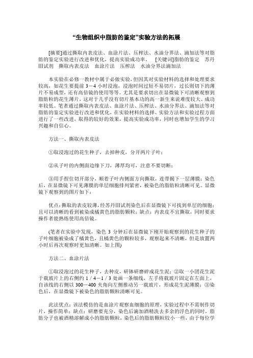
方法一、撕取内表皮法①取浸泡过的花生种子,去掉种皮,分开两片子叶;②从子叶的内侧面边缘下刀,薄厚均可,注意不要切断;③用手捏住切开部分,顺着子叶内侧面方向撕取,连带揭下一层薄膜;染色后,在显微镜下可见薄膜的单层细胞排列紧密,被染色的脂肪粒清晰可见。
显微镜下观察到的图片如下:优点:撕取的表皮较薄,经苏丹Ⅲ试剂染色后在显微镜下可找到单层的细胞;且可以清晰的看到被染成橘黄色的脂肪颗粒;缺点:内表皮不宜撕取,同时要求操作者能熟练使用高倍镜。
(笔者在实验中发现,染色3分钟后在显微镜下刚开始观察到的花生种子的子叶细胞被染成了橘黄色,且橘黄色的颗粒较多,观察起来不清晰,但是放置两小时后再次观察时更加清晰。
如上图)方法二、血涂片法①取浸泡过的花生种子,去种皮,研钵研磨碎成花生泥;②取一小团花生泥于载玻片上的右侧约1/4-1/3处画一条细线,左手将载玻片固定在左面上,自该线的右侧以300-400夹角向左侧推动另一载玻片,形成花生泥薄膜;③染色后,在显微镜下被染色的脂肪颗粒清晰可见。
此法优点:该法模仿的是血涂片观察血细胞的原理,实验过程中不需制作切片,操作简单;缺点:研磨要充分,染色后滴加酒精洗去多余的浮色的同时,脂肪分子也被酒精溶解成小的脂肪颗粒,染色后的脂肪颗粒较小一些,由于每位学生研磨花生时研磨的程度不同,观察到的结果也不尽相同,如有的学生研磨的不充分,细胞重叠在一起,染色后观察不到单层花生细胞,导致学生个体间的实验误差较大,同时此法要求操作者能熟练使用高倍镜。
显示脂肪的染色方法及其应用

诊断局部感染,收入外科治疗。
使用抗生素治疗1周,右臀部肿胀减轻,右大腿肿胀明显,10日后再行血常规检测,白细胞38.8×10⁹/L,血红蛋白95g/L,血小板30×109/L,淋巴细胞0.03,单核细胞0.10,中性粒细胞0.87,未作涂片镜检,故无幼稚细胞报告,而临床考虑为血液病,做骨髓穿刺,诊断为急性早幼粒细胞性白血病。
临床检验人员应熟悉急性白血病的各种临床表现特点,当血红蛋白、血小板减少,白细胞超越正常参考值,或直方图异常的血标本均应涂片镜检,防止各类白血病细胞漏检。
1.2 人员素质与高科技之间的差距 目前,部分临床检验工作者,对临床使用的进口血液分析仪性能不能全面深入了解和熟练掌握。
如我院最新引进的迈瑞BC-3000plus型全自动血液分析仪在1m in内可测出19项参数,可做出标本含义报告,该机采用国内普遍使用的电阻抗分析原理,依据血细胞经溶血剂作用后,由核和胞质颗粒结构大小决定白细胞分群结果。
由于部分检验人员对仪器性能缺乏足够的认识,误认为仪器白细胞分群结果可代替镜检分类,致白血病细胞漏检情况时有发生[3]。
如1例36岁女性患者,因鼻、牙龈出血来我院就诊,经2次血常规检查,血红蛋白110g/L,血小板32×109/L,白细胞11×109/L,淋巴细胞0.68,单核细胞0.22,中性粒细胞0.10,未作幼稚细胞报告。
医师初诊原发性血小板减少性紫癜,入院治疗第7天骨穿诊断为急性淋巴细胞性白血病,此时外周血幼稚细胞达0.36。
1.3 缺乏高度的责任心 各类白血病的诊断主要依据实验室血液和骨髓象的分析,为得出准确的结果,要求检验人员必须具有高度的责任心,熟练掌握仪器性能、操作规程,检查血液分析结果时才能做到全面、细心,及时发现白血病细胞。
目前,由于受检验人员编制的限制,各大医院的血常规检测工作量较大,如果责任心不强,仅仅是机械的操作,或仅满足简单数字分析,则极易引起白血病漏诊、误诊。
秀丽隐杆线虫脂肪染色的方法比较

秀丽隐杆线虫脂肪染色的方法比较冯婉娟;黄文明;许想平;吴政星【摘要】Obesity and its related diseases are affecting an increasing number of people.Many key fat regulatory genes and fat metabolism pathways found in mammals are conserved inC.elegans.Therefore,C.elegans is a powerful genetic model for fat biology research.Different methods were used to quantitate the fat stores,and the results turn out different.According to the test in fixed worms during fasting or gene mutation,the dye such as Oil Red O,Sudan Black B,Nile Red and C1-BODIPY-C12 could quantitate fat stores correctly.%肥胖及其相关的疾病影响了越来越多的人.在哺乳动物中调节脂肪代谢的因子和脂肪代谢途径在线虫中也是保守存在的,因此线虫是脂肪研究的良好动物模型.在研究线虫脂肪代谢中,存在多种脂肪染色方法,而不同方法所表征的脂肪含量存在一定的差异,通过各种染色方法检测基因突变或者饥饿引起的脂肪含量变化.实验结果表明,采用油红、苏丹黑、尼罗红染料的固定染色法均能够正确地表征线虫的贮存脂肪.【期刊名称】《生命科学研究》【年(卷),期】2012(016)001【总页数】6页(P9-14)【关键词】线虫;脂肪染色;荧光染料;油红;苏丹黑【作者】冯婉娟;黄文明;许想平;吴政星【作者单位】华中科技大学生命科学与技术学院中国湖北武汉430074;华中科技大学生命科学与技术学院中国湖北武汉430074;华中科技大学生命科学与技术学院中国湖北武汉430074;华中科技大学生命科学与技术学院中国湖北武汉430074【正文语种】中文【中图分类】Q504Fat metabolic pathways are conserved between C.elegans and mammals.These pathways include fatty acid synthesis,fatty acid elongation and desaturation,mitochondrial and peroxisomal β-oxidation of fatty acids,and amino acid metabolism[1~5].In addition,C.elegans store fat in droplets in their intestinal cells and hypodermal cells[4],and these fat stores can be directly visualised by microscopy in intact animals due to the transparent bodies of C.elegans.Thus,C.elegans is a powerful system for analyzing the mechanisms of fat storage.There are several methods for examining fat storage and metabolism in C.elegans.An early method for visualising fat storage is using a classic lipophilic dye,Sudan Black B,to stain fixed animals[6,7].In using this stain,lipid droplets become visiible in intestinal and hypodermal tissues.Oil Red O is also a classic lipophilic dye that stains lipid droplets red[8].Aside from colourimetric dyes,another way to stain fat is through the use of lipophilic fluorescent dyes.For example,Nile Red and C1-BODIPY-C12 both fluoresce when in hydrophobicenvironments and thus are used to stain intracellular lipid droplets and can be used to examine fat content in intactliving animals[9,10].When fed C1BODIPY-C12,worms show fluorescence not only fat stores but also other organelles containing lipids.Though these mentioned dyes label fat storage compartments,there is controversy surrounding them,mainly that their results are inconsistent[9,11].An alternative dye-labelling assay for quantifying fat stores is coherent anti-Stokes Raman scattering(CARS)microscopy[12,13],which is a label-free chemicalimagingtechniquethatrelieson intrinsic molecular vibration as a contrast mechanism.Both hypodermal and intestinal fat stores are visualized using this technique.However,the disadvantage of CARS microscopy is that specialised equipment is required and that the assay is very expensive.In this paper,we confirmed that Nile Red and C1-BODIPY-C12 could stain fat storage as efficiently as Oil Red O in fixed worms.Then,we used fixed staining with different dyes to label fat stores in well-fed and fastingC.elegans,where fat stores are present orconsumed,respectively.Finally,using different dyes we tested several mutants which are insulin pathway related or a nuclear hormone receptor of stly,we compared the use of different dyes in fat storage quantification to facilitate researchers who could benefit from such analysis.Nematode strains were obtained from the C.elegans geneticcentre(CGC)unless otherwise stated.All strains were maintained at20℃us ing standard methods[14].The strains used in this study were as follows:wild-type(N2),daf-2(e1370),nhr-49(gk405)and ZXW3 hkdEx3[sur-5::atgl-1::gfp;Rol-6].Oil Red O staining was performed as previously described[9].To permeabilize the cuticle,worms were suspended and washed twice with PBS(phosphate buffered saline)and then suspended in 120 μL ofPBS.Next,an equal volume of 2× MRWB(Modified Ruvkun’s witches brew)buffer containing 2%paraformaldehyde was added,and animals in suspension were rocked for an hour(composition:160 mmol/L KCl,40 mmol/L NaCl,14 mmol/L Na2EGTA,1 mmol/L spermidine HCl,0.4 mmol/L spermine,30 mmol/L Na PIPES at pH 7.4,0.2%βmercaptoethanol).After permeabilizing,worms were re-suspended and dehydrated in60%isopropanol for 15 min at room temperature.After allowing worms to settle,isopropanol was removed,and approximately 1 mL of 60%Oil Red O solution(Cat.No.09755,Sigma-Aldrich,St.Louis,MO,USA)was added to each sample.Samples were incubated overnight while rocking.Oil Red O was prepared as follows:0.5 g of Oil Red O powder was dissolved in 100 mL of isopropanol and equilibrated for several days.Animals were mounted and imaged using an Olympus microscope outfitted with DIC optics.For Sudan Black staining,young adult animals were fixed in2%paraformaldehyde in M9 buffer(3 g KH2PO4,6 g Na2HPO4,5 g NaCl,1 mL 1 mol/L MgSO4,adding H2O to 1 L)with rocking for an hour.Fixed worms were then washed with M9 and dehydrated through an ethanol series (25%,50%,and 70%ethanol).Staining was performed overnight in a 50%saturated solution of Sudan black B in 70%ethanol[15].Stained animals were visualised with an Olympus microscope outfitted with DIC optics.Approximately 500~1 000 nematodes were suspended in 1 mL of water,and 50 μL of freshlyprepared 10% paraformal dehyde solution was then added.Animals and paraformaldehyde solutions were mixed and rocked for an hour.Afterward,rocking was stopped,and worm solutions were allowed to settle.Then,1 mL of 100 μg/L Nile Red in M9 was added to the worm pellet,and the entire solution was incubated for 6 h at room temperature with occasional gentle agitation.Worms were allowed to settle again and then were washed once with M9 buffer.After most of the staining solution had been removed,the fixed worms were mounted onto 2%agarose pads for microscopic observation and photography[15].A stock solution (2 g/L)was made by dissolving C1-BODIPY-C12 in DMSO (Dimethyl sulfoxide).The stock solution was then diluted in 1X PBS to a final concentration of 100 μg/L C1-BODIPYC12,and fixed worms were incubated 6 h in the working solution.After most of the staining solution had been removed,the fixed worms were mounted onto 2%agarose pads for microscopic observation and photography[16].Fixe d Nile Red and C1-BODIPY-C12 stained worms were visualised using an Olympus IX 71 inverted microscope (Olympus,Japan)using a 40x objective(Olympus UPlanSApo series).Fourteen bit images were taken with a CCD camera(Andor DV885).Nile Red yellow (referred to as Nile Red 580)was visualised with 586/20 nm emission filters with excitation light of 491 nm.Image analysis was performed using Image J software(Wayne Rasband,USA).A recent study claimed that Nile Red and C1-BODIPY-C12 did not show fatstorage staining[12].When C.elegans are fed Nile Red,the dye accumulates in lysosome-related organelles[17].Therefore,it was concluded that fixed Nile Red is a better proxy for fat storage visualisation than fed Nile Red.To confirm that fixed C1-BODIPY-C12 and fixed Nile Red are both good indicators of fat stores,we co-localised fixed Nile Red and fixed C1-BODIPY-C12 images.As shown in Fig.1a,we found that fixed Nile Red and fixed C1-BODIPY-C12 co-labelled a population of structures in gut epithelial organelles.Additionally,fixed Nile Red co-localised with Oil Red O and ATGL-1::GFP (Fig.1b and 1c).ATGL-1 encodes a homology of mammalian adipose triglyceride lipase,which is a lipid dropletmarker[18].So fixed Oil Red O staining is a reliable method to measure fat stores.Thus fixed Nile Red and fixed C1-BODIPY-C12 are sufficient dyes to visualise fat stores properly in C.elegans models.We also conducted similar assays which fed Nile Red and ATGL-1::GFP were assessed for co-localisation.According to Fig.1d,fed Nile Red was not co-localised with ATGL-1::GFP,it suggested a poor indicator of fat stores did fed Nile Red act. Mammals consume fat stores to fulfil their energy requirements upon starvation.We used different dyes to examine fat storage changes between well-fed and starved worms.After 12 h of starvation,worms were fixed and stained using Sudan Black B,Oil Red O,Nile Red and C1-BODIPYC12,respectively.In Oil Red O stained worms,well-fed animals had notable staining,while starved animals exhibited reduced staining(Fig.2).Sudan Black-stained worms show similar results(Fig.2).As for fixed Nile Red worms,the fluorescence intensity was brighter in well-fed wormsthan in 12 h starved worms (Fig.2).The situation was the same in fixed C1-BODIPY-C12-stained worms (Fig.2).Taken together,fasting caused fat storage consumption in C.elegans just as in mammals,and different dye-labelled assays showed the reflected fat store consumption in starved worms compared to well-fed worms.The insulin pathway is an important signal pathway to control lipid metabolism.The gene daf-2 encodes an insulin-receptor.Mutation of this gene results in a temperature-sensitive and constitutive dauer(a diapause stage of nematode worms whereby the larva can survive harsh conditions)formation.When daf-2 mutants are grown at permissive temperatures,the adults exhibit increased lifespan and enhanced fat storage[15].To test if the different fat staining assays can reflect the role of DAF-2 in neutral fat mass regulation,we examined daf-2(e1370)mutants using different dye-labelled assays.According to Fig.3,fat content stained by Oil Red O and Sudan Black B in daf-2 loss-of-function mutants were much higher than that of the wild type.Fluorescence dye assays with Nile Red and C1-BODIPY-C12 were performed and fluorescence intensities of fat stores were then quantitated.Results in Fig.4 showed that fluorescence intensity increased in daf-2 mutants compared to that of wild-type worms in both fluorescence dye assays.This increase suggested that the lack of functional daf-2 caused accumulation of fat stores as previously described[15].Taken together,these results suggest that different assays can represent fat masses in C.elegans.The gene nhr-49 encodes a nuclear hormone receptor (NHR),and a recent study indicated that nhr-49 acts as akey regulator of fat usage by modulating fat consumption and fatty acid composition in C.elegans[3,19,20].However,this conclusion rested on the Nile Red live fed staining and was later proven to be a poor indicator of fat content.Therefore,we tested the fat stores of nhr-49 mutants in several assays.Our data showed that Oil Red O and Sudan Black-labelled nhr-49 mutants exhibited normal fat stores as in wild-type animals.We also labelled wild-type and nhr-49 mutants with Nile Red and C1-BODIPY-C12.Results were similar to Oil Red O and Sudan Black staining.This similarity might be due to the fact that NHR-49 protein regulates fatty acid metabolism and only negligibly affects fat stores.Different assays using dye labels could be used to monitor fat storage changes caused by fasting or gene mutation[17].In this paper,we compared fat stores in well-fed and starved worms and specific genetic mutants.As anticipated,fat stores were consumed when wormsfasted.Additionally,all dye-labelled assays used in this study showed increases in fat stores in daf-2 mutants.It is universally acknowledged that daf-2 mutants show this phenotype.For nhr-49 mutants,some research has suggested that nhr-49 animals display abnormally high fatcontent[3,19].However,this conclusion depends on the Nile Red staining in which the dye was fed to animals,which has been proven to be a poor indicator of fat content[17].We tested the fat stores of nhr-49 mutants in several assays and concluded that nhr-49(gk405)mutants exhibit normal abilities to store fat.We summarised different assays of fat labelling with dyes in Table 1.Generally,Oil Red O,Sudan Black B,fixed Nile Red,and fixed C1-BODIPY-C12 are successful in accurately representing fat stores.However,classical fixed Sudan black dye has been shown easily to unsuccessfully label fat stores in C.elegans due to the ethanol-based wash-ing steps during the fixing procedure[15].Oil Red O was supported as an appropriate method to stain and quantify the main fat stores in C.elegans[9].The limitation of this method is that fixation can be variable and difficult to quantify.Certain studies in-dicated that fluorescent dyes,such as Nile Red and C1-BODIPY-C12,could be used to examine fat content in intact living animals.The advantage of Nile Red is the ability to use it in high-throughput screens designed to identify gene inactivations as-sociated with fat reduction or accumulation.Howev-er,more recent research has suggested that fluores-cent dyes fed to worms could not represent neutral lipid content in live worms[12].Therefore,we chose C1-BODIPY-C12 and Nile Red to stain fat stores when used in fixative assays.Both fixed Nile Red and fixed C1-BODIPY-C12 staining performed with short incubation times led to very efficient staining of lipid droplets.Additionally,C1-BODIPY-C12 is more specific than Nile Red staining of lipid droplets,and this method may be suitable for screening assays.Acknowledgement:We thank Caenorhabditis Genetic Centre for wild-type,nhr-49(gk405),and daf-2(e1370)stains and Roy R for sur-5::atgl-1::gfp construct.[1]MCKAY R M,MCKAY J P,AVERY L,et al.C elegans:a model for exploring the genetics of fat storage[J].Developmental Cell,2003,4(1):131-142.[2]WANG J,KIM S K.Global analysis of dauer gene expression in Caenorhabditis elegans[J].Development,2003,130(8):1621-1634.[3]Van GILST M R,HADJIVASSILIOU H,JOLLY A,et al.Nuclear hormone receptor NHR-49 controls fat consumption and fatty acid composition inC.elegans[J].PLoS Biology,2005,3(2):e53.[4]ASHRAFI K.Obesity and the regulation of fatmetabolism[J].WormBook,2007,1-20.[5]HOLT S J,RIDDLE D L.SAGE surveys C.elegans carbohydrate metabolism:evidence for an anaerobic shift in the long-lived dauerlarva[J].Mechanisms of Ageing and Development,2003,124(7):779-800.[6]RALSER M,BENJAMIN I J.Reductive stress on life span extension inC.elegans[J].BMC Research Notes,2008,(1):19.[7]OGG S,RUVKUN G.The C.elegans PTEN homolog,DAF-18,acts in the insulin receptor-like metabolic signaling pathway[J].MolecularCell,1998,2(6):887-893.[8]SOUKAS A A,KANE E A,CARR C E,et al.Rictor/TORC2 regulates fat metabolism,feeding,growth,and life span in Caenorhabditiselegans[J].Genes&Development,2009,23(4):496-511.[9]O'ROURKE E J,SOUKAS A A,CARR C E,et al.C.elegans major fats are stored in vesicles distinct from lysosome-related organelles[J].Cell Metabolism,2009,10(5):430-435.[10]ASHRAFI K,CHANG F Y,WATTS J L,et al.Genome-wide RNAi analysis of Caenorhabditis elegans fat regulatory genes[J].Nature,2003,421(6920):268-272.[11]WANG M C,O'ROURKE E J,RUVKUN G.Fat metabolism links germline stem cells and longevity in C.elegans[J].Science,2008,322(5903):957-960.[12]YEN K,Le TT,BANSAL A,et al.A comparative study of fat storage quantitation in nematode Caenorhabditis elegans using label and labelfree methods[J].PLoS One,2010,5(9):e12810.[13]KLAPPER M,EHMKE M,PALGUNOW D,et al.Fluorescence-based fixative and vital staining of lipid droplets in Caenorhabditis elegans reveal fat stores using microscopy and flow cytometry approaches[J].Journal of Lipid Research,2011,52(6):1281-1293.[14]BRENNER S.The genetics of Caenorhabditiselegans[J].Genetics,1974,77(1):71-94.[15]KIMURA K D,TISSENBAUM H A,LIU Y,et al.daf-2,an insulin receptor-like gene that regulates longevity and diapause in Caenorhabditiselegans[J].Science,1997,277(5328):942-946.[16]ZHANG S O,BOX A C,XU N,et al.Genetic and dietary regulation of lipid droplet expansion in Caenorhabditis elegans[J].Proceeding of the National Academy of Sciences of the United States of America,2010,107(10):4640-4645.[17]BROOKS K K,LIANG B,WATTS J L.The influence of bacterial diet on fat storage in C.elegans[J].PLoS One,2009,4(10):e7545.[18]NARBONNE P,ROY R.Caenorhabditis elegans dauers need LKB1/AMPK to ration lipid reserves and ensure long-termsurvival[J].Nature,2009,457(7226):210-214.[19]Van GILST M R,HADJIVASSILIOU H,YAMAMOTO K R.A Caenorhabditiselegans nutrient response system partially dependent on nuclear receptor NHR-49[J].Proceeding of the National Academy of Sciences of the United States of America,2005,102(38):13496-13501.[20]HORIKAWA M,SAKAMOTO K.Fatty-acid metabolism is involved in stress-resistance mechanisms of Caenorhabditis elegans[J].Biochemical and Biophysical Research Communications,2009,390(4):1402-1407.【相关文献】[1]MCKAY R M,MCKAY J P,AVERY L,et al.C elegans:a model for exploring the genetics of fat storage[J].Developmental Cell,2003,4(1):131-142.[2]WANG J,KIM S K.Global analysis of dauer gene expression in Caenorhabditiselegans[J].Development,2003,130(8):1621-1634.[3]Van GILST M R,HADJIVASSILIOU H,JOLLY A,et al.Nuclear hormone receptor NHR-49 controls fat consumption and fatty acid composition in C.elegans[J].PLoSBiology,2005,3(2):e53.[4]ASHRAFI K.Obesity and the regulation of fat metabolism[J].WormBook,2007,1-20.[5]HOLT S J,RIDDLE D L.SAGE surveys C.elegans carbohydrate metabolism:evidence for an anaerobic shift in the long-lived dauer larva[J].Mechanisms of Ageing and Development,2003,124(7):779-800.[6]RALSER M,BENJAMIN I J.Reductive stress on life span extension in C.elegans[J].BMC Research Notes,2008,(1):19.[7]OGG S,RUVKUN G.The C.elegans PTEN homolog,DAF-18,acts in the insulin receptor-like metabolic signaling pathway[J].Molecular Cell,1998,2(6):887-893.[8]SOUKAS A A,KANE E A,CARR C E,et al.Rictor/TORC2 regulates fatmetabolism,feeding,growth,and life span in Caenorhabditiselegans[J].Genes&Development,2009,23(4):496-511.[9]O'ROURKE E J,SOUKAS A A,CARR C E,et al.C.elegans major fats are stored in vesicles distinct from lysosome-related organelles[J].Cell Metabolism,2009,10(5):430-435.[10]ASHRAFI K,CHANG F Y,WATTS J L,et al.Genome-wide RNAi analysis of Caenorhabditis elegans fat regulatory genes[J].Nature,2003,421(6920):268-272.[11]WANG M C,O'ROURKE E J,RUVKUN G.Fat metabolism links germline stem cells and longevity in C.elegans[J].Science,2008,322(5903):957-960.[12]YEN K,Le TT,BANSAL A,et al.A comparative study of fat storage quantitation innematode Caenorhabditis elegans using label and labelfree methods[J].PLoSOne,2010,5(9):e12810.[13]KLAPPER M,EHMKE M,PALGUNOW D,et al.Fluorescence-based fixative and vital staining of lipid droplets in Caenorhabditis elegans reveal fat stores using microscopy and flow cytometry approaches[J].Journal of Lipid Research,2011,52(6):1281-1293.[14]BRENNER S.The genetics of Caenorhabditis elegans[J].Genetics,1974,77(1):71-94.[15]KIMURA K D,TISSENBAUM H A,LIU Y,et al.daf-2,an insulin receptor-like gene that regulates longevity and diapause in Caenorhabditiselegans[J].Science,1997,277(5328):942-946.[16]ZHANG S O,BOX A C,XU N,et al.Genetic and dietary regulation of lipid droplet expansion in Caenorhabditis elegans[J].Proceeding of the National Academy of Sciences of the United States of America,2010,107(10):4640-4645.[17]BROOKS K K,LIANG B,WATTS J L.The influence of bacterial diet on fat storage inC.elegans[J].PLoS One,2009,4(10):e7545.[18]NARBONNE P,ROY R.Caenorhabditis elegans dauers need LKB1/AMPK to ration lipid reserves and ensure long-term survival[J].Nature,2009,457(7226):210-214.[19]Van GILST M R,HADJIVASSILIOU H,YAMAMOTO K R.A Caenorhabditis elegans nutrient response system partially dependent on nuclear receptor NHR-49[J].Proceeding of the National Academy of Sciences of the United States of America,2005,102(38):13496-13501.[20]HORIKAWA M,SAKAMOTO K.Fatty-acid metabolism is involved in stress-resistance mechanisms of Caenorhabditis elegans[J].Biochemical and Biophysical Research Communications,2009,390(4):1402-1407.Using Different Dyes to Label Storage Fat in C.elegans。
第四次实验 脂类化学—苏丹III染色法

在铺片时应掌握好力度,若用力过大,则肠系膜容易被撕破,铺片不成功;若用力过小,则细胞不能被铺呈单层,影响染色及观察。
【实验步骤】
1、断头法处死小鼠,置于解剖盘中,剪开腹腔,用镊子提起小肠,将盖玻片紧贴于肠系膜,用剪刀连同盖玻片和其上粘附的肠系膜剪下,反扣与载玻片上,由盖玻片一边滴入甲醛,使其渗入盖玻片与载玻片间的空隙里,固定20min。
2、吸蒸馏水滴入载玻片与盖玻片之间冲洗,用滴管不断吸去液体,以除去固定液。
3、用70%乙醇溶液代替蒸馏水重复步骤2的操作,再进行一次冲洗。
苏丹染料是偶氮染料,它对脂类的显示是一种简单的物理变化。
苏丹染料是一种脂溶性染料,易溶于乙醇但更易溶于脂肪,所以当含有脂肪的标本与苏丹染料接触时,苏丹染料即脱离乙醇而溶于该含脂肪结构中而使其显色。
脂肪染料一般选用有机溶剂做溶剂,丙酮和乙醇对染料和脂肪都是很好的溶剂,这样可以染色大的脂肪积累块,但是小的脂肪滴会溶解。60%异丙醇当溶剂,可以减轻脂类的溶解。丙二醇或磷酸三乙酯不会溶解脂类物质,但是能溶解染料,是比较理想的溶剂。用这些溶剂配的染料溶液要过滤以去掉沉淀,防止蒸发,因蒸发会引起染料在材料中积累。常用相同溶剂洗掉多余的染料,然后再用水洗,可以防止多余的染料在材料中沉淀。
㈡实验中注意事项有:
1.在抓握小鼠时,应迅速掐住其两耳之间的皮肤,使其头部不可扭动,并用同一只手掐住其背部至尾巴的皮肤,使小鼠整个腹部皮肤紧绷,不能反击。
实验报告 脂类的化学——苏丹三染色

姓名 xxxx 班级 xxxxxx 同组人 xxxxxxxx 科目细胞生物学实验题目脂类的化学——苏丹Ш染色组别第2组一.实验目的1.熟悉脂类的显示技术。
2.了解脂类在细胞中的分布。
二.实验原理脂肪是体内储存能量和供给能量的重要物质,根据其性质可以分为中性脂肪、脂肪酸、胆固醇、鞘磷脂等。
很多细胞都含有脂肪,游离状态的脂肪呈小滴状悬浮于细胞质内,比较显著的如肝细胞。
脂肪小滴可以集合,将细胞质及细胞核挤到一旁,如脂肪细胞。
脂肪不溶于水,易溶于浓乙醇、苯、氯仿和乙醚等,因此制作脂类标本一般不用石蜡切片,而用冰冻切片或者铺片法以保存脂类,固定多用甲醛类固定液。
其染色方法有脂溶性染料显示法、化学显示法和特异染色法等。
脂肪染料一般选用有机溶剂做溶剂,丙酮和乙醇对染料和脂肪都是很好的溶剂,这样可以染色大的脂肪积累块,但是小的脂肪滴会溶解。
60%异丙醇当溶剂,可以减轻脂类的溶解。
丙二醇或磷酸三乙酯不会溶解脂类物质,但是能溶解染料,是比较理想的溶剂。
用这些溶剂配的染料溶液要过滤以去掉沉淀,防止蒸发,因蒸发会引起染料在材料中积累。
常用相同溶剂洗掉多余的染料,然后再用水洗,可以防止多余的染料在材料中沉淀。
用锇酸固定的脂肪不溶于无水乙醇、二甲苯等类似的液体,可用于石蜡切片,但是脂肪的标本一般不用石蜡切片或火棉胶包埋,而用如下方法:冰冻切片,明胶包埋冰冻切片,铺片法。
本实验中使用的脂溶性染料显示法利用苏丹染料中的苏丹III、苏丹IV或者苏丹黑等溶于脂类,而使脂类显色的原理显示脂类,使用时,要注意选择溶剂,要求既要溶解苏丹染料,又不溶掉脂肪。
苏丹染料是偶氮染料,它对脂类的显示是一种简单的物理变化。
苏丹染料是一种脂溶性染料,易溶于乙醇但更易溶于脂肪。
当它与含有脂类的标本接触时,苏丹染料即脱离乙醇而溶于该含脂结构中使其显色。
三.实验仪器及试剂1.仪器解剖盘,解剖剪,镊子,盖玻片,载玻片,显微镜,胶头滴管2.试剂苏丹Ш染液,甲醛钙溶液,70%乙醇溶液,蒸馏水3.材料小白鼠一只四.实验步骤1.断头法处死小鼠,置于解剖盘中。
三种脂肪组织脱脂法的应用比较

三种脂肪组织脱脂法的应用比较摘要:目的:探讨三种脱脂法对脂肪组织脱脂效果的比较。
材料与方法:我院外科手术切除的乳腺组织,大网膜,脂肪化淋巴结组织各30例。
运用3种脱脂法进行脱脂结果:常规脱水法脱水不良,制片困难。
丙酮热浴组制片优良率为80%,改良脱水法制片优良率为100%。
结论:通过三种脱脂法比较,改良脱水机法效果最好。
关键词:常规脱水脱脂,丙酮热浴,改良脱水机中图分类号:R361.2文献标识码:B人体脂肪组织结构特殊,含大量油脂和水分,在日常工作中,若脱水脱脂不彻底,就会造成制片困难,染色不佳,诊断医师不能对疾病做出准确的判断,无法为临床提供更直观的理论依据。
为了改善这种现象,我院病理科对常规脱水机脱脂法;丙酮热浴按压脱脂法,改良脱水机脱脂法,三种脱脂方法进行了深入探讨。
现介绍如下:1.资料与方法1.1.一般资料选取2022年1月至2023年1月新疆维吾尔自治区人民医院外科手术切除的乳腺组织,含脂肪大网膜,脂肪化淋巴结组织90例,乳腺组织30例,含脂肪大网膜30例,脂肪化淋巴结30例。
均将三类标本常规取材,包埋,切片,染色,随后显微镜下观察制片效果。
1.2. 试剂与材料 10%中性福尔马林,丙酮,75%乙醇,85%乙醇,95%乙醇,100%乙醇,二甲苯,石蜡,纱布。
1.3. 设备新苗恒温水浴箱,徕卡全封闭脱水机,徕卡包埋机,徕卡2235切片机,徕卡染色封片一体机。
1.4. 方法(1)取材和分组:分别将乳腺组织,含脂肪大网膜,脂肪化淋巴结组织进行常规取材,各取30块,其中乳腺,大网膜组织取材大小为:2.5cm×1.5cm×0.2cm。
三种组织均固定于10%中性福尔马林中15小时,三类标本各取10块一组,常规脱水机处理的组织为A组,丙酮热浴处理后的组织为B 组,改良脱水机处理的组织为C组。
(2)将B组组织块提前从固定液中捞出,放入玻璃广口瓶中,倒入丙酮,并置于65℃恒温水浴箱内,待丙酮沸腾后开始计时30min,时间到后,取出组织,用纱布按压出多余水分和油脂。
实验报告脂类的化学——苏丹三染色

一.实验目的1.熟悉脂类的显示技术。
2.了解脂类在细胞中的分布。
二.实验原理脂肪是体内储存能量和供给能量的重要物质,根据其性质可以分为中性脂肪、脂肪酸、胆固醇、鞘磷脂等。
很多细胞都含有脂肪,游离状态的脂肪呈小滴状悬浮于细胞质内,比较显著的如肝细胞。
脂肪小滴可以集合,将细胞质及细胞核挤到一旁,如脂肪细胞。
脂肪不溶于水,易溶于浓乙醇、苯、氯仿和乙醚等,因此制作脂类标本一般不用石蜡切片,而用冰冻切片或者铺片法以保存脂类,固定多用甲醛类固定液。
其染色方法有脂溶性染料显示法、化学显示法和特异染色法等。
脂肪染料一般选用有机溶剂做溶剂,丙酮和乙醇对染料和脂肪都是很好的溶剂,这样可以染色大的脂肪积累块,但是小的脂肪滴会溶解。
60%异丙醇当溶剂,可以减轻脂类的溶解。
丙二醇或磷酸三乙酯不会溶解脂类物质,但是能溶解染料,是比较理想的溶剂。
用这些溶剂配的染料溶液要过滤以去掉沉淀,防止蒸发,因蒸发会引起染料在材料中积累。
常用相同溶剂洗掉多余的染料,然后再用水洗,可以防止多余的染料在材料中沉淀。
用锇酸固定的脂肪不溶于无水乙醇、二甲苯等类似的液体,可用于石蜡切片,但是脂肪的标本一般不用石蜡切片或火棉胶包埋,而用如下方法:冰冻切片,明胶包埋冰冻切片,铺片法。
本实验中使用的脂溶性染料显示法利用苏丹染料中的苏丹III、苏丹IV或者苏丹黑等溶于脂类,而使脂类显色的原理显示脂类,使用时,要注意选择溶剂,要求既要溶解苏丹染料,又不溶掉脂肪。
苏丹染料是偶氮染料,它对脂类的显示是一种简单的物理变化。
苏丹染料是一种脂溶性染料,易溶于乙醇但更易溶于脂肪。
当它与含有脂类的标本接触时,苏丹染料即脱离乙醇而溶于该含脂结构中使其显色。
三.实验仪器及试剂1.仪器解剖盘,解剖剪,镊子,盖玻片,载玻片,显微镜,胶头滴管2.试剂苏丹Ш染液,甲醛钙溶液,70%乙醇溶液,蒸馏水3.材料小白鼠一只四.实验步骤1.断头法处死小鼠,置于解剖盘中。
剪开腹腔,用镊子提起小肠将盖玻片紧贴于肠系膜,并在其下放置一载玻片,用剪将连同载玻片和其上粘着的肠系膜取下。
脂肪染色

小鼠肠系膜细胞脂肪苏丹Ⅲ染色实验目的:取小鼠肠系膜用脂类染料苏丹Ⅲ染色,观察肠系膜血管周围脂肪细胞的形态和分布,理解苏丹Ⅲ脂类染色的基本原理,熟悉脂类染色的操作方法。
实验原理:脂类细胞化学的主要目的是研究细胞中脂类物质的成分变化以及分布。
在动物细胞中,脂肪是动物体主要的储能物质,很多种细胞都含有脂肪。
通常,细胞中的脂肪和类脂体混合物以游离的液滴状态悬浮在细胞质中,比如肝细胞。
在脂肪含量很高的脂肪细胞中,游离的脂肪液滴可以聚集在一起,占据大部分细胞质空间,将细胞质、细胞核挤到细胞边缘在小鼠肠系膜毛细血管和淋巴管周围,常有单层白色脂肪细胞(对应棕色脂肪细胞)存在这些脂肪细胞的作用主要是以甘油三酸酯和胆固醇酯的形式储存毛细血管从小肠中吸收的部分脂质,待机体需要时再将贮存的脂肪释放到血液中,在特定组织降解并氧化供能。
褐色脂肪细胞由于本身含有大量线粒体,可在脂肪细胞内氧化脂类供能。
细胞中的脂肪不溶于水,易溶于乙醇、氯仿、乙醚等有机溶剂,因此,对脂肪细胞的固定、染色不能使用脂溶剂。
脂肪细胞的固定常使用甲醛类固定剂如甲醛钙,染色使用脂溶性染料如苏丹Ⅲ、苏丹Ⅳ和苏丹黑。
理想脂溶性染料的溶剂应该仅能溶解染料,不溶解脂肪。
脂类染色最常用的染料是苏丹系列染料,本实验即使用苏丹Ⅲ为脂肪显色。
苏丹Ⅲ是一种橙红色偶氮染料,由于其在脂肪中的溶解度高于在乙醇中的溶解度,当用70%乙醇溶解的苏丹Ⅲ饱和溶液浸染脂肪细胞时,苏丹Ⅲ会从70%乙醇中脱离,溶解并集中在脂肪液滴中,使脂肪细胞着色(橘黄色)。
以70%乙醇作为苏丹Ⅲ的溶剂可以减少乙醇对脂肪细胞中脂肪液滴的溶解,染色较大的脂肪块。
染色的主要过程不涉及化学变化。
材料、试剂:小鼠,苏丹Ⅲ70%乙醇饱和溶液(室温),70%乙醇,甲醛钙固定液。
实验方法:将完整展开的小鼠肠系膜连同小肠平铺在载玻片上,以盖玻片盖住肠系膜,剪去多余肠管后,滴加足量甲醛钙固定液固定20min用蒸馏水冲洗玻片肠系膜,除去固定液。
- 1、下载文档前请自行甄别文档内容的完整性,平台不提供额外的编辑、内容补充、找答案等附加服务。
- 2、"仅部分预览"的文档,不可在线预览部分如存在完整性等问题,可反馈申请退款(可完整预览的文档不适用该条件!)。
- 3、如文档侵犯您的权益,请联系客服反馈,我们会尽快为您处理(人工客服工作时间:9:00-18:30)。
carbon wax embedding section, o il red 0 staining; Group B: routine frozen section, o il red 0 staining; group C: osmic acid fixin g, par
ห้องสมุดไป่ตู้
Results affin embedding, HE counterstaining.
第25卷第3 期
2016 年 6 月
中国组织化学与细胞化学杂志
CHINESE JOURNAL OF HISTOCHEMISTRY AND CYTOCHEMISTRY
Vol.25.N 〇.3 June. 2016
技术与方法
三种不同脂肪染色方法的比较
陈敬文^■游淑源王天娲金洪涛罗金风
( 深 圳 市 人 民 医 院 病 理 科 ,深 圳 5 1 8 0 2 0 )
m ic acid staining in fat staining. By comprehensively comparing the advantages and disadvantages of these methods, this study was to fig
Methods ure out a more suitable method fo r fat staining in daily pathological work and scientific research.
[Abstract]
Chen Jingwen, You Shuyuan, Wang TianW a, Jin Hongtao, Luo Jinfeng
(Department o f Pathology, Shenzhen People^ s Hospital, Shenzhen
China)
Objective To investigate the application of carbowax (polyethylene gxycol) embedding section, frozen section and os-
且不适宜大批量制作切片。为了解决这些问题,我们分 国 ),甲醛-惩液(4 0 % 甲 醛 10m l,蒸 馏 水 90m l,氯 化
had in fat staining, and could make up fo r the shortcomings and lim itations o f the conventional frozen section. These two techniques were
considered as applicable in pathological work and scientific research. [K eyw ords] Fat staining; carbowax embedding; frozen section; osmic acid stain
Group A : red fat droplets, blue nuclei; Group B: red fat droplets, blue nuclei; Group C:
Conclusion black fat droplets.
Osmic acid staining and carbon wax embedding had both advantages as frozen section and paraffin section
〔摘要〕 目 的 探 讨 碳 蜡 (聚 乙 二 醇 )包 埋 技 术 、冰冻切片及锇酸染色在脂肪染色中的应用,综合比较几种方法的优缺点, 以便在病理工作及科研工作中能找到更适合的脂肪染色方法。方 法 取 新 鲜 脂 肪 组 织 及 肝 组 织 ,每 份 标 本 各 取 3 块 组 织 ,分
为 A 、B 、C 三组:A 组 和 B 组标本分别进行碳蜡包埋切片和常规冰冻切片,油 红 0 法染色;C 组以锇酸浸染组织块后进行石蜡
包 埋 切 片 及 H E 复 染 。结 果 A 组切片脂肪滴呈红色,细胞核呈蓝色;B 组切片脂肪滴呈红色,细胞核呈蓝色;C 组切片脂肪滴
呈黑色。结 论 锇 酸 染 色 和 碳 蜡 包 埋 技 术 在 脂 肪 染 色 中 ,具有冰冻切片和石蜡切片的优点,弥补了常规冰冻切片脂肪染色的
缺 点 和 局 限 性 ,在 病 理检验及科研中具有一*定 应 用 价 值 。
〔关 键 词 〕脂 肪 染 色 ;碳 蜡 包 埋 ;冰 冻 切 片 ;锇 酸 染 色
〔中图分类号〕R 361.2
〔文 献 标 识 码 〕A
D 01:1 0 .1 6 7 0 5 /j. cnki. 1004-1850. 2016.03.014
Comparison of three different fat staining methods
值。脂肪染色时常用冰冻切片作苏丹幾色,賊 跡 2 主要的仪器及试剂
的效果['但因其在染色过程中不能接触到有机溶剂,
Leical9 0 0 冰 冻 切 片 机 和 Leica2 2 4 5 石 蜡 切 片 机
封片时采用水溶性封固剂,切片染色后不能长久保存, (Lciea公 司 、德 国 ),0 C T 包 埋 剂 (Sandon公 司 、英
Fresh adipose tissue and liv e r
tissue samples were taken. Three pieces o f samples were taken from each specimen and divided into three groups ( A , B, C ). Group A :
HE 在病理诊断和科研工作中, 染色切片中出现的 组 织 ,每份标本取3 块 组 织 ,分 为 A 、B 、C 三 组 ,A 组
大小不等的空泡性质,常需用脂肪染色来鉴别;在某些 进行碳蜡包埋油红〇染 色 ,B 组进行冰冻切片油红
疾病的诊断及鉴别诊断方面,脂肪染色也具有一定的价 〇染 色 ,C 组进行锇酸块染色。
