过表达前列腺特异性膜抗原和荧光素_省略_C3稳定细胞系的建立及成瘤性鉴定_张玲玲
荧光免疫生物素放大
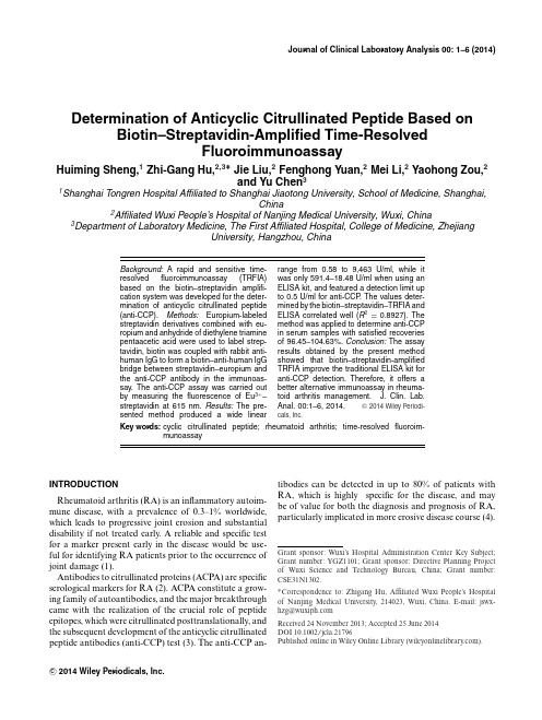
Journal of Clinical Laboratory Analysis00:1–6(2014) Determination of Anticyclic Citrullinated Peptide Based on Biotin–Streptavidin-Amplified Time-ResolvedFluoroimmunoassayHuiming Sheng,1Zhi-Gang Hu,2,3∗Jie Liu,2Fenghong Yuan,2Mei Li,2Yaohong Zou,2and Yu Chen31Shanghai Tongren Hospital Affiliated to Shanghai Jiaotong University,School of Medicine,Shanghai,China2Affiliated Wuxi People’s Hospital of Nanjing Medical University,Wuxi,China 3Department of Laboratory Medicine,The First Affiliated Hospital,College of Medicine,ZhejiangUniversity,Hangzhou,ChinaBackground:A rapid and sensitive time-resolvedfluoroimmunoassay(TRFIA) based on the biotin–streptavidin amplifi-cation system was developed for the deter-mination of anticyclic citrullinated peptide (anti-CCP).Methods:Europium-labeled streptavidin derivatives combined with eu-ropium and anhydride of diethylene triamine pentaacetic acid were used to label strep-tavidin,biotin was coupled with rabbit anti-human IgG to form a biotin–anti-human IgG bridge between streptavidin–europium and the anti-CCP antibody in the immunoas-say.The anti-CCP assay was carried out by measuring thefluorescence of Eu3+–streptavidin at615nm.Results:The pre-sented method produced a wide linear range from0.58to9,463U/ml,while itwas only591.4–18.48U/ml when using anELISA kit,and featured a detection limit upto0.5U/ml for anti-CCP.The values deter-mined by the biotin–streptavidin–TRFIA andELISA correlated well(R2=0.8927).The method was applied to determine anti-CCPin serum samples with satisfied recoveriesof96.45–104.63%.Conclusion:The assayresults obtained by the present methodshowed that biotin–streptavidin-amplifiedTRFIA improve the traditional ELISA kit foranti-CCP detection.Therefore,it offers abetter alternative immunoassay in rheuma-toid arthritis b.Anal.00:1–6,2014.C 2014Wiley Periodi-cals,Inc.Key words:cyclic citrullinated peptide;rheumatoid arthritis;time-resolvedfluoroim-munoassayINTRODUCTIONRheumatoid arthritis(RA)is an inflammatory autoim-mune disease,with a prevalence of0.3–1%worldwide, which leads to progressive joint erosion and substantial disability if not treated early.A reliable and specific test for a marker present early in the disease would be use-ful for identifying RA patients prior to the occurrence of joint damage(1).Antibodies to citrullinated proteins(ACPA)are specific serological markers for RA(2).ACPA constitute a grow-ing family of autoantibodies,and the major breakthrough came with the realization of the crucial role of peptide epitopes,which were citrullinated posttranslationally,and the subsequent development of the anticyclic citrullinated peptide antibodies(anti-CCP)test(3).The anti-CCP an-tibodies can be detected in up to80%of patients with RA,which is highly specific for the disease,and may be of value for both the diagnosis and prognosis of RA, particularly implicated in more erosive disease course(4). Grant sponsor:Wuxi’s Hospital Administration Center Key Subject;Grant number:YGZ1101;Grant sponsor:Directive Planning Project of Wuxi Science and Technology Bureau,China;Grant number:CSE31N1302.∗Correspondence to:Zhigang Hu,Affiliated Wuxi People’s Hospital of Nanjing Medical University,214023,Wuxi,China.E-mail:jswx-hzg@Received24November2013;Accepted25June2014DOI10.1002/jcla.21796Published online in Wiley Online Library().C 2014Wiley Periodicals,Inc.2Sheng et al.Time-resolvedfluoroimmunoassay(TRFIA),a nonra-dioimmunoassay,offers better sensitivity than other tradi-tional FIA methods due to the uniquefluorescence prop-erties of lanthanide chelates and the time-resolved mea-surement mode,which allows the specificfluorescence to be measured after the backgroundfluorescence has al-ready declined(5).Bioanalytical assays based on TRFIA have made a substantial impact on routine and research areas(6).Up to now,the use of Eu3+-labeled streptavidin(Eu3+–streptavidin)and time-resolvedfluorometry has been well suited as an indirect label for its relatively high affinity to biotin and low nonspecific binding properties.As a tetrameric structure with four binding sites for biotin,it can be used for amplifying signal(7).Moreover,a univer-sal detection reagent,europium chelate,as afluorescent marker has attracted much attention for high-throughput screening of large populations of biologically active sam-ples(8,9).In this study,the europium chelate of Eu3+–streptavidin was prepared and used as a signal generation reagent after its binding to the biotin moieties of the surface immuno-complex.A novel,rapid,biotin–streptavidin-amplified TRFIA for the determination of anti-CCP was developed and is described here.This method was applied to deter-mine anti-CCP in human serum samples with satisfactory results.MATERIALS AND METHODSChemicals and InstrumentationAuto DELFIA1235,Eu3+-labeled regent,and Eu3+-labeled enhancement solution were purchased from Wallac Oy(Turku,Finland).The96-well polystyrene microtiter plates and recombinant CCP antigen were ob-tained from AESKU(Dresden,Germany).Monoclonal rabbit anti-human IgM was purchased from Sigma(St. Louis,MO).N2-[p-isocyanate-benzyl]diethylenetriamine tetraacetic acid(DTTA)was purchased from Sigma Aldrich(St.Louis,MO).Bovine serum albumin(BSA) was purchased from Institute of Biological Products Min-istry of Health(Shanghai,China).Sephadex G25was pur-chased from Pharmacia(Piscataway,NJ).Concentrated wash buffer and enhancement solution were provided by Institute of Nuclear Medicine(Jiangsu,China).Other reagents used were of analytical reagent grade.ELISA Kit and Reference Standard of the Anti-CCP PreparationCommercial ELISA kits of the anti-CCP IgG were ob-tained from AESKU,Dr.Fenning(Kirchzarten,Ger-many),and ORGENTEC(Mainz,Germany),respec-tively.Reference standard of the anti-CCP was obtained from AESKU.Preparation of Solid AntigenEach well of polystyrene microtiter plates was coated with100μl of recombinant CCP antigen diluted with coating buffer(pH9.6,50mmol/l Na2CO3-NaHCO3) by incubating at4°C overnight.The coating buffer was discarded.After three times wash,some binding sites not occupied by the coating buffer were then blocked by250μl 3g/l BSA(Institute of Biological Products Department of Health,Shanghai,China)in phosphate buffer solu-tion(PBS;pH7.4)and the plates were incubated at4°C overnight.After the blocking buffer was discarded,the plates were vacuum-dried and stored at−20°C until anal-ysis.Biotin Labeling of Anti-Human IgGOne milligram of N-hydroxysuccinimidobitin (Promega,Madison,Wisconsin)was added to0.5mg of a1mg/ml solution of rabbit anti-human IgG(St. Louis,MO)in pH7.2,0.05M PBS.The reaction mixture was incubated at37°C for1h and then dialyzed overnight at4°C with PBS.The biotin-labeled antibody was stored at−20°C until use.Eu3+-Labeled Streptavidin PreparationEu3+labeling of streptavidin(Sigma)was performed us-ing a Eu3+-labeling Kit(EG&G-Wallac,Turku,Finland) strictly according to the manufacture’s instructions.One milligram of streptavidin was loaded on a PD10column and eluted using carbonate buffer(pH8.5,50mmol/l Na2CO3-NaHCO3)containing0.155mol/l NaCl.Then streptavidin was mixed with0.2mg Eu3+-DTPAA(di-ethylenetriamine pentaacetic acid;Sigma Aldrich)fol-lowed by vigorous stirring at30°C for20h.The resulting mixture was then fractionated on a Sepharose CL-6B(1×40cm;Pharmacia)with pH7.8,80mmol/l Tris-HCl buffer.The absorbance values of the elution were measured at280nm to obtain protein concentra-tions.After purification,equal amount of AR glycerin was added before subpackaging and stored at−20°C until use.Anti-CCP Antibody AssayThe procedures for BAS-TRFIA of serum anti-CCP antibody were performed using an indirect ELISA tech-nique.Hundred microliters of serum diluted with assay buffer was pipetted into each coated microtiter well,and incubated for30min at25°C.The plates were washed fourb.Anal.Determination of Anticyclic Citrullinated Peptide3times with wash buffer,then100μl of rabbit anti-humanIgG was added to each well,and incubated with shakingfor30min at25°C.The plates were washed again fourtimes,then pipetted with100μl buffer,diluted with Eu3+-labeled streptavidin to each well,and incubated with shak-ing for15min.After washing six times with wash buffer,100μL of enhancement solution was pipetted into eachwell.The plates were shaken for5min.Eu3+fluorescenceintensity(cps)was measured with auto DELFIA1235.The concentrations of anti-CCP antibody in respectivesamples were determined based on the calibration curvesusing Multicalc software program.Evaluation on the Kits(1)Precision testing.Three pools of mixed serum speci-mens with high,intermediate,and low concentrationof anti-CCP antibody were subpackaged and stored at −20°C.Mixed with the serum specimens and evaluat-ing their concentrations with samples,the coefficientof variations(CVs)was calculated.(2)Linear range testing.Serum from patient with thehighest anti-CCP antibody concentration was dilutedin a twofold serial dilution manner,and the anti-CCPantibody concentrations of diluted specimens weredetected using the established BAS-TRFIA kit and acommercial ELISA one,respectively.The linear curveof dilution concentration was obtained with dilutionmultiple as the horizontal abscissa andfluorescentintensity as the vertical ordinate.(3)Sensitivity testing.The sensitivity of the assay wasback-calculated by the obtained meanfluorescentcounts(n=20)of the zero standard plus2SD inthe calibration curve.(4)Clinical applications.The specificity of anti-CCP an-tibody detection was evaluated by testing serum from52healthy blood donors,32systemic lupus erythe-matosus(SLE),27Sjogren’s syndrome(SS),10sclero-derma,20mixed connective tissue disease(MCTD),23multiple sclerosis(MS)patients.(5)Correlation test.The anti-CCP antibody concentra-tions of32serum specimens from patients with RAwere detected using the established BAS-TRFIA kitand a commercial ELISA kit,respectively,and thenthe values obtained were analyzed.(6)Stability testing.New BAS-TRFIA kit and threeELISA kits were put into the37°C water box for7days and then compared with the one stored in rou-tine conditions.(7)Coefficient of recovery.Three sera with high antibodyconcentrations were diluted with sample buffer andassayed in the new TRFIAkit.Fig.1.Serial dilutions of a specimen with the highest anti-CCP IgG concentration were tested with TRFIA and ELISA,respectively.Double Y-axis curve was determined according to dilution strength(X-axis)as well as BAS-TRFIAfluorescence intensity and ELISA absorbance(the two Y-axes).Statistical AnalysesData were analyzed using the SPSS13.0software.Re-gression analysis and analysis of variance were used to calculate correlation and test linearity,respectively.In-terassay and intra-assay variation was calculated using the CV.The statistical significance level was set at0.05. RESULTSLinearityA strong positive specimen from9,081to0.55U/ml was diluted and the serial dilutions with the established TRFIA as well as ELISA were detected.The curve of de-tectable range of the two methods is shown in Figure1,in which we observe that for the established TRFIA kit there was a good linear range within9,463–0.58U/ml,whereas it was within591.4–18.48U/ml when using an ELISA kit,indicating that the method of TRFIA we established has a wider detectable range than the commercial ELISA. Calibration Curve for Determination of Anti-CCP IgGAs shown in Figure2,the time-resolvedfluorescence intensities are directly proportional to the concentrations of anti-CCP IgG.The concentrations of anti-CCP IgG were2,365.8,591.44,147.86,36.96,9.24,0.58U/ml.The line equation for the calibration curve of anti-CCP IgG was Y=3.79071+0.79505X+0.01094X2,where X is the concentration of anti-CCP IgG(GPL U/ml)and Y is fluorescence intensity.The sensitivity of the assay,defined as the mean signal of the zero standard plus2SD,was 0.5U/ml.b.Anal.4Sheng et al.Y : A x i s T i t l eX:The concentration for anti-CCP IgG (U/ml)Fig.2.Standard curves for anti-CCP IgG by TRFIA at different concentrations (2,365.8,591.44,147.86,36.96,9.24,0.58U/ml).TABLE 1.Intrabatch and Interbatch Precision AnalysisBAS-TRFIA (U/ml)ELISA (U/ml)Mean ±SDCV (%)Mean ±SD CV (%)HighIntrabatch (n =20)241.5±7.63 3.16242.8±14.86 6.12Interbatch (n =10)240.9±10.33 4.29243.1±17.647.26IntermediateIntrabatch (n =20)72.7±2.78 3.8272.6±5.948.18Interbatch (n =10)72.3±3.38 4.6873.1±7.269.93LowIntrabatch (n =20)14.15±0.60 4.2414.32±1.389.61Interbatch (n =10)14.23±0.714.9714.37±1.5410.74Assay PrecisionThe precision of the assay was also analyzed by mea-suring three pools of mixed serum specimens with high,intermediate,and low concentration of anti-CCP IgG 20times in one series (intra-assay)and in duplicate in eight different series,and data were compared with those ob-tained by the ELISA kit.As shown in Table 1,the intra-assay CVs detected by TRFIA at mean anti-CCP IgG concentrations of 241.5,72.7,14.15U/ml were 3.16%,3.82%,and 4.24%,respectively,and the interassay CVs at mean concentrations of 240.9,72.7,14.23U/ml were 4.29%,4.68%,and 4.97%,respectively.In comparison,the intra-assay CVs detected by ELISA at mean concen-trations of 242.8,72.6,14.32U/ml were 6.12%,8.18%,and 9.61%,respectively,and the interassay CVs at mean concentrations of 243.1,73.1,14.37U/ml were 7.268%,9.93%,and 10.74%,respectively.Obviously,the TRFIA kit we established for anti-CCP IgG detection was better than the commercial ELISA kit in intra-and interassay precision with either rank of concentration.TABLE 2.Specificity of Anti-CCP Antibody in Different Types of PatientsCase and Negative control 15Positive ࣙSpecificity groupn U/ml U/ml(%)Healthy blood donors 5251198.1SLE 3231196.9SS2726196.3Scleroderma 10100100MCTD 2019195.0MS2322195.7Clinical ApplicationEach well of polystyrene microtiter plates was coated with recombinant CCP antigen.The specificity of anti-CCP antibody detection was evaluated by testing serum from 52healthy blood donors,32SLE,27SS,10sclero-derma,20MCTD,and 23MS patients.Cross-reactivities are shown in Table 2.Correlation With ELISAConcentration results of 32serum samples determined by TRFIA were compared with those obtained by an ELISA kit.The scatter plots showed that the two methods correlated well (Fig.3)and the correlation coefficient of the results obtained from the two assays was 0.8927.Stability of the TRFIA KitsThe assay kits were stored at 4°C and water bathed at 37°C for 7days.Then,the difference of fluores-cence intensity or absorbance—that is decline rate ofb.Anal.Determination of Anticyclic Citrullinated Peptide5Fig.3.Correlation between the BAS-TRFIA and ELISA. TABLE3.Stability of the BAS-TRFIA and ELISAGroups4°Cpreserve7days37°Cwater bath7daysThe declinedrate ofcombination(%)ELISA1(abs) 2.56 1.6336.3 ELISA2(abs) 2.39 1.6929.3 ELISA3(abs) 2.71 2.2317.7 BAS-TRFIA(cps)2,451,2362,153,41512.1 combination—was measured both by TRFIA and ELISA.We observed that it exhibited no visible change except for a decline by12.1%for TRFIA(17.7%,29.3%, 36.3%for the commercial ELISA)in the rate of combina-tion when compared with the one stored in routine con-ditions,suggesting that the established assay kit had good stability and could be stored at4°C for1year(Table3). Moreover,the accuracy decreased as time was extended by ELISA,but after enhancement solution was added the detection offluorescent results was not affected up to the next day.Thus,it can be seen that TRFIA is more stable.Coefficient of RecoveryThe recoveries of thefirst sample were100.98%, 102.25%,and96.45%,respectively.The recoveries of the second sample were100.46%,98.18%,and101.59%, respectively.The recoveries of the third sample were 104.63%,97.18%,and98.17%,respectively.The average recovery rate calculated by the average of nine individ-ual values by the formula of observed concentrations×100/expected concentration in Table4)was99.98%.TABLE4.Coefficient of Recovery:Three Sera With High Anti-body Concentrations Were Diluted With Sample Buffer and As-sayed in the New BAS-TRFIA KitObserved ExpectedSample Dilution(U/ml)(U/ml)O/E(%) 11:100266.41:200134.5133.2100.981:40068.166.6102.251:80032.133.396.40 21:100175.61:20088.287.8100.461:40043.143.998.181:80022.321.95101.59 31:10088.51:20046.344.25104.631:40021.522.12597.181:80010.8611.062598.17DISCUSSIONTo facilitate early diagnosis,a serological marker of RA that is detectable very early in the disease is needed.As mentioned above,several recent studies have shown that anti-CCP can be detected very early in the course of RA and may be helpful in early diagnosis.Herein,we developed a new sandwich TRFIA with biotin–streptavidin system as a quantitative and rapid immunological assay for measuring anti-CCP in clini-cal serum samples,since the new method combined the advantages of both high sensitivity of traditional TR-FIA and high affinity of biotin to streptavidin tetrameric bond.Moreover,the detectable signal can be amplified because of the tetrameric structure with four binding sites of streptavidin for biotin.The experimental results indi-cated that the new TRFIA is a promising immunofluoro-metric method with highly sensitive and specific,mean-while,the assay provided wider dynamic working ranges and better reproducibility.In Figure1,TRFIA demon-strated a good linear range within9,463–0.58U/ml,by contrast,within591.4–18.48U/ml of an ELISA kit.The expanded detection range will improve earlier detection and higher sensitivity in RA diagnosis(10,11).Further-more,the serologic-positive patients will be treated timely and effectively,subsequently,preventing irreversible joint damage(12).By comparison,the new assay has satisfied correlation with general commercial ELISA kit with the R2value 0.8927(Fig.3),moreover,its stability performance is bet-ter than the commercial ones,which will guarantee the more stable yield(Table3).In sum,the new TRFIA with biotin–streptavidin system as a quantitative and rapid im-munological assay for measuring anti-CCP is a promising alternative assay for RA diagnosis and monitor.b.Anal.6Sheng et al.CONFLICT OF INTERESTAll authors declare that they have no competing inter-ests.REFERENCES1.Raptopoulou A,Sidiropoulos P,Katsouraki M,Boumpas DT.Anti-citrulline antibodies in the diagnosis and prognosis of rheumatoid arthritis:Evolving concepts.Crit Rev Clin Lab Sci 2007;44(4):339–363.2.van Gaalen FA,Linn-Rasker SP,van Venrooij WJ,et al.Au-toantibodies to cyclic citrullinated peptides predict progression to rheumatoid arthritis in patients with undifferentiated arthritis:A prospective cohort study.Arthritis Rheum2004;50(3):709–715. 3.van Venrooij WJ,van Beers JJ,Pruijn GJ.Anti-CCP antibod-ies:The past,the present and the future.Nat Rev Rheumatol 2011;7(7):391–398.4.Forslind K,Ahlm´e n M,Eberhardt K,Hafstr¨om I,Svensson B;BARFOT Study Group.Prediction of radiological outcome in early rheumatoid arthritis in clinical practice:Role of antibodies to cit-rullinated peptides(anti-CCP).Ann Rheum Dis2004;63(9):1090–1095.5.Du L,Cheng S,Wang S.Determination of diethylstilbe-strol based on biotin-streptavidin-amplified time-resolvedfluoro-immunoassay.Luminescence2012;27(1):28–33.6.Hu Z,Liu J,Y e Y,Zhou Y,Yu L.Detection of anticardiolipinantibody igm by sm(3+)-labeled time-resolvedfluoroimmunoassay.J Immunoassay Immunochem2013;34(3):255–265.7.Suonpaa M,Markela E,St˚a hlberg T,Hemmil¨a I.Europium-labelled streptavidin as a highly sensitive universal label.Indirect time-resolved immunofluorometry of FSH and TSH.J Immunol Methods1992;149(2):247–253.8.Qin QP,Lovgren T,Pettersson K.Development of highlyfluores-cent detection reagents for the construction of ultrasensitive im-munoassays.Anal Chem2001;73(7):1521–1529.9.Huhtinen P,Soukka T,L¨ovgren T,H¨a rm¨a H.Immunoassayof total prostate-specific antigen using europium(III)nanoparti-cle labels and streptavidin-biotin technology.J Immunol Methods 2004;294(1–2):111–122.10.van Genderen FT,Gorus FK,Pipeleers DG,van Schravendijk CF.Sensitive and specific time-resolvedfluorescence immunoassay of rat C-peptide for measuring hormone secretory and storage capacity of beta-cells in vivo and in vitro.Endocrinology2013;154(5):1934–1939.11.Farid S,Azizi G,Mirshafiey A.Anti-citrullinated protein antibodiesand their clinical utility in rheumatoid arthritis.Int J Rheum Dis 2013;16(4):379–386.ndewe RB,Boers M,Verhoeven AC,et al.COBRA combina-tion therapy in patients with early rheumatoid arthritis:Long-term structural benefits of a brief intervention.Arthritis Rheum 2002;46(2):347–356.b.Anal.。
结肠肿瘤前细胞系的建立及应用
结肠肿瘤前细胞系的建立及应用曹海龙;许梦雀;鄢芳;王邦茂【摘要】结肠肿瘤前细胞即永生化结肠上皮细胞系(IMCE),来源于 Immorto 小鼠和腺瘤性结肠息肉病基因(Apc)min/+小鼠杂交后子代小鼠的结肠上皮细胞,其表型正常,可发生恶性转化,已成为肠道肿瘤发生发展相关研究较为理想的细胞模型。
此文主要就该细胞系的建立及其在肠道肿瘤发生机制和筛选防治药物等领域中的应用进展作一综述。
【期刊名称】《国际消化病杂志》【年(卷),期】2016(036)001【总页数】4页(P31-33,53)【关键词】结直肠癌;结肠肿瘤前细胞;腺瘤性结肠息肉病基因【作者】曹海龙;许梦雀;鄢芳;王邦茂【作者单位】300052 天津医科大学总医院消化科;300052 天津医科大学总医院消化科;300052 天津医科大学总医院消化科;300052 天津医科大学总医院消化科【正文语种】中文·综述·近年来,结直肠癌(CRC)的发病率明显增加。
现有研究已发现与CRC发生密切相关的一些重要的基因突变,如腺瘤性结肠息肉病基因(Apc)、K-ras、p53以及错配修饰基因等[1]。
目前仍有必要探讨这些基因突变如何影响正常结肠上皮细胞的表型,含有基因突变的肿瘤前细胞如何发生恶性转化,以及阐明已知CRC中发生变化的分子之间协同作用等。
由于体外分化的人正常结肠上皮细胞的培养非常困难,目前国内外研究仍多以结肠癌细胞作为相关研究模型[2-3],显然这些肿瘤细胞并不完全符合上述研究要求。
结肠肿瘤前细胞即永生化结肠上皮细胞系(IMCE),来源于Immorto小鼠和Apcmin/+小鼠杂交后子代小鼠的结肠上皮细胞,其表型正常,可发生恶性转化,已成为肠道肿瘤发生发展相关研究较为理想的细胞模型[4]。
本文主要就该细胞系的建立及其应用进展作一综述。
Immorto小鼠及Apcmin/+小鼠的建立为结肠肿瘤前细胞系的建立提供了可能。
Immorto小鼠携带了一种温度敏感的猴病毒40(SV40)大T抗原基因(tsA58)的突变形式,使之可置于干扰素-γ(IFN-γ)诱导的H-2Kb启动子(H-2Kb-tsA58)控制下。
库普弗细胞的研究进展

第4l卷第2期2010年4月解剖学报ACTAANATOMICASINICAV01.41,No.2Apr.2010‘331。
库普弗细胞的研究进展张强伟1’2徐存拴1’2。
(1.河南师范大学生命科学学院;2.河南省-科技部共建细胞分化调控国家重点实验室培育基地,河南新乡453007)[摘要]库普弗细胞(KCs)是肝脏的非实质细胞。
具有分泌细胞因子及吞噬、免疫等功能,亦与多种肝脏疾病,如肝损伤、肝纤维化、肝硬化等密切相关。
本文简要总结了近几年有关库普弗细胞的生理功能及与肝病关系等方面的研究进展。
[关键词】库普弗细胞;细胞因子[中图分类号】Q28[文献标志码]A[DOI]10.3969/j.issn.0529—1356.2010.02.035ResearchprogressonKupffercellsZHANGQiang.wei。
・2.XUCurt.shuanlf2。
(J.CollegeofLifeSe如twe,胁’ilartNormalUniversity;2.研LaboratoryforCellDifferentiationRegulation,肌’llallXinxiang453007,China)[Abstract]Kupffercellisamemberofthelivernonparenehymalcells,itparticipatesinavarietyofphysiologicalactivitiesthroughphagocytosis,secretingcytokines,antigen—presentingpathways,andiscloselyrelatedtoavarietyofdiseasessuchaslivercancer,liverfibrosis,liverinjury.Thispaperpresentstheprogressofresearchonphysiologicalfunctionsofkupffercellsanditsrelationswithliver.[Keywords]LiverKupffercell;Cytokine自1876年发现库普弗细胞(kupffercells,KCs)以来,至今已有130多年。
BLTR-4永生化细胞系的建立及生物学特性初步鉴定

BLTR-4永生化细胞系的建立及生物学特性初步鉴定郑闪;郭素萍;何祖根;程书钧;高燕宁【期刊名称】《中国医学科学院学报》【年(卷),期】2004(026)005【摘要】目的建立膀胱尿路上皮永生化细胞系,并初步鉴定其生物学特性.方法以Fugene-6TM为载体,将含人乳头瘤病毒16(HPV-16)E6、E7基因的HPV-16K质粒,体外转染原代培养的正常胎儿膀胱尿路上皮细胞.运用PCR、免疫组织化学及细胞特征鉴定等方法,对所得BLTR-4细胞系中HPV-16 E6、E7基因的存在情况及该细胞系生物学特性进行初步鉴定.结果 BLTR-4细胞系是含有HPV-16 E6、E7基因并表达上述两种蛋白的尿路上皮源性细胞系.该细胞在体外已传70余代,细胞倍体数发生了二倍体至四倍体再至非整倍体的改变,生长特性符合永生化细胞系.结论BLTR-4细胞系是膀胱尿路上皮永生化细胞系,该细胞系含有HPV-16 E6、E7基因.BLTR-4细胞系的建立为研究高危型HPV感染与膀胱移行细胞癌(TCC)关系提供了体外实验模型.【总页数】6页(P543-548)【作者】郑闪;郭素萍;何祖根;程书钧;高燕宁【作者单位】中国医学科学院,中国协和医科大学,肿瘤医院病理科,北京,l00021;Department of Carcinogenesis,Cancer Hospital, CAMS and PUMC, Beijing;中国医学科学院,中国协和医科大学,肿瘤医院病理科,北京,l00021;Department of Carcinogenesis,Cancer Hospital, CAMS and PUMC,Beijing;Department of Carcinogenesis,Cancer Hospital, CAMS and PUMC, Beijing【正文语种】中文【中图分类】R737.14【相关文献】1.永生化人肝细胞系的建立和生物学特性研究 [J], 张然星;李宓;张文健;顾锋;门秀丽;叶丽亚;房青;徐梅;高福云;娄晋宁2.人卵巢癌永生化细胞系BUPH:OVSC-2的建立及其生物学特性研究 [J], 冯捷;刘广芝;付天云;叶雪;姚煜3.永生化绵羊附睾上皮细胞系的建立及其生物学特性分析 [J], 宋慧子; 栾兆进; 王兆琛; 杜炜; 赵勇超; 张家新4.人类黑色素细胞瘤永生化细胞系的建立及生物学特性鉴定 [J], 曾先捷;蔡捷;姚洁民;黄英华;郭春雨;马赫;李小平;邝晓聪5.人卵巢浆液性囊腺癌永生化细胞系BUPH∶OVCA-3的建立及其生物学特性 [J], 刘广芝;冯捷;付天云;叶雪;昌晓红因版权原因,仅展示原文概要,查看原文内容请购买。
胸腺肽α1的作用机制和临床应用
""""""""""""""""""""""""""""""""""""""""""""""""
[ 关键词] 胸腺肽 !! ;作用机制;临床应用 [ 中图分类号] D&,’- !’+ + + [ 文献标识码] E 功能而调节免疫系统的,概括起来大致有以下几个方面: ! - !" 免疫调节作用 " ( ! ) 诱导和促进胸腺细胞的分化和成 熟:<!! 可促进胸腺内的骨髓干细胞转化为 < 淋巴细胞,并 + + 胸腺肽 !! ( :;F@/264 8=3;8>! ,<!! ) 是 G/H 等
或不存在的条件下,<!! 可促进骨髓来源的树突状细胞 ( M. ) 成熟分化并增强其功能,而 M. 直接影响 < 细胞的成熟和分
[(] [#] 化 。研究表明 ,<!! 不但促进表达 .M) [ 的树突状细胞
!" #!! 的作用机制
<!! 是由 ’% 个 氨 基 酸 组 成 的 多 肽,其 组 成 为:EC>N10> E23>E=8>O8=>E23><;0>N10>N10>P=9>J=1><;0><;0>GF2>E23>G19>GF2> P=9>GF2>GF2>P=9>O8=>O8=>P=9>P=9>E=8>P=9>E24>QR。 相 对 分 子 质量 ( !0 ) 为 & !"% ,SJ 为 )- ’ 。美国 NC6.=/41 公司生产的药品 日达仙 ( T858U64 ) 是一种人工化学合成的 <!! ,每支产品含 <!! !- ( @7,供皮下注射。该产品的 3R 为 (- % ,血浆半衰期 约 ’ ;。每次注射后使外周血中 <!! 的浓度增加约 *" V !"" 倍, ’ ; 后可达 *" "7 W G。在慢性乙型肝炎 ( .RX ) 和慢性丙型 的治疗中,每周 ’ 次注射,在肿瘤患者化疗过程 肝炎 ( .R.)
T细胞条件性敲除Spi1基因小鼠的繁育及鉴定
Fabricationandperformanceevaluationofhigh modulusandhigh strengthsilkfibroinguidedboneregenerationmembraneLiaoXiaoyu1,FangHui1,YangFeiyu1,ZouDuohong1,2(1College&HospitalofStomatology,AnhuiMedicalUniversity,KeyLaboratoryofOralDiseasesResearchofAnhuiProvince,Hefei 230032;2ShanghaiNinthPeople'sHospital,ShanghaiJiaoTongUniversitySchoolofMedicine,ShanghaiJiaoTongUniversitySchoolofStomatology,ShanghaiKeyLaboratoryofStomatology,Shanghai 200011)Abstract Objective TodevelopahighmodulusandhighstrengthbiodegradablesilkfibroinGBRmembranetoaddresstheissueofmaintainingthespaceforboneregenerationintherepairofosseousdefects.Methods Afterpurifyingsilkfibroinprotein,membranematerialswerepreparedusingevaporation hotpressingmethod.Thephysi calandchemicalpropertiesandbiologicalperformanceofthemembraneswereevaluatedusingstretchingtests,invitrosimulations,andcellco culturingmethods.Results AsilkfibroinGBRmembranewassuccessfullyfabrica ted,resultinginasimulateddegradationrateof35 3%after12hinvitro.Thewet stateelasticmodulusreached45MPa,whilethetensilestrengthreached8 39MPa.Furthermore,thecellsurvivalratewasnearly100%after7days.Conclusion ThebiodegradableGBRmembraneproducedinthisstudypossesseshighmodulusandstrength,aswellasexcellentbiocompatibility,offeringapromiseasafoundationforaddressingthebonedefectre pairandbonespacemaintenance.Keywords bonedefectrepair;maintainingthespaceforboneregeneration;guidedboneregenerationmem brane;silkfibroin;evaporation heatpressingmethod;osteogenicdifferentiation;tensilestrength网络出版时间:2024-04-1213:52:17 网络出版地址:https://link.cnki.net/urlid/34.1065.R.20240410.1008.006T细胞条件性敲除Spi1基因小鼠的繁育及鉴定王卉卉,朱向玲,吴旭铭,张慧茹,周园园,王安琪,刘 崇,涂佳杰摘要 目的 繁育T细胞条件性敲除Spi1基因的小鼠并对其进行鉴定,为进一步探索Spi1编码蛋白PU 1的作用提供研究基础。
中药复方对肝癌移植瘤小鼠肿瘤干细胞表面标志c-kit和CD_(133)表达的影响
公 司 , 格 证 号 :C K( ) 0 2 0 0 ;— i C 1 合 S X 京 2 0 — 0 1 c kt D3 、 3
抗 体 购 自 snacu ;免 疫组 化检 测 MaVs n试 at rz x io i
剂 盒 购 自福 州迈 新公 司 ;解 毒 消 瘾饮 ( 白花蛇 舌 草、 山慈 姑 、 枯 草 、 参 ) 扶 正 抑 瘤方 ( 芪 、 夏 苦 、 黄 灵
中发现 。具 有提 高围手 术 期 消化 道 肿瘤 患 者免 疫 功 能 、 善 生存 质 量 、 长患 者 生存 期 的作 用 。前 改 延 期基 础研 究 表 明该 复 方具 有提 高荷 瘤小 鼠免疫 功 能, 抑制 血管生 长 因子 V G E F表 达 的作用 。我 们应
用 免疫组 化 方法 检 测皮 下 移植 瘤 小 鼠肿 瘤 组织 中
多种恶 性 肿瘤 中已得 到 初步 证 实 。其 表达 变 化可 反 映肿 瘤干 细胞 的 表达 水平 [4 11 - 。中药复 方解 毒 消 - 瘕 饮和 扶正 抑瘤 方 在长 时间 、大 样本 的临 床 观察
消 癜饮 。术后 给予 扶 正抑 瘤 方 。设 计 给药 方 式 如 下 : 胃 1 毒 消 瘾 饮 后 接 种 肝癌 细 胞 , 灌 0d解 接种 第 2天 开 始灌 胃 1 正 抑瘤 方 , 理 盐 水 作 为 0d扶 生 药物 对 照 。具 体方 案 如 下 : 组 接种 肝 癌 细胞 , A 灌 胃生 理盐 水 ; 接种 肝癌 细胞 , 胃 2种 中药 。 B组 灌 122 动 物模 型 建 立 取 H2 __ 2传 代 第 7天 的腹
中 图分 类 号 :2 55 R 8 . 文献标志码 : A 文章 编 号 :0 4 5 2 (0 0 0 — 08 0 10 — 6 7 2 1 ) 3 0 1— 4
- 1、下载文档前请自行甄别文档内容的完整性,平台不提供额外的编辑、内容补充、找答案等附加服务。
- 2、"仅部分预览"的文档,不可在线预览部分如存在完整性等问题,可反馈申请退款(可完整预览的文档不适用该条件!)。
- 3、如文档侵犯您的权益,请联系客服反馈,我们会尽快为您处理(人工客服工作时间:9:00-18:30)。
DOI:10.13423/ki.cjcmi.008110
前列腺癌( prostate cancer,PCa) 是严重威胁男性 健康的常见泌尿系肿瘤。在西方国家,PCa 占男性癌 。 在 我 国 随 着 人 口 老 龄 化, 症死 因 的 第 二 位 PCa 的发病率和死亡率也逐年升高。 前列腺特异性膜 抗原( prostate specific membrane antigen,PSMA ) 是一
( 第四军医大学: [ 摘 要] 目的
1
2 免疫学教研室, 西京医院泌尿外科,陕西 西安 710032 )
+ PC3 细胞系,并将 利用慢病毒感染的方法建立能稳定表达前列腺特异性膜抗原 ( PSMA) 和荧光素酶的 PSMA -
化成单细胞悬液; 进行细胞计数后,分至各流式管, 使每管细胞数为 1 ˑ 10 个。 每种细胞均设置空白对 照组、同型对照组和 PSMA 抗体组。 空白组加入流 式细胞术分选液,同型对照组加入人 IgG,PSMA 抗 体组加入 PE 标记的 PSMA 抗体,4ħ 孵育 30 min。 随后,除 PSMA 抗体组外,每管加入 100 μL PE 标记 4ħ 避光孵育 30 min。洗涤后, 的抗人 IgG, 用流式细 胞仪进行检测。 1. 2. 3 PC3 细胞最佳嘌呤霉素筛选剂量的确定 取 生长状态良好的 PC3 细胞,消化成单细胞悬液,均匀 0. 5、 1、 1. 5、 2) μg / mL 嘌 次日, 加入( 0、 铺至 6 孔板, 48 h 细胞全部死亡的最 呤霉素,每天观察细胞状态, 低剂量即为最佳筛选剂量。 1. 2. 4 的建立 PSMA PC3 和 PSMA PC3 稳定转染细胞系 取生长状态良好的 PC3 细胞,消化成单细
2. 5 ˑ 107 个 / mL。 取 0. 2 mL 分别接种于每只裸鼠的右 下肢皮下,各接种 5 只; 接种 2 周后,用生物发光成
+ PC3 像法( bioluminescence imaging,BLI) 检测 PSMA - PC3 细胞的成瘤情况。 细胞和 PSMA -
胞用 含 100 mL / L 胎 牛 血 清 的 RPMI1640 培 养 基, PC3 细胞和 DU145 细胞用含 100 mL / L 胎牛血清的 DMEM 培养基,于 37ħ 、50 mL / L CO2 的孵箱中培 养,定期换液传代。 1. 2. 2 况 流式细胞术检测 PCa 细胞上 PSMA 的表达情 取生长状态良好的上述前列腺癌细胞,将其消
[3 ] 种前列腺上皮细胞特异性表达的 2 型跨膜糖蛋白 。 研究表明,PSMA 在 PCa 细胞及 PCa 组织中的表达均 [1 - 2 ]
难。PSMA 阴性的 PC3 细胞可以在裸鼠体内有效成 瘤,是一种理想的体内研究模型。 为使 PC3 细胞表 达 PSMA,我们利用表达 PSMA 的慢病毒感染 PC3 细 胞,并经 嘌 呤 霉 素 筛 选 获 得 了 稳 定 表 达 PSMA 的 PSMA + PC3 细胞。此外,我们还给 PC3 细胞同时感 染了表达荧光素酶的慢病毒,以便在体内对肿瘤的 定位和生长进行实时的观察。 1 材料和方法 1 . 1 材料 2、LNCaP、PC3、DU145 细胞为第 人前列腺癌 C4四军医大学免疫学教研室保存; RPMI1640、DMEM / F12 培养液购自 Gibco 公司; 表达 PSMA 的慢病毒和表达 萤光素酶的慢病毒由上海吉凯生物公司包装; 新生 胎 牛 血 清 购 自 中 国 医 学 科 学 院; 藻 红 蛋 白 ( phycoerythrin,PE ) 标 记 的 小 鼠 抗 人 PSMA 抗 体 ( PSMAPE) 购自 Biolegend 公司; 小 鼠 对 照 IgG 购 自 R&D 公 司; 兔 抗 人 PSMA 抗 体 购 自 Cell Signaling Technology 公司; HRP 标记的羊抗兔 IgG 购自 Abcam 公 司; 细胞培养皿( 瓶) 为 Thermo Nunc 公司产品; D荧光 素钾盐购自 GoldenBio 公司。体 质 量 为 25 30 g 的 4 6 周龄雄性裸鼠购自第四军医大学动物实验中心。
5 + -
PSMA 的表达情况。 2 2. 1 结果 C42 和 LNCaP 细胞表达 PSMA,而 PC3 和 将 PE 标记的抗 PSMA 的抗体与 4 种前列腺癌细 2 细胞和 胞孵 育 后, 流 式 细 胞 术 检 测 发 现, C4LNCaP 细胞上 PSMA 表达阳性,而 PC3 细胞和 DU145 细胞为 PSMA 阴性细胞( 图 1 ) 。
4期
张玲玲,等. 过表达前列腺特异性膜抗原和荧光素酶的 PC3 稳定细胞系的建立及成瘤性鉴定
515
98% 以上的 细 胞 中 能 够 检 测 到 强 的 荧 光 信 号,而 PSMA - PC3 细胞中检测不到荧光信号,提示成功建
其接种到裸鼠皮下检测其成瘤能力 。方法
包装表达 PSMA 的慢病毒和表达荧光素酶的慢病毒 ,将其感染 PC3 细胞后,用嘌呤
+ PC3 细胞和仅表达荧光素酶的 PSMA - PC3 细胞; 利用流式细胞 建立能够稳定表达 PSMA 和荧光素酶的 PSMA 霉素进行筛选, + PC3 和 PSMA - PC3 细胞接种于裸鼠皮下建立荷瘤模型 ,利用生物发光成像法观察其在 术检测 PSMA 的表达情况。将 PSMA + PC3 和 PSMA - PC3 肿瘤组织中 PSMA 的表达情况。 结果 体内的成瘤情况,利用免疫组织化学染色法检测 PSMA -
+ PC3 细 胞 和 PSMA - PC3 细 胞。 用 PE 得到 PSMA -
PSMA - PC3 细胞,将它们消化成单细胞悬液,进行细 胞计数后,用无血清培养基重悬细胞,使细胞数为
标记抗人 PSMA 抗体分别与这 2 种细胞孵育后, 用流
+ PC3 细胞中 式细胞术进行检测。 结果显示,PSMA -
远远高于正常前列腺上皮,其表达水平与格里森评 分呈正相关,并且在绝大多数的转移灶都呈阳性表
[4 - 6 ] 。 这 些 表 达 特 点 使 得 PSMA 被 认 为 是 达 PCa 免疫诊断和免疫治疗的理想靶点[7 - 8]。 多种靶向 PSMA 的研究已经确认了 PSMA 作为
前列腺癌特异性诊断和治疗靶点的有效性,特异性 识别 PSMA 的导向肽、适配子以及针对 PSMA 的抗 体,都 显 示 出 比 较 理 想 的 靶 向 诊 断 和 抗 肿 瘤 活 [9 - 11 ] 。特别是近年来基于抗体的肿瘤免疫治疗研 性 究发展迅速,被认为是继手术治疗、化疗及放疗之后 [12 ] 的第四大抗肿瘤治疗策略 。 因此,靶向 PSMA 的 抗体用于 PCa 治疗的研究被认为具有良好的应用前 2 细胞和 景。但是,由于 PSMA 阳性的前列腺癌 C4LNCaP 细胞恶性程度较低,不易在裸鼠体内成瘤,这 对以 PSMA 为靶点的靶向治疗研究带来了一定的困
6
1. 2. 6 的表达
HE 和免疫组织化学染色检测肿瘤中 PSMA 将上述荷瘤裸鼠在接种后第 30 天,麻醉后
+ PC3 和 PSMA - PC3 肿瘤组织, 处死; 分别取 PSMA -
制成石蜡切片。取 2 份切片,一份切片进行脱蜡后, 加入苏木素染色 5 min,流水冲洗,加入盐酸酒精分 化 30 s,之后在自来水中浸泡 15 min,再加入伊红染 色 2 min,常规脱水后封片。另一份切片脱蜡后加入 30 mL / L H2 O2 ,室温放置 10 min,灭活除内源性过氧 化物酶。加入山羊血清室Байду номын сангаас封闭 30 min,再加入兔 抗人 PSMA 抗体 ( 1 ʒ100 ) ,湿盒内 4ħ 孵育过夜; 次 日,将切片用 PBS 洗涤后,加入 HRP 标记的山羊抗 兔 IgG( 1ʒ200 ) ,室温孵育 30 min; 洗涤后,DAB 显色 2 3 min,用 苏 木 素 衬 染 后 封 片, 显 微 镜 下 观 察
△
同为第一作者
* 通讯作者,温伟红,E-mail: wenweih@ fmmu. edu. cn; 杨安钢,E-mail: agyang@ fmmu. edu. cn
514 1. 2 1. 2. 1 方法 PCa 细胞系的培养 C42 细胞和 LNCaP 细
33 ( 4 ) 细胞与分子免疫学杂志 ( Chin J Cell Mol Immunol) 2017 ,
33 ( 4 ) 细胞与分子免疫学杂志 ( Chin J Cell Mol Immunol) 2017 ,
513 文章编号: 1007 - 8738 ( 2017 ) 04 - 0513 - 04
·论著·
过表达前列腺特异性膜抗原和荧光素酶的 PC3 稳定细胞系的建立及成瘤性鉴定
1 1 2 2 2 2 1 1 2 1* 1* 张玲玲 △ ,吴介恒 △ ,韩东晖 ,史圣甲 ,杨发 ,谢品 ,席文锦 ,郭张燕 ,秦卫军 ,杨安钢 ,温伟红
+ PC3 细 胞 和 取 生 长 状 态 良 好 的 PSMA - +
图1 情况
流式 细 胞 术 检 测 4 种 前 列 腺 癌 细 胞 上 PSMA 的 表 达
2. 2
PSMA + PC3 和 PSMA - PC3 稳定转染细胞系 用表达 PSMA 的慢病毒和 / 或表达萤光素酶的慢
的建立 病毒感染 PC3 细胞后,用嘌呤霉素筛选并扩大培养,
+ PC3 肿瘤上 PSMA 的表达为阳性。结论 学染色检测证实 PSMA + PC3 细 成功建立了能稳定表达 PSMA 和荧光素酶的 PSMA -
胞系,并在裸鼠体内证实其可有效成瘤 ,为靶向 PSMA 的研究提供了有用的细胞和动物模型 。 [ 关键词] PSMA; 慢病毒; 前列腺癌; 靶向治疗; 动物模型 [ 中图分类号] R737. 25 [ 文献标志码] A