间充质干细胞培养方法
骨髓间充质干细胞总结

2003年8月,为给大家提供一个网上交流干细胞研究经验的平台,我们干细胞版设立了骨髓间充质干细胞培养讨论区,经过近三个月的讨论学习,我们既学习丰富了自己的知识体系,也对间充质干细胞尤其是分离培养方面有了更为翔实的认识,为了大家阅读的方便,我们决定把本版中的相关内容,同时参考部分书目和文献,做一总结。
一、骨髓间充质干细胞的分离目前常用的分离MSC的方法有全骨髓法和密度梯度离心法,全骨髓法即根据干细胞贴壁特性,定期换液除去不贴壁细胞,从而达到纯化MSC的目的。
密度梯度离心法即根据骨髓中细胞成分比重的不同,提取单核细胞进行贴壁培养。
随着对MSC表面抗原认识的深入,有人利用免疫方法如流式细胞仪法、免疫磁珠法等对其进行分离纯化,但经过流式或磁珠分选后的细胞出现了增殖缓慢等一些问题,加之耗费较大和技术的难度,在某种程度上限制了这些方法的广泛应用。
1. 直接培养法(全骨髓培养法)1987年,Friedenstein等发现在塑料培养皿中培养的贴壁的骨髓单个核细胞在一定条件下可分化为成骨细胞、成软骨细胞、脂肪细胞和成肌细胞,而且这些细胞扩增2 0-30代后仍能保持其多向分化潜能,这类细胞即为骨髓间充质干细胞(BMSC),其工作对今后MSC的研究具有重要意义,不仅证实了骨髓MSC的存在,而且创建了一种体外分离和培养MSC的简便可行的方法,得到了广泛的应用。
culture-spirit采用直接贴壁法,24-36小时首次换液,换液时用PBS洗两次,7 -10天传第一代,以后2-3天传代。
培养基采用Hyclone的DMEM/F-12(1:1),血清是天津TBD的FBS(顶级),得到了较好的培养结果。
布兰卡根据自己培养大鼠MSC的经验,详细介绍了实验步骤:(1)接种后60-80分钟,换液去除悬浮细胞(2)原代培养24h,48h各换液一次(3)观察细胞情况,在原代培养7天左右时,如观察到成片的典型形态的细胞,在瓶底用Marker笔标记,0.25%胰酶消化,镜下观察控制,约5-10分钟(室温太低时应放置到孵箱中),加入全培养基终止消化,瓶体朝上,吸管轻轻吹打4-8分钟,尤其是标记部位。
3D培养间充质干细胞
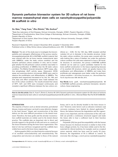
Dynamic perfusion bioreactor system for3D culture of rat bone marrow mesenchymal stem cells on nanohydroxyapatite/polyamide 66scaffold in vitroXu Qian,1Fang Yuan,1Zhu Zhimin,2Mo Anchun31State Key Laboratory of Oral Diseases,Sichuan University,Chengdu,610041,People’s Republic of China2Department of Prosthodontics,West China College of Stomatology,Sichuan University,Chengdu,610064,People’s Republic of China3Department of Oral implant,West China College of Stomatology,Sichuan University,Chengdu,610041,People’s Republic of ChinaReceived3September2012;revised4December2012;accepted11December2012Published online in Wiley Online Library().DOI:10.1002/jbm.b.32894Abstract:The aim of the study was to investigate the biocom-patibility and osteogenic effectiveness of the porous nanohy-droxyapatite/polyamide66(n-HA/PA66)scaffold material that was cultured with the rat bone marrow mesenchymal stem cells(rBMSCs),under the static culture condition and the dynamic perfusion culture condition in vitro,and to investi-gate whether the3D perfusion culture condition was better in provoking proliferation of rBMSCs than the3D static culture condition.The Methyl thiazolyl tetrazolium(MTT)assay,alka-line phosphatase(ALP)activity assay,Osteocalcin(OCN) assay and scanning electron microscope(SEM)were used to observe the proliferation and differentiation of rBMSCs.The samples were respectively harvested at1st,3rd,7th,14th,and 21st days and effect comparisons were made between the two of the culture conditions.The results showed that values of MTT,ALP,and OCN were increased continuously and revealed a significant difference between the two culture con-ditions(p<0.05).On the14th day,SEM revealed calcified nodules2–8l m in diameter in the lamellar structure.Under the static culture condition,the pores were covered with the cells looking like a piece of blanket,but under the perfusion culture condition the cells were observed to have a3D lamel-lar structure.In conclusion,the porous n-HA/PA66scaffold material can be used as a good candidate material for the bone scaffold construction in the tissue engineering because of its excellent3D structure,which can greatly improve the proliferation and differentiation of rBMSCs and make them proliferate and osteogenesis even better under the perfusion culture condition.V C2013Wiley Periodicals,Inc.J Biomed Mater Res Part B:Appl Biomater00B:000–000,2013.Key Words:bone graft,nanohydroxyapatite/polyamide66 (n-HA/PA66)scaffold,porosity,perfusion bioreactor,dynamic culture,tissue engineeringHow to cite this article:Qian X,Yuan F,Zhimin Z,Anchun M.2013.Dynamic perfusion bioreactor system for3D culture of rat bone marrow mesenchymal stem cells on nanohydroxyapatite/polyamide66scaffold in vitro.J Biomed Mater Res Part B2013:00B:000–000.INTRODUCTIONThe majority of factors such as dental extraction,periodontal disease,trauma and tumor can lead to some defective changes in the hard tissues.1Repair and reconstruction of the bone defects are the most difficult procedures in the dental treat-ment.Therefore,the bone grafts and the alveolar reconstruc-tion scaffold materials are widely used in the dental clinical practice.The scaffolds play an important role to direct and provide support to the growth of cells and serve as a matrix of the tissue formation during the bone tissue regeneration.This kind of role has well been recognized and the scaffolds have widely been used in the bone tissue engineering.Hydroxyapatite(HA)is an ideal bioactive material because of its excellent osteoconductivity and osteoinductivity.The composition and structure are similar to those of the natural bones,and it can be directly bonded to the bone tissues in vivo.2However,some factors such as abrasion resistance and corrosion resistance have limited its clinical application.3,4 Hence,the balance between the degradation and the regener-ation is very important,and the construction of the cells scaf-fold can provide a continuous structural support and make the bones able to bear a sufficient load in their function performances.Polyamide(PA)has good biocompatibility with the human tissues because its chemical structure is similar to that of the bone collagen protein.So,it has been widely used in the bio-materials for clinical medicine,for example,used as a surgical suture material,which has been used for nearly50years. However,the degradation of PA is difficult to control.To overcome this shortage,a novel porous nanohydroxyapatite/Correspondence to:M.Anchun;e-mail:moanchun@ or Z.Zhimin;e-mail:zzhimin@V C2013WILEY PERIODICALS,INC.1polyamide 66(n-HA/PA66)has been developed,which is syn-thesized from hydroxyapatite and polyamide,foamed by the thermal pressing and the injection molding techniques.5As our previous article stated,its great porosity and proper pore size can facilitate the cell seeding,survival,growth,prolifera-tion and differentiation in vivo.6In order to provide an enough theoretical support for its clinical application,we should per-form a further research on the n-HA/PA66scaffold in vitro .The bone tissue engineering is a promising technique,which can promote the development related to the bone scaffold materials,seed cells,culture bioreactor,and so forth.All the researches are based on a new concept of creating a similar microenvironment for the bone formation and har-vesting the biggest biological activity of the tissue engi-neered bones.7The bone marrow mesenchymal stem cells (BMSCs),also called the bone marrow stromal cells (BMSCs),have become the most potential seed cells because of their relative easiness to obtain,their amplification phenotype sta-bility,and their avoidance of excessive bone abnormal prolif-eration and avoidance of the related medical ethics dis-pute.8–10In addition,the culture bioreactors can imitate the living environment in vivo ,such as the rotating wall bioreac-tor and the perfusion bioreactor.11,12Since the biomechani-cal stimulation is an important influence factor of inducing BMSCs to differentiate into the osteoblastic cells.The bio-reactor systems can alleviate the nutrient transfer limita-tion,13upregulate the osteoblastic markers,13,14and increase the calcium deposition.15Therefore,this study was designed to investigate the reactiveness of the rBMSCs in vitro to eval-uate the biocompatibility and the osteogenic effectiveness of the n-HA/PA66scaffold material under the static culture condition and the dynamic perfusion culture condition in vitro ,and to investigate whether the dynamic culture condi-tion can better provoke the proliferation of rBMSCs in vitro .MATERIALS AND METHODSMaterials and characterizationThe porous n-HA/PA66scaffold materials (Figure 1)were synthesized from nanohydroxyapatite and polyamide,foamed by the thermal pressing and the injection moldingtechniques.5The bending strength,tensile strength and compressive strength of the composite with 64.5%n-HA content were 90–95,75–85,and 110–125MPa,respectively,which were similar to those of the natural bone (80–100,60–120,50–140MPa).5,16The elastic modulus of the com-posite was 5–7GPa,which was similar to that of the natural bone (3–25GPa).The pore size was 300-500l m in diame-ter and the porosity was 65–75%.There were 40pieces of materials to guarantee the comparability of the testing data.Mesenchymal stem cell donorAll animal procedures were performed according to the guidelines made by Animal Ethics Committee of Sichuan University.All experiments were conformed to the interna-tional guidelines on the ethical use of animals.We mini-mized the number of the rats used in the study and mini-mized their sufferings as much as possible.rBMSCs were isolated from the 4-week-old male or female Sprague Dawley (SD)rats 140–160g in weight,which had been purchased from Experimental Animal Cen-ter of Sichuan University.Isolation and culture of rBMSCsrBMSCs were harvested from the femur and the tibiae of the SD rats according to the protocol modified by Soleimani and Nadri.17The femur and the tibiae were placed in peni-cillin-streptomycin (Gibco,100U/ml)for 5minutes.Then,the bone marrow was flushed by Minimum Essential Me-dium Alpha Medium (a -MEM,Gibco,USA),which was sup-plemented by 10%(v/v)FBS (Gibco,Australia),using an 18gauge needle.Then,the cell suspension was transferred to the tissue culture flasks in a 37 C-5%CO 2incubator.The medium was poured half and the fresh medium was added 24hours later.After 48hours,the medium was replaced by the fresh medium,and the non-adherent cells were removed but the adherent cells were left for the culture in a mono-layer.The initial adherent cells were defined as Passage 0(P0)cells.The culture medium was refreshed every 3days until the adherent cells became confluent.Then,the P0cells were trypsinized (0.25%trypsin,Sigma,USA),washedandFIGURE 1.The surface topograph of the n-HA/PA66scaffold material.A:The material size was prepared as 16mm in diameter and 1.5mm in thickness for each experiment.One hole 2.5mm and three holes 1.5mm in diameter were drilled in the material,which better benefited the cul-ture medium passing through.Then,sterilization was performed by irradiation of Co 60.B:The material was magnified 10times for better obser-vation on the pores.n-HA/PA66:nanohydroxyapatite/polyamide 66.[Color figure can be viewed in the online issue,which is available at .]2QIAN ET AL.DYNAMIC PERFUSION BIOREACTOR SYSTEMplated into the other tissue culture flasks.rBMSCs were cul-tured to Passage 3(P3)for use in the following experimen-tal procedures.Flow cytometryP3rBMSCs were trypsinized and stained with monoclonal antibodies (CD44,CD29,CD45,CD11b,CD34)at room tem-perature for 30min.9,10Then the P3rBMSCs were per-formed by flow cytometry (China)analysis to identify the phenotypes before the experiments using.Mesenchymal multipotential differentiationP3rBMSCs were trypsinized,washed,and plated into the other tissue culture flasks with the osteogenic and adipo-genic inductive a -MEM medium (Gibco,USA)at a density of 2Â104cells/cm 2in a 37 C-5%CO 2incubator for the Alzarin red staining and oil red staining.Cell proliferation assayP3rBMSCs were trypsinized,washed and seeded on the 40pieces of n-HA/PA66scaffold material at a density of 5Â104cells /cm 2with osteogenic inductive medium in a 37 C-5%CO 2incubator for 24h.When the cells were adhered to the scaffold closely,they were equal separatedfor the following two culture conditions:the static culture condition (Figure 2)and the dynamic perfusion culture con-dition (Figures 3and 4),both with osteogenic inductive me-dium.Then,those sample cells underwent the following examinations:MTT assay,ALP activity,OCN detection assay,and SEM.Analysis of cell proliferation by MTT assayThe MTT assay is a colorimetric test for measuring the cell viability,cell proliferation,and cytotoxicity.The MTT substance is reduced by mitochondrial dehydrogenase in the living cells to a blue-colored formazan precipitate.This pre-cipitate can be dissolved by dimethylsulfoxide (DMSO,sigma,USA)and monitored by Thermo Scientific Varioskan Flash (Finland),a proxy for the cell proliferation.18In brief,MTT was first prepared as a stock solution of 5g/L in phosphate buffer and was filtered.MTT was added on the 1st ,3rd ,7th ,14th and 21st days respectively and dissolved by DMSO repeatedly.Then wash precipitate in the pores as much as possible and take out the scaffolds,the data were analyzed as absorbance at 570nm for the comparison between the static culture condition and the dynamic perfusion culturecondition.FIGURE 2.The stainless wire bent to fix the material to prevent its floating in the culture medium.Before rBMSCs were seeded,the material was soaked in the medium for 48h.rBMSCs:the rat bone marrow mesenchymal stem cells.[Color figure can be viewed in the online issue,which is available at.]FIGURE 3.Structure of the dynamic perfusion bioreactor.The dynamic perfusion bioreactor consisting of a peristaltic pump,a tubing circuit,a medium reservoir and a perfusion cartridge.The system was divided into two parts,that is,the medium circulation part and the gas exchange part.In the medium circulation part,the peristaltic pump perfused the medium out of the medium reservoir and,then,into the perfusion car-tridge.After that,the medium returned to the medium reservoir and experienced this circulation repeatedly.The flow velocity of the peristaltic pump was regulated at 2mL/min.In the gas exchange part,the mixed air contained 5%CO 2,21%O 2and 75%N 2according to the requirement of the cell culture.The mixed air passed through the bacterial filter at a speed of 60bubbles per minute.The safety bottle was placed at the end of the circulation part to guarantee the airway unobstructed.[Color figure can be viewed in the online issue,which is available at .]ORIGINAL RESEARCH REPORTJOURNAL OF BIOMEDICAL MATERIALS RESEARCH B:APPLIED BIOMATERIALS |MONTH 2013VOL 00B,ISSUE 003ALP activity assayALP decomposes p-nitrobenzene phosphate (PNPP)into p-nitrophenol (PNP)under the relevant conditions,which reflects the degree of the cell activity according to the shade of yellow of PNP .The ALP activity was performed at 405nm using PNPP (Sigma,USA)as the substrate.The optical density (OD)value reflected the PNP generation at each minute.HTS 7000Plus High-Efficient Analyzer (USA)determined the OD value every 60seconds for 4times and calculated the absorbance changes (D A).According to the formula,the ALP activity (U/ml)was fig-ured out.The total protein content was determined by the Coo-massie brilliant blue method.Then,the ALP activity was expressed as U/mg (unit per mg of protein).Briefly,all the sam-ples in the static culture group and the dynamic culture group were collected respectively on the 1st ,3rd ,7th ,14th and 21st days.Cells were rinsed two times with PBS followed by trypsini-zation and then scraped into ddH 2O.All samples were followed by three cycles of freezing and thawing.The ALP activity values were determined by the method mentioned above.OCN detection assayThe OCN enzyme linked immunosorbent assay (ELISA)kits were used to detect the OCN levels.In the two culture groups,the culture medium was collected for determination of the OCN levels.The samples were harvested respectively on the 1st,3rd,7th,14th,and 21st days.Absorbance of each well was measured at 450nm by HTS 7000Plus.SEM observationThe samples were harvested respectively on the 1st,3rd,7th,14th,and 21st days under the two culture conditions.Then,the samples were soaked in 2.5%glutaraldehydeovernight,and the alcohol gradient dehydration was pre-pared for the SEM observation.The samples were sliced into two pieces to be convenient for observing the pores inside and the surface of both sides of scaffolds.Statistical analysisThere were at least 90pieces of materials to guarantee the comparability of the testing data,half for static condition and half for dynamic condition.Each 3pieces for ALP assay,MTT assay and SEM observation on 1,3,7,14,and 21days.The samples were harvested respectively.All the data were expressed as mean 6standard error (SE).Comparisons were performed by a paired two-tailed distribution test between the two culture conditions.Values of p <0.05were considered statistically significant.RESULTSCharacterization of rBMSCsCulture expanded rBMSCs grew as slender spindle-like or star-like cells [Figure 5(A)].Under permissive culture condi-tions,rBMSCs underwent osteogenic and adipogenic differ-entiation,which confirming their bona-fide MSC phenotype [Figure 5(B–D)].The majority of rBMSCs at P3were positive for CD44(99.6%)and CD29(99.8%),while a few cells were positive for CD45(4.8%),CD11b (0.6%)and CD34(0.4%)(Figure 6).It indicated that high-purity rBMSCs could be harvested and used in the next experiment.MTT assayIn Figure 7,the curves indicated a rising pattern for the MTT values of the two culture results.In spite of the MTT value on the 3th day of the static culture seemed alittleFIGURE 4.A,B:The scaffolds seeded with rBMSCs,which were separated by the bent stainless wires to prevent the cell injury due to the fric-tion between the materials.C:There were 10pieces of materials inside one perfusion culture chamber.To guarantee the comparability of the testing data,the materials located at the same position in the perfusion cartridge should be sent to the same test determination.Each 3pieces for ALP assay,MTT assay and SEM observation on 1,3,7,14and 21days.The samples were harvested respectively.ALP:alkaline phosphatase,MTT:Methyl thiazolyl tetrazolium,SEM:scanning electronic microscopy.[Color figure can be viewed in the online issue,which is available at .]4QIAN ET AL.DYNAMIC PERFUSION BIOREACTOR SYSTEMgreater than that of the dynamic perfusion culture,but no statistically significant difference was found between the two cultures (p >0.05);however,observations on the 14th and the 21st days showed that the MTT value of the dynamic culture exceeded the static culture,and there was a significant difference between the two groups (p <0.05).ALP assayFigure 8showed that on the 7th day the peak of ALP values were observed under the two culture conditions but after that time the falling trend of the values was found under the both conditions.The statistical analysis showed that there was significant difference in the ALP values according to the observations on the 3rd,7th,14th,and 21st days except for the observation on the 1st day (p <0.05).OCN assayFigure 9showed that the OCN values obtained under the two-culture conditions showed a rising trend during the ob-servation period.Under the two culture conditions,the OCN values were significantly greater on the 21st day than on the 3rd,7th,and 14th days (p <0.05).A statistically signifi-cant difference was found in the OCN values between the two-culture conditions on the 3rd day (p ¼0.0075),14th day (p ¼0.007)and 21st day (p ¼0.001);however,on the 7th day no significant difference was found between the two culture conditions (p ¼0.703).SEM observationThe SEM results from the static culture revealed that rBMSCs uniformly spread on the surface of scaffold after 24h of culture [Figure 10(A)].The pseudopods stretched on the pore walls,the pore size of which was 300–500l m in diameter,and the porosity was 65–75%.6On the 3rd day af-ter the cell seeding,the cells began to grow on the edge of the pores,proliferated well and connected with the other cells [Figure 10(B)].On the 7th day,the cells on the pore edge proliferated layer upon layer,looking like apavedFIGURE 5.Characterization of rBMSCs.A:Primary rBMSCs were slender spindle-like or star-like cells.B:Mineralized nodules formed after 14days of the mineralization inductive culture.C:rBMSCs were induced for 21days of the ontogenetic inductive culture.Mineralized nodules were reddish orange dyed by the Alzarin red staining.D:rBMSCs were induced for 5days of the adipogenic inductive culture.Lipid Droplet was red-dish dyed by oil red staining.E:rBMSCs grew well at the margin of the n-HA/PA66scaffold.The viability of rBMSCs around n-HA/PA66was strong though calcification was occasionally found.[Color figure can be viewed in the online issue,which is available at.]FIGURE 6.rBMSCs phenotype identification by flow cytometry.The majority of rBMSCs at P3were positive for CD44(99.6%)and CD29(99.8%),while a few cells were positive for CD45(4.8%),CD11b (0.6%)and CD34(0.4%).[Color figure can be viewed in the online issue,which is avail-able at .]ORIGINAL RESEARCH REPORTJOURNAL OF BIOMEDICAL MATERIALS RESEARCH B:APPLIED BIOMATERIALS |MONTH 2013VOL 00B,ISSUE 005blanket.However,the cells at the bottom and on the walls of the pores had less nutrition supply.Therefore,the cells became smaller in number or even died [Figure 10(C)].On the 14th day,the pores were completely covered by the cells looking like a paved blanket [Figure 10(D)].The SEM results revealed that under the dynamic perfu-sion culture condition,rBMSCs were better adhered to the surface of the scaffolds.The cells spread on the rim and the inner wall of the pores after the 24-h perfusion culture.On the 3rd day,the proliferated cells spread on the deep pores and connected with each other [Figure 11(A)].rBMSCs became denser and denser in arrangement,and they were proliferated along with the porous stereoscopic structure af-ter 7days [Figure 11(B)].The cells growing vigorously for 14days remained at the bottom and on the inner wall of the pores.In the static culture group,the pores were covered by the cells looking like a paved blanket;however,in the dynamic perfusion culture group,the cells were shaped like the 3D lamellar structure which simulated to the shape of scaffold [Figure 11(C)].Majority of the pores were opened and unobstructed,so that the exchange of air and nutrients could be guaranteed.In the lamellar structure,calcified nod-ules 2-8l m in diameter could be observed [Figure 11(D)].DISCUSSIONSBMSCs are not only involved in the hematopoietic stem cell survival and differentiation but also have a multiplex differ-entiation potential.Since BMSCs are relatively few in the bone marrow,they have to be purified and cultivated to meet the bone tissue engineering requirement.The purifica-tion process usually includes the cell adherence separation,density gradient centrifugation,19immune magnetic beads separation,20and flow cytometry.21The monoclonal anti-body magnetic beads separation system and the flow cytom-etry require not only good experimental conditions but also a great amount of bone marrow;therefore,the maintaining of the BMSCs activity is relatively difficult.The gradient density centrifugation can harvest highly pure cells but the centrifugal speed and time have to be exactly adjusted in the different environmental conditions.So,in this study we used a mature method,i.e.,the cell adherence separation for amplification of BMSCs.This method had been proved effec-tive in the previous studies.BMSCs were proliferated on the walls but the hematopoietic cells were proliferated in the medium.Non-adherent cells were removed and the adher-ent cells were left for the culture in a monolayer along with the procedure of refreshing the medium.At last,BMSCs were trypsinized for 3or 4passages and they were puri-fied.22The majority of BMSCs at P3phenotype identification by flow cytometry indicated that high-purity BMSCs could be harvested and used in the experiment.The natural bone material was considered as nanocompo-site material,which was composed of collagen,apatite,and water;one-third was inorganic and two-thirds was organic.The bending strength,tensile strength and compressive strength of the porous n-HA/PA66scaffold material were 90–95,75–85,and 110–125MPa,respectively.5The porous n-HA/PA66scaffold material was close to the natural bone material that has the similar bending strength,tensile strength,and compressive strength (80–100,60–120,50–140MPa).16There-fore,this kind of composite can be well matched to that of the human bones.5PA had good biocompatibility with the human tissues,probably because of its similar chemical structure to that of the collagen protein.The polarity of the amide keys and the carboxy of PA could guide the tissue and cell growth,and promote the osteoid formation and mineralization.5PA had an excellent biological activity,biocompatibility and bone conduc-tion property.23n-HA/PA66also had excellentmechanicalFIGURE 7.The MTT values obtained under the static culture condi-tion and the dynamic perfusion culture condition for 1,3,7,14,and 21days.*p <0.05.[Color figure can be viewed in the online issue,which is available at.]FIGURE 8.The ALP values obtained under the static culture condition and the dynamic perfusion culture condition for 1,3,7,14,and 21days.*p <0.05.[Color figure can be viewed in the online issue,which is available at.]FIGURE 9.The OCN values obtained under the static culture condi-tion and the dynamic perfusion culture condition for 1,3,7,14,and 21days.*p <0.05.[Color figure can be viewed in the online issue,which is available at .]6QIAN ET AL.DYNAMIC PERFUSION BIOREACTOR SYSTEMcompatibility,and its mechanical strength came mainly from the hydrogen bond between carboxyl of n-HA and acylamino of PA66.The content of n-HA more or less affected the stability of the hydrogen bond and the crystallization rate of PA66,and thus affected the strength of n-HA/PA66.n-HA/PA66foamed by the thermal pressing and the injection molding techniques had the uniform pore size (diameter,300-500l m),which pro-vided a lasting stress-absorbent core framework.19The specific surface area of the porous n-HA/PA66scaffold had a large con-tact region,which facilitated adhesion,differentiation,prolifera-tion and metabolization of the cells.6The 3D scaffold structure effectively stimulated the osteoblast differentiationandFIGURE 10.The SEM observation on rBMSCs under the static culturecondition.FIGURE 11.The SEM observation on rBMSCs under the dynamic perfusion culture condition.ORIGINAL RESEARCH REPORTJOURNAL OF BIOMEDICAL MATERIALS RESEARCH B:APPLIED BIOMATERIALS |MONTH 2013VOL 00B,ISSUE 007benefited the osteoprogenitor cell proliferation and migra-tion.24,25Another experiment successfully proved that the3D porous scaffold effectively promoted the extracellular matrix synthesis,and its structure was similar to the natural bone structure.26After rBMSCs were seeded on the scaffolds,they were cultured under the two conditions,that is,the static culture condition and the dynamic perfusion culture condition. Under the static culture condition,rBMSCs were proliferated on the surface or out of the scaffolds because of the gravity action.27rBMSCs in the scaffolds only accounted for25%of the initially cultivated rBMSCs because the culture medium and the gas molecules could not be effectively transferred to the deep pores,so that the cell proliferation and mineral-ization only existed in the scaffold surface120–250l m in depth21;therefore,there was little or no bone formation at the centre of the scaffolds.28The structure of the3D scaf-fold met the tissue engineering requirement,but the static culture condition became an unfavorable factor.22In order to imitate the living environment in vivo,quite a few bio-reactors were developed.Glowacki et al.performed some experiments to prove the dynamic perfusion culture condi-tion able to increase the viability and the functions of BMSCs.29The cells could be seeded well-proportionally inside and outside of the scaffolds by the3D dynamic perfu-sion culture condition,which could promote oxygen and nutrients to enter the scaffolds so that the sufficient prolif-eration of the cells inside the scaffolds could be guaranteed.Many of the previous researches showed that the proper shear force and the mechanical distractive force could pro-mote the BMSCs osteogenic differentiation.There were many force application systems used for this purpose,for example,the four-point bending model,30the rotating wall bioreactor,31and the perfusion bioreactor.29The dynamic perfusion bioreactor used in this experiment perfused oxy-gen and carbon dioxide into the pores to guarantee the cul-ture medium circulation and nutrition exchanges.We chose aflow velocity of2ml/min to promote the cell proliferation and differentiation in our previous experiment because that the velocity was not so fast to cause rBMSCs away from the scaffolds.Compared with the3D static culture condition in this experiment,the3D dynamic perfusion culture condition could make the values of MTT,ALP,and OCN increase con-tinuously.However,the MTT value on the3th day of the static culture seemed a little greater than that of the dynamic perfusion culture,but observations on the14th and the21st days showed that the MTT value of the dynamic culture exceeded the static culture condition obvi-ously.The ALP activity examination could show a significant difference between the two-culture conditions on the3rd, 7th,14th,and21st days.The OCN value obtained under the dynamic perfusion culture condition was greater than that obtained under the static culture condition on the14th and 21st days.All the results indicated that the3D dynamic per-fusion culture condition could induce osteogenesis more effectively,which were similar to the results from other pre-vious researches.32The SEM results showed that under the static culture condition,the mass of the cells was shaped like a paved blanket on the scaffold surface,and most of the pores were covered by the so-called blanket.It was difficult tofind rBMSCs existing in the pores,but some dead cells were observed there.The reason for thosefindings was probably that the material exchange level was relatively low in that place,so that the cells had to proliferate on the nu-trient-rich liquid surface,forming the structure like a paved blanket.However,under the dynamic perfusion culture con-dition,rBMSCs formed a3D layer structure,and the cells could be clearly observed at the bottom or on the sides of the pore walls.Some liquid was observed to beflowing across the pores,so that the cells could obtain sufficient nutrients,which was a beneficial factor for the cell prolifer-ation.Some calcified nodules2-8l m in diameter could be found in the lamellar structure on the14th day,which revealed that porous n-HA/PA66could increase the calcium matrix deposition and the differentiation speed of the late-stage osteoblast.In addition,n-HA/PA66with65%-75% interconnected porosity had a greater calcium deposition rate than the other materials with a lower porosity.As the preosteoblast line was seeded in the scaffold that was9 mm in diameter and5mm in height,the central oxygen concentration would drop to0%on the5th day of the cul-ture[33].The cell death was observed subsequently in those areas of the scaffold.When the demineralized bone matrix scaffold was placed under the dynamic perfusion cul-ture condition,the central oxygen concentration increased to4%.Although this oxygen concentration level was still lower than that under the bulk medium condition(20%),it was still enough for prevention of the cell death.13 CONCLUSIONSThe innovative bone tissue-engineering porous n-HA/PA66 scaffold material is an ideal three-dimensional micro-struc-ture material for sufficient proliferation of rBMSCs.The po-rous n-HA/PA66scaffold material is confirmed to be a good candidate as a bone scaffold material used in the tissue en-gineering.And the dynamic perfusion culture condition can greatly improve proliferation and osteogenic effectiveness of rBMSCs on porous n-HA/PA66scaffold. ACKNOWLEDGMENTSThe authors are grateful to Ms.Li Xiaoyu at the State Key Labo-ratory of Oral Diseases(West China College of Stomatology) for her assistance in the cell culture and Dr.Zhu Zhimin(West China College of Stomatology)for her assistance in the experi-ment implementation.REFERENCES1.Mecall RA,Rosenfeld AL.The influence of residual ridge resorp-tion patterns onfixture placement and tooth position.1.Int J Periodontics Restorative Dent1991;11:8–23.2.Woodard JR,Hilldore AJ,Lan SK,Park CJ,Morgan AW,EurellJA,Clark SG,Wheeler MB,Jamison RD,Wagoner Johnson AJ.The mechanical properties and osteoconductivity of hydroxyapa-tite bone scaffolds with multi-scale porosity.Biomaterials2007;28:45–54.3.Strocchi R,Orsini G,Lezzi G,Scarano A,Rubini C,Pecora G,Piat-telli A.Bone regeneration with calcium sulfate:Evidence for8QIAN ET AL.DYNAMIC PERFUSION BIOREACTOR SYSTEM。
不同培养方法对骨髓间充质干细胞贴壁和纯度的影响

[ 要 】 目的 摘
探讨 骨髓 问 充 质 干 细 胞 ( C ) 想 的 分 离 、 养 方 法 。 方 法 将 M C 分 别 于 普 通 培 养 瓶 、 MS s理 培 Ss 胶 原 培 养 瓶 及 C 培 养 瓶 细 胞 贴 壁 , 况 o o 情
c=培养 瓶 、 原 培 养瓶 中 培 养 , 察 三批 标 本 细 胞 贴 壁 情 况 ; 3个 批 次 标 本 细 胞 分 别 用 I M、60和 M snu 0 6 0 胶 观 将 MD 14 e cl e t 培 养 液 培 养 , 察 细 胞 纯 化 情 况 ; 式 细胞 术 检 测 细 胞 表 面 标 志 。 结 果 观 流 明 显 好 于 普 通 培养 瓶 , 后 者 费 用 低 ; sn u 培 养 体 系 能 获得 高 纯 度 M C , 传 代 后 细 胞 不 易 分 化 。 流 式 细 胞 且 Me C t e l S s且 术 示 三 种 培 养体 系 中细 胞 表 型 均 表 现 为 C 3 ( 、 D 5 一)C 7 ( +)C 4 ( +)C g ( +) 结 论 D 4 一)C 4 ( 、 D 1 + 、D4 + 、 DO + 。 临 床 提 供 了 实 验方 法 及依 据 。 【 键 词 】 人 骨髓 间充 质 干 细 胞 ; 胞 培 养 关 细 [ 图分 类 号 】 o l 中 83 [ 文献 标 识 码 ] A [ 章 编 号 】 10 . 6 (084 . 1.3 文 022 X 2 0 )00 50 6 0 应 用
送检中检验实验室间充质干细胞质量标准

送检中检验实验室间充质干细胞质量标准1. 引言充质干细胞(mesenchymal stem cells, MSCs)作为一种多功能的细胞类型,具有广泛的临床应用前景。
随着充质干细胞的应用越来越广泛,不同实验室之间对于充质干细胞质量标准的一致性要求也逐渐提升。
本文旨在确立送检中检验实验室间充质干细胞质量标准,以促进充质干细胞研究的进一步发展。
2. 充质干细胞质量标准的选择为了确保实验室间充质干细胞质量标准的一致性,需要选择合适的评估指标。
以下是一些常用的充质干细胞质量标准指标:- 表面标记物:CD105、CD73、CD90阳性;CD45、CD34、CD14、CD11b、CD79a、CD19阴性。
- 多向分化潜能:具备成骨、成脂、成软骨等分化能力。
- 增殖能力:具备较高的增殖速度和活力。
- 免疫抑制功能:能够抑制免疫细胞的活化和增殖。
3. 充质干细胞质量标准的实验方法为了确定充质干细胞质量标准的实验方法,可以参考以下步骤:1. 充质干细胞的培养和扩增:选择合适的培养基和培养条件,确保充质干细胞的良好扩增。
2. 充质干细胞的表面标记物鉴定:通过流式细胞术或免疫荧光染色法,鉴定充质干细胞的表面标记物。
3. 充质干细胞的多向分化潜能鉴定:通过体外培养分化实验,观察充质干细胞在不同诱导条件下的分化能力。
4. 充质干细胞的增殖能力评估:通过MTT法等细胞增殖实验,评估充质干细胞的增殖能力。
5. 充质干细胞的免疫抑制功能评估:通过淋巴细胞增殖实验或ELISA法,评估充质干细胞的免疫抑制功能。
4. 实验室间质检的建立和维护为了确保实验室间充质干细胞质量标准的一致性,需要建立和维护质检体系。
以下是一些建议:- 建立标准化的实验操作流程和质检标准,确保每个实验室的操作规范一致。
- 定期组织质检活动,通过对比不同实验室的质检结果,发现和解决质检过程中的问题。
- 建立质检记录和数据库,记录每次质检的结果和不良事件的处理情况。
送检中检验机构间充质干细胞质量标准

送检中检验机构间充质干细胞质量标准引言充质干细胞(mesenchymal stem cells,MSCs)作为一种多潜能的干细胞,被广泛应用于组织工程、再生医学以及免疫治疗等领域。
由于充质干细胞的质量直接关系到其临床应用的效果和安全性,各个检验机构在进行充质干细胞的质量检验时需要遵循统一的标准。
本文档旨在制定一份统一的充质干细胞质量标准,以确保不同检验机构之间的一致性。
标准内容1. 充质干细胞定义充质干细胞为一类具有自我更新和多向分化潜能的成人软组织起源的凋亡细胞。
2. 充质干细胞分离与培养2.1 分离方法:推荐使用胶原酶及胰酶对组织进行消化分离。
分离过程中需注意细胞的存活率和纯度。
2.2 培养条件:充质干细胞应在适当的培养基中进行培养,包括适宜的营养液、培养基浓度及PH值等。
3. 充质干细胞鉴定3.1 表面标记物:充质干细胞常表达CD73、CD105、CD90,并不表达CD34、CD45和CD11b等表面标记物。
3.2 多向分化潜能:充质干细胞应具备成骨、成脂以及成软骨的分化潜能。
4. 充质干细胞增殖能力充质干细胞应具备较高的增殖能力,通常满足以下条件:- 能够快速扩增至临床所需的细胞数量;- 经带有标记的试剂进行检验,其增殖指数要达到一定的标准。
5. 充质干细胞免疫学特性5.1 免疫抑制作用:充质干细胞应具备对淋巴细胞等免疫细胞的抑制作用。
5.2 免疫抗原性:充质干细胞应具备较低的免疫原性,以减少免疫排斥反应。
结论本文档提供了送检中检验机构间充质干细胞质量标准的基本内容,以确保对充质干细胞的质量检验具有一致性。
各个检验机构应根据实际需求,在此基础上进行进一步的细节制定,以提高充质干细胞的质量控制水平。
在追求高质量代码的同时,本文档已经超过了800字,如需进一步扩展内容或有其他要求,请告知。
小鼠骨髓间充质细胞原代培养及传代

小鼠骨髓间充质细胞原代培养及传代之flamerking图文版[精华]很多战友发email或PM向我询问小鼠MSC的培养方法,我零零散散地一个个回复过几次,由于我平时时间不是很多,所以回答地时候有些内容写的不是很详细。
今天又有一个战友PM我,询问我培养方法。
看来做小鼠MSC的战友越来越多。
一遍遍地重复写很费时间(想粘贴复制又找不到上次写的在哪~惨!),所以我想还是发贴公布一下。
其实我小鼠MSC在我的课题中不是主角,只是起一个control的作用,因此我在这方面的经验可能没有一些专门做小鼠MSC的战友丰富,不过我还是很愿意把我的经验写出来供大家参考,希望能够起到“抛砖引玉”的作用。
欢迎各位同道对我的方案进行批评和指正,与大家一起分享你们的经验和智慧。
提纲一,简版主要步骤及操作要点。
二,完整版1. 试剂及材料准备2. 动物手术及取材3. 原代细胞培养4. 细胞传代5. 讨论时间关系,今天先给个简版。
我先顶一下,由于我还是新手,故不能说是经验介绍,只好讲让你们指出缺点,谢谢斑竹1. 试剂及材料准备MEM,10%FCS,手术器械,培养瓶,200目滤网,5ml注射器2. 动物手术及取材CR小鼠1只,酒精浸泡5分钟,取股,胫,肱骨,用基培冲出骨髓,滤过,1000转离心,完培重悬,接种于25厘米培养瓶3. 原代细胞培养:,3天后首次换液,自己发现一周贴壁细胞就铺满达90%,自行传代4. 细胞传代:消化下来很少,能下来的增长很慢5. 讨论:没经验欢迎斑竹的继续讲解,希望有经验的老师们能不啬指教羽之无限版主,能有空再讲讲吗这几天,我的原代就是不长,42天了,25平方厘米的培养瓶还未铺慢,不能传代真是天天盼着你啊呵呵,不好意思,最近我的sony笔记本又坏了,上次坏了几次维修部修不好,给我换了台新的,结果以用了没2个月又不能启动了,真是麻烦,我的资料都在里面,最近实验又忙,没空去维修部……不过明天我就去了。
……呵呵,跑题了。
间充质干细胞流程
间充质干细胞流程下载温馨提示:该文档是我店铺精心编制而成,希望大家下载以后,能够帮助大家解决实际的问题。
文档下载后可定制随意修改,请根据实际需要进行相应的调整和使用,谢谢!并且,本店铺为大家提供各种各样类型的实用资料,如教育随笔、日记赏析、句子摘抄、古诗大全、经典美文、话题作文、工作总结、词语解析、文案摘录、其他资料等等,如想了解不同资料格式和写法,敬请关注!Download tips: This document is carefully compiled by the editor. I hope that after you download them, they can help yousolve practical problems. The document can be customized and modified after downloading, please adjust and use it according to actual needs, thank you!In addition, our shop provides you with various types of practical materials, such as educational essays, diary appreciation, sentence excerpts, ancient poems, classic articles, topic composition, work summary, word parsing, copy excerpts,other materials and so on, want to know different data formats and writing methods, please pay attention!间充质干细胞(MSCs)是一类具有多重潜能的成体细胞,在干细胞领域具有重要的应用前景。
脐带间充质干细胞
脐带间充质干细胞扩增培养方法
采用无血清培养基(可含10%受者灭活血清)将 细胞调成悬液,按2×10^4/cm^2接种于培养 瓶, 37℃、5%的CO2条件下培养4d后全量换液, 吸弃非贴壁细胞。每3d换液,细胞覆盖瓶底80% 时,0.25%胰蛋白酶-1mmol/L EDTA消化,按1: 3传代。培养3周左右,胰酶消化细胞,细胞悬液 2000r/min离心5min,弃上清后加入生理盐水混匀, 2000r/min离心5min,重复2次。
脐带间充质干细胞培养结果
新分离的脐带单个核细胞呈纺锤形
原代培养6h后有大量细胞贴壁,贴 壁细胞呈梭形、多角形
培养7天细胞呈长梭形,细胞平行或 漩涡状排列。培养10天细胞可达 80%融合。传代后的细胞形态无明显 改变,排列更加整齐。
脐带间充质干细胞在临床上的应用
1.能具有较强的免疫调节作用,可用于治疗 红斑狼疮和硬皮病等自身免疫性疾病,降 低细胞或器官移植后的免疫排斥反应,提 高细胞或器官移植的成功率。
酶消化法
培养方法
组织块贴壁法
酶消化法
• 去除血管,将脐带组织剪成1~3cm大小组织块, 用无血清培养基洗2次,将组织浸泡在0.1%胶原 酶中,37℃消化4~6小时。加入生理盐水(消化 液体积的2倍),2000r/min离心5min,弃上清, 加入2.5%胰蛋白酶,37℃处理30min,加入生理 盐水(消化液体积的2倍),100目滤网过滤,滤 液2000r/min离心5min,弃上清。重复两次。细胞 计数,将细胞调成一定浓度用于培养。
组织块贴壁法
• 取新鲜健康脐带,用PBS冲洗干净后,用剪刀镊 子剔去血管,剥出里面的华通氏胶,将所得组织 充分剪碎至1 mm^3大小,加入α-MEM培养液置 于37℃,5%的CO2培养箱培养,培养液中含10% FBS,100U/ml青霉素,100U/ml链霉素。脐带组 织培养5-7天后,可见有部分细胞从组织块周围爬 出,形态呈细小的梭形,一周后,细胞开始迅速 增殖,形成大小不等的细胞集落。
一种pbmc和msc共培养的方法及其应用
一种pbmc和msc共培养的方法及其应用1. 引言PBMC(外周血单个核细胞)和MSC(间充质干细胞)是目前研究较为广泛的细胞类型之一。
PBMC是体内免疫系统的主要组成部分,具有免疫调节和抗肿瘤等功能;而MSC则具有自我更新、多向分化和免疫调节等特性。
PBMC和MSC共培养能够发挥两者的协同效应,有望在疾病治疗和再生医学领域发挥重要作用。
2. 共培养方法(1)PBMC和MSC分离:从人体外周血中分离PBMC和从骨髓、脂肪组织等源头分离MSC,并进行细胞计数和鉴定。
(2)细胞培养:将PBMC和MSC分别培养至适当的浓度和状态,如PBMC可采用RPMI-1640培养基,MSC可采用DMEM/F12培养基,并添加适当的生长因子和补充物。
(3)共培养体系建立:将PBMC和MSC以一定比例混合,常用比例为1:1或2:1,将混合细胞悬液转移到共培养培养基中。
(4)培养条件调节:调整培养条件如温度、湿度、CO2浓度等,提供适宜的生长环境。
(5)共培养时间:根据具体实验设计,共培养时间可在数小时至数天不等。
(6)细胞采集:根据实验需要,采集共培养体系中的细胞,如细胞上清液、细胞沉淀等。
3. 共培养方法的应用(1)免疫治疗:PBMC和MSC共培养后,PBMC的活性和功能会得到调节和改善。
共培养体系中的MSC具有免疫抑制作用,可抑制PBMC 的过度激活和炎症反应,从而在免疫治疗中发挥作用。
例如,在移植物抗宿主病(GVHD)的治疗中,共培养的PBMC和MSC可通过调节免疫应答,减轻移植物对宿主组织的攻击。
(2)组织再生:PBMC和MSC共培养后,MSC的多向分化能力可以促进组织再生和修复。
共培养体系中的MSC可分化为成骨细胞、软骨细胞等,同时PBMC中的细胞因子也能促进MSC的分化和增殖。
这一方法在骨折修复、软骨修复等领域具有潜在应用前景。
(3)肿瘤治疗:PBMC和MSC共培养后,MSC的免疫调节能力可增强PBMC对肿瘤细胞的攻击。
骨髓间充质干细胞的体外培养
A l t o ilfLzo ei l oo e Lzo . i un6 60 , hn f ie H s t u uM d a Clg ,ahu S ha 0 0 C i i f ad pa o h c l c 4 a 【 bt c】 O jcv T sbi f s l m t dw i pr eadprybn a o e nhmls m cl ( S s A s at r bet e oe alha e ie e o h hs aa n ui oem r wm s ey a t esM C ) i t s ab h c e t f r e e
・
l O2 ・ 6
川 学 20 08年 l 第 2 ( 1 ) S ha ei l or l20 ,o.9 N .2 2月 9卷 第 2期 i lM dc un ,08 V12 . o 1 c tl aJ a
论 著
红 , 昌平 , 晓琳 杜 李 钟
( 州 医 学 院 附 属 医 院 消 化 内科 , 川 泸 州 660 ) 泸 四 400
【 摘要 】 目的 建立骨髓 问充质干 细胞( C) MS s体外分 离纯化 的方法 , 索 M C 体外培 养的最佳方法 , 探 Ss 为细胞工程 学提供 细胞来源。方 法 ①M C 的分 离、 Ss 培养 : 在无茵条件下取 3— 4周龄 Wia 雄性大鼠四肢骨的骨髓细胞 , sr t 采用裂解
及 荧光 染料 后 见 C 4 、 D 0染 色呈 阳 性 ,D 5染 色呈 阴性 反 应 。 结 论 采 用 裂 解 红 细 胞 与 贴 壁 培 养 法相 结 合 分 离 、 D4 C g C4 纯
化 M C, S s 可得 到 活 性 和 纯 度 均较 好 的 M C 。 S s
- 1、下载文档前请自行甄别文档内容的完整性,平台不提供额外的编辑、内容补充、找答案等附加服务。
- 2、"仅部分预览"的文档,不可在线预览部分如存在完整性等问题,可反馈申请退款(可完整预览的文档不适用该条件!)。
- 3、如文档侵犯您的权益,请联系客服反馈,我们会尽快为您处理(人工客服工作时间:9:00-18:30)。
间充质干细胞培养方法 1. 间充质干细胞MSC基本形态 2. 干细胞应用与干细胞调控。 3. 间充质干细胞MSC生长过程 4. 间充质干细胞MSC培养的合适气体环境 5. 细胞培养板的选择 6. 如何选用细胞培养基 7. 如何维持培养液 p H 8. 血清与干细胞的培养 9. 胎牛血清(F B S )是否需要灭活 10. 细胞的细菌、真菌污染及排除 11. 细胞培养污染的预防 12. 使用胰蛋白酶时加入 E DTA的目的是什么 13. 胶原酶的种类和选型 14. 胶原酶 V S胰酶 15. 干细胞的种类和表面标记 16. 间质干细胞培养原理概述 17. 间质干细胞成脂和成骨诱导分化 18. 干细胞老化的表现和处理 19. 细胞传代消化过程指导 20. 冷冻保护剂作用和选择 21. 细胞冻存指导 22. 干细胞冷冻和复苏 23. 移植细胞的基因修饰 1.间充质干细胞MSC基本形态 体外培养细胞根据它们在培养器皿是否能贴附于支持物上生长特征,可分为贴附型生长细胞,常表现为成纤维型细胞和上皮细胞。悬浮型细胞在培养中悬浮生长。 间充质干细胞MSC基本形态:形态与成纤维细胞类似,细胞在支持物表面呈梭形或不规则三角形生长,细胞中央有卵圆形核,胞质向外伸出2-3 厘米个长短不同的突起。可看到细胞成螺旋状生长。
2.干细胞应用与干细胞调控 干细胞的调控是指给出适当的因子条件,对干细胞的增殖和分化进行调控,使之向指定的方向发展。
2.1内源性调控 干细胞自身有许多调控因子可对外界信号起反应从而调节其增殖和分化,包括调节细胞不对称分裂的蛋白,控制基因表达的核因子等。另外,干细胞在终末分化之前所进行的分裂次数也受到细胞内调控因子的制约。 (1)胞内蛋白对干细胞分裂的调控 干细胞分裂可能产生新的干细胞或分化的功能细胞。这种分化的不对称是由于细胞本身成分的不均等分配和周围环境的作用造成的。细胞的结构蛋白,特别是细胞骨架成分对细胞的发育非常重要。如在果蝇卵巢中,调控干细胞不对称分裂的是一种称为收缩体的细胞器,包含有许多调节蛋白,如膜收缩蛋白和细胞周期素A。收缩体与纺锤体的结合决定了干细胞分裂的部位,从而把维持干细胞性状所必需的成分保留在子代干细胞中。 (2)转录因子的调控 在脊椎动物中,转录因子对干细胞分化的调节非常重要。比如在胚胎干细胞的发生中,转录因子Oct4 是必需的。Oct4 是一种哺乳动物早期胚胎细胞表达的转录因子,它诱导表达的靶基因产物是FGF-4 等生长因子,能够通过生长因子的旁分泌作用调节干细胞以及周围滋养层的进一步分化。Oct4 缺失突变的胚胎只能发育到囊胚期,其内部细胞不能发育成内层细胞团。另外白血病抑制因子(LIF)对培养的小鼠ES 细胞的自我更新有促进作用,而对人的成体干细胞无作用,说明不同种属间的转录调控是不完全一致的。又如 Tcf/Lef 转录因子家族对上皮干细胞的分化非常重要。Tcf/Lef 是Wnt 信号通路的中间介质,当与β-Catenin 形成转录复合物后,促使角质细胞转化为多能状态并分化为毛囊。
2.2外源性调控 除内源性调控外,干细胞的分化还可受到其周围组织及细胞外基质等外源性因素的影响。 (1)分泌因子 间质细胞能够分泌许多因子,维持干细胞的增殖,分化和存活。有两类因子在不同组织甚至不同种属中都发挥重要作用,它们是TGFβ家族和Wnt 信号通路。比如TGF 家族中至少有两个成员能够调节神经嵴干细胞的分化。最近研究发现,胶质细胞衍生的神经营养因子(GDNF)不仅能够促进多种神经元的存活和分化,还对精原细胞的再生和分化有决定作用。GDNF 缺失的小鼠表现为干细胞数量的减少,而 GDNF的过度表达导致未分化的精原细胞的累积。Wnts 的作用机制是通过阻止β-Catenin 分解从而激活Tcf/Lef 介导的转录,促进干细胞的分化。比如在线虫卵裂球的分裂中,邻近细胞诱导的Wnt 信号通路能够控制纺锤体的起始点和内胚层的分化。 (2)膜蛋白介导的细胞间的相互作用 有些信号是通过细胞-细胞的直接接触起作用的。β-Catenin 就是一种介导细胞粘附连接的结构成分。除此之外,穿膜蛋白Notch及其配体Delta 或Jagged 也对干细胞分化有重要影响。在果蝇的感觉器官前体细胞,脊椎动物的胚胎及成年组织包括视网膜神经上皮、骨骼肌和血液系统中,Notch 信号都起着非常重要的作用。当Notch 与其配体结合时,干细胞进行非分化性增殖;当Notch 活性被抑制时,干细胞进入分化程序,发育为功能细胞。 (3)整合素(Integrin)与细胞外基质 整合素家族是介导干细胞与细胞外基质粘附的最主要的分子。整合素与其配体的相互作用为干细胞的非分化增殖提供了适当的微环境。比如当β1整合素丧失功能时,上皮干细胞逃脱了微环境的制约,分化成角质细胞。此外细胞外基质通过调节β1整合素的表达和激活,从而影响干细胞的分布和分化方向。
2.3干细胞的可塑性 越来越多的证据表明,当成体干细胞被移植入受体中,它们表现出很强的可塑性。通常情况下,供体的干细胞在受体中分化为与其组织来源一致的细胞。而在某些情况下干细胞的分化并不遵循这种规律。1999 年 Goodell 等人分离出小鼠的肌肉干细胞,体外培养5 天后,与少量的骨髓间质细胞一起移植入接受致死量辐射的小鼠中,结果发现肌肉干细胞会分化为各种血细胞系。这种现象被称为干细胞的横向分化(trans-differentiation )。关于横向分化的调控机制目前还不清楚。大多数观点认为干细胞的分化与微环境密切相关。可能的机制是,干细胞进入新的微环境后,对分化信号的反应受到周围正在进行分化的细胞的影响,从而对新的微环境中的调节信号做出反应。
3.间充质干细胞MSC生长过程 潜伏期→指数增生期→停滞期 (1)潜伏期(latent phase) 细胞接种后,先经过一个在培养液中呈悬浮状态的悬浮期。此时,细胞质回缩, 胞体呈圆球形,然后细胞贴附于载体表面,称贴壁,悬浮期结束。细胞贴壁速度与细胞种类, 培养基成分,载体的理化性质等密切相关。一般情况下,原代培养细胞贴壁速度慢,可达10-24 小时或更多, 而传代细胞系贴壁速度快, 通常10-30分钟即可贴壁。细胞贴壁后还需经过一个潜伏阶段,才进入生长和增殖期。原代培养细胞潜伏期长,约24-96 小时或更长, 连续细胞系和肿瘤细胞潜伏期短,仅需6-24 小时。
(2)指数增生期(logarithmic growth phase) 这是细胞增殖最旺盛的阶段,分裂相细胞增多。指数增生期细胞分裂相数量可作为判定细胞生长是否旺盛的一个重要标志。通常以细胞分裂相指数(Mitotic index, MI )表示,即细胞群中每1000 个细胞中的分裂相数。一般细胞的分裂指数介于 0.1%-0.5% ,原代细胞分裂指数较低,而连续细胞和肿瘤细胞分裂相指数可高达3%-5%。指数增生期的细胞活力最好时期,是进行各种实验最佳时期,也是冻存细胞的最好时机。在接种细胞数量适宜情况下,指数增生期持续 3-5 天后,随着细胞数量不断增多、生长空间减少,最后细胞相互接触汇合成片。正常细胞相互接触后能抑制细胞运动,这种现象称接触抑制现象(contact inhibition)。而恶性肿瘤细胞无接触抑制现象,能继续移动和增殖,导致细胞向三维空间扩展,使细胞发生堆积(piled up)。细胞接触汇合成片后,虽然发生接触抑制,但只要营养充分,细胞仍能进行增殖分裂,因此细胞数仍然在增多。但是,当细胞密度进一步增大,培养液中营养成分减少,代谢产物增多时,细胞因营养枯竭和代谢产物的影响,导致细胞分裂停止,这种现象称密度抑制现象(Density Inhibition)。
(3)停滞期(Stagnate phase) 细胞数量达到饱和密度后,如不及时进行传代,细胞就会停止增殖,进入停止期。此时细胞数持平,故也称平台期(Plateau phase)。停滞期细胞虽不增殖,但仍有代谢活动。如不进行分离传代,细胞会因培养液中营养耗尽、代谢产物积聚、pH 下降等因素中毒,出现形态改变,贴壁细胞会脱落,严重的会发生死亡,因此,应及时传代。
4.间充质干细胞MSC培养的合适气体环境 干细胞相关的培养液都必须在 5% CO2 的气体环境中培养使用。否则会对细胞产生影响。气体是哺乳动物细胞培养生存必需条件之一,所需气体主要有氧气和二氧化碳。氧气参与三羧酸循环,产生供给细胞生长增殖的能量和合成细胞生长所需用的各种成分。开放培养时一般把细胞置于95%空气加5%二氧化碳混合气体环境中。二氧化碳既是细胞代谢产物也是细胞生长繁殖所需成分,它在细胞培养中的主要作用在于维持培养基的pH 值。大多数细胞的适宜pH 为7.2-7.4,偏离这一范围对细胞培养将产生有害的影响。一般情况下,细胞耐酸性比耐碱性大一些,在偏酸环境中更利于细胞生长。
5.细胞培养板的选择 细胞培养板依底部形状的不同可分为平底和圆底(U 型和V 型);培养孔的孔数有6、12、24、48、96、384、1536 孔等;根据材质的不同有Terasaki 板和普通细胞培养板。具体选择时根据培养细胞的类型、所需培养体积及不同的实验目的而定。
(1)平底和圆底(U 型和V 型)培养板的区别和选择 不同形状的培养板有不同用途。培养细胞,通常是选用平底的,这样便于镜下观测、有明确的底面积、细胞培养液面高度相对一致。因此做MTT 等实验时,无论是贴壁和悬浮细胞,一般选用平底板。测吸光值一定要使用平底的培养板。要特別注意材质,标示“Tissue Culture (TC) Treated”是养细胞用的。 U 型或V 型板,一般在某些特殊要求時才使用。如在免疫学方面,当做两种不同淋巴细胞混合培养時,需要二者相互接触刺激,这时一般会选用U 型板,因为细胞会由于重力的作用而聚集在很小的范围内內。圆底培养板还会用于同位素掺入的实验,需要用细胞收集仪收集细胞的培养,如“混合淋巴细胞培养”等。V型板常用做细胞杀伤、免疫学血凝集实验。细胞杀伤这种实验也可用U 型板替代