epilepsy
课件癫痫_PPT幻灯片
有
发作持续时间
短
长
暗示诱发发作
罕见
常见
发作其EEG异常
几乎均可见到 常见
发作后EEG异常
通常
罕见
癫痫的治疗
Treatment
癫痫处置一般原则
多药
新诊断癫痫
第1药
第2药 40%
难治疗
47% 无发作
13% 无发作
考虑手术
理想的抗癫痫药治疗
针对患者选药
药物因素 作用谱 疗效 耐受性 安全性
药物相互作用 药代动力
癫痫的病因
癫痫根据病因
在新 氧,
分为三类
得
婴儿 育异
素
儿童
发育
成年 酒精
症状性癫痫 特发性癫痫 隐源性癫痫
老年
症状性癫痫常见病因
Symptomatic epilepsy
高热惊厥
肿瘤 Tumour
中毒
toxicosis
外伤 Injure
*脑血管病 Cerebrovascular
diseases
convulsion caused by Intense fever
癫痫的定义
Definition of epilepsy
流行病学
Epidemiology
癫痫与癫痫发作
Seizure and Epilepsy
癫痫发作
癫痫
中国癫痫病现状
Total population 1,306,313,812
Life time prevalence 7.0 / 1000
Active epilepsy
目录
第一节 概述 第二节 癫痫病因及发病机制 第三节 癫痫的分类 第四节 癫痫的临床表现 第五节 癫痫的诊断及鉴别诊断 第六节 癫痫的治疗 第七节 癫痫持续状态 第八节 癫痫的预后
PPAR--γ激动剂对SE大鼠神经元保护作用的研究的开题报告

PPAR--γ激动剂对SE大鼠神经元保护作用的研究的开题报告一、研究背景癫痫( epilepsy)是一种常见的神经系统发病,具有复杂的发病原因和危害。
其中,发作性癫痫(SE)作为急危重症,具有高危高死亡的特点,目前仍缺乏有效的治疗手段。
研究表明,SE的发生与神经元的损伤、神经细胞凋亡以及发炎反应等密切相关,针对这些病理生理过程的干预可能成为SE治疗的新途径。
PPAR-γ(peroxisome proliferator activated receptor gamma)是一种转录因子,在炎症反应、细胞分化和代谢过程中发挥重要作用。
研究发现,PPAR-γ激动剂可以减少SE大鼠神经元死亡、减轻神经元凋亡和神经炎症反应等神经损伤过程。
因此,本研究将探索PPAR-γ激动剂对SE大鼠神经元保护作用的机制和途径,为临床治疗SE提供新的思路。
二、研究目标通过建立SE大鼠模型,观察PPAR-γ激动剂对SE大鼠神经元的保护作用,并探索其机制和途径。
三、研究方法1.建立SE大鼠模型:采用利用高剂量大麻碱注射的方法诱发SE大鼠模型。
2.实验组设计:将大鼠随机分为以下四组:正常对照组、SE模型组、PPAR-γ激动剂组、PPAR-γ激动剂+SE模型组。
其中,PPAR-γ激动剂组和PPAR-γ激动剂+SE模型组采用PPAR-γ激动剂进行治疗。
3.观察指标:观察各组大鼠的神经元损伤程度、神经元凋亡水平以及炎症反应情况等指标,并进行比较分析。
四、预期结果1.建立SE大鼠模型成功。
2.治疗PPAR-γ激动剂能够降低SE大鼠神经元死亡率、减轻神经元凋亡和神经炎症反应。
3.探究PPAR-γ激动剂保护神经元的机制和途径。
五、研究意义本研究通过探索PPAR-γ激动剂对SE大鼠神经元保护作用的机制和途径,为SE的临床治疗提供新的思路和方法。
此外,该研究也为神经系统疾病相关机制的深入研究提供参考和借鉴。
小儿急性惊厥(英文)
Partial seizures: there is initial activation of pare of one cerebral hemisphere
Generalized seizures
Absence seizure: brief unawareness lasting a few seconds;
no loss of posture; immediate recovery; may be very frequent; associated with automatisms.
Myoclonic seizures: repaid, brief, usually isolated jerks of
A convulsion is a subtype of seizure in which motor activity occurs.
Can be provoked in individuals who do not have epilepsy ( examples of provoking insults including fever,trauma, hypoglycaemia and hypoxia)
the limbs, neck or trunk.
Tonic seizures: a generalised increase in tone Tonic-clonic seizures: tonic phase of rigidity with loss of
SEIZURE AND EPILEPSY 癫痫与癫痫发作
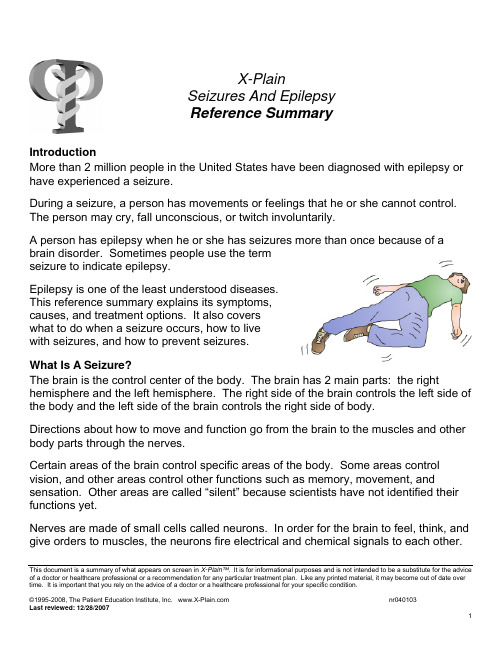
X-PlainSeizures And EpilepsyReference SummaryIntroductionMore than 2 million people in the United States have been diagnosed with epilepsy or have experienced a seizure.During a seizure, a person has movements or feelings that he or she cannot control. The person may cry, fall unconscious, or twitch involuntarily.A person has epilepsy when he or she has seizures more than once because of a brain disorder. Sometimes people use the termseizure to indicate epilepsy.Epilepsy is one of the least understood diseases.This reference summary explains its symptoms,causes, and treatment options. It also coverswhat to do when a seizure occurs, how to livewith seizures, and how to prevent seizures.What Is A Seizure?The brain is the control center of the body. The brain has 2 main parts: the right hemisphere and the left hemisphere. The right side of the brain controls the left side of the body and the left side of the brain controls the right side of body.Directions about how to move and function go from the brain to the muscles and other body parts through the nerves.Certain areas of the brain control specific areas of the body. Some areas control vision, and other areas control other functions such as memory, movement, and sensation. Other areas are called “silent” because scientists have not identified their functions yet.Nerves are made of small cells called neurons. In order for the brain to feel, think, and give orders to muscles, the neurons fire electrical and chemical signals to each other. This document is a summary of what appears on screen in X-Plain™. It is for informational purposes and is not intended to be a substitute for the advice of a doctor or healthcare professional or a recommendation for any particular treatment plan. Like any printed material, it may become out of date over time. It is important that you rely on the advice of a doctor or a healthcare professional for your specific condition.Seizures happen because of abnormal electrical activity in the brain.Depending on the area of the brain affected, a person having a seizure will experience different symptoms. For instance, if an area controlling a muscle is affected, the muscle may become still or jerk uncontrollably.A person may have only one seizure in their whole life. Epilepsy is when the patient has recurrent seizures due to an underlying disease of the brain.CausesAbout 1/2 of all seizures have no known cause. The other 1/2 are linked to a disease or injury of the brain.During development and the first few years of childhood, the brain undergoes a lot of growth. During this growth, the brain is at danger of certain diseases due to infections, poor nutrition, and poor supply of oxygen. Some of these diseases are associated with epilepsy.The neurons of the brain develop into complex webs of wires. Defects in wiring during brain development could lead to epilepsy.After a head injury due to an accident or a stroke, the brain repairs itself by making new wiring. If the new wiring is abnormal, it could causeseizures.Disease of the brain, such as hydrocephalus andmeningitis, could cause epilepsy.Poisoning of the brain, such as lead and carbonmonoxide poisoning, could lead to seizures.Exposure to street drugs and overdoses ofantidepressants could also lead to seizures.Older people sometimes develop diseases of the brain, such as brain tumors, strokes, infections and bleeding. These types of diseases could lead to epilepsy.Some types of epilepsy tend to run in families, suggesting hereditary causes.This document is a summary of what appears on screen in X-Plain™. It is for informational purposes and is not intended to be a substitute for the advice of a doctor or healthcare professional or a recommendation for any particular treatment plan. Like any printed material, it may become out of date over time. It is important that you rely on the advice of a doctor or a healthcare professional for your specific condition.Types Of SeizuresDue to the complexity of the brain and its function, there are more than 32 types of seizures. There are many different words used to describe seizures; some of them include:• Convulsions• Epileptic attacks• Tonic-clonic seizures.Most seizures last from a few seconds to a few minutes and stop naturally.Seizures that limit themselves to one part of the brain are called partial seizures. These seizures are usually labeled using the area of the brain that they started from. In partial seizures, the person may experience sudden feelings of joy or sadness orsudden sensations of smell, hearing, or vision.Another kind of partial seizure is called a complex partialseizure. During this kind of seizure, the patient may displayabnormal repetitive behaviors, such as blinking, moving in acircle, striking out at walls or moving an arm or leg withoutbeing able to control the movement.Seizures that spread to the rest of the brain are calledgeneralized seizures. These seizures may cause the person to:• Lose consciousness• Fall• Have muscle spasms• Have jerking muscles all over the body or• Stare into space, losing contact with reality for a few seconds.Not all people who have a seizure have epilepsy. Some people have just one seizure at some point in their life and never have another one.Sometimes a child may have a seizure during an illness with a high fever. Most of the time, these seizures do not recur unless there has been damage to the brain.When a person has a seizure, the brain shows abnormal electrical activity that can be recorded by doctors on a special machine called an EEG.This document is a summary of what appears on screen in X-Plain™. It is for informational purposes and is not intended to be a substitute for the advice of a doctor or healthcare professional or a recommendation for any particular treatment plan. Like any printed material, it may become out of date over time. It is important that you rely on the advice of a doctor or a healthcare professional for your specific condition.Some people have seizure-like behavior without any abnormal electrical activity. These are called non-epileptic seizures or pseudo-seizures. They may occur due to psychological reasons, such as stress or need for attention.Some people can tell when they are about to have a seizure because they have a specific feeling before the seizure starts; this is called an “aura”. The most common aura is the smell of burnt rubber.DiagnosisDoctors take a medical history, do blood tests, and use a variety of other medical tests to determine whether a person has epilepsy.A test commonly used to diagnose epilepsy is called an electroencephalogram, or EEG. This tests records brain waves. In most cases of epilepsy a doctor can determine if the brain has abnormal electrical activity associated with a seizure by reading the EEG.During an EEG, electrodes are placed on the scalp and brain waves are measured. The test is painless. The doctor may also want to do an EEG while the patient issleeping.A doctor may use a magneto-encephalogram, orMEG. The purpose of this test is similar to an EEG,except that it measures magnetic signals in the braininstead of electrical signals. Because of thisdifference, it does not require electrodes and candetect signals from deeper areas of the brain thanthe EEG can.The doctor may also request a brain scan in order to see structures inside the brain. Examples of brain scans are MRI, CT, and PET scans. These allow the doctor to see structures, such as tumors or cysts, which could be causing the seizures.Treatment OptionsMore than 80% of patients with epilepsy can have their seizures controlled with medication.This document is a summary of what appears on screen in X-Plain™. It is for informational purposes and is not intended to be a substitute for the advice of a doctor or healthcare professional or a recommendation for any particular treatment plan. Like any printed material, it may become out of date over time. It is important that you rely on the advice of a doctor or a healthcare professional for your specific condition.After diagnosing the type of seizure, the doctor can recommend one of about twodozen different drugs that are available to control seizures. This usually depends onthe type of seizures, the patient’s age and medical condition.The doctor usually prescribes a drug and adjusts its dosage based on the patient.Usually the doctor starts at a low dosage and increases it if needed, after each bloodtest.Based on the patient’s diagnosis, the doctor will determine whether or not the patientcan stop using the medication. This depends on subsequent EEG tests, as well ashow long the patient has been free of seizures.If a medication is stopped suddenly, the patient may have more seizures that areharder to treat.The side effects of anti-epilepsy drugs are relatively minor and include:•Fatigue• Weight gain• Dizziness and• Depression.If your doctor recommends an anti-epilepsy drug, he or she willdiscuss the benefits and risks of it.Some patients may be allergic to certain drugs. These patientsshould contact the doctor immediately if they develop any kind ofrash.In some cases of childhood seizures, a special diet that is rich in fat and low in sugarcan help to reduce the frequency of seizures. This type of treatment should be doneunder the supervision of a healthcare provider to make sure the child gets propernutrition.When medical treatment fails to control the seizures, a brain surgery may beconsidered. Brain surgery for seizures tries to remove the part of the brain that isresponsible for abnormal electrical signals; this is the part that causes the seizures.However, these operations only work in less than 1/2 of all seizure patients.This document is a summary of what appears on screen in X-Plain™. It is for informational purposes and is not intended to be a substitute for the adviceof a doctor or healthcare professional or a recommendation for any particular treatment plan. Like any printed material, it may become out of date overtime. It is important that you rely on the advice of a doctor or a healthcare professional for your specific condition.Another operation, called a vagal nerve stimulator, may be suggested. During this operation the surgeon inserts an electronic device under the skin in the upper left chest. The device stimulates a big nerve in the neck. This nerve is known as the vagus nerve. The stimulation helps some patients to have seizures less often. This operation is not ideal for all seizure patients.Living With EpilepsyWhen seizures are controlled, most epileptic patients can have a normal life. However, patients with seizures that are not well controlled need to take precautions that may affect their daily living.Patients with uncontrolled seizures may not be able to drive, or operate hazardous machinery. Most states will not issue a driver’s license to someone with epilepsy unless the person can document that he or she has been seizure-free for a certain period of time. The length of this period varies from state from state.Jobs and hobbies may have to be limited to those that are not dangerous to the person in case he or she loses consciousness or attention for a few moments. Examples of jobs and hobbies that may need to be avoided are:• Flying an airplane• Motor racing• Skydiving• Mountain climbing.Other activities and sports may be possible with supervision, such as: • Swimming• Sailing• Riding bicycles.A lot of activities and sports are safe for a person with epilepsy, like jogging and volleyball. Contact sports should be avoided, since even minor trauma could set off a seizure. Therefore, it might not be wise for seizures patients to play football or hockey.The effects of uncontrolled seizures are more distinct in children and young adults who may not be able to proceed with their education in a normal manner.Since some anti-epilepsy medications interfere with memory and concentration, children with epilepsy may need extra time to learn and complete their homework.This document is a summary of what appears on screen in X-Plain™. It is for informational purposes and is not intended to be a substitute for the advice of a doctor or healthcare professional or a recommendation for any particular treatment plan. Like any printed material, it may become out of date over time. It is important that you rely on the advice of a doctor or a healthcare professional for your specific condition.Women with a seizure disorder can get pregnant. However, they Array should stay on the medication prescribed by their doctor throughoutthe pregnancy. The neurologist should be made aware of anupcoming planned pregnancy. He or she may switch anti-epilepticmedications, as some of these medications are known to increasethe risks of abnormalities in the baby.People with epilepsy should not drink alcoholic beverages or do illicitdrugs; doing so increases the chances of seizures significantly.Epilepsy is NOT associated with mental retardation.By law, people with epilepsy in the United States cannot be deniedemployment, access to education, or recreational activities.Moreover, epilepsy does not affect a patient’s sexual capabilities.Dealing With A SeizureSeizures can last from just a few seconds up to a few minutes. The greatest majorityof seizures stop on their own. However, in rare cases seizures can last hours,requiring urgent medical attention.If you notice a person having a seizure, protect the person from harm until he or sheregains awareness and control.The following are some tips that can decrease the chances of injury during a seizure. Lower the patient into a reclining position on the floor or a flat surface.Put something soft under their head.Turn the head gently to one side to prevent any vomit from being sucked into the lungsas the person breathes.If the person is confused during a seizure and is moving around, remove anything fromthe area that may cause injury to the patient or to others, such as a pan of boilingwater or a hot iron.During a seizure, do NOT do the following:Do not force anything into their mouth.This document is a summary of what appears on screen in X-Plain™. It is for informational purposes and is not intended to be a substitute for the advice of a doctor or healthcare professional or a recommendation for any particular treatment plan. Like any printed material, it may become out of date over time. It is important that you rely on the advice of a doctor or a healthcare professional for your specific condition.Do not give them water or medicine until the seizure is over.Do not try to stop the jerking movement.People with epilepsy can lead full, active lives and usually return to normal activity aftera seizure. However, in some cases, the seizure is prolonged or the patient does notregain consciousness between seizures. In either case you should call 911 if thishappens.If the person stops breathing, call 911 and perform CPR.SummaryEpilepsy is a disorder of the brain that affects millions of Americans. Seizures aresigns of epilepsy but not all seizure-like symptoms are due to epilepsy.There are many types of seizures and epilepsies. It isimportant to see a doctor for the correct diagnosis andappropriate treatment.This document is a summary of what appears on screen in X-Plain™. It is for informational purposes and is not intended to be a substitute for the adviceof a doctor or healthcare professional or a recommendation for any particular treatment plan. Like any printed material, it may become out of date overtime. It is important that you rely on the advice of a doctor or a healthcare professional for your specific condition.Thanks to medical advances, we understand epilepsymuch better than we did 50 years ago. Epilepsy is not amental illness nor is it mental retardation.Most cases of epilepsy can be controlled withmedications, allowing the person to live a normalproductive life without any reason for shame or embarrassment.。
海词

海词编辑部快捷功能功能设置改进词条简洁模式求助词友(=hemiconvulsions, hemiplegia, epilepsy) 半惊厥、半瘫痪、癫痫Google提供的广告目录导读海词讲解例句与词语英英解释8.11MPA国贸系统班晚班开班 - 泰祺mpa英语国贸系统班火热开班免费试听,预约热线:58698175/95属于你的暑假专有学堂真理追寻者大学生理财五招我们为何意志薄弱邻近词语|查看全部例句与词语【邻近词语】HFC abbr. high-f... HHBS Hereford Her...hfg abbr. =heavy... HHC Home Health ...HG =Hand Genera... hhcc higher-harmo...HGH =Human Growt... HHD abbr. <拉>Hum...hgo (=hepatic gl... hhdws heavy handy ...美丽的芭蕾爱情【英语漫画】如此强悍的人事2011情侣卫衣125元/套印尼流行“卧轨治疗法”缩略词收藏|分享到:微课堂,人生如课堂,我们有时候是学生,在课堂上学习知识;有时候是老师,与他人分享经验。
在学习和分享中,我们不断成长。
您的生词本登录登录后添加单词将生词储存到您的生词本!海词推荐这个夏天留学热![新概念]你修炼到第几本[考研英语]名师课堂免费外教测试英语水平!学好英语口语有妙招!【游戏】卑鄙的我拼图小笨霖英语笔记新托福口语大冲关2012海词考研专题【号外】海词考试版块全新上线7月海词最热英文歌曲TOP 10【海词课堂】名师授课在线解疑热门阅读∙大牌云集裘德·洛新片曝光宣传海报∙奶茶刘若英宣布结婚∙比老公更能吸金的女明星∙你有“选择焦虑症”吗?∙雅思小作文高分必备句型∙贝克汉姆裸身显露新纹身∙英国女王”惊现伯明翰街头乞讨∙西班牙八旬女公爵弃巨财嫁61岁公务员∙中国航母首出航,"航母"英语怎么说?∙作文高分关键:如何提高雅思大作文用手机进行查词用手机访问"海词"随时随地,手机查词>>立即体验了解●手机海词词典1.0版发布(免费下载)联系我们- 关于我们- 隐私保护- 特色功能- 工具使用- 诚聘英才©2003-2011 海词词典() - 自2003 年11 月27日开始服务。
epilepsy
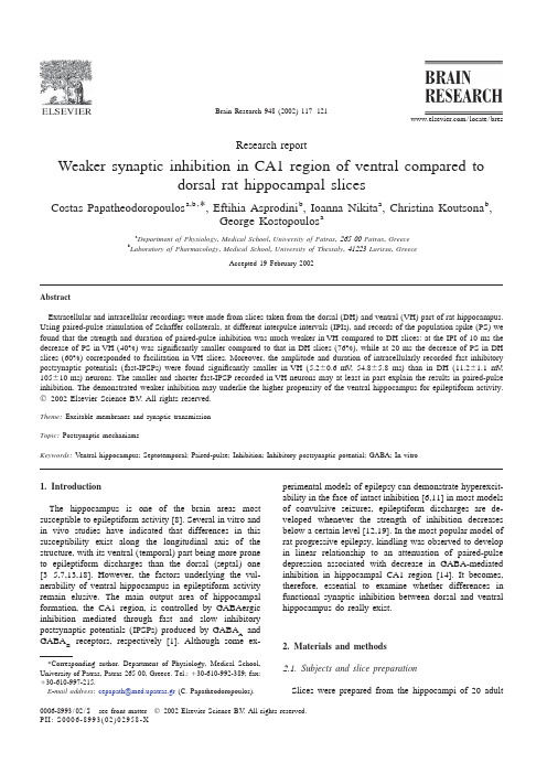
Brain Research 948(2002)117–121/locate/bresResearch reportW eaker synaptic inhibition in CA1region of ventral compared todorsal rat hippocampal slicesa,b ,b a b *Costas Papatheodoropoulos ,Eftihia Asprodini ,Ioanna Nikita ,Christina Koutsona ,aGeorge Kostopoulos aDepartment of Physiology ,Medical School ,University of Patras ,26500Patras ,Greece bLaboratory of Pharmacology ,Medical School ,University of Thessaly ,41223Larissa ,GreeceAccepted 19February 2002AbstractExtracellular and intracellular recordings were made from slices taken from the dorsal (DH)and ventral (VH)part of rat ing paired-pulse stimulation of Schaffer collaterals,at different interpulse intervals (IPIs),and records of the population spike (PS)we found that the strength and duration of paired-pulse inhibition was much weaker in VH compared to DH slices:at the IPI of 10ms the decrease of PS in VH (40%)was signi ficantly smaller compared to that in DH slices (76%),while at 20ms the decrease of PS in DH slices (60%)corresponded to facilitation in VH slices.Moreover,the amplitude and duration of intracellularly recorded fast inhibitory postsynaptic potentials (fast-IPSPs)were found signi ficantly smaller in VH (5.260.6mV ,54.865.8ms)than in DH (11.261.1mV ,105610ms)neurons.The smaller and shorter fast-IPSP recorded in VH neurons may at least in part explain the results in paired-pulse inhibition.The demonstrated weaker inhibition may underlie the higher propensity of the ventral hippocampus for epileptiform activity.©2002Elsevier Science B.V .All rights reserved.Theme :Excitable membranes and synaptic transmission Topic :Postsynaptic mechanismsKeywords :Ventral hippocampus;Septotemporal;Paired-pulse;Inhibition;Inhibitory postsynaptic potential;GABA;In vitro1.Introductionperimental models of epilepsy can demonstrate hyperexcit-ability in the face of intact inhibition [6,11]in most models The hippocampus is one of the brain areas most of convulsive seizures,epileptiform discharges are de-susceptible to epileptiform activity [8].Several in vitro and veloped whenever the strength of inhibition decreases in vivo studies have indicated that differences in this below a certain level [12,19].In the most popular model of susceptibility exist along the longitudinal axis of the rat progressive epilepsy,kindling was observed to develop structure,with its ventral (temporal)part being more prone in linear relationship to an attenuation of paired-pulse to epileptiform discharges than the dorsal (septal)one depression associated with decrease in GABA-mediated [3–5,7,13,18].However,the factors underlying the vul-inhibition in hippocampal CA1region [14].It becomes,nerability of ventral hippocampus in epileptiform activity therefore,essential to examine whether differences in remain elusive.The main output area of hippocampal functional synaptic inhibition between dorsal and ventral formation,the CA1region,is controlled by GABAergic hippocampus do really exist.inhibition mediated through fast and slow inhibitory postsynaptic potentials (IPSPs)produced by GABA and A GABA receptors,respectively [1].Although some ex-B 2.Materials and methods*Corresponding author.Department of Physiology,Medical School,2 .1.Subjects and slice preparationUniversity of Patras,Patras 26500,Greece.Tel.:130-610-992-389;fax:130-610-997-215.E -mail address :cepapath@med.upatras.gr (C.Papatheodoropoulos).Slices were prepared from the hippocampi of 20adult0006-8993/02/$–see front matter ©2002Elsevier Science B.V .All rights reserved.PII:S0006-8993(02)02958-X118C.Papatheodoropoulos et al./Brain Research948(2002)117–121male Wistar rats.Animals were deeply anesthetized with where they were maintained at a constant temperature of ether and decapitated immediately after they stopped3260.28C.They were continuously humidified with mixed breathing.The brain was submerged in chilled(2–48C)gas95%O and5%CO,and perfused with ACSF22artificial cerebrospinalfluid(ACSF)and the two hip-containing(in mM):124NaCl;4KCl;2MgSO;2CaCl;42 pocampi were excised ing a McIlwain tissue 1.25NaH PO;26NaHCO;10glucose;at pH7.4,243chopper500m m thick slices were prepared by cutting equilibrated with95%O and5%CO.22transversally the dorsal and the ventral part of hippocam-pus(as shown in insert of Fig.1B).The slices were2.2.Recordingsimmediately transferred in an interface type chamber,Recordings of extracellular and intracellular synapticpotentials were made in CA1stratum pyramidale.Forextracellular recordings a homemade single carbonfiber(diameter7m m)electrode was used.For intracellularrecordings,glass micropipettesfilled with2M potassiumacetate with resistances of80–120M V were used.Synap-tic potentials were evoked by Schaffer collateral stimula-tion using either steel or tungsten made bipolar electrodes,which were placed in CA1stratum radiatum at a distanceof approximately0.5mm from the recording electrode,andelectrical stimuli were delivered at a frequency of0.05Hz.2.3.DrugsDrugs used included the antagonists of ionotropicexcitatory amino acid receptors4-hydroxyquinoline-2-car-boxylic acid(kynurenic acid,1mM),6-cyano-7-nitro-quinoxaline-2,3-dione disodium(CNQX,10m M),D-(2)-2-amino-5-phosphonopentanoic acid(AP5,50m M),andthe antagonists of GABA receptors2-hydroxysaclofenB(200m M),and(3-aminopropyl)(diethoxymethyl)phos-phinic acid(CGP35348,0.5mM).All drugs wereobtained from Tocris,UK.2.4.Statistical analysisValues throughout text are expressed as mean6S.E.M.Paired and unpaired t-tests were used for statistical com-parisons in the same group,and between the two groups ofslices,respectively.parison of paired-pulse inhibition(PPI)between dorsal(DH) 3.Resultsand ventral(VH)hippocampal slices.PPI was studied by pairing oneconditioning stimulus,which evoked a PS,with a second one,of equal3.1.Extracellular recordingsintensity,after an interpulse interval(IPI),and calculating the percentchange of the second PS with respect to thefirst one.(A)Examples offield records in CA1st.pyramidale from one DH and one VH slice The evokedfield potentials from23dorsal hippocampal showing the different suppressing effect of PPI on the second PS.(DH)and23ventral hippocampal(VH)slices were Superimposed traces,of averages of four sweeps,at the IPIs of10,20,40examined and compared.The maximum population spike and80ms are shown.(B)Percentage change of PS at different IPIs(PS)amplitude was similar between DH(5.6560.47mV, produced by paired-pulse stimulation.Symbols indicate mean6S.E.M.n523)and VH(6.260.57mV,n523)slices.In order to from10DH and22VH slices.Asterisks demark the statisticallysignificant differences of mean values between dorsal and ventral slices.quantify the strength of inhibition on pyramidal neuron At short IPIs corresponding to the action of fast-IPSP(8–40ms),VH excitability the orthodromic paired-pulse stimulation(PPS) slices showed much less inhibition of PS than DH ones.At longer IPIs protocol was used.According to this protocol two con-paired-pulse facilitation prevailed,which was much more prominent in secutive stimuli of equal intensity were delivered at DH than VH slices.The insert shows that transverse slices(solid lines)varying interpulse intervals(IPIs)to Schaffer collaterals, were taken from only restricted areas(lines with arrowheads)of the twopoles of hippocampus.with thefirst stimulus(conditioning)evoking a75%of theC.Papatheodoropoulos et al./Brain Research948(2002)117–121119 maximal PS(Fig.1A).The activation of inhibition with value at80ms(38.7610.6%).Facilitation was observedthe conditioning stimulus produces a depression of the also in VH slices between20and100ms.However,at second stimulus(test)PS.The strength of the so-produced longer IPIs a slight but significant(at400–800ms,P, paired-pulse inhibition(PPI)was quantified by calculating0.01)depression of PS was observed.This could be the percent change of second(test)PS with respect to the attributed to the GABA-mediated slow inhibition[9]Bfirst one.Additionally to inhibition,double orthodromic revealed as prominent in VH slices due to the absence of stimulation produces a facilitatory effect in excitatory paired-pulse facilitation in VH slices at long IPIs[17]. postsynaptic potentials,which tends to increase the am-plitude of PS.The time-courses of facilitation and inhibi-3.2.Intracellular recordingstion largely overlap demonstrating,however,differentrelative effectiveness on PS at each IPI.This balance of Two series of intracellular experiments were made in potencies of each phenomenon along IPIs determines the DH and VH neurons.In thefirst series,recordings were interval at which an overall PPI appears to be replaced by made in standard conditions,while in the second,record-paired-pulse facilitation.ings were obtained following blockade of excitation andThe effect of PPS on synaptic responses at IPIs ranging GABA-mediated slow inhibition.Under standard con-Bfrom4to1400ms was tested in10dorsal and22ventral ditions the resting membrane potential(RMP)and the slices.PPS strongly suppressed PS at the very short IPIs of input resistance(Ri)were similar between the two groups 4and6ms,in both DH and VH slices(decrease greater of neurons(262.660.7mV,n517,61.769.4M V,n512 than85%,Fig.1B.The results at IPIs1200and1400ms in DH and264.061.1mV,n513,59.068.0M V,n55in were quite similar to those at1000ms).This suppression VH neurons).Multiphasic postsynaptic potentials consist-could be the result of the relative refractory period on ing of excitatory postsynaptic potential(EPSP),fast-IPSP neuronalfiring.The suppression of PS at the IPIs of10–20and slow-IPSP were elicited by using orthodromic stimula-ms,corresponding to the interval of the most intense action tion subthreshold for action potential.Similar intensities of the fast-IPSP[2],was robust in the DH slices,being were used for stimulation of DH and VH neurons 60%at20ms and reaching a maximum at10ms(11.762.1V and10.862.6V,respectively).Postsynaptic (76.269.6%).On the contrary,VH presented significantly potentials were recorded at a membrane potential(MP)of less inhibition at10ms(4067.7%decrease of PS),and256.960.9mV(n511)in DH and259.261.8mV(n512) facilitation at20ms(comparison of percent inhibition or in VH neurons.Evoked EPSPs,subthreshold for triggering facilitation,at10and20ms,in DH versus VH slices:action potentials,had comparable amplitudes in DH P,0.01).At relatively long IPIs the depressant effect of(6.261.5mV,n511)and VH neurons(7.862.5mV,n57).the GABA-mediated slow inhibition on PS could be However,the peak amplitude of both fast and slow-IPSPs Bexpected[9].However,since slow inhibition is generated was significantly smaller in VH compared to DH neurons mainly in dendrites[2,15],its contribution in controlling(Table1and traces on the left in Fig.2).neuronal excitability/discharge is somewhat restricted[10]In the presence of a cocktail,containing the antagonistsand could additionally be covered by the effect of paired-of excitatory amino acid receptors and GABA receptors,Bpulse facilitation.Thus,at relatively long IPIs($40ms),and using stimulation intensities producing maximal re-when presumably the inhibitory action of the fast-IPSP had sponse,monosynaptic fast-IPSPs were elicited.Under declined,facilitation of PS was evident as the prevalent these conditions the values of RMP(264.061.17mV, event.In DH slices PPS produced significant facilitation of n510in DH and264.960.6mV,n510in VH neurons) PS at the IPIs of80–1400ms(P,0.05),with maximal and Ri(52.763.8M V,n510in DH and53.167.3M V,Table1Intracellular measures in dorsal and ventral hippocampal neuronsPassive properties Fast-IPSP Slow-IPSP RMP Input resistance Reversal potential Peak amplitude Peak latency Duration at half-amplitude Peak amplitude (mV)(M V)(mV)(mV)(mS)(mS)(mV)DH263.1160.657.665.4276.961.3411.261.112.360.620.861.6105.0610.110.160.9c c c a b b b a(27)(22)(14)(11)(11)(10)(11)(11)VH264.460.6555.265.3272.662.22 5.260.69.760.718.262.154.865.8 6.561.3c c c a b b b a(23)(14)(8)(13)(11)(11)(7)(7)a bP,0.01P,0.05P,0.001P,0.05The number of cells is indicated in parentheses.a Measures taken in the absence of drugs.b Measures taken in the presence of antagonists of ionotropic excitatory amino acid receptors and GABA receptors.Bc Measures taken under either a or b conditions.120C.Papatheodoropoulos et al./Brain Research948(2002)117–121Fig.2.Postsynaptic inhibition of CA1pyramidal neurons is reduced in VH slices as compared to DH slices.Recordings(averages of4–5sweeps)weremade in standard ACSF(traces on the left)and in the presence of cocktail containing antagonists of ionotropic excitatory amino acid receptors and GABAB receptors(traces on the right).Fast and slow-IPSPs in VH neurons exhibited smaller amplitude and duration,as compared to DH neurons,when slices wereperfused with standard ACSF(traces on the left).This difference of fast-IPSPs remained in the presence of cocktail-containing ACSF.Artifacts aretruncated for clarity.Subthreshold stimulation strengths were used in standard conditions and stimulations producing maximal responses were delivered incocktail-containing ACSF.Data were obtained from one DH and two VH neurons.n59)were not significantly different when compared to 4.Discussionthose recorded in standard conditions.The input resistanceof the neurons was determined by injecting hyperpolariz-In this study we demonstrated a significant difference of ing current step commands of increasing amplitude(250synaptic inhibition in CA1region between DH and VH ms,20.1to21.0nA)at the resting membrane potential of slices.By using the protocol of paired-pulse stimulation each cell,and calculating the slope of the linear portion of we tested the effect of inhibition on the pyramidal cell the resulting steady-state current–voltage plot.The mean excitability in the two groups of slices.The strength of this value of reversal potential(RP)of fast-IPSPs was slightly effect was quantified by the percent suppression of PS and less negative in VH neurons(273.263.0,n56)than in the range of IPIs at which suppression of PS was observed, DH(277.461.6mV,n57).Since the amplitude of fast-was taken as an index of the duration of PPI.In contrast to IPSPs increases upon membrane depolarization,and is DH slices,in which robust PPI was observed at the IPIs therefore more easily visualized and measured,fast-IPSPs expectedly corresponding to the time-window in which thewere evoked by injecting direct current(d.c.),the MP close inhibitory action of GABA-mediated fast-IPSP is maxi-Ato260mV.The MPs at which fast-IPSPs were recorded in mal(10–20ms),VH slices showed much weaker or even DH and VH neurons were259.260.5mV(n511)and absent PPI at these IPIs,suggesting significant difference 258.161.5mV(n510),respectively.As found in standard in functional GABA-mediated fast inhibition between DHAconditions,the amplitude of fast-IPSPs was significantly and VH slices.In view of the relatively weaker paired-smaller in VH than in DH neurons(Table1and traces on pulse facilitation of PS(and of EPSP[17])in VH com-the right in Fig.2).Since the values of RMP,Ri and RP of pared to DH slices,and given the known overlap in time fast-IPSPs obtained in the presence of cocktail were not between facilitation and inhibition[14]it is assumed that significantly different from those found in standard con-PPI in VH may be even smaller than described here since it ditions,the respective mean values of these measures is relatively less‘covered’by facilitation.Orthodromicindicated in Table1were calculated after grouping the stimulation elicited monosynaptic GABA-mediated fast-Anumbers from both experimental situations.In the presence IPSPs produced in the dendrites and soma of pyramidal of cocktail two additional characteristics of the fast-IPSP neurons,and slow-IPSPs produced by the activation ofwere also attained,its duration measured at half-maximal GABA receptors located mostly on dendrites[2,15].TheBamplitude and the peak latency measured as the time from peak amplitude of both fast and slow-IPSPs was sig-the artefact to peak amplitude.The duration of maximally nificantly smaller in VH compared to DH neurons.Theevoked fast-IPSP was significantly smaller in VH than infinding of smaller in amplitude and shorter in durationDH neurons(see Table1).The peak latency was accord-fast-IPSPs in VH neurons is consistent with the extracellu-ingly shorter in VH neurons but not significantly rly estimated differences in PPI and could effectivelyC.Papatheodoropoulos et al./Brain Research948(2002)117–121121[3]C.Borck,J.G.Jefferys,Seizure-like events in disinhibited ventral explain these differences between DH and VH slices.Theslices of adult rat hippocampus,J.Neurophysiol.82(1999)2130–slow-IPSP having limited effect on neuronal excitability is2142.expected to moderately affect the PS at relatively long IPIs[4]A.C.Bragdon,D.M.Taylor,W.A.Wilson,Potassium-induced epi-in the PPS paradigm.Indeed,neurons from DH slices,at leptiform activity in area CA3varies markedly along the septotem-IPIs80–1000ms,which coincide temporally with the poral axis of the rat hippocampus,Brain Res.378(1986)169–173.[5]R.Elul,Regional differences in the hippocampus of the cat.I. activation of GABA-mediated slow-IPSP,exhibitedBSpecific discharge patterns of the dorsal and ventral hippocampus synaptic facilitation that may mask any possible effect ofand their role in generalized seizures,Electroencephalogr.Clin. GABA-mediated inhibition.In contrast to DH,neuronsB Neurophysiol.16(1964)470–488.from VH slices exhibited no effect of PS.This is in line[6]M.Esclapez,J.C.Hirsch,R.Khazipov,Y.Ben-Ari,C.Bernard, with our previous results demonstrating absence of short-Operative GABAergic inhibition in hippocampal CA1pyramidalneurons in experimental epilepsy,A94 term facilitation of EPSP in VH slices[17].It should also(1997)12151–12156.be noted that the values of amplitude of the GABA-B[7]M.Gilbert,R.J.Racine,G.K.Smith,Epileptiform burst responses in mediated IPSP have been taken only under control con-ventral vs.dorsal hippocampal slices,Brain Res.361(1985)389–ditions,i.e.,without pharmacological isolation this slow391.component of IPSP could be to some extent‘contami-[8]J.D.Green,The hippocampus,Physiol.Rev.44(1964)561–608.[9]M.J.Higgins,T.W.Stone,Bicuculline-resistant paired-pulse inhibi-nated’by the preceding fast IPSP.tion in the rat hippocampal slice,Br.J.Pharmacol.109(1993) The difference in inhibition between DH and VH slices1164–1168.could be attributed to several factors,such as a relatively[10]G.Karlsson,C.Kolb,A.Hausdorf,C.Portet,M.Schmutz,H.R. less extensive inhibitory innervation on pyramidal neurons,Olpe,GABAB receptors in various in vitro and in vivo models of or to differences concerning the GABA receptors(number,epilepsy:a study with the GABAB receptor blocker CGP35348,Neuroscience47(1992)63–68.density,postsynaptic placement,or even affinity for[11]G.K.Kostopoulos,Spike-and-wave discharges of absence seizures GABA).However,no such data are available till now.as a transformation of sleep spindles:the continuing development of Moreover,recent observations from our laboratory showa hypothesis,Clin.Neurophysiol.111(2000)S27–38.lower levels of muscimol binding in CA1st.oriens-[12]K.Krnjevic,GABA-mediated inhibitory mechanisms in relation to pyramidale of VH compared to DH slices(unpublished epileptic discharges,in:H.H.Jasper,N.M.van Gelder(Eds.),BasicMechanisms of Neuronal Hyperexcitability,Alan R.Liss,New data),suggesting differences in the density of GABAAYork,1983,pp.249–280.receptors between the two regions.Although we prepared[13]P.H.Lee,C.W.Xie,D.V.Lewis,W.A.Wilson,C.L.Mitchell,J.S. dorsal and ventral slices identically,we cannot exclude theHong,Opioid-induced epileptiform bursting in hippocampal slices: possibility of selective destruction of distinct inhibitory higher susceptibility in ventral than dorsal hippocampus,J.Phar-ramifications,functionally relevant,in VH slices,which macol.Exp.Ther.253(1990)545–551.[14]F.H.Lopes da Silva,W.Kamphuis,M.Titulaer,M.Vreugdenhil,W.J. could contribute to the differences in inhibition.However,Wadman,An experimental model of progressive epilepsy:the comparative anatomical data supporting a different ramifi-development of kindling of the hippocampus of the rat,Ital.J. cation pattern of inhibitory cells between the dorsal andNeurol.Sci.16(1995)45–57.ventral part of hippocampus are not available until now.[15]N.R.Newberry,R.A.Nicoll,A bicuculline-resistant inhibitory post-Our results broaden the range offindings supporting the synaptic potential in rat hippocampal pyramidal cells in vitro,J.Physiol.(London)348(1984)239–254.concept of differentiation between dorsal and ventral[16]C.Papatheodoropoulos,G.Kostopoulos,Decreased ability of rat hippocampus at the level of local synaptic circuitstemporal hippocampal CA1region to produce long-term potentia-[3,4,7,13,16,17,19],and provide a physiological basistion,Neurosci.Lett.279(2000)177–180.which may explain,to some extent,the propensity of[17]C.Papatheodoropoulos,G.Kostopoulos,Dorsal–ventral differentia-ventral hippocampus for epileptic activity.tion of short-term synaptic plasticity in rat CA1hippocampal region,Neurosci.Lett.286(2000)57–60.[18]R.Racine,P.A.Rose,W.M.Burnham,Afterdischarge thresholds andkindling rates in dorsal and ventral hippocampus and dentate gyrus, ReferencesCan.J.Neurol.Sci.4(1977)273–278.[19]R.D.Traub,es,J.G.Jefferys,Synaptic and intrinsic conduct-[1]B.Alger,Gating of GABAergic inhibition in hippocampal pyrami-ances shape picrotoxin-induced synchronized after-discharges in thedal cells,Ann.N.Y.Acad.Sci.627(1991)249–263.guinea-pig hippocampal slice,J.Physiol.(London)461(1993) [2]B.E.Alger,R.A.Nicoll,Feed-forward dendritic inhibition in rat525–547.hippocampal pyramidal cells studied in vitro,J.Physiol.(London)328(1982)105–123.。
医学英语阅读:癫痫
医学英语阅读:癫痫epilepsydefinitionepilepsy is a brain disorder involving recurrent seizures.causes, incidence, and risk factorsepilepsy is a disorder involving repeated seizures ofany type. seizures ("fits") are episodes of disturbed brain function that cause changes in attention and/or behavior.they are caused by abnormal electrical excitation in the brain.sometimes, seizures are related to a temporary condition, such as exposure to drugs, withdrawal from certain drugs, or abnormal levels of sodium or glucose in the blood. in such cases, repeated seizures may not recur once the underlying problem is corrected.in other cases, injury to the brain (e.g., stroke orhead injury) causes brain tissue to be abnormally excitable.in some people, an inherited abnormality affects nerve cellsin the brain, which leads to seizures. in some cases, nocause can be identified.some of the more common causes of seizures include:* idiopathic (no identifiable cause)o usually begin between age 5 and 20o can occur at any ageo no other neurologic abnormalities presento often a family history of epilepsy or seizures* developmental or genetic conditions present at birth, or injuries near birth —— in this case, the seizures usually begin in infancy or early childhood* metabolic abnormalitieso may affect people of any ageo diabetes complicationso electrolyte imbalanceso kidney failure, uremia (toxic accumulation of wastes)o nutritional deficiencieso phenylketonuria (pku) —— can cause seizures in infantso other metabolic diseases, such as inborn error of metabolismo use of cocaine, amphetamines, alcohol, or certain other recreational drugso withdrawal from alcoholo withdrawal from drugs, particularly barbiturates and benzodiazepines* brain injuryo may affect any age, but most common in young adultso most likely to occur if the brain membranes are damagedo seizures usually begin within 2 years after the injury o early seizures (within 2 weeks of injury) —— do not necessarily indicate that chronic (ongoing) seizures (epilepsy) will develop* tumors and brain lesions that occupy space (such as hematomas)o may affect any age, more common after age 30o partial (focal) seizures most common initiallyo may progress to generalized tonic-clonic seizures* disorders affecting the blood vessels (such as stroke and tia)o most common cause of seizures after age 60* degenerative disorders (senile dementia alzheimer type, or similar organic brain syndromes)o mostly affect older people* infectionso may affect people of all ageso may be a reversible cause of seizureso brain infections like meningitis and encephalitis can produce seizureso brain abscess。
electroencephalography 翻译
electroencephalography 翻译基本解释●Electroencephalography:脑电图●/ɪˌlek.trəʊ.enˌsef.əˈlɒɡ.rə.fi/●n. 使用电极记录大脑电活动的技术变化形式●n. 复数形式:electroencephalographies具体用法●n.:o使用电极记录大脑电活动的技术o例句:●Electroencephalography is a non-invasive method to recordelectrical activity of the brain, which is crucial for diagnosingepilepsy and other brain disorders.●脑电图是一种非侵入性的方法,用于记录大脑的电活动,这对于诊断癫痫和其他脑部疾病至关重要。
●The development of electroencephalography has greatlyenhanced our understanding of brain functions and neurological disorders.●脑电图的发展极大地提高了我们对大脑功能和神经系统疾病的理解。
●Researchers use electroencephalography to study the brain'sresponse to different stimuli, providing insights into cognitive processes.●研究人员使用脑电图来研究大脑对不同刺激的反应,从而提供对认知过程的见解。
●Electroencephalography is often used in sleep studies tomonitor brain activity during different sleep stages.●脑电图常用于睡眠研究,以监测不同睡眠阶段的大脑活动。
EpilepsyandSeizures
EPILEPSY AND SEIZURESINTRODUCTIONThere are many different types of seizures disorders. Seizures can be caused by drugs or alcohol, or they can be caused by withdrawal from drugs or alcohol. They can also be caused by head injury, infection, pregnancy, low blood sugar, fever, a lack of oxygen to the brain, damage to the brain by a stroke, a brain tumor, or certain psychiatric disorders. But the most common cause of seizure disorders is epilepsy. Epilepsy is a disease of the brain and the nervous system. Epilepsy, as with the other seizure disorders, causes seizures. Seizures are also called convulsions. Epileptic seizures are usually caused by a part of the brain – a seizure focus – that sends out violent and uncoordinated electrical signals; in some cases of epilepsy, however, there is no specific seizure focus. In response to the erratic, uncoordinated, and intense electrical signals, the patient with epilepsy experiences a seizure.Epileptic seizures are sudden, involuntary movements caused by abnormal brainactivity.Epilepsy is common, and there are actually many types of epilepsy that produce very different signs and symptoms. Approximately 1% of the population – approximately 300,000 people – has epilepsy. There are some surgical procedures that can help if seizures are not controlled with medical treatment. However, most people with epilepsy must take medications to control the disease and they must take the drugs for their entire life because there is no cure for epilepsy.OBJECTIVESWhen the student has finished this module, he/she will be able to:1. Identify three causes of seizure disorders.2. Identify the most common cause of seizures.3. Identify the correct definition of an epileptic seizure.4. Identify the correct names of an epileptic seizure5. Identify the three stages of an epileptic seizure.6. Identify the correct name for the signs/symptoms that immediately precede a seizure.7. Identify signs and symptoms of a seizure.8. Identify the two biggest risks to a patient during an epileptic seizure.9. Identify the three basic treatments for epilepsy.10. Identify two goals to remember when someone is having a seizure.SEIZURES AND THE NERVOUS SYSTEMThe nervous system is divided into three parts: the brain, the spinal cord and the peripheral nerves. The brain and the nervous system are the “command center” of all the body’s activities.The brain is inside the skull, and at the base of the skull at the level of the neck, the spinal cord begins. The spinal cord is a long body of nervous tissue attached to the brain. It is inside the spine, and all along the length of the spinal cord, the peripheral nerves– long, thin strands of nervous tissue like wires – leave the spinal cord and travel to the organs (heart, lungs, liver, kidneys, glands, etc) and to every part of the body. Peripheral nerves also travel back through the spinal cord to the brain.The nervous system coordinates and controls our conscious functions (for example, the ability to speak, move, etc.) and our unconscious functions (for example, our heart rate and breathing). All of this happens by way of electrical signals that start in the brain, and travel through the spinal cord and the peripheral nerves to the heart, muscles, lungs, etc. The brain takes in information from the internal and external environment by way of the peripheral nerves. In response to the information it receives, the brains sends out electrical signals through the peripheral nerves to various organs and parts of the body. These electrical signals are basically messages, or instructions, and the body responds to these signals.For example, if someone is running to catch a bus, the brain receives information that that person needs more oxygen and that more blood is needed in the legs. In response, the brain and the nervous system sends out signals that increase the rate of breathing and make the heart pump harder and faster. The brain and the nervous system also send out signals that control our conscious functions such as speech and movement.Learning Break: Basically, every activity of the human body, conscious or unconscious, is controlled by the brain and the nervous system.Normally, the electrical signals the brain and nervous system use to control and coordinate are organized and purposeful. However, people with epilepsy have an area or areas in their brain that send out messages that are disorganized and cause harm.WHAT ARE THE CAUSES OF EPILEPSY?There are many types of epilepsy and there are many types of seizures. The type of epilepsy that will be discussed here is epilepsy that occurs in adults and does not have an obvious cause. (Remember, some seizures can be cause by infection, trauma, etc.) This type of epilepsy is called idiopathic, and no one knows what causes idiopathic epilepsy.Idiopathic epilepsy is basically caused by an area or areas of the brain that send out powerful, disorganized electrical signals.However, researchers do know what is happening in the brain during an epileptic seizure. In the normal brain (and in the brain of someone who has epilepsy, as well), there is always a lot of electrical activity going on. Some of this electrical activity is stimulating and some is inhibiting, and there is a constant balance between the two. For example, the blood vessels that supply the brain, kidneys, heart, etc. can open or close, but most of the time they just need to stay open at a certain diameter to make sure enough blood gets through to the organs. Maintaining the blood vessels at that diameter dependson a balance between the stimulating impulses (which would open them) and the inhibiting impulses (which would close them).An epileptic seizure occurs when there is an imbalance between the stimulating brain signals and the inhibiting brain signals. There is an area or areas of the brain that suddenly and unpredictably sends out stimulating signals that are extremely powerful and disorganized; another to view this is as an electrical “storm.” The inhibiting signals are simply not strong enough to balance them out, and the body responds with an epileptic seizure.Learning Break: If you have ever touched a live electrical wire, then you have an approximate idea of what an epileptic seizure is like. The strong, sudden, and unexpected electrical current – which is very much like the strong, sudden electrical stimulus of an epileptic seizure – can knock you down, and it will certainly cause you to move in a very erratic and uncontrolled manner.WHAT ARE THE SIGNS AND SYMPTOMS OF AN EPILPETIC SEIZURE?Just as there are many types of seizure disorders, there are many types of seizures. The type of seizure that idiopathic epilepsy causes – the type of seizures that are very dramatic and very frightening – are called tonic-clonic seizures. They are also called grand mal seizures. An epileptic seizure episode has three parts.In the period of time immediately before the seizure, many people will some signs and symptoms that are a warning that the seizure is about to happen. These signs and symptoms are called the aura.These signs and symptoms can be very subtle. Some times the person will simply become very quiet and stare blankly; he/she looks as if they are thinking very deeply. Some times you might notice that the person begins to tremor and lose some coordination.Learning Break: The aura is different for each person, and many times – but not always – the person with epilepsy can recognize his/her aura and will know that a seizure is imminent. Not everyone will have an aura.The next part of the seizure episode is the actual seizure. A tonic-clonic seizure is an unforgettable event to witness. These seizures are called tonic-clonic because they cause intense muscular tension (tonic, as in muscle tone) and rapid muscle contractions (clonus). A tonic-clonic seizure can be identified by these signs and symptoms.•Loss of consciousness.•Loss of bladder control.•Rapid muscular contractions and movements alternating with periods of intense muscular tension.Learning Break: People with a tonic-clonic seizure lose consciousness. You may hear someone say that it impossible to have a seizure and remain awake. But there are seizure disorders in which the person can have a seizure and not lose consciousness. However,these seizure disorders do not cause the sudden, violent, and dramatic muscle and body movements of a tonic-clonic seizure.The person is unconscious and thrashing violently. The back will be arched and the arms and legs will move back and forth rapidly and out of control; this is the clonic period. If the person is standing, he/she will lose balance and fall. After the sudden, intense movements, the person will become very stiff and rigid because of the intense muscular tension; this is the tonic period. The person may lose control of his/her bladder or bowels. Most seizures do not last very long. After 30 to 60 seconds, the seizure will end spontaneously.After the seizure has ended, the person enters the third stage of a seizure episode. This is called the postictal period. During this time, the patient sleeps very deeply and cannot be aroused. The postictal period may be lengthy, and when the person wakes up, he/she may remember the aura period, but will not remember the events of the seizure itself. The person may be confused and disoriented.ARE EPILEPTIC SEIZURES DANGEROUS?For the most part, a single epileptic seizure in and of itself is not highly dangerous. There is some evidence that people with epilepsy have a shortened life expectancy. There is also some evidence that people with epilepsy have a higher risk of sudden death, death that is not related to a seizure.But although the seizure itself is not particularly dangerous, remember that a seizure causes sudden, unpredictable loss of consciousness and violent, uncontrollable muscle contractions and movements. The physical safety of the patient is definitely at risk. The patient is unconscious and thrashing violently. It is not unusual for someone to fall and strike his/her head, break bones, and become bruised and cut.Aside from the physical safety of the patient, the other big risk during a tonic-clonic seizure is the lack of oxygen. During a tonic-clonic seizure, the rapid, intense muscular contractions and the intense muscular tension prevent the chest from expanding and contracting. The person having a tonic-clonic seizure essentially stops breathing. For young, healthy individuals, this can be tolerated. But for older people or people who have cardiac or respiratory problems, the lack of oxygen can be dangerous.Learning Break: Many people with idiopathic epilepsy have single seizures. However, some people with idiopathic epilepsy have a condition called status epilepticus. In status epilepticus, there are multiple tonic-clonic seizures, one after another. At times, someone with status epilepticus can have periods of seizure activity that can last for many hours. This is very dangerous and requires aggressive treatment.WHAT ARE THE TREATMENTS FOR EPILEPSY?People with idiopathic epilepsy must realize that there is no cure for this disease. There may be long periods during which someone with epilepsy does not have a seizure. But the pattern of seizures and the number of seizures someone will have cannot beaccurately predicted. Idiopathic epilepsy is treated using medications, surgery, and to a lesser degree, a special diet.Medications are the cornerstone of treating idiopathic epilepsy. Some of the most common medications use to treat idiopathic epilepsy are (The generic name is followed by the trade name in parentheses):•Phenytoin (Dilantin®)•Valproic acid (Depakote®)•Carbamazapine (Tegretol®)•Levetiracetam (Keppra®)•Lamotrigine (Lamictal®)•Gabapentin (Neurontin®)•Topirimate (Topamax®)•Oxcarbazepine (Trileptal®)These are all oral medications taken by mouth and most often take two or three times a day. As with any medication, it can take time to adjust the dosage. Some of the medications such as phenytoin, carbamazapine, and valproic acid are adjusted by periodically checking the level of the drug in the blood.Learning Break: Many people find it difficult to take these medications faithfully. They have unpleasant side effects, many people need to take a combination of several of these drugs, and they must be taken indefinitely. If someone has not had a seizure in a long time, it can be easy for them to decide to stop taking their phenytoin, valproic acid, etc. Approximately 30% of all people with idiopathic epilepsy will not respond to medications. Surgery can be used to either a) remove the area of the brain that is causing the seizures, or b) interrupt the nerve pathways that transmit the seizure impulses. If the seizure focus and/or the nerve pathways can be clearly identified, and no important parts of the brain or nervous system will be harmed by the surgery, it can be a good option. Many people who have surgery for epilepsy no longer have seizures and do not need medications.For a few people – mostly children – a special diet called the ketogenic diet may be very helpful. This diet contains a lot of fat and very few carbohydrates (e.g., breads, starches, etc). When fats are digested, one of the breakdown products is ketones. No one is sure why, but when someone with epilepsy uses ketones for energy, the incidence of seizures can be dramatically reduced.TAKING CARE OF SOMEONE HAVING A SEIZUREMost people with idiopathic epilepsy live a normal life, but there is always a possibility of a tonic-clonic seizure. As a health care professional, you are responsible for your patient’ safety, so you need to know what to do if/when someone has a seizure. The two most important goals to focus on and remember when someone is having a tonic-clonic seizure are a) remain calm, and b) Stay with the patient and protect the patient.•Remain calm: This is easy to say, but difficult to do. It can be especially difficult to stay calm if there are non-medical people witnessing the seizure; they willalmost certainly become excited and can add to the confusion. The simplest way to remain calm and focused is to remember this fact: For the great majority of people, the seizure will only last a minute or so and the patient will not beharmed.•Check your watch or a clock and note the time.•Call for help, but if you cannot do this without leaving the patient, don’t leave the patient.•Do not try and insert anything into the patient’s mouth; he/she is not in danger of swallowing the tongue.•Do not try and give the patient anything to eat or drink during the seizure or during the postictal period.•Do not try and restrain the patient. It is impossible to do and you may injure the patient or yourself.•Remove any objects nearby that the patient may hit during the seizure.•Try and protect the patient’s head. This can be done by placing a pillow, folded blanket, etc. under the head.•If the person is on the floor, gently try and roll him/her on the side so they do not aspirate secretions into the lungs.•After the postictal period, the patient may be confused and disoriented. Although he/she is awake, you must still make sure the patient’s safety is monitored.In summary, your goals are to stay calm and keep the patient safe, and that can be done with some knowledge and some basic common sense.。
小儿病毒性脑炎继发癫痫的危险因素分析
临床医学小儿病毒性脑炎继发癫痫的危险因素分析王冬玲,王胜男,张乐滨州市中心医院特检科,山东滨州251700[摘要]目的探讨小儿病毒性脑炎(Viral Encephalitis, VE)继发癫痫(Epilepsy, EP)的危险因素。
方法方便选取2022年1月—2023年7月滨州市中心医院收治的98例VE患儿为研究对象,统计EP发生情况,并进行单因素分析与多因素Logistic回归分析以明确VE继发EP的危险因素。
结果 98例VE患儿中,26例(26.53%)继发EP;EP组家族EP史、痫性发作次数>2次、未使用抗癫痫药物比例较非EP组更高,差异有统计学意义(P均<0.05);多因素Logistic回归分析结果显示,家族EP史(OR=2.369)、痫性发作次数(OR=3.415)是小儿VE继发EP的独立危险因素(P均<0.05),使用抗癫痫药物(OR=0.127)是小儿VE继发EP的保护因素(P<0.05)。
结论小儿VE继发EP的危险因素主要包括家族EP史、痫性发作次数等,使用抗癫痫药物可降低继发EP的发生率。
[关键词]小儿病毒性脑炎;癫痫;危险因素[中图分类号]R512 [文献标识码]A [文章编号]1674-0742(2024)02(b)-0044-03 Analysis of Risk Factors of Epilepsy Secondary to Viral Encephalitis in ChildrenWANG Dongling, WANG Shengnan, ZHANG LeDepartment of Special Inspection, Binzhou Central Hospital, Binzhou, Shandong Province, 251700 China [Abstract] Objective To investigate the risk factors of epilepsy (EP) secondary to viral encephalitis (VE) in children. Methods A total of ninety eight cases of VE children admitted to Binzhou Central Hospital from January 2022 to July 2023 were conveniently selected as the study objects to analyze the incidence of EP. Univariate analysis and multivari⁃ate Logistic regression analysis were performed to identify the risk factors of VE secondary EP. Results Of 98 children with VE, 26 (26.53%) had secondary EP. The family history of EP, the number of epileptic seizures>2 times and the proportion of no use of antiepileptic drugs in EP group were higher than those in non-EP group,the difference was sta⁃tistically significant (all P<0.05). Multivariate Logistic regression analysis showed that family EP history (OR=2.369) and the number of epileptic seizures (OR=3.415) were independent risk factors for secondary EP in children with VE (all P<0.05). The use of antiepileptic drugs (OR=0.127) was a protective factor for secondary EP in children with VE (P<0.05). Conclusion The risk factors of secondary EP in children with VE mainly include family EP history, the num⁃ber of epileptic seizures >2 times. The use of antiepileptic drugs can reduce the incidence of secondary EP.[Key words] Pediatric viral encephalitis; Epilepsy; Risk factor病毒性脑炎(Viral Encephalitis, VE)是以儿童为主要发病群体,主要由病毒感染引起的神经内科常见疾病,对脑部组织、神经等具有较强的损伤性,可引发一系列并发症[1]。