TREM1 (Human) ELISA Kit
尿足细胞Nephrin分子测定在狼疮性肾炎中的临床意义
甘肃医药2021年40卷第5期Gansu Medical Journal ,2021,Vol.40,No.5狼疮性肾炎(lupus nephritis ,LN )是系统性红斑狼疮常见的并发症之一[1]。
临床上判断肾功能损害严重程度以肾活检病理表现作为金指标。
狼疮性肾病的病理类型可发生转型,由于肾活检是一种有创的检查方法,受医疗条件和患者自身条件的限制,重复肾活检受到限制。
目前大量关于足细胞的基础研究表明,尿足细胞的浓度可间接评估肾脏损伤严重程度,检测尿足细胞可作为一种无创、可追踪观察的检查手段[2],成为肾脏疾病重要观察和预测指标。
本研究探讨了尿足细胞Nephrin 分子在狼疮性肾炎诊疗中的临床意义。
现报道如下。
1资料与方法1.1一般资料选取2016年1月至2020年1月我院肾内科收治的经肾活检病理确诊并表现为肾病综合征的LN 患者46例为研究对象,年龄19~51岁,平均(32.1±10.6)岁,其中男8例,女38例。
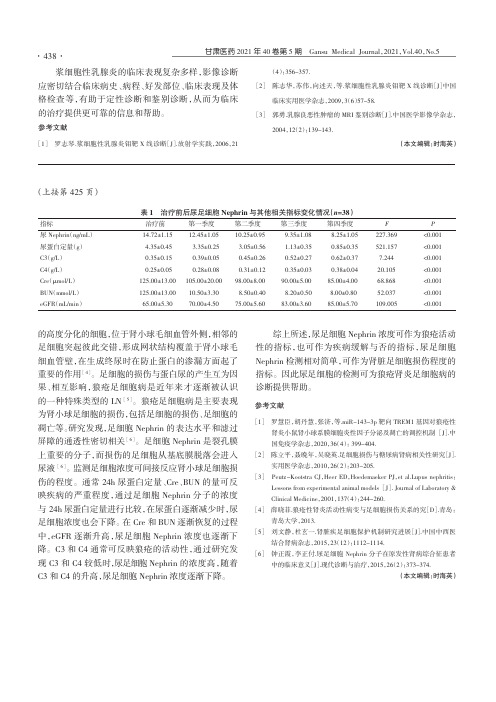
治疗方案均为醋酸泼尼松(1mg/kg )联合环磷酰胺(0.8g/m 2),对该46例患者在治疗前及每季度进行肾小球滤过率(eGFR )、24h 尿蛋白定量,补体C3和C4、血沉、尿足细胞的检测。
排除出现少尿、无尿进展至急性肾衰竭者。
1.2方法1.2.1尿足细胞Nephrin 分子的浓度测定。
留取晨尿20mL ,1500r/min 离心10min ,吸管收集上清液,-20℃保存,用于尿足细胞Nephrin 分子的测定。
采用酶联免疫吸附法(ELISA )测定尿足细胞Nephrin 分子的浓度(试剂盒由上海素尔生物科技有限公司提供),按照试剂盒说明书进行操作,测定OD 值并绘制标准曲线,计算样品的实际浓度。
1.2.224h 尿蛋白定量的测定。
采集患者24h 尿液,混匀,记录尿量,送检验科行24h 尿蛋白定量测定。
空腹12h 后取血查血肌酐(Scr )、血清尿素氮(BUN )、eGFR 、补体C3、C4,由我院检验科完成。
整理slr试验阳性

文件编号:9D-CB-56-9E-B6slr试验阳性整理人尼克梅毒血清初筛试验及其生物学假阳性反应尹建奇河北省人民医院皮肤科,石家庄 050051人体感染梅毒螺旋体后,患者体内产生一种抗心磷脂的抗体,又称反应素。
在体外,用心磷脂(cardiolipin)作为抗原检测梅毒患者血清中的抗心磷脂的抗体,称为梅毒血清反应素试验,即梅毒血清初筛试验。
目前常用的梅毒血清初筛试验主要有性病研究实验室(venereal disease research laboratory , VDRL)试验、快速血浆反应素环状卡片试验(rapid plasma reagin circle card test , RPR)、不加热血清反应素试验(unheated serum reagin test ,USR)、甲苯胺红试验(tolulized red unheated serum test ,TRUST)。
由于它们所用的心磷脂抗原不是梅毒螺旋体本身或其菌体成分,所以也称为非梅毒螺旋体抗原血清试验。
梅毒血清初筛试验检测梅毒敏感性高,但在某些疾病状态下可发生假阳性。
梅毒治疗以后,梅毒血清反应素试验滴度下降,直至阴转,因而可以作为监测病情和观察疗效的指标。
1抗心磷脂抗体的概况1.1梅毒感染中抗心磷脂抗体的产生机理对于梅毒感染中抗心磷脂抗体的产生机理,目前尚不完全清楚[1]。
1942年,Mary 从牛心组织中分离纯化出一种能与梅毒患者血清发生反应的抗原,证实这种抗原为一种带阴电荷的磷脂,因此将其命名为心磷脂。
心磷脂属于甘油磷脂,化学名为二磷脂酞甘油,是由2分子磷脂酸和1分子甘油结合而成,是脂质中唯一具有抗原性的磷脂。
心磷脂广泛存在于各种细胞线粒体内膜上。
长期以来,一直推测梅毒螺旋体感染后,造成宿主组织细胞的破坏,宿主细胞的线粒体膜释放出心磷脂,刺激机体产生抗心磷脂抗体[2]。
但并不知道这种抗原性心磷脂究竟来源于人体哪种或哪些组织和细胞。
Proteintech Rat TNF-alpha Sandwich ELISA Kit 说明书

Rat TNF-alpha Sandwich ELISA Kit DatasheetPlease read it entirely before use Catalogue Number:KE20001Size: 96TSensitivity:7.7 pg/mLRange: 15.6-1000 pg/mLUsage:For the quantitative detection of rat TNF-alpha concentrations in cell culture supernatant.This product is for research use only and not for use in human or animal therapeutic or diagnostic.1/8Table of content page1. Background32. Principle33. Required Materials44. Kit Components and Storage45. Safety Notes46. Sample Collection and Storage47. Regent Preparation58. Assay Procedure Summary69. Validation Data79.1 Standard curve79.2 Precision79.3 Recovery89.4 Sensitivity89.5 Linearity810. References82/81. BackgroundTNF, as also known as TNF-alpha, or cachectin, is a multifunctional proinflammatory cytokine that belongs to the tumor necrosis factor (TNF) superfamily. It is expressed as a 26 kDa membrane bound protein and is then cleaved by TNF-alpha converting enzyme (TACE) to release the soluble 17 kDa monomer, which forms homotrimers in circulation. It is produced chiefly by activated macrophages, although it can be produced by many other cell types such as CD4+ lymphocytes, NK cells, neutrophils, mast cells, eosinophils, and neurons. It can bind to, and thus functions through its receptors TNFRSF1A/TNFR1 andTNFRSF1B/TNFBR. This cytokine is involved in the regulation of a wide spectrum of biological processes including cell proliferation, differentiation, apoptosis, lipid metabolism, and coagulation. rat and human TNF-alpha share 79% amino acid sequence identity. Unlike human TNF-alpha, the rat form is glycosylated. In rat deficiency of this gene is associated with defects in response to bacterial infection, with defects in forming organized follicular dendritic cell networks and germinal centers, and with a lack of primary B cell follicles.2. PrincipleSandwich ELISA structure (Detection antibodylabeled with biotin)A capture antibody is pre-coated onto the bottom of wellswhich binds to analyte of interest. A detection antibodylabeled with biotin also binds to the analyte. Streptavidin-HRPbinds to the biotin. TMB acts as the HRP substrate and thesolution color will change from colorless to blue. A stopsolution containing sulfuric acid turns solution yellow. Thecolor intensity is proportional to the quantity of bound proteinwhich is measurable at 450 nm with the correction wavelengthset at 630 nm.3. Required Materials3.1 A microplate reader capable of measuring absorbance at 450 nm with the correction wavelength set at 630 nm.3.2 Calibrated, adjustable precision pipettes and disposable plastic tips. A manifold multi-channel pipette is recommended for large assays.3.3 Plate washer: automated or manual.3.4 Absorbent paper towels.3.5 Glass or plastic tubes to prepare standard and sample dilutions.3.6 Beakers and graduated cylinders.3.7 Log-log or semi-log graph paper or computer and software for ELISA data analysis. A four-parameter logistic (4-PL) curve-fit is recommended.3/84. Kit Components and StorageMicroplateMicroplate - antibody coated 96-well microplate (8 well × 12 strips) 1 plate Unopened Kit:Unopened Kit: Store at 2-8°C for 6 months or -20°C for 12 months.Opened Kit:Opened Kit: All reagents stored at 2-8°C for 7 days.Please use a new standardfor each assay.Protein standardProtein standard - 1000 pg/bottle; lyophilized 2 bottles Detection antibody, biotinylated (100×)Detection antibody, biotinylated (100×) - 120 μL/vial*1 vial Streptavidin-horseradish peroxidase (HRP) (100×)Streptavidin-horseradish peroxidase (HRP) (100×) - 120 μL/vial* 1 vial Sample Diluent PT 1-ef Sample Diluent PT 1-ef - 30 mL/bottle 1 bottle Detection DiluentDetection Diluent - 30 mL/bottle 1 bottle Wash Buffer Concentrate (20×)Wash Buffer Concentrate (20×) - 30 mL/bottle 1 bottle Tetramethylbenzidine Substrate (TMB)Tetramethylbenzidine Substrate (TMB) - 12 mL/bottle 1 bottle Stop Solution Stop Solution - 12 mL/bottle 1 bottlePlate Cover Seals4 pieces * Centrifugation immediately before use5. Safety Notes5.1 Avoid any skin and eye contact with Stop Solution and TMB. In case of contact, wash thoroughly with water.5.2 Do not use the kit after the expiration date.5.3 Do not mix or substitute reagents or materials from other kit lots or other sources.5.4 Be sure to wear protective equipment such as gloves, masks and goggles during the experiment.5.5 When using an automated plate washer, adding a 30 second soak period following the addition of Wash Buffer to improve assay precision6. Sample Collection and Storage6.1 Cell Culture Supernatant: Remove particulates by centrifugation for 5 minutes at 500xg and assay immediately or aliquot and store samples at ≤ -20℃. Avoid repeated freeze-thaw cycles.4/87. Regent Preparation7.1 Wash Buffer (1X):7.1 Wash Buffer (1X): If crystals have formed in the concentrate, warm to room temperature and mix gently until the crystals have completely dissolved. Add 30 mL of Wash Buffer Concentrate(20X) to 570 mL deionized or distilled water to prepare 1X Wash Buffer.7.2 7.2 Detection Antibody Detection Antibody (1X): (1X): Dilute 100X Detection Antibody 1:100 using Detection Diluent prior to assay. Suggested 1:100dilution: 10 μL 100X Detection Antibody + 990 μL Detection Diluent (Centrifuge the 100 X Detection Antibody solution for a few seconds prior to use).7.3 7.3 Streptavidin-HRP Streptavidin-HRP (1X) (1X): : Dilute 100X Streptavidin-HRP 1:100 using Detection Diluent prior to assay. Suggested 1:100dilution: 10 μL 100X Streptavidin-HRP + 990 μL Detection Diluent (Centrifuge the 100X Streptavidin-HRP solution for a few seconds prior to use).7.4 Sample Dilution:7.4 Sample Dilution: Different samples should be diluted with corresponding Sample Diluent, samples may require further dilution if the readout values are higher than the highest standard OD reading. Variations in sample collection, processing and storage may affect the results of the measurement.Recommended Dilution for different sample types: 1:2 or 1:4 is recommended for cell culture supernatant.7.5 Standard Serial Dilution:Add 1 mL Sample Diluent PT 1-ef in protein standard.5/88. Assay Procedure SummaryBring all reagents to room temperature before use (Detection antibody and Bring all reagents to room temperature before use (Detection antibody and Streptavidin-HRP Streptavidin-HRP can be used immediately). To avoid cross-contamination, change pipette tips between additions of each standard level,between sample additions, and between reagent additions. Also, use separate reservoirs for each reagent.8.1 Take out the required number of microplate strips and return excess strips to the foil pouch containing the drying reagent pack and reseal; store at 4°C immediately. Microplate strips should be used in one week.8.2 Preset the layout of the microplate, including control group,standard group and sample group, add 100 μL of each standard and sample to the appropriate wells.(Make sure sample addition is uninterrupted and completed within 5 to 10 minutes, It is recommended to assay all standards, controls, and samples in duplicate).8.3 Seal plate with cover seal, pressing it firmly onto top of microwells. Incubate the plate for 2 hours at 37°C.8.4 Wash1) Gently remove the cover seal. Discard the liquid from wells by aspirating or decanting. Remove any residual solution by tapping the plate a few times on fresh paper towels.2) Wash 4 times with 1X Wash Buffer, using at least 350-400 μL per well. Following the last wash, firmly tap plates on fresh towels 10 times to remove residual Wash Buffer. Avoid getting any towel fibers in the wells or wells drying out completely.8.5 Add 100 μL of 1X Detection Antibody solution (refer to Reagent Preparation7.2) to each well. Seal plate with cover seal and incubate for 1 hour at 37°C.8.6 Repeat wash step in 8.4.8.7 Add 100 μL of 1X Streptavidin-HRP solution (refer to Reagent Preparation7.3) to each well. Seal plate with cover seal and incubate the plate for 40 minutes at 37°C .8.8 Repeat wash step in 8.4.8.9 Signal development: Add 100 μL of TMB substrate solution to each well, protected from light. Incubate for 15 to 20 minutes. Substrate Solution should remain colorless until added to the plate.8.10 Quenching color development: Add 100 μL of Stop Solution to each well in the same order as addition of the TMB substrate. Mix by tapping the side of the plate gently. NB: Avoid skin and eye contact with the Stop solution.8.11 Read results: Immediately after adding Stop solution read the absorbance on a microplate reader at a wavelength of 450nm. If possible, perform a double wavelength readout (450 nm and 630 nm).8.12 Data analysis: Calculate the average of the duplicate readings (OD value) for each standard and sample, and subtract the average of the zero standard absorbance. Construct a standard curve by plotting the mean absorbance for each standard on the y-axis against the concentration on the x-axis, use four-parameter logistic curve- fit (4-PL) analysis to do this. If the samples have been diluted, the OD readout from the standard curve must be multiplied by the dilution factor used. 1Standard and Samples 100 µL 120 min 4 times Cover Wells incubate at 37°C 2Diluent Antibody Solution 100 µL 60 min 4 times Cover Wells incubate at 37°C 3Diluent HRP Solution 100 µL 40 min 4 times Cover Wells incubate at 37°C 4TMB Substrate 100 µL 15-20 min Do not wash Incubate in the dark at 37°C 5Stop Solution100 µL0 minDo not wash-6Read plate at 450 nm and 630 nm immediately after adding Stop solution. DO NOT exceed 5 minutes.6/8(pg/mL)O.D Average Corrected00.0830.0890.086-15.60.1610.1790.1700.08431.250.2360.2470.2420.15662.50.4120.4050.4090.3231250.6330.6510.6420.556250 1.0331.0391.0360.950500 1.5651.6791.622 1.5361000 2.2552.3562.306 2.220Intra-assay PrecisionSample n Mean (pg/mL)SD CV% 120288.817.9 6.2 220598.940.2 6.7 3201,088.179.87.3Inter-assay PrecisionSample n Mean (pg/mL)SD CV% 124238.216.97.1 224374.034.29.2 324716.937.2 5.29. Validation Data9.1 Standard curveThese standard curves are provided for demonstration only. A standard curve should be generated for each set of samples assayed.9.2 PrecisionIntra-assay PrecisionIntra-assay Precision (Precision within an assay) Three samples of known concentration were tested 20 times on one plate to assess intra-assay precision.Inter-assay PrecisionInter-assay Precision (Precision between assays) Three samples of known concentration were tested in 24 separate assays to assess inter-assay precision.7/89.3 RecoveryThe recovery of rat TNF-alpha spiked to three different levels throughout the range of the assay in cell culture supernatant was evaluated.Cell culture supernatant 1:29392-94 1:47571-779.4 SensitivityThe minimum detectable dose of rat TNF-alpha is 7.7 pg/mL. This was determined by adding two standard deviations to the concentration corresponding to the mean O.D. of 20 zero standard replicates.9.5 LinearityTo assess the linearity of the assay, three samples were spiked with high concentrations of rat TNF-alpha in cell culture supernatant and diluted with Sample DiluentSample Diluent to produce samples with values within the dynamic range of the assay.1:2Average% of Expected105 Range (%)102-1071:4Average% of Expected99 Range (%)94-1031:8Average% of Expected96 Range (%)95-961:16Average% of Expected90 Range (%)89-9210. References1. Agbanoma G. et al. (2012) J Immunol. 188: 1307-17.2. Kriegler M. et al. (1988) Cell. 53: 45-53.3. Theiss AL. et al. (2005) J Biol Chem. 280: 36099-109.4. Swardfager W. et al. (2010) Biol Psychiatry. 68:930-41.5. Locksley RM.et al. (2001) Cell. 104(4):487-501.6. provided by RefSeq, Jun 2013.8/8。
人褪黑素(melatonin)ELISA试剂盒使用说明书

人褪黑素 (melatonin)ELISA试剂盒使用说明书本试剂仅供研究使用目的:本试剂盒用于测定人血清,血浆及相关液体样本中褪黑素(melatonin)的含量。
实验原理:本试剂盒应用双抗体夹心法测定标本中人褪黑素(melatonin)水平。
用纯化的人褪黑素(melatonin) 抗体包被微孔板,制成固相抗体,往包被单抗的微孔中依次加入褪黑素(melatonin),再与HRP标记的褪黑素(melatonin)抗体结合,形成抗体-抗原-酶标抗体复合物,经过彻底洗涤后加底物TMB显色。
TMB在HRP酶的催化下转化成蓝色,并在酸的作用下转化成最终的黄色。
颜色的深浅和样品中的褪黑素(melatonin)呈正相关。
用酶标仪在450nm 波长下测定吸光度(OD值),通过标准曲线计算样品中人褪黑素(melatonin)浓度。
样本处理及要求:1. 血清:室温血液自然凝固10-20分钟,离心20分钟左右(2000-3000转/分)。
仔细收集上清,保存过程中如出现沉淀,应再次离心。
2. 血浆:应根据标本的要求选择EDTA或柠檬酸钠作为抗凝剂,混合10-20分钟后,离心20分钟左右(2000-3000转/分)。
仔细收集上清,保存过程中如有沉淀形成,应该再次离心。
3. 尿液:用无菌管收集,离心20分钟左右(2000-3000转/分)。
仔细收集上清,保存过程中如有沉淀形成,应再次离心。
胸腹水、脑脊液参照实行。
4. 细胞培养上清:检测分泌性的成份时,用无菌管收集。
离心20分钟左右(2000-3000转/分)。
仔细收集上清。
检测细胞内的成份时,用PBS(PH7.2-7.4)稀释细胞悬液,细胞浓度达到100万/ml左右。
通过反复冻融,以使细胞破坏并放出细胞内成份。
离心20分钟左右(2000-3000转/分)。
仔细收集上清。
保存过程中如有沉淀形成,应再次离心。
5. 组织标本:切割标本后,称取重量。
加入一定量的PBS,PH7.4。
人抑癌基因Beclin1BECN1酶联免疫分析ELISA

人抑癌基因Beclin 1(BECN1)酶联免疫分析(ELISA)试剂盒使用说明书本试剂仅供研究使用目的:本试剂盒用于测定人血清,血浆及相关液体样本中抑癌基因Beclin 1(BECN1)的含量。
实验原理:本试剂盒应用双抗体夹心法测定标本中人抑癌基因Beclin 1(BECN1)水平。
用纯化的人抑癌基因Beclin 1(BECN1)抗体包被微孔板,制成固相抗体,往包被单抗的微孔中依次加入抑癌基因Beclin 1(BECN1),再与HRP标记的抑癌基因Beclin 1(BECN1)抗体结合,形成抗体-抗原-酶标抗体复合物,经过彻底洗涤后加底物TMB显色。
TMB在HRP酶的催化下转化成蓝色,并在酸的作用下转化成最终的黄色。
颜色的深浅和样品中的抑癌基因Beclin 1(BECN1)呈正相关。
用酶标仪在450nm波长下测定吸光度(OD值),通过标准曲线计算样品中人抑癌基因Beclin 1(BECN1)含量。
样本处理及要求:1. 血清:室温血液自然凝固10-20分钟,离心20分钟左右(2000-3000转/分)。
仔细收集上清,保存过程中如出现沉淀,应再次离心。
2. 血浆:应根据标本的要求选择EDTA或柠檬酸钠作为抗凝剂,混合10-20分钟后,离心20分钟左右(2000-3000转/分)。
仔细收集上清,保存过程中如有沉淀形成,应该再次离心。
3. 尿液:用无菌管收集,离心20分钟左右(2000-3000转/分)。
仔细收集上清,保存过程中如有沉淀形成,应再次离心。
胸腹水、脑脊液参照实行。
4. 细胞培养上清:检测分泌性的成份时,用无菌管收集。
离心20分钟左右(2000-3000转/分)。
仔细收集上清。
检测细胞内的成份时,用PBS(PH7.2-7.4)稀释细胞悬液,细胞浓度达到100万/ml左右。
通过反复冻融,以使细胞破坏并放出细胞内成份。
离心20分钟左右(2000-3000转/分)。
仔细收集上清。
保存过程中如有沉淀形成,应再次离心。
人的肌钙蛋白T(TNT)酶联免疫吸附测定试剂盒 说明书
Uscn Life Science Inc.Wuhan网址:电话:+862784259552传真:+862784259551E-mail:***************人的肌钙蛋白T(TnT)酶联免疫吸附测定试剂盒使用酶联免疫吸附测定试剂盒使用说明说明说明书书产品编号:E1820Hu规格:96T本试剂盒仅供体外研究使用,不用于临床诊断!预期应用本酶联免疫吸附测定试剂盒运用双抗体夹心ELISA 法定量测定人血清、血浆或其它相关生物液体中TnT 含量。
本试剂盒试剂盒未提供但需自备的设备及试剂未提供但需自备的设备及试剂1、450±10nm 滤光片的酶标仪(建议仪器使用前提前预热)2、单道和多道微量加液器及吸头3、稀释样品的EP 管4、蒸馏水或去离子水5、吸水纸6、盛放洗液的容器试剂盒的储存及有效期所有试剂均按试剂瓶标签上所示保存。
请注意,收到试剂盒后请尽快将标准品、检测溶液A 、检测溶液B 以及96孔板保存于-20。
开封后的酶标板要密封加干燥剂后保存于-20,避免潮湿。
有效期为6个月。
实验原理将TnT 抗体包被于96孔微孔板中,制成固相载体,向微孔中依次加入标准品和标本,其中的TnT 与连接于固相载体上的抗体结合,洗板之后加入生物素化的TnT 抗体,将未结合的生物素化抗体洗净后,加入HRP 标记的亲和素,再次彻底洗涤后加入底物(TMB)显色。
TMB在过氧化物酶的催化下转化成蓝色,并在酸的作用下转化成最终的黄色。
颜色的深浅和样品中的TnT呈正相关。
用酶标仪在450nm波长下测定吸光度(值),计算样品浓度。
标本的采集与与保存标本的采集1、血清:将收集于血清分离管的全血标本在室温放置30分钟或4过夜,然后1000g离心20分钟,取上清即可检测,或将上清置于-20或-80保存,但应避免反复冻融。
2、血浆:用EDTA或肝素作为抗凝剂采集标本,并将标本在采集后的30分钟内于2-81000g离心15分钟,取上清即可检测,或将上清置于-20或-80保存,但应避免反复冻融。
四正柏生物 人类TNF-α ELISA试剂盒说明书
REV20190712仅供研究,不用于临床诊断。
客服热线: 400-7060-959﹡技术支持邮箱: **************公司官网: 目录简介 ......................................................................................................................................................................... - 3 -检测原理 ................................................................................................................................................................. - 3 -试剂盒组分 ............................................................................................................................................................. - 4 -储存条件 ................................................................................................................................................................. - 5 -其他实验材料 ......................................................................................................................................................... - 5 -注意事项 ................................................................................................................................................................. - 5 -样本收集处理及保存方法 ..................................................................................................................................... - 6 -试剂准备 ................................................................................................................................................................. - 6 -操作步骤 ................................................................................................................................................................. - 7 -操作流程图 ............................................................................................................................................................. - 8 -操作要点提示 ......................................................................................................................................................... - 8 -结果判断 ................................................................................................................................................................. - 9 -结果重复性 ........................................................................................................................................................... - 10 -灵敏度 ................................................................................................................................................................... - 10 -特异性 ................................................................................................................................................................... - 10 -参考文献 ............................................................................................................................................................... - 10 -该产品由北京四正柏生物科技有限公司研制。
小鼠(Mouse)凝血酶受体(thrombin receptor,TR)ELISA试剂盒说明书
本试剂盒只能用于科学研究,不得用于医学诊断小鼠(Mouse)凝血酶受体(TR/PAR-1)ELISA检测试剂盒使用说明书检测原理试剂盒采用双抗体一步夹心法酶联免疫吸附试验(ELISA)。
往预先包被多聚ADP核糖聚合酶1(TR/PAR-1)抗体的包被微孔中,依次加入标本、标准品、HRP标记的检测抗体,经过温育并彻底洗涤。
用底物TMB显色,TMB在过氧化物酶的催化下转化成蓝色,并在酸的作用下转化成最终的黄色。
颜色的深浅和样品中的多聚ADP核糖聚合酶1(TR/PAR-1)呈正相关。
用酶标仪在450nm波长下测定吸光度(OD 值),计算样品浓度。
样品收集、处理及保存方法1.血清:使用不含热原和内毒素的试管,操作过程中避免任何细胞刺激,收集血液后,3000转离心10分钟将血清和红细胞迅速小心地分离。
2.血浆:EDTA、柠檬酸盐或肝素抗凝。
3000转离心30分钟取上清。
3.细胞上清液:3000转离心10分钟去除颗粒和聚合物。
4.组织匀浆:将组织加入适量生理盐水捣碎。
3000转离心10分钟取上清。
5.保存:如果样本收集后不及时检测,请按一次用量分装,冻存于-20℃,避免反复冻融,在室温下解冻并确保样品均匀地充分解冻。
自备物品1.酶标仪(450nm)2.高精度加样器及枪头:0.5-10uL、2-20uL、20-200uL、200-1000uL3.37℃恒温箱操作注意事项1.试剂盒保存在2-8℃,使用前室温平衡20分钟。
从冰箱取出的浓缩洗涤液会有结晶,这属于正常现象,水浴加热使结晶完全溶解后再使用。
2.实验中不用的板条应立即放回自封袋中,密封(低温干燥)保存。
3.浓度为0的S0号标准品即可视为阴性对照或者空白;按照说明书操作时样本已经稀释5倍,最终结果乘以5才是样本实际浓度。
4.严格按照说明书中标明的时间、加液量及顺序进行温育操作。
5.所有液体组分使用前充分摇匀。
试剂盒组成名称96孔配置48孔配置备注微孔酶标板12孔×8条12孔×4条无标准品0.3mL*6管0.3mL*6管无样本稀释液6mL3mL无检测抗体-HRP10mL5mL无20×洗涤缓冲液25mL15mL按说明书进行稀释底物A6mL3mL无底物B6mL3mL无终止液6mL3mL无封板膜2张2张无说明书1份1份无自封袋1个1个无注:标准品(S0-S5)浓度依次为:0、50、100、200、400、800μmol/L试剂的准备20×洗涤缓冲液的稀释:蒸馏水按1:20稀释,即1份的20×洗涤缓冲液加19份的蒸馏水。
大鼠褪黑素(MT)酶联免疫吸附测定试剂盒使用说明书
2022年修订第一版(本试剂盒仅供体外研究使用,不用于临床诊断!)产品货号:E-EL-R0031c产品规格:96T/48T/24T/96T*5Elabscience 大鼠褪黑素(MT)酶联免疫吸附测定试剂盒使用说明书Rat MT(Melatonin) ELISA Kit使用前请仔细阅读说明书。
如果有任何问题,请通过以下方式联系我们:销售部电话技术部电话************电子邮箱(销售)********************电子邮箱(技术)**************************网址:具体保质期请见试剂盒外包装标签。
请在保质期内使用试剂盒。
联系时请提供产品批号(见试剂盒标签),以便我们更高效地为您服务。
Copyright ©2021-2022 Elabscience Biotechnology Co.,Ltd. All Rights Reserved目录用途 (3)基本性能 (3)检测原理 (3)试剂盒组成及保存 (4)试验所需自备物品 (5)样品收集方法 (5)注意事项 (6)■ 试剂盒注意事项 (6)■ 样品注意事项 (6)样本稀释方案 (6)检测前准备工作 (7)操作步骤 (8)结果判断 (10)技术资源 (10)典型数据 (10)性能 (11)■ 精密度 (11)■ 回收率 (11)■ 线性 (11)声明 (12)Intended use (13)Character (13)Test principle (13)Kit components & Storage (14)Other supplies required (15)Sample collection (15)Note (16)■ Note for kit (16)■ Note for sample (16)Dilution Method (17)Reagent preparation (17)Assay procedure (18)Calculation of results (20)Technical resources (20)Typical data (20)Performance (21)■ Precision (21)■ Recovery (21)■ Linearity (21)Declaration (22)用途该试剂盒用于体外定量检测大鼠 血清、血浆或其他相关生物液体中MT浓度。
人转化生长因子β1TGF
人转化生长因子β1(TGF-β1)酶联免疫分析试剂盒使用说明书本试剂仅供研究使用目的:本试剂盒用于测定人血清,血浆及相关液体样本中转化生长因子β1(TGF-β1)的含量。
实验原理:本试剂盒应用双抗体夹心法测定标本中人转化生长因子β1(TGF-β1)水平。
用纯化的人转化生长因子β1(TGF-β1)抗体包被微孔板,制成固相抗体,往包被单抗的微孔中加入转化生长因子β1(TGF-β1),再与HRP标记的转化生长因子β1(TGF-β1)抗体结合,形成抗体-抗原-酶标抗体复合物,经过彻底洗涤后加底物TMB显色。
TMB在HRP酶的催化下转化成蓝色,并在酸的作用下转化成最终的黄色。
颜色的深浅和样品中的转化生长因子β1(TGF-β1)呈正相关。
用酶标仪在450nm波长下测定吸光度(OD值),通过标准曲线计算样品中人转化生长因子β1(TGF-β1)的含量。
样本处理及要求:1. 血清:室温血液自然凝固10-20分钟,离心20分钟左右(2000-3000转/分)。
仔细收集上清,保存过程中如出现沉淀,应再次离心。
2. 血浆:应根据标本的要求选择EDTA或柠檬酸钠作为抗凝剂,混合10-20分钟后,离心20分钟左右(2000-3000转/分)。
仔细收集上清,保存过程中如有沉淀形成,应该再次离心。
3. 尿液:用无菌管收集,离心20分钟左右(2000-3000转/分)。
仔细收集上清,保存过程中如有沉淀形成,应再次离心。
胸腹水、脑脊液参照实行。
4. 细胞培养上清:检测分泌性的成份时,用无菌管收集。
离心20分钟左右(2000-3000转/分)。
仔细收集上清。
检测细胞内的成份时,用PBS(PH7.2-7.4)稀释细胞悬液,细胞浓度达到100万/ml左右。
通过反复冻融,以使细胞破坏并放出细胞内成份。
离心20分钟左右(2000-3000转/分)。
仔细收集上清。
保存过程中如有沉淀形成,应再次离心。
5. 组织标本:切割标本后,称取重量。
加入一定量的PBS,PH7.4。
- 1、下载文档前请自行甄别文档内容的完整性,平台不提供额外的编辑、内容补充、找答案等附加服务。
- 2、"仅部分预览"的文档,不可在线预览部分如存在完整性等问题,可反馈申请退款(可完整预览的文档不适用该条件!)。
- 3、如文档侵犯您的权益,请联系客服反馈,我们会尽快为您处理(人工客服工作时间:9:00-18:30)。
www.abnova.com TREM1 (Human) ELISA Kit
Catalog Number KA1772 96 assays Version: 02
Intended for research use only KA1772 2 / 10
Table of Contents Introduction ...................................................................................................... 3 Principle of the Assay .................................................................................................... 3 General Information ......................................................................................... 4 Materials Supplied ......................................................................................................... 4 Storage Instruction ........................................................................................................ 4 Materials Required but Not Supplied ............................................................................. 4 Assay Protocol ................................................................................................. 5 Reagent Preparation ..................................................................................................... 5 Assay Procedure ........................................................................................................... 6 Data Analysis.................................................................................................... 7 Calculation of Results .................................................................................................... 7 Performance Characteristics ......................................................................................... 7 Resources ......................................................................................................... 9 Troubleshooting ............................................................................................................. 9 Plate Layout ................................................................................................................ 10 KA1772 3 / 10
Introduction Principle of the Assay The TREM-1 (Human) ELISA Kit kit is an in vitro enzyme-linked immunosorbent assay for the quantitative measurement of human TREM-1 in serum (human TREM-1 concentration is pretty low in normal serum, it may not be detected in this assay), plasma, cell culture supernatants and urine. This assay employs an antibody specific for human TREM-1 coated on a 96-well plate. Standards and samples are pipetted into the wells and TREM-1 present in a sample is bound to the wells by the immobilized antibody. The wells are washed and biotinylated anti-human TREM-1 antibody is added. After washing away unbound biotinylated antibody, HRP-conjugated streptavidin is pipetted to the wells. The wells are again washed, a TMB substrate solution is added to the wells and color develops in proportion to the amount of TREM-1 bound. The Stop Solution changes the color from blue to yellow, and the intensity of the color is measured at 450 nm. KA1772 4 / 10
General Information Materials Supplied Component Amount TREM-1 Microplate (Item A): 12 strips x 8 wells coated with anti-human TREM-1. 96 wells Wash Buffer Concentrate (20x) (Item B): 20x concentrated solution. 25 ml Standards (Item C): Recombinant human TREM-1 2 vials Assay Diluent A: Diluent buffer, 0.09% sodium azide as preservative. For Standard/Sample (serum/plasma) diluent 30 ml
Assay Diluent B: 5x concentrated buffer. For Standard/Sample (serum/plasma samples/cell culture medium/urine) diluent. 15 ml
Detection Antibody TREM-1 (Item F): Biotinylated anti-human TREM-1 (each vial is enough to assay half microplate). 2 vials
HRP-Streptavidin concentrates (Item G): 500x concentrated HRP-conjugated Streptavidin. 200 μl
TMB One-Step Substrate Reagent (Item H): 3, 3’, 5, 5’- tetramethylbenzidine (TMB) in buffered solution. 12 ml
Stop Solution (Item I): 0.2 M sulfuric acid. 8 ml
Storage Instruction May be stored for up to 6 months at 2 to 8°C from the date of shipment. Standard (recombinant protein) should be stored at -20°C or -80°C (recommended at -80°C) after reconstitution. Opened Microplate Wells or reagents may be stored for up to 1 month at 2 to 8°C. Return unused wells to the pouch containing desiccant pack, reseal along entire edge. Note: the kit can be used within one year if the whole kit is stored at -20°C. Avoid repeated freeze-thaw cycles.
Materials Required but Not Supplied Microplate reader capable of measuring absorbance at 450 nm. Precision pipettes to deliver 2 μl to 1 ml volumes. Adjustable 1-25 ml pipettes for reagent preparation. 100 ml and 1 liter graduated cylinders. Absorbent paper. Distilled or deionized water. Log-log graph paper or computer and software for ELISA data analysis. Tubes to prepare standard or sample dilutions.