ORIGINAL ARTICLE Gene expression profiles of AML derived stem cells similarity to hematopoi
诱导骨髓间充质干细胞向成骨细胞和成软骨细胞分化

・文章要点・
该实验选用小香猪作为动物模型, 一是取骨髓非常 方便,用常规骨穿针穿刺即可获得骨髓并可反复抽取; 二是利用小香猪可进行 F9’ 同体移植实验 究结果比较容易用于人的研究。 能否寻找到一种比成骨细胞或成软骨细胞更原始,更具有 增殖能力,同时又能向成骨或成软骨细胞分化的自体细胞尤为 (;+,+*4(Q;12 重要。作者利用小香猪骨髓来源的间充质干细胞 ,在体外分离培养成功诱导了 F9’ 向成骨和成 ,7+; 4+22 ,F9’) 软骨细胞分化, 为进一步的研究打下了基础。 & &= & 材料与方法 骨髓间充质干细胞分离与培养
・论著・
(解放军第三军医大学全军复合伤研究所, 蒙, 刘晓宏, 罗成基, 程天民 重庆 ?%%%@# )
(F9’ ) 向成骨细胞和成软骨细胞诱导分化的能力。 方法 选用早期贴壁培养的小香 摘要: 目的 探讨小香猪骨髓间充质干细胞 猪骨髓 F9’ , 在传代两三次后加入条件培养基, 观察了细胞的形态学变化, 同时检测碱性磷酸酶、 钙盐沉积以及细胞外基质和 GG 型 (HFI) 、 地塞米松和 ! J 磷酸甘油钠的作用下, 胶原的表达。 结果 F9’ 在骨形成蛋白 两三天细胞从梭形变为圆形或类圆形, 细胞体 碱性磷酸酶染色为阳性, 分泌颗粒更加明显, 核固红染色 积增大, &! K &D L 时细胞集落的表面可见有颗粒状物质分泌, !% K !D L 时, 为阳性, 表现出成骨细胞的特点; 在培养体系中加入地塞米松和转化生长因子 J !& 后两三天, 可见细胞由类成纤维状变为圆形或椭 圆形状, 有的呈肾性, 体积增大, 表现为软骨细胞分化的特点。 结论 在一定 &% L 后可见细胞周围有基质分泌, GG 型胶原染色为阳性, 培养条件下成功诱导小香猪骨髓 F9’ 向成骨细胞和成软骨细胞分化 8 为我们进一步的研究打下基础。 关键词: 间充质干细胞; 分化; 成骨细胞; 成软骨细胞 中图分类号: 文献标识码: 文章编号: 5@&# M &"A& J D$!" B !%%! E &# J !"#$ J %!
专家点评CellRes刘兵兰雨团队解密人类造血干细胞起源

专家点评CellRes刘兵兰雨团队解密人类造血干细胞起源造血干细胞(hematopoietic stem cell,HSC)能够在体内产生所有类型的血液细胞,并且通过自我更新和多系分化维持个体整个生命周期的血液系统功能。
虽然HSC的发生过程在斑马鱼和小鼠等动物模型已经被充分揭示,但受限于研究技术的缺乏和研究材料的稀缺,目前对人类早期胚胎造血发育的认识仍十分有限。
研究人员通过异种移植的功能分析,确认了人类的HSC于胚胎卡内基阶段(Carnegie stage,CS)14(妊娠后32天)最早出现在主动脉-性腺-中肾(aorta–gonad–mesonephros,AGM)区域。
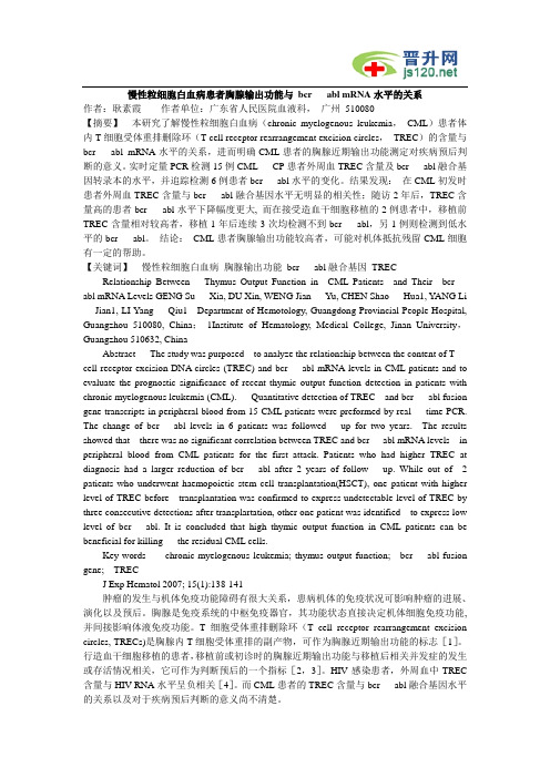
与胎肝和脐带血中HSC类似,AGM区的HSC也具有CD45 CD34 表型【1,2】(图1)。
图1. 人类胚胎造血发育事件概览【2】功能学和组织学实验证实,人类胚胎的第一个HSC产生于背主动脉腹侧壁【3,4】。
鉴于人类HSC与血管内皮细胞存在着紧密的时空联系,并且共享一些表面标志【4】,因此推测人类HSC与小鼠类似,均起源于生血内皮细胞(hemogenic endothelial cell,HEC)【5】。
目前研究认为,HEC表达内皮基因和生血特异的转录因子RUNX1,但不表达典型造血表面标志如CD43和CD45【6,7】。
HEC的特化是血管内皮细胞选择HSC命运的关键步骤,然而,由于HEC数量稀少、发育过程转瞬即逝,对于内皮造血转化的认识,尤其是对HEC的精准识别,成为造血发育研究领域的重点和难点。
鉴于临床的巨大需求和血液细胞来源有限性之间的矛盾,血液系统尤其是HSC的再生研究显得尤为重要。
研究者一直致力于利用多能干细胞高效诱导功能性HSC,但罕见成功的报道,表明对于HSC胚胎发生的时序特征及调控机制的认识仍有待深入,HSC的再生研究依然任重道远【8】。
2019年9月9日,解放军总医院第五医学中心刘兵研究组与暨南大学基础医学院兰雨研究组合作在Cell Research杂志在线发表了题为Tracing the first hematopoietic stem cell generation in human embryo by single-cell RNA sequencing的研究论文。
GW4869抑制骨髓间充质干细胞外泌体的释放

GW4869抑制骨髓间充质干细胞外泌体的释放孙淑丽;樊毫军;肖培欣;丁辉;王敬;刘子泉;刘晋阳;王雪;石沙;吕琪【摘要】背景:间充质干细胞来源外泌体与间充质干细胞本身具有相似的功能,但外泌体释放的调控机制依然不是很清楚.目的:探讨中性鞘磷脂酶抑制剂GW4869对骨髓间充质干细胞来源外泌体释放的抑制作用及其对细胞增殖的影响.方法:将骨髓间充质干细胞分为正常对照组,GW4869处理24 h组和GW4869撤药24 h组(GW4869预处理24 h,撤药后继续培养24 h),分别收集各组条件培养基,采用超速离心法提取外泌体,Western blot法鉴定外泌体表面标志蛋白CD63、TSG101的表达,Nano-sight检测外泌体粒径分布及浓度,BCA法测定外泌体总蛋白水平,活细胞成像系统观察GW4869对骨髓间充质干细胞增殖的影响.结果与结论:①Western blot显示外泌体阳性表达标志蛋白CD63、TSG101;②纳米示综分析证实释放的外泌体大小约114 nm;③与正常对照组相比,GW4869处理24 h组和GW4869撤药24 h组外泌体释放减少差异有显著性意义(P < 0.01),而GW4869预处理24 h组和GW4869撤药24 h组差异无显著性意义(P >0.05);④与正常对照组相比,GW4869及其溶剂二甲基亚砜对骨髓间充质干细胞增殖影响差异无显著性意义(P > 0.05);⑤结果表明,GW4869可以显著抑制骨髓间充质干细胞外泌体的释放,并且在撤药24 h内依然保持抑制作用,GW4869对骨髓间充质干细胞增殖无影响.%BACKGROUND: Bone marrow mesenchymal stem cells (BMSCs) and BMSCs-derived exosomes have similar functions, but the regulatory mechanism underlying the release of exosomes is still unclear. OBJECTIVE: To investigate the role of GW4869, an inhibition of neutral sphingomyelinase 2, in the release of exosomes in BMSCs and the influence of GW4869 on BMSCs proliferation. METHODS: Rat BMSCs weredivided into three groups: normal control group, 24-hour GW4869 treatment group and withdrawal of GW4869 for 24 hours group (24-hour GW4869 treatment followed by 24-hour successive culture with drug withdrawal). Cultured cells were collected to extract exosomes by ultracentrifugation. Western blot was used to detect exosome-associated proteins CD63 and tumor susceptibility gene 101 (TSG101). The concentration and size distribution of exosomes were measured using nanoparticle tracking analysis. BCA was used to test the level of total proteins in exosomes. Live cell imaging system was used to observe the influence of GW4869 on BMSCs proliferation. RESULTS AND CONCLUSION: (1) Western blot results showed that exosomes expressed marker proteins such as CD63, TSG101. (2) Findings from the nanoparticle tracking analysis confirmed that the size of released exosomes was about 114 nm. (3) Significantly reduced release of exosomes was found in the two treatment groups compared with the normal control group (P < 0.01), but there was no significant difference between 24-hour GW4869 treatment group and withdrawal of GW4869 for 24 hours group (P > 0.05). (4) No significant difference in the proliferation of BMSCs was found among the three groups (P > 0.05). To conclude, 24-hour W4869 can inhibit the release of exosomes by BMSCs and this inhibitory effect is still sustained within 24 hours after drug withdrawal. However, GW4869 has no influence on the proliferation of BMSCs.【期刊名称】《中国组织工程研究》【年(卷),期】2018(022)001【总页数】6页(P26-31)【关键词】外泌体;GW4869;骨髓间充质干细胞;干细胞【作者】孙淑丽;樊毫军;肖培欣;丁辉;王敬;刘子泉;刘晋阳;王雪;石沙;吕琪【作者单位】武警后勤学院附属医院,救援医学研究所,天津市 300162;武警后勤学院附属医院,医教部,天津市 300162;武警后勤学院附属医院,救援医学研究所,天津市 300162;天津大学灾难医学研究院,天津市 300072;武警后勤学院附属医院,救援医学研究所,天津市 300162;天津大学灾难医学研究院,天津市 300072;武警后勤学院附属医院,救援医学研究所,天津市 300162;武警后勤学院附属医院,救援医学研究所,天津市 300162;武警后勤学院学员2旅,天津市 300309;天津大学灾难医学研究院,天津市 300072【正文语种】中文【中图分类】R394.20 引言 Introduction外泌体是活细胞分泌的直径30-150 nm的脂质双分子层囊泡[1]。
AML的WHO分型

形态学特点
大原始细胞、胞浆有假Chediak Higashi大 颗粒及空泡,Auer小体易见,粒系病态造 血 (Pelget Huet异常),胞浆呈均一性粉红色,嗜 酸粒细胞增加,强的MPO活性。有时可见 嗜碱粒细胞和肥大细胞增多。
伴t(8;21)(q22;q22)(AML1-ETO)AML
形态学和细胞化学:
多系病态造血改变(>50% 2系细胞) : 中性粒细胞Pelget Huet畸形 红系巨幼变、多核、环状铁粒幼 小巨核等
伴多系病态造血的AML
伴多系病态造血的AML
免疫表型:CD34+、CD13+、CD33+常伴CD56 和 /或CD7表达,原始细胞MDR-1表达增高.
遗传学:和MDS相似:-5/5q-、-7/7q-、+8、+9、+11、 del11q、del12p、-18、+19、del(20q)、+21、t(2;11)、 t(1;7)、inv(3)(q21q26),t(3;3)(q21;q26)伴Plt增高, t(3;5)(q25;q34)伴多系病态造血,而血小板正常。
免疫表型:表达1至多种髓系抗原(CD13、CD33、 CD117),但B、T淋系抗原均阴性,CD34、 CD38、HLA-DR常阳性
遗传学:无特异性异常,常有复杂异常(+13、+8、 +4、-7)
细胞起源:髓系分化早期阶段造血干细胞 预后:CR率低,易早期复发,生存期短
微分化AML(相当于M0) (血片)
定义:
伴粒系和单核系分化并有嗜酸粒细胞异常的AML。 占10%~12%的AML,青年人多见。粒细胞肉瘤。 主要见于FAB分型M4E0型
形态学和细胞化学:
白血病Ly+AML型和My+ALL型预后因素的临床研究 (13)
急性白血病骨髓基质细胞Connexin 43的表达及细胞间通讯功能研究作者:刘耀,张曦,司英健,高蕾,高力,陈幸华作者单位:第三军医大学新桥医院血液科,重庆400037【摘要】本研究探讨急性白血病和正常骨髓基质细胞中连接蛋白43(connexin 43, Cx43)的表达及细胞间隙连接通讯(gap junction intercellular communication, GJIC)功能的变化及二者细胞通讯功能之间的差异。
体外培养正常及急性白血病骨髓基质细胞,采用细胞免疫组织化学方法及计算机灰度检测观察二者之间Cx43表达的变化,采用细胞划痕染料传输技术比较两者之间GJIC功能的差异。
结果显示,体外培养的急性白血病骨髓基质细胞Cx43的表达较正常骨髓基质细胞Cx43的表达明显减低,GJIC功能减弱。
结论:急性白血病骨髓基质细胞间通讯功能较正常骨髓基质细胞明显减低。
【关键词】急性白血病Connexin 43 Expression and Interacellular Communicating Function in Acute Leukemia Bone Marrow Stroma Cells LIU Yao*,ZHANG Xi*,SI Ying Jian,GAO Lei,GAO Li,CHEN Xing Hua Department of Hematology,Xinqiao Hospital,The Third Military Medical University,Chongqing 400037,ChinaAbstract This study was purposed to investigate the connexin 43(Cx43)expression level in acute leukemia bone marrow stromal cells (ABMSCs)and normal bone marrow stromal cells (NBMSCs), and to explore the difference in communicating functions between these cells. The Cx43 expression levels of ABMSCs and NBMSCs were detected by using immunohistochemistry and computer gray scale assay,and the difference of gap junction intercellular communication(GJIC)was examined through dry transfer technique. The results showed that expression level of Cx43 in ABMSCs was lower than that in NBMSCs and its function of GJIC in ABMSCs was also weaker than that in NBMSCs. It is concluded that cell cell communication function is lowered in ABMSCs.Key words Cx43; GJIC; acute leukemia; bone marrow stromal cellJ Exp Hematol 2007; 15(4):679-682造血基质细胞所形成的造血微环境(hematopoietic microenvironment,HME)是造血干/祖细胞定殖、分化、发育和成熟的场所[1]。
白血病Ly+AML型和My+ALL型预后因素的临床研究 (42)
慢性粒细胞白血病患者胸腺输出功能与bcr abl mRNA水平的关系作者:耿素霞作者单位:广东省人民医院血液科,广州510080【摘要】本研究了解慢性粒细胞白血病(chronic myelogenous leukemia,CML)患者体内T细胞受体重排删除环(T cell receptor rearrangement excision circles,TREC)的含量与bcr abl mRNA水平的关系,进而明确CML患者的胸腺近期输出功能测定对疾病预后判断的意义。
实时定量PCR检测15例CML CP患者外周血TREC含量及bcr abl融合基因转录本的水平,并追踪检测6例患者bcr abl水平的变化。
结果发现:在CML初发时患者外周血TREC含量与bcr abl融合基因水平无明显的相关性;随访2年后,TREC含量高的患者bcr abl水平下降幅度更大, 而在接受造血干细胞移植的2例患者中,移植前TREC含量相对较高者,移植1年后连续3次均检测不到bcr abl,另1例则检测到低水平的bcr abl。
结论:CML患者胸腺输出功能较高者,可能对机体抵抗残留CML细胞有一定的帮助。
【关键词】慢性粒细胞白血病胸腺输出功能bcr abl融合基因TREC Relationship Between Thymus Output Function in CML Patients and Their bcr abl mRNA Levels GENG Su Xia, DU Xin, WENG Jian Yu, CHEN Shao Hua1, Y ANG Li Jian1, LI Yang Qiu1 Department of Hemotology, Guangdong Provincial People Hospital, Guangzhou 510080, China;1Institute of Hematology, Medical College, Jinan University,Guangzhou 510632, ChinaAbstract The study was purposed to analyze the relationship between the content of T cell receptor excision DNA circles (TREC) and bcr abl mRNA levels in CML patients and to evaluate the prognostic significance of recent thymic output function detection in patients with chronic myelogenous leukemia (CML). Quantitative detection of TREC and bcr abl fusion gene transcripts in peripheral blood from 15 CML patients were preformed by real time PCR. The change of bcr abl levels in 6 patients was followed up for two years. The results showed that there was no significant correlation between TREC and bcr abl mRNA levels in peripheral blood from CML patients for the first attack. Patients who had higher TREC at diagnosis had a larger reduction of bcr abl after 2 years of follow up. While out of 2 patients who underwent haemopoietic stem cell transplantation(HSCT), one patient with higher level of TREC before transplantation was confirmed to express undetectable level of TREC by three consecutive detections after transplartation, other one patient was identified to express low level of bcr abl. It is concluded that high thymic output function in CML patients can be beneficial for killing the residual CML cells.Key words chronic myelogenous leukemia; thymus output function; bcr abl fusion gene; TRECJ Exp Hematol 2007; 15(1):138-141肿瘤的发生与机体免疫功能障碍有很大关系,患病机体的免疫状况可影响肿瘤的进展、演化以及预后。
牙周膜干细胞的表型分析

牙周膜干细胞的表型分析贺慧霞;刘洪臣;王东胜;鄂玲玲;马攀【摘要】目的:分析人牙周膜干细胞(PDLSCs)的表型特点,为分离、鉴定和应用PDLSCs提供有效途径.方法:采用免疫磁珠法分离PDLSCs,制备细胞爬片.分别采用免疫荧光、免疫细胞化学染色法检测PDLSCs表面蛋白STRO-1、CD44、Vimentin、CK、COL-Ⅰ、BSP的表达.收集PDLSCs,采用RT-PCR法检测PDLSCs Scleraxis mRNA、Col Ⅰ及CAP mRNA的表达情况.结果:PDLSCs表达间充质干细胞表面标志STRO-1、CD44,强阳性表达间充质来源细胞表面蛋白Vimentin,弱表达Col-Ⅰ,不表达上皮细胞标志蛋白CK、成骨细胞相对特异性蛋白BSP.RT-PCR也显示PDLSCs表达韧带组织特异性基因Scleraxis和Col-Ⅰ,不表达成牙骨质细胞特异性蛋白CAP.结论:本实验所分离的PDLSCs具有间充质来源的、牙周组织特异性的、未分化细胞的表型特点,该特点可作为PDLSCs分离和鉴定的重要依据.%Objective: To analyze the phenotypic characteristics of human periodontal ligament stem cells(PDLSCs), and to provide new way for understanding and further application of PDLSCs. Materials and Methods. PDLSCs were isolated using an immunomagnetic bead selection system. The slides of the cells were prepared and immunofluorescence and immunocytochemical straining were performed to detect the expression of specific proteins including STRO-1, CD44, Vimentin, CK, COL-I, BSP, respectively. In addition, PDLSCs were collected to detect the the gene expression of Scleraxis mRNA, Col I mRN and CAP mRNA by RT-PCR techniques. Results: PDLSCs expressed the surface markers of mes-enchymal stem cells such as STRO-1 and CD44, and positively expressedthe specific proteins of cells originated from mes-enchymal tissue including Vimentin, weakly expressed CoL-I, and negatively expressed the CK which is expressed specially in epithelial cells. PDLSCs were also negatively expressed the marker of osteocytes of BSP. Results from RT-PCR showed that these cells positively expressed the specific marker of tendon tissue-Scleraxis, specific marker of membrane-Collagen I, and negatively expressed the specific marker of cementocyte-CAP. Conclusion: PDLSCs isolated by immunomagnetic bead selection system in this study showed the phenotypes of mesenchymal origination, periodontal specificity and undifferentiated cells. These characteristics may provide important evidences for isolation, identification, and futher application for PDLSCs.【期刊名称】《口腔颌面修复学杂志》【年(卷),期】2011(012)005【总页数】4页(P257-260)【关键词】牙周膜干细胞;表型;间充质【作者】贺慧霞;刘洪臣;王东胜;鄂玲玲;马攀【作者单位】解放军总医院口腔医学研究所北京100853;解放军总医院口腔医学研究所北京100853;解放军总医院口腔医学研究所北京100853;解放军总医院口腔医学研究所北京100853;解放军总医院口腔医学研究所北京100853【正文语种】中文【中图分类】R780.2在生理状态下,牙周膜干细胞(periodontal ligament stem cells,PDLSCs)的主要功能是维持牙周膜中细胞更新和凋亡的动态平衡;在病理情况下,则主要是参与牙周损伤后的再生修复、生理性改建过程。
原发耐药急性髓系白血病伴费城染色体阳性一例并文献复习_A_Case_of_Philadelphia_

World Journal of Cancer Research 世界肿瘤研究, 2021, 11(4), 121-125 Published Online October 2021 in Hans. /journal/wjcr https:///10.12677/wjcr.2021.114016原发耐药急性髓系白血病伴费城染色体阳性 一例并文献复习王梦莹1,2,许 晗1,2,冯献启2*1青岛大学医学部,山东 青岛 2青岛大学附属医院血液科,山东 青岛收稿日期:2021年8月30日;录用日期:2021年9月15日;发布日期:2021年9月29日摘 要费城染色体阳性急性髓系白血病(Ph +AML)是一种罕见的AML 亚型,2016年修订的世界卫生组织(WTO)髓系恶性肿瘤将其归类为预后不良伴有遗传学异常的AML 暂定型。
Ph +AML 是有别于慢性髓系白血病的一组高度复杂的异质性疾病,对化疗反应差,易复发,预后不佳,具有独特的临床和血液学特点。
关键词急性髓系白血病,费城染色体,原发耐药A Case of Philadelphia Chromosome-Positive Acute Myeloid Leukemia (Ph +AML) of Primary Drug Resistance and Relevant Literature ReviewMengying Wang 1,2, Han Xu 1,2, Xianqi Feng 2*1Medical Science Center of Qingdao University, Qingdao Shandong 2Department of Hematology, The Affiliated Hospital of Qingdao University, Qingdao ShandongReceived: Aug. 30th , 2021; accepted: Sep. 15th , 2021; published: Sep. 29th , 2021 *通讯作者。
- 1、下载文档前请自行甄别文档内容的完整性,平台不提供额外的编辑、内容补充、找答案等附加服务。
- 2、"仅部分预览"的文档,不可在线预览部分如存在完整性等问题,可反馈申请退款(可完整预览的文档不适用该条件!)。
- 3、如文档侵犯您的权益,请联系客服反馈,我们会尽快为您处理(人工客服工作时间:9:00-18:30)。
ORIGINALARTICLEGeneexpressionprofilesofAMLderivedstemcells;similaritytohematopoieticstemcells
HGal1,2,3,NAmariglio2,LTrakhtenbrot2,JJacob-Hirsh2,OMargalit2,AAvigdor4,ANagler4,STavor5,LEin-Dor3,TLapidot6,EDomany3,GRechavi2andDGivol1
1DepartmentofMolecularCellBiology,WeizmannInstituteofScience,Rehovot,Israel;2DepartmentofPediatricHemato-
Oncology,ChaimShebaMedicalCenter,Tel-HashomerandSacklerSchoolofMedicine,Tel-AvivUniversity,Tel-Aviv,Israel;3DepartmentofPhysicsofComplexSystems,WeizmannInstituteofScience,Rehovot,Israel;4TheDivisionofHematologyand
BoneMarrowTransplantation,ChaimShebaMedicalCenter,Tel-Hashomer,Israel;5DepartmentofHematology,Tel-Aviv
SouraskyMedicalCenter,Tel-Aviv,Israeland6DepartmentofImmunology,WeizmannInstituteofScience,Rehovot,Israel
Tumorscontainafractionofcancerstemcellsthatmaintainthepropagationofthedisease.TheCD34þCD38Àcells,isolatedfromacutemyeloidleukemia(AML),wereshowntobeenrichedleukemicstemcells(LSC).WeisolatedtheCD34þCD38ÀcellfractionfromAMLandcomparedtheirgeneexpressionprofilestotheCD34þCD38þcellfraction,usingmicroarrays.Wefound409genesthatwereatleasttwofoldover-orunder-expressedbetweenthetwocellpopulations.TheseincludeunderexpressionofDNArepair,signaltransductionandcellcyclegenes,consistentwiththerelativequiescenceofstemcells,andchromosomalaberrationsandmutationsofleukemiccells.ComparisonoftheLSCexpressiondatatothatofnormalhematopoieticstemcells(HSC)revealedthat34%ofthemodulatedgenesaresharedbybothLSCandHSC,supportingthesuggestionthattheLSCoriginatedwithintheHSCpro-genitors.WefocusedontheNotchpathwaysinceJagged-2,aNotchligandwasfoundtobeoverexpressedintheLSCsamples.WeshowthatDAPT,aninhibitorofgamma-secretase,aproteasethatisinvolvedinJaggedandNotchsignaling,inhibitsLSCgrowthincolonyformationassays.IdentificationofadditionalgenesthatregulateLSCself-renewalmayprovidenewtargetsfortherapy.Leukemia(2006)20,2147–2154.doi:10.1038/sj.leu.2404401;publishedonline12October2006Keywords:stemcells;microarray;colonyformation;Notch
IntroductionAcutemyeloidleukemia(AML)isaheterogeneousdiseasewithvariationsinthecellmarkers,chromosomalaberrations,mutations,responsetotherapyandprognosis.IncreasingevidencesuggestthatAMLandothermalignanciesaresustainedbyaminortumorsubpopulationwithself-renewalpotential,referredtoas‘cancerstemcells’or‘leukemicstemcells’(LSC).Thesecellsdrivethegrowthanddisseminationoftheleukemia.1–4Recentstudieshaveextendedthismodeltoother
typesofcancerssuchasbreastcancer5andGlioblastomamultiforme.6Theadvancesintheanalysisoftheoriginofthe
leukemiccellscamefromstudiesonengraftmentofpatient’sderivedleukemiaintotheimmunodeficientNOD-SCIDmice,7,8
andfollowingthepropagationofthedisease.9ThesestudiesledtotheidentificationofCD34þCD38ÀcellsastheLSCfractionofAML.Inaddition,ahighpercentageofCD34þCD38Àstem
cellsatdiagnosiswasdirectlycorrelatedwithpoorsurvivalandsignificantlycorrelatedwithahighminimalresidualdiseasefrequency,especiallyafterchemotherapy.10
AnimportantpropertythatissharedbybothLSCandnormalHSCistheirabilityofself-renewal,whichenablesthemaintenanceoftheleukemiccloneontheonehandorthenormalhematopoiesisontheotherhand.3,4,11,12Somemole-
cularmechanismsresponsibleforselfrenewal,likeBmi-1,NotchandWntsignalingpathways,werefoundtobesharedbybothHSCandLSC.4,13,14ThissuggeststhattheoriginofLSC
maybeatthehematopoietcstemcelllevel.2Theexistenceofcancerstemcellsisofmajorclinicalrelevancesincetheiruniqueproperties,suchasslowmitosis,increasedmultidrugresistanceandlowerexpressionofFas/Fas-LandFas-inducedapoptosis,mayenablethemtoescapetherapy15thatisbasedon
markersorphenotypeoftheentireAMLpopulation.WethereforesoughttoexaminethegeneexpressionprofilesoftheLSCwithintheAMLcellpopulationtofurtherunderstandtheirbiologyandidentifynewtargetgenesthatmaintainandcharacterizeLSC.ForthispurposeweisolatedCD34þCD38ÀandCD34þCD38þcellpopulationsfromfiveAMLpatientsandanalyzedtheirgeneexpressionprofilesusingAffymetrixHu133Amicroarrays.Wefound409genesthatwereoverorunderexpressedbetweenthetwopopulations,sortingthemodulatedgenesshowedacleardistinctionbetweenthecellpopulations.TheGO(GeneOntology)analysisofthemodu-latedgenesinLSCrevealedadecreasedexpressionofDNArepair,signaltransductionandcellcycleregulatinggenesandanincreasedexpressionofgenesrelatedtoproteindegradation,celladhesion,transcriptionandtheNotchpathway.WethereforetriedtoinhibitthegrowthofLSCcoloniesinmethylcellulosebyinhibitingtheNotchpathway,anapproachthatmaybeofclinicalvalue.UnderstandingthemechanismofactionofgenesinvolvedinLSChassignificantimplicationsforfutureresearchandwillpotentiallyleadtonewtargetsfortherapyanddiagnostics.