电泳技术要求PDF
电泳漆技术标准

二、电泳漆的工艺性能。电泳漆在使用过程中的工艺稳定性才是关键。电泳 漆的槽液工艺参数如下:
项
目
单
位
指
标
PH 值(25℃) 电导率(25℃) 固体份 溶剂含量 灰份(黑) 灰份(灰) MEQ 值 施工温度 施工电压 施工时间 电泳电压 泳透力 (一汽钢管法 210V) 库仑效率 击穿电压 L 效果 重溶性 加热减量 沉淀性 超滤液 PH 值 超滤液固体份 阳极液 PH 值 阳极液电导率 槽液更新周期 月 % μm/cm % % % % 毫克当量/100g
40—60(60 )
≥5 ≥10
% H cm 级 mm 小时 小时 小时 小时 小时 小时
0
≤2 ≥540 ≥900(1000) ≥300 ≥90 ≥1 ≥48
使用两年后涂层完整
注:我厂现有设备限定的工艺参数如下: 1. 电泳电压:100—250V 2. 电泳时间:150—180S 3. 烘道烘干温度:< 180℃ 4. 烘道烘干时间:< 30min
℃
5.8—6.6
≤1600
15—18 2.5—4.5 9±4 21±3 30±4 28—32 170—250 150—210 170—250
≥80 ≥25 >280
V S V % mg/c V
水平面与垂直面涂膜外观无明显差别 % %
≤8 ≤8
24h 静止,工作液不分层,颜料沉淀≤50mL/L,易搅起 5.8—6.8 0.4—0.7 2.5—5 600—1000
电泳漆的技术要求
电泳漆主要考虑两个方面的因素:涂料的漆膜性能和工艺性能。 一、电泳漆膜的性能,必须达到如下要求: 项 颜色 外观 膜厚 光泽度 铅笔硬度 冲击强度 附着力 柔韧性 耐盐雾性(工件) 耐盐雾性(试片) 耐水性 耐酸性 耐碱性 耐机油性 耐候性 目 指 标 单 位 检 测 方 法 目测 目测 μm
Q-RT 011-2008电泳涂层要求(1)

宁波锐泰机械制造有限公司企业标准Q/RT 011-2008电泳涂层要求2008-12-17发布 2008-12-20实施 宁波锐泰机械制造有限公司发布前 言本标准对《电泳涂层的主要性能指标》进行了修订,统一了标准的行文格式。
本标准对金属零(部)件电泳涂层的常规检验项目与特殊检验项目进行了区分。
本标准增加的内容有:耐盐雾试验、电泳涂装L形试板试验效果的判定级别、泳透力板耐盐雾试验判定以及划格试验判定级别。
本标准自实施之日起代替《电泳涂层的主要性能指标》。
本标准由宁波锐泰机械制造有限公司工程技术部提出,由宁波锐泰机械 制造有限公司标准化委员会归口。
本标准主要起草人: 李春艳本标准首次发布日期:2007年7月11日,修订日期2008年12月17日。
宁波锐泰机械制造有限公司企业标准电泳涂层要求 Q/RT 011-20081 范围本标准规定了金属零(部)件电泳涂层要求及试验方法。
本标准适用于电泳涂层验收。
2 规范性引用文件下列标准中的条款通过本标准的引用而成为本标准的条款。
凡是注日期的引用文件,其随后所有的修改单(不包括勘误的内容)或修订版均不适用于本标准。
凡是不注日期的引用文件,其最新版本适用于本标准。
JB/T 10242-2001 阴极电泳涂装通用技术规范QC/T 484-1999 汽车油漆涂层3 金属零(部)件电泳涂层常规检验项目及试验方法3.1 外观金属零(部)件电泳涂层外观均匀、平整、光滑,无杂质、缩孔、凹陷、起皱、掉皮、涂料痕迹,无露底。
3.2 涂层厚度涂层厚度要求:20±5 μm。
按照GB/T1764-79规定进行,检测仪器用杠杆千分尺(精确度为2μm)或磁性测厚仪(测定范围0~500μm、精确度为2μm)。
3.3附着力—划格试验按GB/T9286规定进行,划格间距为1mm,试验后评定结果达0级或1级。
0级:切割边缘完全平滑,无一格脱落。
1级:在切口交叉处有少许涂层脱落但交叉切割面积受影响不能明显大于5%。
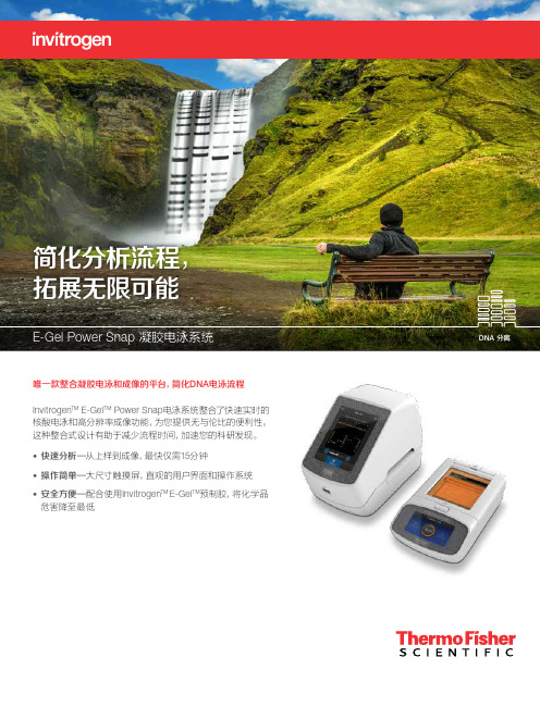
Invitrogen E-Gel Power Snap电泳系统说明书

唯一款整合凝胶电泳和成像的平台,简化DNA 电泳流程 Invitrogen TM E-Gel TM Power Snap 电泳系统整合了快速实时的核酸电泳和高分辨率成像功能,为您提供无与伦比的便利性。
这种整合式设计有助于减少流程时间,加速您的科研发现。
• 快速分析—从上样到成像,最快仅需15分钟• 操作简单—大尺寸触摸屏,直观的用户界面和操作系统 • 安全方便—配合使用Invitrogen TM E-Gel TM 预制胶,将化学品危害降至最低台式设计,快速便捷Invitrogen TM E-Gel TM Power Snap式设备,配备有蓝光透射仪,操作安全,此外,它还自带琥珀色滤光片,可用于对预染了SYBR TM Safe 或 SYBR TM Gold染料的脂糖凝胶中的样品进行实时样品追踪。
方案,适用于不同类型的E-Gel琼脂糖凝胶。
高分辨率数码相机快速简单完成凝胶成像无需外接电源直接由下方电泳设备供电方便转移图片文件32GB海量存储凝胶图片选择适合的E-Gel 预制琼脂糖凝胶类型E-Gel Powe Snap 电泳设备与以下E-Gel 预制琼脂糖凝胶兼容,仪器内预设针对不同凝胶类型的运行程序方案。
参考下表,选择适合的E-Gel 预制琼脂糖凝胶类型。
了解更多,请登录thermofi/egels 。
E-Gel SizeSelect II 预制琼脂糖凝胶最为巧妙的DNA 凝胶纯化方法,二代测序的得力助手Invitrogen TM E-Gel ™ SizeSelect ™ II 琼脂糖凝胶是二代测序的得力助手,无需切胶和纯化试剂盒,即可从凝胶中回收您感兴趣的DNA 条带,适用于<1kb DNA 片段的分离,纯化的DNA 片段可直接用于二代测序平台文库的构建或下游克隆实验。
上样电泳回收目标条带通过三个简单的步骤完成DNA 的凝胶纯化E-Gel SizeSelect II 琼脂糖凝胶为双梳凝胶。
将样品加到上面一排孔中,开始电泳,直至条带迁移至底部一排孔中。
电泳漆技术说明

目录一、公司简介二、电泳原理三、规范工艺流程四、产品简介五、全系列丙烯酸聚氨酯电著产品简介六、全系列产品规格七、全系列涂膜性能八、补给剂资料九、管理对策十、问题点与对策十一、各型号产品说明书二、电泳涂装原理:阴极电泳涂料是一种具有半水溶型和悬体系特征的多组份体系,在电场作用下,已中和的阳离子树脂携带的涂料粒子在阴极进行电沉积,其过程包括电泳、电解、电沉积、电渗四种现象。
1、电泳:悬浮在极性液体中的带电粒子由于电场影响而发生泳动的现象。
阳离子型树脂粒子向作为阴极的工件移动。
2、电解:液体中的氢离子和氢氧根离子在直流电源的作用下,生成氢气和氧气,阴极界面PH值升高。
3、电沉积:阳离子涂料粒子在吸引它的电极(阴极)上被还原为中性不溶脂,析出粘附,形成不溶解的涂膜。
过程如下:H2O≒H++OH-阳极:2H++2OH—- 2e→H2O+1/2O2↑+2H+阴极:2OH-+2H++2e→H2↑+2OH-R1 R 1∣∣~NH++OH-→~N↓+H2O ∣∣R 2 R24、电渗:在外电场力作用下,涂膜内部所含的水份从涂膜中渗析出来移向槽液,进行内聚部分脱水。
电渗使亲水涂膜变为憎水涂膜,脱水使涂膜致密化。
由于上述四个过程是连续进行的,获得的涂膜在烘干之前含水量在10%以下,可以直接进行水洗、烘干,最后形成连续均一的品质优良的涂膜。
三、规范工艺流程:1.环氧体系电泳流程:高温脱脂(加超声波)→常温脱脂→水洗三次→酸洗除锈→水洗三次→流动水洗→表调→磷化→水洗三次→流动水洗→纯水洗两次→电泳涂装→回收水洗三次→纯水浸洗两次→纯水喷淋→脱水剂洗/吹洗→烘干固化→成品包装2.透明有色体系电泳流程:电镀工件(抛光工件)→化学除油(加超声波)→水洗两次→电解除油→水洗三次(溢流水洗)→中和(弱酸洗)→水洗三次(溢流水洗)→纯水洗两次→电泳涂装(透明及各色)→回收纯水洗两次→纯水洗三次(溢流)→脱水剂洗→滴水(吹净水珠)→烘烤→成品包装四、产品简介:1.环氧体系电泳涂料:本公司之环氧体系电泳涂料目前主要以色浆、乳液双组份供应市场。
SDS-PAGE电泳的标准操作规程

SDS-PAGE电泳的标准操作规程(编号:039)1、目的及适用范围SDS-PAGE电泳的分离原理,是根据大多数蛋白质都能与阴离子表面活性剂十二烷基硫酸钠(SDS)按重量比结合成复合物,使蛋白分子所带的负电荷远远超过天然蛋白分子的净电荷,消除了蛋白分子的电荷效应,使蛋白按分子大小分离,主要用于蛋白质分子量的测定、蛋白质混合组分的分离和蛋白质亚基组分的分析等方面。
2、主要仪器恒压或恒流电源、垂直板、制胶模具3、试剂及配制方法试剂配制方法或要求H2O 电阻率不低于18.2 MΩ.cm1.5 M Tris-HCl 1.5 moL/L Tris-HCl缓冲液,pH 8.830% Acrylamide 30% 丙烯酰胺溶液(避光保存)10% SDS 10% SDS溶液10% APS 10% 过硫酸铵溶液(临用前配制)TEMED TEMED溶液1 M Tris-HCl 1 moL/L Tris-HCl缓冲液,pH 6.815.1g、甘氨酸94 g、SDS 5 g,水稀释至1 L,pH 8.35×电极缓冲液 Tris2×Loading buffer 100 mM Tris-HCl(pH6.8)、4% SDS、0.2% 溴酚蓝、20% 甘油,用于非还原SDS-PAGE电泳,如用于还原SDS-PAGE电泳,则再加2% β-巯基乙醇5×Loading buffer 250 mM Tris-HCl(pH6.8)、10% SDS、0.5% 溴酚蓝、50% 甘油,用于非还原SDS-PAGE电泳,如用于还原SDS-PAGE电泳,则再加5% β-巯基乙醇考马斯亮蓝染色液 0.1%考马斯亮蓝R250、25%异丙醇、10%冰醋酸,水稀释至1 L考马斯亮蓝脱色液醋酸100 mL、乙醇50 mL、水850 mL4、操作步骤4.1 制胶:取合适的密封垫片和两块已清洁的干燥的玻璃板,组装成灌胶的模具,按下表制成分离胶,灌入模具内一定高度(一般离玻璃板上端3.5 cm),小心加水或异丙醇封顶(约0.5 cm高),室温下聚合(温度不同,聚合时间不同,20℃大约需30 min)。
[指南]核酸电泳的注意事项
![[指南]核酸电泳的注意事项](https://img.taocdn.com/s3/m/a356bd4a3a3567ec102de2bd960590c69ec3d863.png)
核酸电泳的注意事项1. 琼脂糖:不同厂家、不同批号的琼脂糖,其杂质含量不同,影响DNA的迁移及荧光背景的强度,应有选择地使用。
2. 凝胶的制备:凝胶中所加缓冲液应与电泳槽中的相一致,溶解的凝胶应及时倒入板中,避免倒入前凝固结块。
倒入板中的凝胶应避免出现气泡,以免影响电泳结果。
3. 电泳缓冲液:为保持电泳所需的离子强度和pH,应常更新电泳缓冲液。
4. 样品加入量:一般情况下,0.5cm宽的梳子可加0.5μg的DNA量,加样量的多少依据加样孔的大小及DNA中片段的数量和大小而定。
当加样孔大时,样品上样量应相应加大,否则会造成条带浅甚至辩认不清;反之则应适当减少加样量,但是上样量过多会造成加样孔超载,从而导致拖尾和弥散,对于较大的DNA此现象更明显。
5. DNA样品中盐浓度会影响DNA的迁移率,平行对照样品应使用同样的缓冲条件以消除这种影响。
DNA迁移率取决于琼脂糖凝胶的浓度,迁移分子的形状及大小。
采用不同浓度的凝胶有可能分辨范围广泛的DNA分子,制备琼脂糖凝胶可根据DNA分子的范围来决定凝胶的浓度。
小片段的DNA电泳应采用聚丙烯酰胺凝胶电泳以提高分辨率。
回收率低或为零1. 漂洗缓冲液中没加入乙醇.在使用前应确保乙醇已加入漂洗液PW中。
2. 吸附材料上有乙醇残留.洗脱时硅胶膜或硅胶树脂上有漂洗缓冲液残留,因含乙醇会降低洗脱效率。
在洗脱前可通过再次离心或置于50℃烤箱中5-10分钟,彻底去除漂洗缓冲液。
3. 洗脱缓冲液pH值偏低.DNA只在低盐buffer中才能被洗脱,如洗脱缓冲液EB (10 mM Tris·Cl, pH 8.5)或水。
洗脱效率取决于pH值。
最大洗脱效率在pH7.0-8.5间。
当用水洗脱时确保其pH值在此范围内。
4. 洗脱液加入位置不正确.洗脱液应加在硅胶膜中心部位以确保洗脱液会完全覆盖硅胶膜的表面,达到最大洗脱效率。
DNA质量不好洗脱产物含有乙醇残留.洗脱时硅胶膜或硅胶树脂上有漂洗缓冲液残留,会使洗脱产物中含有乙醇,影响下游操作。
琼脂糖凝胶电泳法(agarosegelelectrophoresis)

琼脂糖凝胶电泳法(agarose gel electrophoresis)agarose gel electrophoresisThe principle of 1. agarose gel electrophoresisAgarose gel electrophoresis is a standard method for isolation, identification and purification of DNA fragments. The technique is simple and rapid and can be used to distinguish DNA fragments that cannot be isolated by other methods such as density gradient centrifugation. When the dye Ethidium (bromide, EB) is dyed with low concentration fluorescent dye, DNA bands of 1-10ng can be detected at least under ultraviolet light so that the position of DNA fragments in the gel can be determined. In addition, a specific DNA band can be recovered from the electrophoretic gel for subsequent cloning operations.Agarose can be made into various shapes, sizes, and porosities. Agarose gel separated DNA slices with a wide range of sizes. Agarose gels of different concentrations could separate DNA fragments from 200bp to near 50kb. Agarose is commonly used in horizontal devices for electrophoresis under constant electric field with constant intensity and direction.Agarose is mainly used as a solid support substrate in DNA electrophoresis, and its density depends on the concentration of agarose. In the electric field, the negative charge DNA moves towards the anode at the neutral pH, and the migration rate is determined by the following factors:Molecular size of 1. DNA:A linear double stranded DNA molecules in a certain concentration in agarose gel migration rate and molecular weight of DNA is inversely proportional to the logarithm of the molecular, the greater the resistance is greater, more difficult in the gel pores. Therefore, the slower migration.2. agarose concentrationA linear DNA molecule of a given size has a different migration rate at different concentrations of agarose gels. The logarithm of mobility of DNA electrophoresis is linearly related to gel concentration. The choice of gel concentration depends on the size of the DNA molecule. The concentration required for separating DNA fragments less than 0.5kb is 1.2-1.5%, and the concentration of DNA molecules larger than 10KB is 0.3-0.7%, and the concentration of DNA fragments between them is0.8-1.0%.Conformation of 3.DNA moleculeWhen the DNA molecule is in different conformation, it moves in the electric field, not only in relation to the molecular weight, but also to its conformation. The same molecular weight of the linear, open loop and ultra helical DNA in agarose gel moving speed is not the same, ultra helical DNA moving fastest, and linear double stranded DNA move the slowest. As in the electrophoresis of plasmid purity found several gel DNA with plasmid DNA is difficult to determine the cause of different conformation or because of containing other caused by DNA, can be gradually recovered from the agarose gel, DNA, respectively,hydrolysis, using the same restriction enzyme and gel electrophoresis, as in the DNA appear on the map is the same. For the same kind of DNA.4 、 supply voltageAt low voltage, the migration rate of the linear DNA fragment is proportional to the applied voltage. But with the increase of field strength, the migration of DNA with different molecular weight fragment rate will increase with different amplitude, fragment is larger, because the field strength increases caused by migration rate increased more significantly, so the voltage increases, the effective separation range of agarose gel will be reduced. To maximize the resolution of the DNA fragment greater than 2KB, the voltage shall not exceed 5v/cm.5, the presence of embedded dyesEthidium bromide, a fluorescent dye, was used to detect DNA in agarose gels,The dye is embedded between the stacked base pairs, stretching the elongated and notched ring DNA, making it more rigid and reducing the linear DNA mobility by 15%.6. effect of ionic strengthThe composition and ionic strength of electrophoretic buffer affect the electrophoretic mobility of DNA. In the absence of ion existence (such as the misuse of distilled water gel), theconductivity minimum, DNA almost does not move in high ionic strength buffer in (such as the error plus 10 x buffer), has very high conductance and obvious heat production would cause serious gel melting or denaturation of DNA.For natural double stranded DNA, several commonly used electrophoretic buffers are TAE[, EDTA (pH8.0) and Tris- acetic acid], TBE (Tris-, boric acid and EDTA), TPE (Tris-, phosphoric acid and EDTA) are generally formulated as concentrated mother liquor and stored at room temperature.2. steps of agarose gel electrophoresis1. take 5 * TBE buffer, 20ml add water to 200ml, prepare 0.5 * TBE dilution buffer.2. glue preparation: weigh 0.4g agarose, a 200ml conical flask, adding 0.5 50ml * TBE dilution buffer, placed in a microwave oven (or electric heating) to remove all agarose melting, shake, this is the 0.8% agarose gel. During heating, the agarose particles attached to the bottle wall shall be shaken from time to time to enter the solution. Heating should be covered with sealing film to reduce moisture evaporation.3., the preparation of rubber board: plexiglass trough trough at the ends of each with rubber paste (width 1cm) sealed tightly. Place the sealed glue trough on the horizontal support, insert the sample comb, and pay attention to observe the gap between the comb tooth edge and the bottom of the rubber tank to keep the clearance of about 1mm. Adding to the ethidium bromide agarose liquid cooled to 45-60 DEG C in (EB) solution to thefinal concentration of 0.5 g /ml (or not added EB gel after electrophoresis, but EB solution with 0.5 g/ml staining). Absorb a small amount of molten agarose gel with a pipette and seal the inner side of the adhesive. After the agarose solution is coagulated, the remaining agarose is carefully poured into the gel groove to form a uniform adhesive layer. Pour glue when the temperature can not be too low, otherwise the solidification is uneven, the speed can not be too fast, otherwise prone to bubbles. After the gel has been completely solidified, dial the comb and pay attention not to damage the gel at the bottom of the comb. Then add 0.5 * TBE dilution buffer to the surface of the comb, just above the surface of the rubber sheet. Because of the edge effect, there will be some uplift near the sample slot, which prevents the buffer from entering the sample tank, so make sure the sample tank is filled with buffer.4. add sample: take 10 L enzymolysis liquid and mix with 2 l 6 * sample liquid, carefully add into the sample trough with micro liquid gun. If the content of DNA is low, the amount of sample can be increased according to the above proportion, but the total volume can not exceed the capacity of the sample tank. When each sample is added, replace the tip head to prevent contamination. Attention should be paid to handling the sample, avoiding damage to the gel or piercing the gel at the bottom of the sample slot.5. electrophoresis: after finishing the sample, close the electrophoresis cover and turn on the power immediately. The control voltage is kept at 60-100V and the current is above 40mA. When the br blue indicator band is moved to the gum 3/4, theelectrophoresis is stopped at all times.6. dyeing: no EB rubber plate, after electrophoresis, moved into 0.5 g/ml EB solution, dyeing at room temperature for 20-25 minutes.SevenObserve and photograph: adjust the shooting range and focal length of the lens under the long wavelength ultraviolet lamp with the wavelength of 254nm, and observe or dye the electrophoresis gel board with EB added. The presence of DNA showed a reddish orange fluorescence band visible to the naked eye. Finally, print the photos and make the relevant analysis records.Note: 1. observation of DNA can not be separated from ultraviolet transmittance instrument, but ultraviolet light has a cutting effect on DNA molecule. When the DNA is recovered from the glue, the illumination time should be shortened and the long wavelength ultraviolet lamp (300-360nm) should be adopted to reduce the ultraviolet ray cutting DNA.2.EB is a potent mutagen that has potential carcinogenicity and is moderately toxic. Therefore, gloves must be used when preparing and using it, and do not spill EB on the table or on the ground. Any container or article contaminated with EB must be specially treated before it can be washed or discarded.3. when the EB is too much, the gel is too dark and the DNA band is not clear, the gel can be distilled into distilled water andbe re observed after 30 minutes.4. avoid the formation of bubbles when preparing agarose gel5., the sample should be of appropriate concentration and smaller volume, with a micro syringe slowly add samples, point sample, the power must be in a closed state. In order to indicate the distance between electrophoresis and to prevent the sample from floating in the buffer of the electrophoresis bath, the sample buffer containing sucrose or glycerol and indicator (bromo phenol blue, xylene blue, etc.) is often added into the prepared sample. In addition, the salt content in the sample can not be too high, otherwise, the electrophoresis will occur in the disappearance of zones and the uneven front. The content of DNA in the sample should be no less than 0.1 mu g in each zone, and the concentration of DNA is too high, which will widen the electrophoresis zone and change the swimming distance of DNA.6., under the violet light observation, should wear protective glasses or plexiglass mask, so as not to damage the eye.。
SDS-PAGE电泳技术

4.4试剂和溶液(以下所用试剂均为分析纯级)4.4.1 纯化水4.4.2 A液(1.5mol/L Tris-HCl,pH8.8):称取18.17g Tris加80ml纯化水溶解,用6 mol/L 盐酸调pH值至8.8,加纯化水定容至100ml。
4.4.3 B液(30%丙烯酰胺):称取29.1g丙烯酰胺,0.9 g N,N’-甲叉双丙烯酰胺于100ml 烧杯中,加纯化水溶解,用量筒定容至100ml,待其完全溶解后用滤纸过滤,避光4℃保存。
4.4.4 C液(10%SDS):称取10g SDS于100ml烧杯中,加纯化水溶解,用量筒定容至100ml。
4.4.5 D液:10%过硫酸铵溶液(4℃保存有效期为3个月)。
4.4.6 E液(1.0mol/L Tris-HCl,pH6.8):称取12.11g Tris加80ml纯化水溶解,用6 mol/L 盐酸调pH值至6.8,加纯化水定容至100ml。
4.4.7 电极缓冲液母液(5×),称取15.1g Tris,94.0g甘氨酸,5.0g SDS加800ml纯化水溶解,用6mol/L盐酸调pH值至8.3,加纯化水定容至1000ml。
4.4.8 电极缓冲液,量取200ml电极缓冲液母液,加700ml纯化水稀释,再用6 mol/L 盐酸调pH值至8.3,加纯化水定容至1000ml。
4.4.9 供试品缓冲液,称取0.303g Tris,0.189ml盐酸,4ml甘油,0.002g溴酚蓝,0.8g SDS,加纯化水定容至10ml,此溶液用于非还原SDS-PAGE电泳,如用于还原SDS-PAGE电泳,则再加2ml β-巯基乙醇;4℃保存。
4.4.10 考马斯亮蓝染色液,称取1.25g考马斯亮蓝R-250,250ml甲醇,50ml冰醋酸,加纯化水溶解至500 ml。
4.4.11 考马斯亮蓝脱色液,量取150ml 95%乙醇,75ml冰醋酸,加纯化水至1000 ml。
