靶标循环扩增电化学阻抗法检测DNA
催化发夹自组装技术用于miRNA 检测的研究进展

微RNA (microRNA,miRNA)是一类小分子非编码RNA,仅由几十个碱基序列构成,主要调节基因表达,参与了细胞增殖、迁移、凋亡以及癌变等基本细胞生命过程,生物体所患有的很多疾病已被证实与miRNA 的异常表达密切相关[1-2]。
mi-RNA 凭借稳定地存在于人的外周血液中这一优势,被认为是液体活检的重要标志物,临床意义重要。
miRNA 在不同细胞中的表达是异质性的,研究单细胞miRNA 的表达对研究miRNA 介导的调控通路以及miRNA 相关疾病的复杂性和异质性具有重要价值[3-5]。
此外,在面对庞大而复杂的临床样本时,研发出快捷简单、准确有效的miRNA DOI:10.16605/ki.1007-7847.2022.05.0146催化发夹自组装技术用于miRNA 检测的研究进展龙禹同,万里,赵国杰*(中国医科大学生命科学学院,中国辽宁沈阳110122)摘要:微RNA (microRNA,miRNA)是一类小分子RNA,参与了众多的细胞过程,在生命体的生长发育过程中起到了关键作用。
鉴于miRNA 的重要性和结构特殊性,其对于疾病的预测与评估有着深刻的意义。
当前,miRNA 检测技术迅猛发展,其中,催化发夹自组装(catalytic hairpin assembly,CHA)是一项新型核酸恒温扩增技术,具有反应过程无需酶催化、检测灵敏度高特异性强、操作简单方便等优点,在miRNA 的检测领域有着巨大潜力。
本文将着重阐述CHA 技术的检测原理,从靶标识别、信号扩增、信号输出3个方面对基于CHA 技术的miRNA 检测策略进行介绍,并提出该技术当前面临的挑战及前景,旨在为医学、生物信息等相关领域的研究提供进一步参考。
关键词:微RNA (miRNA);催化发夹自组装(CHA);检测中图分类号:Q503文献标志码:A文章编号:1007-7847(2023)01-0086-09收稿日期:2022-05-11;修回日期:2022-08-11;网络首发日期:2022-09-30基金项目:沈阳市中青年科技创新人才支持计划项目(RC190235);中国医科大学大学生创新创业项目(X202210159088)作者简介:龙禹同(2000—),女,辽宁鞍山人,学生;龙禹同和万里对本文的贡献相同,为本文共同第一作者;*通信作者:赵国杰(1978—),男,辽宁沈阳人,博士,中国医科大学教授,主要从事核酸及核苷酸衍生物的生物化学、核酸相关酶学、核酸扩增等方面的研究,E-mail:**************.cn 。
一种检测线粒体核酸酶靶向剪切活性的新方法

一种检测线粒体核酸酶靶向剪切活性的新方法魏迪;高敬;池振奋;张癸荣;聂凌云【摘要】旨在建立一种简便检测线粒体DNA(mtDNA)核酸酶靶向剪切活性的方法。
利用转基因技术,将一段含有两个靶向目标序列(T1、T2)的线粒体DNA 序列随机整合到宿主基因组中,通过实时荧光定量PCR筛选单拷贝或低拷贝的单克隆转基因细胞株。
将含有T1、T2的CRISPR(clustered regularly interspaced short palindromic repeats)/Cas9质粒分别瞬时转染到所选细胞株中,靶向剪切核基因组,在靶向目标序列处造成DNA双链断裂,引发非同源末端连接修复机制,引入插入或缺失突变。
观察测序峰图,证明两个靶向目标序列T1、T2均有剪切效率,且T1高于T2。
建立了一种高效快速检测线粒体核酸酶靶向剪切活性的新方法。
%Mitochondrial DNA(mtDNA)mutation has been associated with human mitochondrial disease. The precise correction of mutated mtDNA is considered an important strategy of mitochondrial therapy. Due to the lack of the complete repair system of DNA damage, a convenient method for detecting activity of mitochondria-targeted nuclease needs to be innovated urgently. Using transgenic technology, a mitochondrial DNA containing two target sequences(T1 and T2)was integrated into host genome randomly. Single or low copy monoclonal transgenic cell lines were selected by real-time fluorescence quantitative PCR. The two CRISPR(clustered regularly interspaced short palindromic repeats)/Cas9 plasmids, which contained T1 and T2 target sequences, were transiently transfected into selected copy monoclonal transgenic cell lines. Target DNA double-strand breaks were caused by CRISPR/Cas9, followedby DNA insertion and deletion mutation via non-homologous end joining repair pathway. The cutting activities of two target sequences were proved by DNA sequencing peaks diagram, it was proved that two target sequences of T1 and T2 had the cutting activity, furthermore, T1 was better than T2. A novel method for rapidly and efficiently detecting activity of mitochondria-targeted nuclease is established.【期刊名称】《生物技术通报》【年(卷),期】2015(000)012【总页数】10页(P81-90)【关键词】线粒体核酸酶;转基因细胞株;靶向修饰;CRISPR/Cas9;实时荧光定量PCR【作者】魏迪;高敬;池振奋;张癸荣;聂凌云【作者单位】河北北方学院,张家口 075000; 中国人民解放军总后勤部卫生部药品仪器检验所,北京 100071;河北北方学院,张家口 075000; 中国人民解放军总后勤部卫生部药品仪器检验所,北京 100071;中国科学院北京基因组研究所,北京 100101;河北北方学院,张家口 075000; 中国人民解放军总后勤部卫生部药品仪器检验所,北京 100071;河北北方学院,张家口 075000; 中国人民解放军总后勤部卫生部药品仪器检验所,北京 100071【正文语种】中文随着测序技术的不断发展,我们获得越来越多的遗传信息。
DNA提取及PCR扩增实验报告之欧阳德创编

PCR扩增及DNA琼脂糖凝胶电泳刘琳1131428 环境科学一、实验目的1.学习并掌握PCR扩增的基本原理与实验技术。
2.对扩增后的DNA进行琼脂糖凝胶电泳试验,并分析相应结果。
二、实验原理1. PCR扩增多聚酶链反应(PCR)技术的原理类似于DNA 的天然复制过程。
在微量离心管中加入适量缓冲液,加入微量模板DNA、四种脱氧核苷酸(dNTP)、耐热Taq聚合酶及两个合成DNA的引物,而后加热使模板DNA在高温下(94℃)变性,双链解链,这是所谓变性阶段。
降低溶液温度,使合成引物在低温(55℃)与模板DNA互补退火形成部分双链,这是所谓退火阶段。
溶液反应温度升至中温(72℃),在Tap酶作用下,用四种dNTP为原料,引物为复制起点,模板DNA的一条双链在解链和退火之后延伸为两条双链,这是延伸阶段。
如此反复,在同一反应体系中可重复高温变性、低温退火和DNA合成这一循环,使产物DNA重复合成,并在重复过程中,前一循环的产物DNA可作为后一循环的模板DNA而参与DNA 的合成,使产物DNA的量按指数方式扩增。
经过30~40个循环,DNA扩增即可完成。
2. DNA琼脂糖凝胶电泳实验DNA分子在高于其等电点的溶液中带负电,在电场中向阳极移动。
在一定的电场强度下,DNA 分子的迁移速度取决于分子筛效应,即分子本身的大小和构型是主要的影响因素。
DNA分子的迁移速度与其相对分子量成反比。
不同构型的DNA分子的迁移速度不同。
该电泳方法以琼脂凝胶作为支持物,利用DNA分子在泳动时的电荷效应和分子筛效应,达到分离混合物的目的。
三、实验材料仪器:PCR扩增仪、0.2ul薄壁管、1.5ml离心管、移液枪、枪头、微波炉、电泳仪、水平电泳槽、制胶版、紫外透射仪。
试剂:TapDNA聚合酶、dNTP、buffer、两种引物、16S全长DNA样本、无菌ddH2O、模板DNA、TBE、琼脂糖、EB、显色剂。
四、实验步骤1. PCR扩增本次试验选择细菌16S rDNA V3区片段进行扩增。
超灵敏电化学及化学发光成像检测DNA

超灵敏电化学及化学发光成像检测DNA纪晗旭1,严枫2,雷建平1,鞠愰先1,*1南京大学生命分析化学教育部重点实验室,南京2100932江苏省肿瘤防治研究所,南京210009Tel./Fax: +86-25-83593593. E-mail: hxju@ 在传染病与遗传病诊断、环境与法医分析等领域中,特定序列的DNA检测具有重要的作用,而高灵敏的特定序列的DNA检测仍是一个巨大的挑战。
本工作利用DNA剪切酶循环放大以及DNA滚环扩增,可以极大地增加DNA检测的灵敏度。
另外,通过片段的特殊设计,引入了DNA催化信标的技术,可以使扩增出的片段同时具有良好的电化学与光学性质,以适应不同的检测需要。
致谢:感谢国家重点基础研究发展计划(2010CB732400),国家重大科技专项(2009ZX10004 -313),国家自然科学基金(20875044, 20821063, 21075055)资助。
参考文献:[1] Bi S.; Li L.; Zhang S. S. Anal. Chem.2010, 82, 9447–9454[2]Kong R. M.; Zhang X. B.; Zhang L. L.; Huang Y.; Lu D. Q.; Tan W. H.; Shen G. L.;Yu R. Q. Anal. Chem.2011, 83, 14–17Ultrasensitive electrochemical and visual chemiluminescencedetection of specific nucleic aidsHanxu Ji1, Feng Yan2, Jianping Lei1, Huangxian Ju1*1MOE Key Laboratory of Analytical Chemistry for Life Science,Nanjing University, Nanjing 210093, 2Jiangsu Institute of Cancer Prevention and Cure, Nanjing 210009 Tel./Fax: +86-25-83593593. E-mail: hxju@ The detection of DNA sequence is critical in various fields such as the diagnosis of infectious and genetic diseases, and environmental and forensic analysis. However, ultrasensitive identification of specific DNA sequence is still great challenge.This study combined target-triggered enzymatic recycling amplification and rolling circle amplification to greatly increase the sensitivity of DNA detection. Using a special designed sequence, a catalytic nucleic acid technology was introduced, which made the amplified fragment show good electrochemical and optical properties. This method could be adapted for various detection techniques.AcknowledgementsThis work was financially supported by the the Important National S&T Specific Project (2009ZX10004-313), National Natural Science Foundation of China (20875044, 20821063, 21075055) and National Basic Research Program (2010CB732400).Reference s[1] Bi S.; Li L.; Zhang S. S. Anal. Chem.2010, 82, 9447–9454[2]Kong R. M.; Zhang X. B.; Zhang L. L.; Huang Y.; Lu D. Q.; Tan W. H.; Shen G. L.;Yu R. Q. Anal. Chem.2011, 83, 14–17。
二代测序实验方案和应用
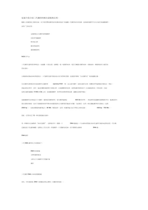
这里为您介绍二代测序的相关流程和应用。
随着人类基因组工程的完成,对于低花费的测序技术的需求促进了高通量二代测序技术的发展。
这些新的测序平台允许进行高通量测序,具有广泛的应用:全基因组从头测序或者重测序目标序列重测序转录组分析微生物组研究基因调控研究NGS序歹y二代测序仪器有很多种组合,在通量、片段长度、准确度、每一轮测序成本、每百万碱基对测序成本、初始成本、规格和技术方面存在存在差异。
从规格和初始成本的角度而言,二代测序仪器可轻松地分类为更窄的范围,也就是所谓的“台式测序仪”和高通量仪器。
台式测序仪使得任何实验室都可以像使用real-time PCR —样,自己进行测序。
这些仪器可以和一些靶标序列富集技术相结合,用在一些临床的应用中,其中:选定的靶标基因用于深度分析,以检测稀有的突变,或者检测多样样本中(比如癌症样本)中的突变。
目前,这些仪器的通量在10 Mb到Gb之间,但是随着硬件,软件和试剂的持续改善,通量也在稳步增加。
高通量测序仪非常适合于大量的,基因组范围的研究,每次测序能测定600 Gb的序列。
一些这样的高通量和高精度的平台,能测定的片段长度相对较短,这对于高重复性的序列和未知基因组的从头测序就可能成为问题。
与此相反,也有一些仪器能测序的片段较长(达到2500 bp ),但是其精度和测序能力(90 Mb)要低很多。
还有一些测序能力位于两者之间的仪器(~800 bp,700 Mb)。
因此,应用决定了哪一种仪器是最合适的。
有一种新的方法被称作“纳米孔测序”。
这种技术中,根据一个DNA链通过一个合成的或者蛋白纳米孔道所引起的电流的改变,可以确定通过这个孔道的碱基。
这理论上可以仅用一步就测序一个完整的染色体,而不需要生成新的DNA链。
DNA测序二代DNA测序的工作流程如下:DNA样本制备文库构建和验证文库分子大规模平行克隆扩增测序二代测序DNA羊本的质量控制首先,评价基因组DNA勺质量是非常必要的(完整性和纯度)。
电化学方法检测DNA碳纳米管修饰电极

应用化学
Vol. 25 No. 9
CH INESE JOURNAL OF APPL IED CHEM ISTRY Sep. 2008
电化学方法检测 D NA碳纳米管修饰电极
龚美娟 a 李 静 a 韩 涛 a 蔡称心 a 陆天虹 a, b 葛存旺 c 杜江燕 a3
© 1994-2009 China Academic Journal Electronic Publishing House. All rights reserved.
1038
应 用 化 学 第 25卷
它试剂均为分析纯 。实验用水均为三次蒸馏水 。
共价键合法目前应用较多的是用盐酸 12乙基 232(32二甲基氨基丙基 )碳二亚胺 ( EDC)和 N 2羟基琥
珀酰亚胺 (NHS)活化剂活化碳纳米管表面的含氧基团 ,以促进 DNA 在 CNTs修饰电极表面的固定 ,但
是对此固定方法进行电化学研究的报道较少 [ 13 ] 。本文将羧基化的 CNTs修饰至 GC 电极表面制成
光敏剂 ,用于肿瘤等疾病的光动力学化学治疗试剂 [9 ] 。此外 , Th具有氧化还原活性 ,电化学方法是研究
Th与 DNA 相互作用的重要手段 [ 10 ] 。但是将 Th作为 DNA 修饰电极的电化学指示剂的报道较少 [ 11 ] , 开
展此方面的研究无疑会对 DNA 修饰电极的电化学研究手段提供更多的选择途径 。
图 2 不同电极在含 Th的磷酸盐缓冲溶液中 的循环伏安曲线
Fig. 2 Cyclic voltammogram s of 20 mmol/L Th in 0. 1 mol/L phosphate buffer solution at ( a) GC,
临床分子生物学检验

第 1~6 章1、现代分子生物学的开端:1953 年,Watson 和 Crick 提出了 DNA 双螺旋构造,标志着现代分子生物学的兴起,为揭开人类生命现象的本质奠定了根底。
2、临床分子生物学检验:是分子生物学技术在临床检验诊断应用中进展起来的,以疾病为中心、以生物分子标志物为靶标的一代临床检验诊断技术,是临床分子生物学的重要组成局部。
3、应用到临床的分子标志物包括基因组 DNA、各种 RNA、蛋白质和各种代谢物。
4、分子标志物:是指可以反映机体生理、病理状态的核酸、蛋白质〔多肽〕、代谢产物等生物分子,是生物标志物的一种类型。
5、核酸分子标志物包括:基因突变,DNA 多态性,基因组 DNA 片段,RNA 和循环核酸等多种形式。
6、DNA 一级构造〔直径,两个碱基之间的距离,一个螺距,一个螺旋有多少个核苷酸〕:DNA 一级构造就是指各核苷酸单体沿多核苷酸链排列的挨次。
7、DNA 二级构造〔右手螺旋—B 型最常见,左手螺旋—Z 型〕:DNA 的二级构造是双螺旋构造,主要特征是①主干链反向平行:DNA 分子是一个由两条平行的脱氧多核苷酸链围绕同一个中心轴盘曲形成的右手螺旋构造,两条链行走方向相反,一条链为5’→3’走向,另一条链为3’→5’走向。
磷酸基和脱氧核糖基构成链的骨架,位于双螺旋的外侧;碱基位于双螺旋的内侧。
碱基平面与中轴垂直。
②侧链碱基互补配对:两条脱氧多核苷酸链通过碱基之间的氢键连接在一起。
DNA 双螺旋的直径为 2nm,一圈螺旋含 10 个碱基对〔一个螺旋有 20 个核苷酸〕,每一碱基平面的轴向距离为 0.34nm,故每一螺距为 3.4nm,每个碱基的旋转角度为36°。
8、DNA 三级构造〔真核生物 DNA 三级构造是染色质或染色体〕:DNA 双螺旋进一步盘曲形成更加简单的构造,称为三级构造。
超螺旋是 DNA 三级构造的最常见的形式。
9、真核生物的 DNA 形成染色质的包装过程〔4 步〕:①形成核小体:构成染色质的根本单位是核小体。
dna体外扩增技术总结

dna体外扩增技术总结《dna体外扩增技术总结》是一篇好的范文,好的范文应该跟大家分享,希望大家能有所收获。
目的的获取及体外扩增技术概述在基因的研究及体外人工复制过程中,我们通常把需要研究的基因称为目的基因。
基因工程流程的第一步就是获得目的DNA片段,如何获得目的DNA片段就成为基因工程的关键问题。
一般来说,目的基因的克隆战略分为两大类:一类是构建感兴趣的生物个体的基因文库,即将某生物体的全基因组分段克隆,然后建立合适的筛选模型从基因组文库中挑出含有目的基因的重组克隆;另一类是利用PCR扩增技术甚至化学法体外直接合成目的基因,然后将之克隆表达,常用的有PCR法、cDNA法及建立基因文库等。
1.PCR法技术及原理聚合酶链式反应(Polymerase Chain Reaction),简称PCR,是一种分子生物学技术,用于放大特定的DNA片段。
它是一种体外酶促合成,扩增特定 DNA片段的方法。
1985年由美国Karray等学者首创了PCR技术,并由美国Cetus公司开发研制。
随着科学技术的发展和突破,PCR技术已在多个领域得到广泛地应用,如微生物检测、兽医学、水产养殖等方面。
PCR技术模拟DNA的天然复制过程,基于DNA的半保留复制原理,其特异性依赖于与靶两端互补的寡核苷酸引物,该原理主要包括3个基本反应过程:变性——退火——延伸。
变性主要是指双链DNA的变性,即双链DNA经加热至93℃左右,其热稳定性降低,配对碱基之间的氢键断裂,使双链DNA成为单链,以便与引物结合。
退火是指单链DNA在温度降低时与引物的复性(称为退火)。
在此过程中,引物与单链DNA模板仍然按照碱基互补的方式配对。
延伸是指引物与DNA模板按碱基互补的原则配对后,在TaqDNA聚合酶的作用下,以dNTP为反应原料,进行体外的半保留复制,合成一条新的与模板DNA链互补的新链。
经重复循环变性——退火——延伸3个过程,就可获得更多的“半保留复制链”,而且这种新链又可成为下次循环的模板。
- 1、下载文档前请自行甄别文档内容的完整性,平台不提供额外的编辑、内容补充、找答案等附加服务。
- 2、"仅部分预览"的文档,不可在线预览部分如存在完整性等问题,可反馈申请退款(可完整预览的文档不适用该条件!)。
- 3、如文档侵犯您的权益,请联系客服反馈,我们会尽快为您处理(人工客服工作时间:9:00-18:30)。
Cite this:DOI:10.1039/c1cc14902d Target recycling amplification for sensitive and label-free impedimetric genosensing based on hairpin DNA and graphene/Au nanocomposites wYing Chen,a Bingying Jiang,b Yun Xiang,*a Yaqin Chai a and Ruo Yuan*aReceived 8th August 2011,Accepted 27th October 2011DOI:10.1039/c1cc14902dThe presence of exonuclease III leads to direct recycling and reuse of the target DNA,which in turn results in substantial signal amplification for highly sensitive,label-freeimpedimetric detection of specific DNA sequences.Graphene is a two-dimensional single layer of sp 2-hybridized carbon sheets.1Since its first isolation in 2004,graphene has attracted great research attention due to its unique properties,such as high surface area,2,3amazing intrinsic mobility,excellent thermal and electrical conductivity.4,5Recently,graphene and graphene-based materials have been increasingly used in biosensor applications.One of the major goals of the biosensor technology is to determine specific DNA sequences for various applications in drug discovery,diagnosis of diseases,forensic and food technology.Currently,specific and sensitive detection of DNA is mainly based on the transduction of hybridization of complementary DNA strands by fluorescence,6–8quartz microbalance,9,10surface plasmon resonance spectroscopy,11,12atomic force microscopy 13and electrochemical (EC)techniques.14–16Among these transduction methods,EC DNA sensors have shown promising potentials for point-of-care diagnostics and other important applications.Being highly sensitive and label-free,electrochemical impedance spectroscopy (EIS)has gained more and more attention as a powerful tool for probing molecular binding events such as DNA hybridizations.17–19The formation of the complexes upon bioaffinity bindings commonly changes the interfacial electron transfer kinetics between the redox probe and the electrode and generates quantitative signal outputs monitored by EIS for biosensing.However,one of the challenges in EIS-based detections is to amplify the signal mon routes for amplified EIS detection of a trace amount of target molecules require modifications of either the target or the probe molecules with labels,20–23which sacrifices the inherent advantages (e.g.simplicity and label-free capability)of this technique.In response,we reportherein a convenient signal amplification strategy for highly sensitive,label-free EIS determination of DNA sequences based on enzymatic target recycling and graphene/AuNP nanocompo-sites,as well as the hairpin DNA probes.The introduction of the catalytic enzyme,exonuclease III (Exo III),into the sensing system leads to direct recycling and reuse of the target DNA,which in turn amplifies the EIS signal.This enables amplified EIS detection of a trace level of specific DNA sequences in a convenient and truly label-free fashion.Our enzyme-assisted target recycling amplification strategy for sensitive and label-free EIS detection of DNA is illustrated in Scheme 1.This involves a one-step EC reduction of a mixture of graphene oxide and HAuCl 4to form graphene/AuNP nanocomposites on a screen printed carbon electrode (see ESI w for details),self-assembly of the hairpin DNA probes on the modified electrode and surface blocking with 6-mercapto-1-hexanol (MCH),addition of the target DNA together with Exo III and EIS measurement of the resulting electrode.The target DNA present hybridizes with the hairpin probe to form a double stranded DNA.At the same time,Exo III specifically cleaves the open,hybridized hairpin DNA and releases the target DNA because of the unique enzymatic property of Exo III,which catalyzes the stepwise removal of mononucleotides from the blunt or recessed 30-hydroxyl termini of a duplex DNA and is inactive to single stranded DNA.Upon enzymatic cleavage of the probe,the released target DNA again hybridizes with the remaining hairpin DNA probe and triggers another target recycling cycle.Therefore,a smallScheme 1Illustration of the enzyme-assisted target recycling for amplified EIS detection of DNA on a graphene/AuNP modified electrode.aKey Laboratory on Luminescence and Real-Time Analysis,Ministry of Education,School of Chemistry and ChemicalEngineering,Southwest University,Chongqing 400715,P.R.China.E-mail:yunatswu@,yuanruo@;Fax:+86-23-68252277;Tel:+86-23-68253172bSchool of Chemistry and Chemical Engineering,Chongqing University of Technology,Chongqing 400054,P.R.Chinaw Electronic supplementary information (ESI)available:Preparation of LBL assemblies and ECL detections.See DOI:10.1039/c1cc14902dChemCommDynamic Article Links/chemcommCOMMUNICATIOND o w n l o a d e d b y S H A N D O N G U N I VE R S I T Y o n 12 N o v e m b e r 2011P u b l i s h e d o n 11 N o v e m b e r 2011 o n h t t p ://p u b s .r s c .o r g | d o i :10.1039/C 1C C 14902DView Online / Journal Homepageamount of target DNA is expected to efficiently remove a large number of hairpin DNA probes from the electrode surface,leading to a substantial decrease in electron transfer resistance(R et )monitored by EIS.The decrease in R et can thus be related to the quantity of the target DNA in the testing samples.The stepwise sensor fabrication process is monitored by EIS and the resulting Nyquist plots are displayed in Fig.1.The EIS data were fitted to a Randles equivalent circuit (inset in Fig.1),which includes the solution resistance (R s ),R et ,the constant phase element (Q dl )and Warburg impedance (W ).In the Nyquist diagram,the diameter of the semicircle reflects the R et of redox conversion of the electroactive marker [Fe(CN)6]3À/4Àon the electrode at certain applied potential.This process is strongly dependent upon any modification to the electrode surface.As can be seen in Fig.1,the R et value decreases significantly after the deposition of the graphene/AuNPs on the electrode surface.This dramatic decrease in R et can be primarily attributed to the excellent electrical conductivity of both the electrochemically reduced graphene oxide 24and AuNPs,25which promotes the electron transfer from the electroactive markers to the electrode.However,after self-assembly of a mixed monolayer of the hairpin DNA probes and MCH on the graphene/AuNP modified electrode,a sub-stantial increase in R et is observed.Such an increase in R et is basically due to two facts.First,the negative charges on the phosphate backbone of the surface immobilized hairpin probes repel [Fe(CN)6]3À/4Àfrom the electrode,leading to an increase in R et .Second,the steric hinderance of the hairpin structure of the probe prevents [Fe(CN)6]3À/4Àfrom approaching the electrode and causes increase in R et .26The combination of these two effects thus results in a substantial increase in R et after formation of the mixed monolayer on the electrode.In order to demonstrate the dramatic signal amplification capability of the proposed label-free assay protocol,we compared our enzyme-assisted target recycling detection approach with the conventional method.From the Nyquist plots in Fig.2,we can see that the presence of 500pM target DNA (without Exo III)results in a small drop (20.7%)in R et (curve b vs.curve a),which is due to the decrease in the steric hinderance of thehairpin DNA probe upon hybridization and easy approaching of [Fe(CN)6]3À/4Àto the electrode.27While in the presence of both target DNA (500pM)and Exo III (5U),the target DNA can be used multiple times and more hairpin DNA probes are removed from the surface due to the catalytic target recycling by Exo III.A substantial decrease (60.0%)in R et is observed (curve c vs.curve a).When no target DNA is present in the testing sample,the addition of Exo III alone causes a negligible effect on R et (curve d).These results clearly suggest significant signal amplification by the incorporation of the enzyme-assisted target recycling in EIS detection of DNA.EIS measurements of the target DNA at various were used to determine the detection limit and dynamic range of the proposed sensor.Typical Nyquist plots of the sensor before and after incubation with the target DNA are displayed in Fig.3.It is obvious that the R et value shows a dependence upon the concentration of the target DNA (Fig.3A).As the concentration of the target DNA increases,the R et value decreases accordingly.The corresponding calibration plot (Fig.3B)of log c vs.D R et (the difference before and after incubation with the target DNA)exhibits a dynamic range from 50fM to 5nM and the limit of detection could be estimated to be 10fM based on the 3s rule.This detection limit is about 20–500-fold than other universal EIS detection strategies.26,28–33Such a low detection limit is primarily due to the involvement of the target recycling signal amplification in the assay protocol.The reproducibility of the proposed method was evaluated by performing a series of six repetitive experiments for the target sequence at the concentration of 500pM,Fig.1Nyquist diagrams for (a)bare SPCE electrode,(b)graphene/AuNP modified SPCE and (c)hairpin DNA and MCH self-assembled graphene/AuNPs/SPCE.Inset:equivalent circuit used to fit the EIS data.EIS measurements were carried out in 0.1M KCl solution containing 5mM (1:1)[Fe(CN)6]3À/4Àwith the range from 10kHz to 50MHz and an alternate voltage of 5mV.Fig.2Nyquist diagrams for sensors incubated with (a)buffer,(b)complementary DNA (500pM),(c)complementary DNA (500pM)with Exo III (5U)and (d)Exo III (5U).Fig.3(A)Nyquist diagrams for sensors incubated with different concentrations of target DNA:(a)5nM,(b)500pM,(c)50pM,(d)5pM,(e)500fM,(f)50fM,(g)blank (0fM target).(B)The resulting calibration plot of log c vs.D R et for the target DNA over the 50fM to 5nM range (error bars:SD,n =3)D o w n l o a d e d b y S H A N D O N G U N I VE R S I T Y o n 12 N o v e m b e r 2011P u b l i s h e d o n 11 N o v e m b e r 2011 o n h t t p ://p u b s .r s c .o r g | d o i :10.1039/C 1C C 14902Dwhich yielded a relative standarddeviation of 7.3%.This indicates that our assay protocol is coupled with good reproducibility.The selectivity of the sensor was examined by incubation with complementary,non-complementary and base mismatch DNAs and the EIS results are shown in Fig.4.The addition of 500pM non-complementary DNA (Fig.4b)to the sensor causes insignificant decrease in R et compared to the blank test (Fig.4a).Despite that the presence of 500pM one-base and three-base mismatch DNAs leads to decrease in R et ,it is not comparable with that of the presence of 500pM target DNA.These results indicate that the label-free impedimetric sensor could effectively distinguish target DNA from non-complementary and base mismatch DNA.Such discrimination between target and non-target DNA suggests the inherent high selectivity of the hairpin DNA probes 26as well as the minimized non-specific adsorption by the surface blocking 34,35and washing.In summary,we have demonstrated a convenient signal amplification strategy for sensitive and selective EIS detection of DNA target on a graphene/AuNP modified electrode.Our approach couples the high sensitivity of the EIS technique with the enzyme-assisted target recycling signal amplification to achieve low femtomolar detection of target gene sequences.Moreover,unlike other endonucleases such as nicking endo-nuclease and Fok I enzyme,which require a specific sequence for enzyme cleavage,Exo III is sequence independent.This unique property of Exo III makes the proposed sensor hold great potential for highly sensitive,selective and simple detection of a wide range of target DNA sequences.This work is supported by National Science Foundation of China (No.20905062and 20675064),Natural Science Foundation Project of Chongqing City (CSTC-2009BB5052),State Key Laboratory of Electroanalytical Chemistry (SKLEAC2010009),China Postdoctoral Science Foundation (20090460715and 201003305),Scientific Research Foundation for the Returned Overseas Chinese Scholars and research funds from Southwest University (SWUB2008078).Notes and references1K.S.Novoselov,A.K.Geim,S.V.Morozov,D.Jiang,Y.Zhang,S.V.Dubonos,I.V.Grigorieva and A.A.Firsov,Science ,2004,306,666.2K.Geim and K.S.Novoselov,Nat.Mater.,2007,6,183.3S.Park and R.S.Ruoff,Nat.Nanotechnol.,2009,4,217.4A.A.Balandin,S.Ghosh,W.Z.Bao,I.Calizo,D.Teweldebrhan,F.Miao and u,Nano Lett.,2008,8,902.5R.F.Service,Science ,2009,324,875.6J.Reichert,A.Csaki,J.M.Kohler and W.Fritzsche,Anal.Chem.,2000,72,6025.7S.Bi,H.Zhou and S.S.Zhang,Chem.Sci.,2010,1,681.8T.A.Taton,G.Lu and C.A.Mirkin,J.Am.Chem.Soc.,2001,123,5164.9F.Patolsky,A.Lichtenstein and I.Willner,J.Am.Chem.Soc.,2001,123,5194.10X.Su,R.Robelek,Y.Wu,G.Wang and W.Knoll,Anal.Chem.,2004,76,489.11C.E.Jordan,A.G.Frutos,A.J.Thiel and R.M.Corn,Anal.Chem.,1997,69,4939.12B.P.Nelson,T.E.Grimsrud,M.R.Liles,R.M.Goodman and R.M.Corn,Anal.Chem.,2001,72,1.13Z.L.Zhang,D.W.Pang,H.Yuan,R.X.Cai and H.Abruna,Anal.Bioanal.Chem.,2005,381,833.14F.R.R.Teles and L.P.Fonseca,Talanta ,2008,77,606.15E.M.Boon, D.M.Ceres,T.Drummond,M.G.Hill and J.K.Barton,Nat.Biotechnol.,2001,18,1096.16J.Wang,X.Cai,G.Rivas and H.Shiraishi,Anal.Chim.Acta ,1996,326,141.17J.Y.Park and S.M.Park,Sensors ,2009,9,9513.18E.Katz and I.Willner,Electroanalysis ,2003,15,913.19C.Berggren, B.Bjarnason and G.Johansson,Electroanalysis ,2001,13,173.20A.Bardea,F.Patolsky,A.Dagan and I.Willner,mun.,1999,21.21F.Patolsky,K.T.Ranjit,A.Kichtenstein and I.Willner,mun.,2000,1025.22F.Patolsky,A.Lichtenstein and I.Willner,Angew.Chem.,Int.Ed.,2000,39,940.23W.M.Hassen,C.Chaix,A.Abdelghani,F.Bessueille,D.Leonard and N.Jaffrezic-Renault,Sens.Actuators,B ,2008,134,755.24M.Zhou,Y.M.Zhai and S.J.Dong,Anal.Chem.,2009,81,5603.25Y.Ding,M.Chen and J.Erlebacher,J.Am.Chem.Soc.,2004,126,6876.26A.Bonanni and M.Pumera,ACS Nano ,2011,5,2356.27M.Steichem and C.Buess-Herman,mun.,2005,7,416.28Y.W.Hu,F.H.Li,X.X.Bai,D.Li,S.C.Hua,K.K.Wang and L.Niu,mun.,2011,47,1743.29N.Zhou,T.Yang,C.Jiang,M.Du and K.Jiao,Talanta ,2009,77,1021.30A.Bonanni,M.J.Esplandiu and M.Del-Valle,Biosens.Bioelectron.,2009,24,2885.31C.Y.Deng,J.H.Chen,Z.Nie,M. D.Wang,X. C.Chu,X.L.Chen,X.L.Xiao,C.Y.Lei and S.Z.Yao,Anal.Chem.,2009,81,739.32W.Zhang,T.Yang,X.Zhuang,Z.Guo and K.Jiao,Biosens.Bioelectron.,2009,24,2417.33Y.Wang,C.J.Li,X.H.Li,Y.F.Li and H.B.Kraatz,Anal.Chem.,2008,80,2255.34T.Herne and M.Tarlov,J.Am.Chem.Soc.,1997,119,8916.35A.Steel,T.Herne and M.Tarlov,Anal.Chem.,1998,70,4670.Fig.4Selectivity investigations against non-target DNA molecules:(a)blank,(b)non-complementary DNA (500pM),(c)three-base mismatch DNA (500pM),(d)one-base mismatch DNA (500pM)and (e)complementary DNA (500pM).D o w n l o a d e d b y S H A N D O N G U N I VE R S I T Y o n 12 N o v e m b e r 2011P u b l i s h e d o n 11 N o v e m b e r 2011 o n h t t p ://p u b s .r s c .o r g | d o i :10.1039/C 1C C 14902D。