
银染操作步骤(精)
银染操作步骤
1. 固定:
电泳结束后,取凝胶放入约 100ml 固定液中,在摇床上室温摇动 20分钟,摇动速度为 60-70rpm 。
固定 40分钟以上甚至过夜可以进一步降低背景。
固定液的配制:依次加入 50ml 乙醇、 10ml 乙酸和 40ml MilliQ级纯水或双蒸水,混匀后即成 100ml 固定液,可大量配制室温存放。
2. 30%乙醇洗涤:
弃固定液,加入 100ml 30%乙醇,在摇床上室温摇动 10分钟,摇动速度为 60-
70rpm 。
30%乙醇的配制:70ml MilliQ级纯水或双蒸水中加入 30ml 乙醇,混匀后即成 100ml 30%乙醇。
3. 水洗涤:(洗 2次
弃 30%乙醇,加入 200ml MilliQ级纯水或双蒸水,在摇床上室温摇动 10分钟,摇动速度为 60-70rpm 。
4. 增敏:(辅染现配现用
弃水,加入 100ml 银染增敏液 (1X,在摇床上室温摇动 2分钟,摇动速度为 60-
70rpm 。
银染增敏液 (1X的配制:99ml MilliQ级纯水或双蒸水中加入 1ml 银染增敏液 (100X,混匀后即为银染增敏液 (1X。
银染增敏液 (1X配制后需在 2小时内使用。
银染增敏液 (100X的配制:即 2.8%硫代硫酸钠溶液,需用黑袋包裹避光保存。
5. 水洗涤 (共 2次 :
弃原有溶液,加入 200ml MilliQ级纯水或双蒸水,在摇床上室温摇动 1分钟,摇动速度为 60-70rpm 。
弃水,再加入 200ml MilliQ级纯水或双蒸水,在摇床上室温摇动 1分钟,摇动速度为 60-70rpm 。
6. 银染:
弃水,加入 100ml 银溶液 (1X,在摇床上室温摇动 10分钟,摇动速度为 60-
70rpm 。
银溶液 (1X的配制:99ml MilliQ级纯水或双蒸水中加入 1ml 银溶液 (100X,混匀后即为银溶液 (1X。
银溶液 (1X配制后需在 2小时内使用, 最好是现配现用。
银溶液(100 X的配制:称取 5g 硝酸银,加水溶解并稀释至 500ml ,摇匀即得 1%的硝酸银溶液,需用黑袋包裹避光保存。
7. 水洗涤:
弃原有溶液,加入 100ml MilliQ级纯水或双蒸水,在摇床上室温摇动 1-1.5分钟,摇动速度
为 60-70rpm 。
注意:水洗涤的时间不能超过 1.5分钟。
8. 显色:
弃水,加入 100ml 银染显色液,在摇床上室温摇动 3-10分钟,直至出现比较理想的预期蛋白条带,摇动速度为 60-70rpm 。
银染显色液的配制:90ml MilliQ级纯水或双蒸水中加入 10ml 银染基本显色液(10X,再加入 0.05ml 银染显色加速液 (2000X, 混匀后即为银染显色液。
银染显色液配制后需在 20分钟内使用。
银染基本显色液 (5X的配制:称取碳酸钠 60g ,加水溶解并稀释至 500ml ,摇匀即得 12%碳酸钠溶液。
9. 终止:加适量 EDTA-2Na(乙二胺四乙酸二钠终止
弃银染显色液, 加入 100ml 银染终止液 (1X, 在摇床上室温摇动 10分钟, 终止时有气体产生属正常现象,产生的气体为二氧化碳。
银染终止液 (1X的配制:95ml MilliQ级纯水或双蒸水中加入 5ml 银染终止液(20X,混匀后即为银染终止液 (1X。
银染终止液 (1X配制后宜当天使用。
10. 水洗涤:
弃银染终止液,加入 100ml MilliQ级纯水或双蒸水,在摇床上室温摇动 2-5分钟,摇动速度为 60-70rpm 。
11. 保存:
可在 MilliQ 级纯水或双蒸水中保存。
或采用适当的方式制备成干胶。
常见问题:
1. 背景太深:
a. 显色时间过长。
通常显色反应会在 10分钟内结束,显色反应时间过长会导致背景很深。
b. 洗涤不充分。
洗涤时间过短,或洗涤液加入的量不足,或者容器过于狭小导致摇动时溶液不易充分混合, 或摇动速度过慢, 导致混匀不充分。
请按照说明书的建议确保各种溶液的用量和作用时间,摇床的推荐速度为 60-70rpm 。
c. 凝胶中原有的缓冲液等未在固定步骤中去除干净。
一方面需确保固定的时间和固定液的用量,另一方面对于不是最常用的 Bis-Tris 缓冲的凝胶需要更长的固定时间以充分去除凝胶中的原有缓冲成分,以降低背景。
d. 水的纯度太低。
需使用大于16 MΩ·cm的高纯度水。
2. 蛋白条带非常浅:
a. 蛋白的半胱氨酸 (Cysteine残基的含量特别低或几乎没有。
半胱氨酸残基的存在对于银染非常重要,半胱氨酸残基的含量过低会导致检测灵敏度下降。
b. 银染后水洗涤时间过长。
在银溶液染色时需严格控制水洗涤的时间,水洗涤的时间不能超过 1.5分钟,否则会导致过多的银离子被洗去,导致检测灵敏度下降。
c. 上样量不足。
本试剂盒检测 BSA 的灵敏度可以达到 0.3ng , 对于不同的蛋白检测灵敏度可能不同。
对于一些蛋白可能需要大于 1ng 的蛋白量才能被检测到。
d. 固定步骤后的洗涤不够充分。
导致少量乙酸残留,影响后续检测。
确保 30%乙醇洗涤和水洗涤的用量和时间,可以适当延长洗涤时间。
3. 凝胶上出现小点或或其它非蛋白的痕迹:
a. 凝胶没有充分被溶液浸没。
请注意选择大小合适的容器,并加入足量的各种溶液,同时需保持适当的混匀速度确保凝胶可以被溶液浸没。
b. 用于银染的容器没有充分洗涤干净。
容器需先用洗涤剂充分洗涤,随后用自来水充分冲洗, 最后用高纯度水再洗涤数次。
该容器最好能专用于银染, 并注意避免各种可能的蛋白污染。
为确保充分洗涤干净,对于耐硝酸的容器,例如玻璃容器,可以在上述洗涤剂及自来水洗涤后用 50%硝酸洗涤,随后用高纯度水充分洗涤。
c. 指纹或其它压痕。
请注意戴手套操作,切勿直接接触皮肤。
操作时请注意尽量勿挤压、折叠或摩擦凝胶。
d. 有金属物质接触凝胶。
金属物质例如金属镊子等接触凝胶会出现非特异性痕迹。
4. 在 60-70 kD处出现一片模糊的蛋白染色背景:
皮肤上脱落的角蛋白 (keratin污染了蛋白样品。
一方面需注意戴手套操作, 另一方面需注意盛放蛋白样品的容器盖子尽量不要敞开, 甚至在取放蛋白样品时在超净台内进行以避免可能的角蛋白污染。
5. 在凝胶的顶端处出现黄色背景:
a. 样品中 DTT 浓度很高。
采用其它适当的还原试剂,或者在许可范围内适当减少 DTT 的用量。
b. 采用 Tris-Glycine-SDS 电泳体系。
Tris-Glycine-SDS 电泳体系中的 Glycine 会导致背景凝胶的顶端出现轻微的黄色背景。
换用 Tris-Tricine-SDS 电泳体系则可显著消除此黄色背景。
6. 银在染色器皿中出现沉淀:
染色器皿中可能含有残余的洗涤剂或上次银染时的残余试剂。
需确保把染色器皿洗涤干净。
DNA和蛋白银染步骤(独门绝技--有图)

DNA银染一、银染试剂500ml/瓶(双蒸水稀释)1、固定液:50ml乙醇2、前处理液(敏化液):5ml HNO33、染色液:0.5g 硝酸银(避光配制)4、显色液:无水Na2CO3 12.5g 甲醛200ul 加水至500ml5、终止液:50ml 冰醋酸二、银染步骤(摇床设置52rpm左右)1、准备好放有双蒸水的塑料盒,将电泳后的变性胶放入盒中在摇床上轻摇1min;2、凝胶固定:倒掉盒中的水,加入100ml左右的固定液,摇床上缓缓摇动10min;3、洗胶:弃固定液,用双蒸水洗2次,每次30S;4、凝胶氧化:弃双蒸水,加入100ml左右前处理液氧化6min;5、洗胶:弃前处理液,用双蒸水洗2次每次10S;6、凝胶染色:弃双蒸水,加入100ml左右的染色液避光摇20min;7、洗胶:弃染色液,用双蒸水洗1次10S;8、凝胶显色:加入100ml显色液摇到看到清晰的条带为止;9、终止:弃显色液,迅速加入终止液100ml左右,摇2-3min;10、洗胶:弃终止液,用双蒸水洗2次每次1min。
三、变性聚丙酰胺凝胶的配制(两块、使用1.0BioRad胶板)1.2%的凝胶可以跑微卫星,和基因组DNA1、尿素:7.2g2、30%聚丙酰胺,4.68ml3、10×TBE,1.5ml (Tris碱108g;硼酸55g;EDTA 40ml 0.5mol/L,Ph=8.0;加水至1L)4、30%AP,35ul5、TEMED,4ul四、下图:左为果蝇正常基因组DNA与损伤后基因组DNA;右为果蝇微卫星电泳12h Treatment24h TreatmentMCT 0 5 10 200 5 10 20Protein银染一、银染试剂500ml/瓶(双蒸水稀释)1、固定液:200ml甲醇,50ml冰醋酸2、前处理液(敏化液):150ml 乙醇,34g醋酸钠,1g硫代硫酸钠(使用前加入)3、染色液:1.25g 硝酸银(避光配制)4、显色液:无水Na2CO3 12.5g 甲醛200ul 加水至500ml5终止液:2g甘氨酸二、银染步骤(摇床设置52rpm左右)1、凝胶固定:准备好放有双蒸水的塑料盒,将电泳后的变性胶放入盒中,加入100ml左右的固定液,摇床上缓缓摇动30min;2、洗胶:弃固定液,用双蒸水洗2次,每次1min;3、凝胶氧化:弃双蒸水,加入100ml左右前处理液氧化30min;4、洗胶:弃前处理液,用双蒸水洗3次每次5min;5、凝胶染色:弃双蒸水,加入100ml左右的染色液避光摇20min;6、洗胶:弃染色液,用双蒸水洗2次每次5min;7、凝胶显色:加入100ml显色液摇到看到清晰的条带为止;8、终止:弃显色液,迅速加入终止液100ml左右,摇10min;9、洗胶:弃终止液,用双蒸水洗2次每次5min。
鞭毛染色液(银染法)
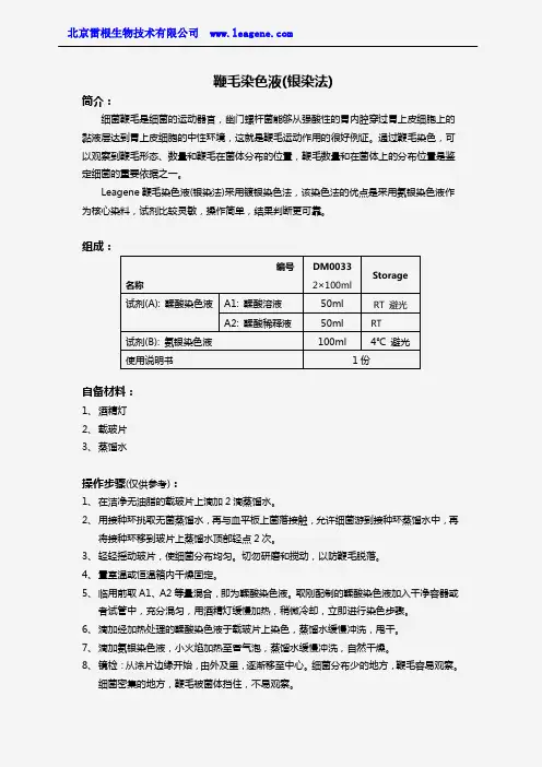
鞭毛染色液(银染法)简介:细菌鞭毛是细菌的运动器官,幽门螺杆菌能够从强酸性的胃内腔穿过胃上皮细胞上的黏液层达到胃上皮细胞的中性环境,这就是鞭毛运动作用的很好例证。
通过鞭毛染色,可以观察到鞭毛形态、数量和鞭毛在菌体分布的位置,鞭毛数量和在菌体上的分布位置是鉴定细菌的重要依据之一。
Leagene 鞭毛染色液(银染法)采用镀银染色法,该染色法的优点是采用氨银染色液作为核心染料,试剂比较灵敏,操作简单,结果判断更可靠。
组成:自备材料:1、 酒精灯2、 载玻片3、 蒸馏水操作步骤(仅供参考):1、 在洁净无油脂的载玻片上滴加2滴蒸馏水。
2、 用接种环挑取无菌蒸馏水,再与血平板上菌落接触,允许细菌游到接种环蒸馏水中,再将接种环移到玻片上蒸馏水顶部轻点2次。
3、 轻轻摇动玻片,使细菌分布均匀。
切勿研磨和搅动,以防鞭毛脱落。
4、 置室温或恒温箱内干燥固定。
5、 临用前取A1、A2等量混合,即为鞣酸染色液。
取刚配制的鞣酸染色液加入干净容器或者试管中,充分混匀,用酒精灯缓慢加热,稍微冷却,立即进行染色步骤。
6、 滴加经加热处理的鞣酸染色液于载玻片上染色,蒸馏水缓慢冲洗,甩干。
7、 滴加氨银染色液,小火焰加热至冒气泡,蒸馏水缓慢冲洗,自然干燥。
8、 镜检:从涂片边缘开始,由外及里,逐渐移至中心。
细菌分布少的地方,鞭毛容易观察。
细菌密集的地方,鞭毛被菌体挡住,不易观察。
编号 名称 DM0033 2×100ml Storage 试剂(A): 鞣酸染色液 A1: 鞣酸溶液 50ml RT 避光 A2: 鞣酸稀释液 50ml RT 试剂(B): 氨银染色液 100ml 4℃ 避光 使用说明书 1份染色结果:菌体深褐色,鞭毛为褐色。
注意事项:1、玻片应洁净,无油污。
2、染色的菌种应连续传代多次,处于生长活跃期。
3、染色过程应小心操作,防止鞭毛脱落。
4、配制好的鞣酸染色液不宜久置,尽量在10min内使用。
5、氨银染色液不稳定,应严格4℃避光保存,出现严重浑浊时应弃用。
基本银染和微波快速银染步骤-中文翻译

基本的银染步骤:(90min)材料:超纯水(>18 megohm/cm全程)、塑料染色托盘、摇床、塑料搅拌棒、10mL 一次性管、干净的玻璃杯、量筒、30%乙醇、100%乙醇、固定液(40%乙醇、10%乙酸)注意事项:1.在拿胶前确定用去离子水清洗橡胶手套。
2.用干净的容器,并标明银染专用。
3.当摇动时,确保容器大小可使胶在里面自由移动,染色时溶液能完全沫过胶面。
4.在拿胶或换溶液时,避免徒手摸胶或用金属物品接触,及避免对胶施加压力。
5.用有铁氟龙图层的棒和干净的玻璃容器准备试剂。
6.避免试剂交叉污染。
7.用新鲜配置的溶液。
样品含有高浓度的DTT:如果你的样品含有高浓度的DTT (>50mM),银染时可能导致条带拖尾和黄色背景。
为避免拖尾按照下面的方法还原和烷基化样品:1. 用新鲜配制的DTT还原你的样品到终浓度为17mM,并在70℃加热10min.2. 用新鲜配制的碘乙酰胺烷基化你的样品到终浓度为35mM,并在70℃加热10min.3. 加不含DTT的SDS样品buffer到还原和烷基化后的样品中。
进行电泳,并按手册进行银染。
在开始之前:用试剂盒中的试剂准备如下溶液用于银染:程序:下面的程序用于8*8厘米预制胶,1.0mm厚。
如果要染两块小胶或1.0mm厚的大胶(18*18),保证孵育时间,将所有溶液的体积加倍。
注意:如果你的胶的尺寸和上面提到的不一样,你可能必须通过调整孵育时间和溶液体积来选择银染策略。
所有的孵育应该在一个旋转摇床上进行,以1圈/秒的速度在室温下进行。
确保每个胶有100ml的溶液。
1. 电泳后,将胶取出至合适尺寸的银染托盘内,用超纯水简单的清洗一下。
2. 用100ml固定液在摇床上缓慢旋转,固定胶20min。
如果你用的是Tricine 胶,那么就固定1小时。
注意:如果没有足够的时间完成银染程序,胶可以在固定液中存放一夜。
长时间的固定在某些情况下可能会改善灵敏度和背景。
快速银染银染步骤及相关药品配制

快速银染银染步骤及相关药品配制银染步骤及相关药品配制一.银染基本步骤1.洗板和擦板电泳槽板先用无水乙醇擦一遍,5分钟后用配制的剥离硅烷(repel)溶液涂抹均匀。
放置15-30分钟玻板(用过的玻板先用NaOH溶液浸泡,直至废胶脱落)先用抹布蘸少许洗洁精在自来水下洗净,再用无水乙醇擦一遍,5分钟后用新鲜配制的亲和硅烷(binding)溶液涂抹均匀。
放置15-30分钟2.装板玻板在下,电泳槽板在上,中间放两根压条(涂抹repel和binding的面相互靠近),对齐后,用三对夹子将波板与电泳槽板夹紧。
3.灌胶用针筒吸取6%的聚丙烯酰胺凝胶溶液,缓慢均匀的从灌胶口将胶灌入,灌好后取下靠近灌胶口的夹子,倒插入梳子,注意不要起汽泡,插好后,再夹上夹子。
凝胶需要1-2个小时。
4.预电泳取下夹子,拔出倒插的梳子,在自来水下将灌胶口冲洗干净,放入电泳槽,卡紧,灌入缓冲液,上下槽各300ml左右,注意用夹子夹紧上槽排放缓冲液的胶管。
80W衡功率电泳半小时。
5.点样用吸管吹干净灌胶口的废胶,将梳子顺插入灌胶口,开始点样,注意不要串样,梳齿不要将胶面弄坏。
6.电泳80W衡功率电泳1小时左右。
7.银染将玻板放入配制好的银染液中,银染半小时左右10.显影从银染液中拿出玻板快速水洗3-5秒,然后放入新鲜配制的显影液中,室温显影,直至条带清晰11.用自来水轻轻冲洗板子正反面几次,洗净残余的NaOH。
二.药品配制1.6%聚丙烯酰胺凝胶(PA):Urea: 420.42gAcr: 60g 丙烯酰胺Bis: 3.16g10×TBE: 50ml加超纯水定容至 1L8% 聚丙烯酰胺PA:尿素 420.42gAcr: 80gBis: 4.212g10*TBE: 100ml加超纯水定容至 1L2. 10×TBETris-base: 216g硼酸:110gEDTA-Na2:14.88g3. binding(亲和硅烷)无水乙醇:2mlBinding原液:8ul冰醋酸:8ul4.Repel (剥离硅烷)三氯甲烷:100mlRepel:2ml5.Loading buffer:甲酰氨:49ml10mM EDTA(PH=8.0):1ml溴酚蓝:0.125g二甲苯青:0.125g7.银染液蒸馏水:1500ml无水乙醇:150ml硝酸银溶液(1g/ml):1500ul 8.显影液蒸馏水:1500ml氢氧化钠:30g甲醛:6ml硫代硫酸钠(10mg/ml):300ul。
蛋白银染
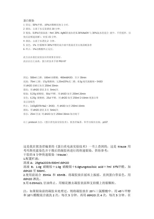
蛋白银染1 固定:50%甲醇,10%冰醋酸浸泡1小时。
2 漂洗:去离子水反复漂洗30分钟。
3 银染:0.8%的银染液(4ml 20% AgNO3滴加到0.36%NaOH与30%氨水的混合液中,不停搅拌,以使沉淀顺速溶解),轻摇15分钟。
4 漂洗:去离子水漂洗2 分钟。
5 显色:1% 柠檬酸和30%甲醛的混合液中震荡直至出现清晰条带6 终止:1%冰醋酸终止反应。
此方法在我们实验室应用效果非常好。
此法出自王家政,蛋白质技术手册P92-97固定:500ml乙醇,100ml冰醋酸,400mldH2O,至少30min浸泡:75ml乙醇,17g醋酸钠,1.25ml25%戊二醛,0.5g硫代硫酸钠·5H2O用dH2O溶解后加至250ml 30min漂洗:用dH2O漂洗3次5min/次银染:0.25g硝酸银,50ul甲醛,用dH2O加至250ml 20min显色:6.25g 碳酸钠,25ul甲醛,用dH2O加至250ml 2-10min视蛋白带显示深棕色终止:3.65gEDTA-Na2·2H2O,用dH2O加至250ml 10min漂洗:用dH2O漂洗3次5min/次保存:25ml甘油用dH2O加至250ml 30min取出晾干以上protocol出自:《蛋白质电泳实验技术》,郭尧君编著,科学出版社出版,p107这是我在郭尧君编著的《蛋白质电泳实验技术》一书上查到的,这是Krause用考玛斯亮蓝染色并干燥后的凝胶再进行的快速银染,供你参考:干胶的5分钟快速银染(Krause)1.配置贮液:溶液A:25gNa2CO3+500ml ddH2O溶液B:1.0g硝酸铵+1.0g硝酸银+5.0gtungstosilisic acid+7ml 37%甲醛,加ddH2O至500ml。
2.使用前混合35mlA和65mlB。
将凝胶放在摇床上振摇,直到蛋白带显色,用ddH2O淋洗。
3.用0.05mol/L甘油终止。
我们实验室用的银染方法

我们实验室用的银染方法我们实验室用的银染方法1.50%甲醇浸泡15分钟(置于摇摆床上,以下省略,本步骤两次,每15分钟更换一次)2.10%甲醇浸泡10分钟3.水漂洗3次,每次数秒4.2μM DTT溶液浸泡20分钟5.0.1%硝酸银溶液浸泡20分钟6.水漂洗3次,每次不超过15秒(每次约150ML)7.显色液漂洗3次,每次不超过15秒(每次约100ML)8.显色液浸泡显色至看到目的蛋白带(约200ML)9.10G柠檬酸定影显色液:15G无水碳酸钠溶于500ml水,用前加入250μL福尔马林搅拌均匀水用双蒸水就可以,纯度越高越好,其他试剂用分析纯,据书上说甲醇用化学纯的比分析纯更灵敏此方法出自冷泉港蛋白质技术指南(实际上是其蛋白质技术会议的总结)灵敏度稍低于分子克隆上老方法,但是非常方便,也比其他方法快速,只需要1.5小时,我染出来的胶非常漂亮,有需要的话可以上传给大家看看一般来说,高背景是由于杂质产生的,银染的水质很重要,最好要用去离子水或双蒸水,装胶的器皿也一定要洗得很干净。
固定、银染之后均需充分的漂洗。
其实银染就是银镜反应,可能是你的样品浓度高,所以白了,浓度更高还会变成一条能反光的带。
固定时间不需加长,我有时来不及还减少固定时间,银染关键是器皿和溶液干净,显色前胶一定要漂干净大板本来效果就差点,浓度越大,条带形状越差,这也很正常,银染薄点的胶效果要好些,因为染的是表面的蛋白1.取下凝胶,蒸馏水冼胶30秒.2. 0.1% 硝酸银染色10分钟.3. 弃去染色液,蒸馏水洗胶1分钟.4.显色液显色15秒,换用新的显色液,显清条带为止.显色液配方: 10克氢氧化钠+0.2克碳酸钠+2m甲醛溶液,定溶到500ml.这个染色方法挺好的,我用过.注意染色时间不能长.材料没什么要求,玻璃会更好一点,但不好搞到,我们是见到什么大小合适就用什么。
液体的量一般是胶体积的5倍,比如13×13×0.1cm的胶,用一百ml一定够,80也行。
银染法的名词解释
银染法的名词解释对于普通人来说,银染法可能是一个陌生的名词,但对于纺织品行业的从业者来说,它却是一个非常重要的工艺。
银染法是一种利用化学方法将银离子引入纺织品中,以达到防菌、抗菌、净化空气等功能的一种技术。
下面,我们将深入探讨银染法的详细内容。
一、银染法的原理银染法利用的是银离子的抗菌性能。
事实上,早在古代,人们就发现银具有抗菌的作用。
在现代,科学家们通过研究发现,银离子能够干扰细菌的细胞膜和核酸的复制,从而抑制细菌的生长和繁殖。
为了将银离子引入纺织品中,银染法应运而生。
二、银染法的流程银染法的流程主要包括:纺织品预处理、银染剂的配制、浸染银染剂、干燥和固定银离子等步骤。
首先,在纺织品进行预处理。
预处理主要包括漂白、洗涤、软化、充电等步骤,以去除纺织品表面的杂质,使其更加适合染色。
接着,配制银染剂。
银染剂通常由银盐和染料组成。
不同的银盐可以产生不同的效果,例如硝酸银可以产生较强的抗菌效果,氧化银则可以产生净化空气的效果。
然后,将纺织品浸泡在银染剂中。
这个步骤中,纺织品会吸收银离子,并且银离子会与纺织品的纤维结合,从而实现功能的目的。
完成浸染后,需要将纺织品进行干燥。
干燥的目的是除去多余的水分,使银离子更好地固定在纺织品上。
最后,采用适当的固定剂将银离子固定在纺织品上。
固定剂能够与银离子进行反应,形成稳定的化学结构,从而增加银离子在纺织品上的使用寿命。
三、银染法的应用银染法在现代纺织品行业中应用广泛。
首先是医疗纺织品领域。
由于银离子的抗菌性能,银染法在生产医用口罩、手术衣等产品时被广泛采用,可以有效地阻止细菌的传播,保护医护人员和患者的健康。
另外,银染法也被应用于家纺领域。
银染法可以制作出带有抗菌功能的床上用品,例如枕头套、被套等。
这些产品可以减少细菌的滋生,为家庭提供一个干净、健康的生活环境。
此外,银染法还可以应用于户外装备领域。
例如,登山服、户外鞋等产品采用了银染法,不仅可以抑制细菌的生长,还有效防止异味产生,让户外运动更加舒适。
银染方法
银染方法1.搪瓷盘中倒入2000ml左右70%乙醇,把胶做好标记卸入乙醇中,摇床上固定15min2.回收乙醇(可重复用3-5次),蒸馏水洗2遍,每遍2-3min,尽可能把水倒净3.190ml蒸馏水中加入3.6%的NaOH4.2ml、20%AgNO33.6ml、氨水2ml混匀配成染色液。
倒入染色液,染色30min。
4.倒掉染色液,蒸馏水洗3遍,每遍2-3min,尽可能把水倒净5.200ml蒸馏水中加入1%柠檬酸钠1ml,甲醛100ul混匀配成显色液。
倒入显色液显色至清晰。
6.倒掉显色液,加蒸馏水停显,并洗2-3遍测序板的硅化处理1:每块玻璃板的面都需硅化处理,以防止凝胶与两块玻璃板紧贴并减少电泳完毕后从胶模中取出凝胶时凝胶发生破裂的可能性。
硅化方法为;将拟硅化的玻璃板置于通风橱中,并戴手套操作,在拟硅化板面上加2-3ml 5%二氯二甲基硅烷(二氯二甲基硅烷5%溶于氯仿中),用纸巾将硅化液涂布均匀,然后用去离子水洗净玻璃板,再用乙醇冲洗后晾干。
如需要从玻璃板上去除原有的硅,可用KOH-甲醇擦拭之。
KOH-甲醇配制为100ml甲醇中加5g片状KOH即可。
测序板的硅化处理2:1.浓度:4%二氯二甲基硅烷2.成分:二氯二甲基硅烷,三氯甲烷4.用途:制备聚丙稀酰胺凝胶时,可用Repel-silane处理小玻板,使凝胶易于剥离。
5.使用方法:测序用玻璃板必须彻底洗净。
先用温水和洗涤剂洗净,用水洗掉洗涤剂,再用去离子水冲洗干净。
最后用乙醇洗板。
1)长玻璃板的处理每次铺胶前均需用亲和硅烷对长玻璃板进行处理。
1、用镜头纸蘸取亲和硅烷溶液少许(1ml左右),涂在长玻璃板上。
要将整块板都涂满、涂匀。
2、4~5分钟后,用95%乙醇洗长玻璃板三次,以去除多余的亲和硅烷。
2)短玻璃板的处理每次铺胶前均需用剥离硅烷对短玻璃板进行硅化处理。
1、处理短玻璃板前先更换手套,以防与亲和硅烷交叉污染。
2、用镜头纸蘸取剥离硅烷溶液适量(1.5ml左右),涂在短玻璃板上。
银染中每步的原理
银染中每步的原理
银染是一种将金属银沉积在物体表面的染色方法,主要用于改变物体的颜色和增加其抗氧化能力。
其原理主要包括以下几个步骤:
1. 表面处理:首先需要对物体表面进行适当的处理,以去除表面的杂质和氧化物,以便银能够更好地沉积在物体表面。
常用的表面处理方法有机械打磨、电化学抛光等。
2. 银溶液制备:制备含有银离子的溶液,通常使用银盐(如硝酸银)和适当的酸性溶液配制而成。
溶液的酸性有助于提供适当的环境,使银离子可以稳定存在,同时可调节酸碱度来控制银沉积的速率和均匀性。
3. 沉积过程:将待染物体放入银溶液中,并加上适当的电压,通过电解作用将银离子还原成金属银,并沉积在物体表面,形成一层均匀的银膜。
电压的选取要根据染色效果和物体材质等因素进行调节。
4. 清洗和后处理:将染色后的物体从银溶液中取出,经过适当的清洗,以去除残留的银离子和其它杂质。
清洗后,还可以进行一些后处理步骤,如漂白、封闭等,以增加染色层的光亮度和耐久性。
总的来说,银染的原理是通过电解作用,将银离子沉积在物体表面,形成一层均匀的银膜。
这一过程的关键是控制电解条件和后处理步骤,以确保银染效果的稳
定和持久。
银染方法
A modified silver staining protocol for visualization of proteins compatible with matrix-assisted laser desorption/ionization and electrospray ionization-mass spectrometryThe growing availability of genomic sequence information,together with improvements in analytical methodology,have enabled high throughput,high sensitivity protein identi-fication.Silver staining remains the most sensitive method for visualization of proteins separated by two-dimensional gel electrophoresis (2-D PAGE).Several silver staining protocols have been developed which offer improved compatibility with subsequent mass spectrometric analysis.We describe a modified silver staining method that is available as a commercial kit (Silver Stain PlusOne;Amersham Pharmacia Biotech,Amersham,UK).The 2-D patterns abtained with this modified protocol are comparable to those from other silver staining methods.Omitting the sensitizing reagent allows higher loading without saturation,which facilitates protein identification and quantita-tion.We show that tryptic digests of proteins visualized by the modified stain afford excellent mass spectra by both matrix-assisted laser desorption/ionization and tandem electrospray ionization.We conclude that the modified silver staining protocol is highly compatible with subsequent mass spectrometric analysis.Keywords:Proteomics /Two-dimensional gel electrophoresis /Silver stain /Mass spectrometry /Protein identification /Matrix assisted laser desorption/ionization ±time of flight /Electrospray ionization ±time of flightEL 4190Jun X.Yan 1Robin Wait 2Tom Berkelman 3Rachel A.Harry 1Jules A.Westbrook 1Colin H.Wheeler 1Michael J.Dunn 11Department of Cardiothoracic Surgery,National Heart and Lung Institute,ImperialCollege School of Medicine,Heart Science Center,Harefield Hospital,Harefield,Middlesex,UK 2Kennedy Institute ofRheumatology,Hammersmith,London,UK 3Amersham Pharmacia Biotech,San Francisco,CA,USAThe increasing availability of genomic sequence informa-tion,together with improvements in protein characteriza-tion by mass spectrometry,have facilitated huge in-creases in the throughput of protein identification.Most commonly,sample components are separated by two-dimensional gel electrophoresis (2-D PAGE)and protein spots are visualized by staining silver,Coomassie blue or SYPRO fluorescent dyes [1±3].Individual spots are then excised from the gel,proteolytically digested,and the masses of the resulting peptides are determined by matrix assisted laser desorption/ionization ±time of flight ±mass spectrometry (MALDI-TOF-MS).The list of peptide masses thus obtained can then be used as a highly spe-cific query to interrogate a protein database [4±9].Recent advances in tandem electrospray ionization-mass spec-trometry (ESI-MS/MS),particularly the development of hybrid quadrupole /orthogonal acceleration TOF instru-ments (Q-TOF),enable routine de novo sequencing of low femtomole levels of peptides [10±13].The ability to determine 10±20amino acid lengths of sequence greatly facilitates cross-species protein identification and retrieval of homologous proteins from genomic and EST data-bases,even when an exactly matching sequence is not present.It is desirable to visualize protein spots in the gel at sensitivities which are roughly comparable to those of the subsequent MALDI-and ESI-MS analyses (usually in the range of nanograms per protein spot).Silver staining has been widely used for this purpose [14,15]since it re-quires relatively inexpensive equipment and reagents and remains one of the most sensitive methods for perma-nently staining proteins in polyacrylamide gels.For protein silver staining,a polyacrylamide gel is soaked in a solution containing soluble silver ions (Ag +)and sub-sequently developed by treatment with a reductant.Pro-tein molecules in the gel promote the reduction of silver ions to metallic silver (Ag 0),which is insoluble and visible.Initial deposition of metallic silver promotes further depo-sition by an autocatalytic process,resulting in exception-ally high sensitivity.There are many published versions of the silver staining process [16,17],which may incorpo-rate,in addition to silver impregnation and development,fixation steps,incubations with sensitivity enhancers (e.g.,glutaraldehyde or formaldehyde),stopping and preservation,and washing steps.The reagents used vary,but the silver reductant is always formaldehyde.The high sensitivity of silver staining comes at the cost of sus-ceptibility to interference from a variety of scources.Correspondence:Dr.Jun X.Yan,Heart Science Center,Hare-field Hospital,Hill End Road,Harefield,Middlesex,UB96JH,UK E-mail:jun.yan@ Fax:+44-(0)1895-828-9003666Electrophoresis 2000,21,3666±3672WILEY-VCH Verlag GmbH,69451Weinheim,20000173-0835/00/1717-3666$17.50+.50/0Exceptional cleanliness must therefore be practiced and reagent and water quality are critical.Silver staining pro-tocols have been developed specifically for visualizingproteins prior to in-gel digestion and mass spectrometric analysis [14,18].Subsequent MS constrains the choice of reagents that can be used during silver staining,because the proteins in the gel must not be chemically modified.Thus many common sensitization reagents (e.g.,glutaraldehyde and strong oxidizing agents)cannot be employed.Since silver staining is a multistep process utilizing numerous reagents,the quality of which is critical,it is often advantageous to purchase a dedicated kit in which the reagents are quality-assured specifically for sil-ver staining.We report here a modified silver staining method that is available as a commercial kit (Silver Stain PlusOne;Amersham Pharmacia Biotech)and we show that it is compatible with subsequent in-gel digestion,MALDI,and ESI analysis.The method is based on that of Heukes-hoven and Dernick [19],but omits the use of glutaralde-hyde in the sensitization step and formaldehyde in the sil-ver impregnation step.The detailed protocol is shown in Table 1.Staining was performed in glass dishes and par-ticular care was taken to avoid contamination by keratin and other extraneous proteins.Electrophoresis 2000,21,3666±3672Silver staining compatible with mass spectrometric analysis 3667Table 1.The modified silver staining protocol using Silver Stain PlusOne kit Step Solution (250mL per gel)Time (min)1.Fix 25mL acetic acid,100mL methanol,125mL milli-Q water 152.Fix25mL acetic acid,100mL methanol,125mL milli-Q water153.Sensitization a)75mL methanol,10ml sodium thiosulfate (5%),17g30sodium acetate 165mL milli-Q water 4.Wash 250mL milli-Q water 55.Wash 250mL milli-Q water 56.Wash 250mL milli-Q water57.Silver a)25mL silver nitrate (2.5%),225mL milli-Q water 208.Wash 250mL milli-Q water 19.Wash 250mL milli-Q water110.Develop6.25g sodium carbonate,100m L formaldehyde,250mL milli-Q water 11.Stop 3.65g EDTA,milli-Q water 1012.Wash 250mL milli-Q water 513.Wash 250mL milli-Q water 514.Wash250mL milli-Q water5a)Omitting the use of glutaraldehyde in the sensitization step and formaldehyde in the sil-ver impregnation step.Working solutions are freshly made immediately prior tostaining.Figure 1.Gel image of normal rat left ventricle using IPG pH 3±10NL 2-D PAGE (12%T)with 100m g total protein loading and silver staining (Owl silver stain kit).P r o t e o m i c s a n d 2-D ENormal human and rat heart left ventricle tissues were used.Sample preparation and2-D PAGE were performed essentially according to Weekes et al.[20].We routinely use100m g total protein loading for analytical gels(pH 3±10NL)and the Owl silver stain kit(Owl Separation Sys-tem,Portsmouth,UK)for visualization.A typical image of one of these gels is shown in Fig.1.To investigate the sensitivity of the PlusOne kit,100,200,300and400m g total protein loading were used.The corresponding gel images are shown in Fig.2.The patterns obtained using the two different kits(Figs.1and2d)are very similar. This,therefore,facilitates comparisons between semipre-parative and analytical gels stained with conventional pro-tocols(e.g.,the Owl kit).Excellent patterns were achieved at higher protein loadings(200,300,and400m g;Fig.2a±c),whereas many spots display negative staining at these loadings when more sensitive staining methods,such as the Owl kit,are used(data not shown).Note that the high-er background obtained from the higher protein loading gels were removed using transform to autoscale the gel image in PDQuest2-D software version6.1(Bio-Rad, Hercules,CA,USA).A400m g total protein loading for IPG3±10strips appears to be optimal in that adequate concentrations of most spots are obtained for MS analy-3668J.X.Yan et al.Electrophoresis2000,21,3666±3672Figure2.Gel images of normal rat left ventricle using IPG pH3±10NL2-D PAGE(12%T)and modi-fied silver staining described in Table1(modified PlusOne silver stain kit)with total protein loading of(a)400m g,(b)300m g,(c)200m g and(d)100m g.sis,while minimizing excessive background and the for-mation of large spot clusters.Higher loadings are possi-ble,however,when using narrow-range IPG strips(data not shown).We investigated the compatibility of the PlusOne modified protocol with ESI-and MALDI-MS.While MALDI is rela-tively tolerant of salts and other contaminants[21,22], the ESI technique is much more susceptible to such inter-ference.Thus,it is necessary to validate the compatibility of the modified stain with both ionization methods.Figure 3shows a gel image of human heart left ventricle(400m g total protein loading)from which20proteins spots were excised for MS characterization.A modified sample prep-aration method was used,which incorporates a destain-ing step[15]to remove silver prior to in-gel digestion with trypsin[14].Aliquots(0.5m L)of the digest supernatant were applied directly to the MALDI target,after which,if necessary,the remainder of the sample was extracted, desalted,and analyzed by ESI-MS/MS.These data are summarized in Table.2.If the results from MALDI mass mapping were ambiguous,ESI-MS/MS was used to gen-erate amino acid sequence data suitable for sequence tag or similarity searching using BLAST.Figure4shows the MALDI mass spectrum of spot19which,when sub-mitted to database searching,retrieved a highly signifi-cant match to ubiquinol cytochrome C reductase.Figure 5a shows a MALDI mass spectrum obtained from spot4, which,when searched,did not find any unambiguous hits.Electrophoresis2000,21,3666±3672Silver staining compatible with mass spectrometric analysis3669Figure3.Gel image of human left ventricle using IPG pH3±10NL2-D PAGE(12%T)with400m gtotal protein loading.The gel was stained using the PlusOne silver stain kit.Protein spots(1±20)labeled on the image were subjected to trypsin digestion and MALDI-TOF-MS or ESI-MS(Table2).This protein was identified by ESI-MS/MS of a doubly charged ion at m/z 777.4,from which 13residues of amino acid sequence were deduced (Fig.5b),which exactly matched the sequence of ATPsynthase a -chain.We conclude that the modified silver staining kit is com-patible with both MALDI-and ESI-MS.Although the sensi-tivity is somewhat lower than other versions,this kit ena-bles higher protein loading,thus facilitating identification by MS.In our laboratory the modified protocol has pro-vided consistent results over a 12-month period and,since the resulting patterns are similar to those produced by the Owl silver stain kit,we have been able to correlate results from semipreparative and analytical gels.Spots of interest are easily located for excision and further charac-terization.A loading of 400m g protein on IPG 3±10strips provides adequate concentrations for successful MALDI analysis of the majority of visible spots.For very low abu-dance proteins,use of the modified stain in conjunction with high protein loadings on narrow pH range IPG strips avoids excessive background staining and spot cluster-ing.We thank the Cardiovascular Disease Group,Rhone-Poulenc Rorer (Collegeville,PA,USA)for providing the rat heart tissue,and Tim Harwood,Amersham PharmaciaElectrophoresis 2000,21,3666±3672Silver staining compatible with mass spectrometric analysis3671T a b l e 2.c o n t i n u e dS p o t E s t i m a t e d I d e n t i f i c a t i o n (a c c e s s i o n n u m b e r )T h e o r e t i c a l A m i n o a c i d P e p t i d e s e q u e n c e i n f o r m a t i o n o b t a i n e d b y E S I -T O F -M S /M S a n a l y s i sN o .p I /M r (D a )p I /M r (D a )c o v e r a g e (%)w i t h M A L D I -T O F -M S 195.7/46200U b i q u i n o l -c y t o c h r o m e c r e d u c t a s e c o m p l e x c o r e p r o t e i n I 5.94/5261939.31.m /z 787.5(3+)A V E L L G D I V Q N C S L E D S Q I E K p r e c u r s o r ,h u m a n (P 31930)2.m /z 901.5(3+)A G Y G P L E Q L P D Y N R3.m /z 1044.3(3+)...F Q G T P L A Q A V E G P S E N V R 4.m /z 1180.7(2+)A V E L L G D I V Q N C S L E D S Q I E K5.m /z 1351.7(2+)Y I I D Q C P A V A G Y Y P I E Q L P D Y N R 205.9/40700A c t i n ,a -c a r d i a c ,h u m a n (P 03996)5.24/4200930.21.m /z 565.9(2+)G Y S F V T T A E R2.m /z 652.7(3+)V A P E E H P T L L T E A P L N P K3.m /z (2+)P y r -E Y D E A G P S I V H RP r o t e i n s p o t s 1±20w e r e e x c i s e d f r o m t h e 2-D g e l s h o w n i n F i g .3a n d a n a l y z e d b y M A L D I -a n d E S I -M S .T h e r e s u l t i n g p e p t i d e m a s s m a p s w e r e s e a r c h e d a g a i n s t S W I S S -P R O T /T r E M B L r e l e a s e 35,u s i n g P r o t e i n P r o b e (M i c r o m a s s ),o r a g a i n s t a n o n r e d u n d a n t d a t a b a s e m a i n t a i n e d b y t h e N a t i o n a l C e n t e r f o r B i o t e c h n o l o g y I n f o r m a t i o n (N C B I )(h t t p ://w w w .n c b i.n l m .n i h .g o v )u s i n g t h e M a s c o t [23]s e a r c h e n g i n e (h t t p ://w w w .m a t r i x s c i e n c e .c o .u k ).A n i n i t i a l m a s s t o l e r a n c e o f 100p p m w a s u s e d ,b u t w a s r e d u c e d t o 50p p m i f e x c e s s i v e n u m b e r s o f h i t s w e r e r e t r i e v e d .A m i n o a c i d s e q u e n c e s o b t a i n e d f r o m E S I -M S /M S w e r e s e a r c h e d a g a i n s t a n o n r e d u n -d a n t d a t a b a s e i n N C B I u s i n g t h e B L A S T p r o g r a m [23].M S /M S s h o w e d t h a t s p o t 1c o n t a i n e d t w o -c o m i g r a t i n g p r o t e i n s .S p o t s 6,8±13,a n d 15±17w e r e n o t a n a l y z e d b y M S /M S b e c a u s e t h e s e a r c h r e s u l t s f r o m t h e M A L D I d a t a w e r e u n a m b i g u o u s .T h e s e q u e n c e c o v e r a g e o f 6,8,9a n d 12a p p e a r s l o w ,b e c a u s e t h e o b s e r v e d p r o -t e i n s p o t s c o r r e s p o n d t o t r u n c a t e d f o r m s ,w h e r e a s t h e c o v e r a g e w a s c a l c u l a t e d f r o m t h e f u l l -l e n g t h s e q u e n c e.Biotech,for conducting this collaboration.JXY acknowl-edges Aventis for their financial support.RAH and JAW thank Proteome Sciences Inc.for their financial support. RW thanks the Wellcome Trust for purchase of the MALDI spectrometer.Work in MJD©s laboratory is sup-ported by the British Heart Foundation.Received May,30,2000References[1]Berggren,K.,Chernolalskaya,E.,Steinberg,T.H.Kemper,C.,Lopez,M.F.,Diwu,Z.,Haugland,R.P.,Patton,W.F.,Electrophoresis2000,21,2509±2521.[2]Steinberg,T.H.,Lauber,W.M.,Berggren,K.,Kemper,C.,Yue,S.,Patton,W.F.,Electrophoresis2000,21,497±508.[3]Steinberg,T.H.,Jones,L.J.,Haugland,R.P.,Singer,V.L.,Anal.Biochem.1996,239,223±237.[4]Henzel,W.J.,Billeci,T.M.,Stults,J.T.,Wong,S.C.,Grim-ley,C.,Watanabe,C.,A1993,90, 5011±5015.[5]Mann,M.,Hojrup,P.,Roeppstorff,P.,Biol,Mass Spectrom.1993,22,338±345.[6]Pappin,D.J.C.,Hojrup,P.,Bleasby,A.J.,Curr.Biol.1993,3,327±332.[7]Liang,X.L.,Bai,J.,Liu,Y.H.,Lubman,D.M.,Anal.Chem.1996,68,1012±1018.[8]Patterson,S.D.,Aebersold,R.,Electrophoresis1995,16,1791±1814.[9]Wheeler,C.H.,Berry,S.L.,Wilkins,M.R.,Corbett,J.M.,Ou,K.,Golley,A.A.,Humphery-Smith,I.,Williams,K.L., Dunn,M.J.,Electrophoresis1996,7,580±587.[10]Morris,H.R.,Paxton,T.,Dell,A.,Langhorne,J.,Berg,M.,Bordoli,R.S.,Hoyes,J.,Bateman,R.H.,Rapid Commun.Mass Spectrom.1996,10,889±896.[11]Shevchenko,A.,Chernushevich,I.,Ens,W.,Standing,K.G.,Thomson,B.,Wilm,M.,Mann,M.,Rapid Commun.Mass Spectrom.1997,11,1015±1024.[12]Borchers,C.,Peter,J.F.,Hall,M.C.,Kunkel,T.A.,Tomer,K.B.,Anal.Chem.2000,72,1163±1168.[13]Kristensen,D.B.,Imamura,K.,Miyamoto,Y.,Yoshizato,K.,Electrophoresis2000,21,430±439.[14]Shevchenko,A.,Wilm,M.,Vorm,O.,Mann,M.,Anal.Chem.1996,68,850±858.[15]Gharahdaghi,F.,Weinberg,C.R.,Meagher,D.A.,Imai,B.S.,Mische,S.M.,Electrophoresis1999,20,601±605.[16]Rabilloud,T.,Electrophoresis1990,11,785±794.[17]Rabilloud,R.,Electrophoresis1992,13,429±439.[18]Arnott,D.,O©Connell,K.L.,King,K.L.,Stults,J.T.,Anal.Biochem.1998,258,1±18.[19]Heukeshoven,J.,Dernick,R.,Electrophoresis1985,6,103±112.[20]Weekes,J.,Wheeler,C.H.,Yan,J.X.,Weil,J.,Eschenha-gen,T.,Scholtysik,G.,Dunn,M.J.,Electrophoresis1999, 20,898±906.[21]Garden,R.W.,Moroz,L.L.,Moroz,T.P.,Shippy,S.A.,Sweedler,J.V.,J.Mass Spectrom.1996,31,1126±1130.[22]Winkler,M.A.,Kundu,S.,Robey,T.E.,Robey,W.G.,J.Chromatogr.A1996,744,177±185.[23]Perkins,D.N.,Pappin,D.J.,Creasy,D.M.,Cottrell,J.S.,Electrophoresis1999,20,3551±3567.[24]Altschul,S.F.,Madden,T.L.,Schäffer,A.A.,Zhang,J.,Zhang,Z.,Miller,W.,Lipman,D.J.,Nuclei Acids Res.1997, 25,3389±3402.[25]Vorm,O.,Mann,M.,J.Am.Soc.Mass Spectrom.1994,5,955±958.3672J.X.Yan et al.Electrophoresis2000,21,3666±3672。