ECM及Matrigel使用手册
Transwell侵袭实验

Transwell侵袭实验1.实验前一天将ECM胶(8-12 mg/ml)(Sigma)放于4℃冰箱中融化过夜2.实验时将所用的枪头在冰上预冷半个小时,ECM胶用预冷的无血清1640按1:9稀释(稀释至1mg/ml),每孔中加入稀释好的ECM胶40μl,放入37℃培养箱中,孵育5h3.水化基底膜:吸出小室中残余液体,每孔加入70ul含无血清1640培养液,37℃,30min,吸去培养基4.对数生长期的细胞,胰酶消化细胞,0.1%血清1640悬浮,计数,稀释至密度为1 106/ml,每空中加200μl细胞悬液5.24孔板下室一般加入600µl含30%新生牛血清的1640培养基6.24h后,弃去孔中培液,PBS洗2遍,甲醇固定20分钟,0.1%结晶紫染色15-20 min,用清水洗3遍以上,用棉签轻轻擦掉上层未迁移细胞7.400倍显微镜下随即五个视野观察细胞,记数个人觉得做Transwell就像小马过河一样,要讲究个体化,不同的细胞侵袭能力不同,胶的稀释倍数、细胞上样量、上下室血清浓度差以及孵育时间也不同。
对于你的问题,我觉的不管是在温箱中放5个小时或者是在超静工作台中风干,其主要目的是为了让Matrigel从液体状变成胶冻状,在温箱中放置5个小时后,拿出来上室中肯定会有液体残留,此时一定要将残留的液体洗干净,然后在水化一下基底膜。
(1) 吸去培养液,用棉签轻轻擦除上室聚碳酸酯膜内侧面贴壁细胞,用0.01MPBS 漂洗两次,4%多聚甲醛固定10 分钟;(2) 吸去固定液,将膜风干,每孔加0.6ml 0.1%结晶紫染液,室温中放置20 分钟;(3) 轻轻甩去染色液,用蒸馏水洗涤各孔,将上室取出,从聚碳酸酯膜内侧面用吸水纸吸干水分,自然干燥;(4) 测定前,将各实验组上室置入新的24 孔板中,每孔加0.6ml 33%醋酸脱色,充分振荡,每组设定5 复孔;同时设置调零孔(33%醋酸);比色:每孔取脱色液150μl 加入96 孔板中,在570nm 处测定OD 值。
GE E-CELL模块用户手册
规 范
3. 规范
3.1 进水要求
单位 ppm ppmCaCO 3 ppm ppm ppm ppm ppm 进水要求 * 使用 E-Calc 计算 < 1.0 ** < 1.0 @ 最小产水量 ~ 标准产水量 < 0.766 @ 最大产水流量 < 0.5 < 0.05 < 0.01 <1.0 未检出 <1.0 未检出 <5
7.
8.
停机程序 ……………………………………………………………………………………… 15 8.1 E-CELL™ 模块在自动操作模式下停机 …………………™ 模块长期停机 ……………………………………………………………… 15 16 16 16 16 17 17 19 20 20 22 22 22 23 25 25 26 27
极水流 GEWPT 淡水产水 整流器
一小部分淡水供给水流经每个E-CELL™ 系统E-CELL™ 模块极水室,并 且在离开系统后将直接排放的水 GE水处理及工艺过程处理 由E-CELL™系统生产的高纯水 直流电源
人身安全防护措施
2. 人身安全防护措施
在E-CELL™ 模块及本使用手册中可以看到以下象形图
TEA - MK-3Pharm
TEA - MK-3 Mini
** 阅读该部分操作说明书 - 浓水的流量取决于回收率与进水硬度。 注意:E-Cell™模块的进水必须是 RO 产水或同等水质的水。
3.2
产水要求
倘若保证进水满足进水水质要求及按照操作说明对系统进行操作, E-Cell™模块可以生产 16MOhm.cm 的产水 (MK-3, MK-3 Mini) 或者 10 MOhm.cm 的产水 (MK-3 Pharm) 。
细胞电融合仪ECM安装操作手册

细胞电融合仪ECM?2001安装/操作手册目录1.检查清点货物及安装2.技术规格3.操作细则4.产生杂交瘤的实验方法5.服务及保修6.附录:电穿孔技术在转基因及动物克隆中的应用第一节检查清点货物及安装1.拆封包装:ECM 2001细胞电融合仪采用纸箱包装,收到货物后,请检查包装完好程度,如果有任何损伤请速与我公司联系。
请小心地拆开包装,将仪器及附件取出,并依据合同清点货物内容及数量,如果有任何外观损伤或者货物与合同有差异,请速与我公司联系。
请保留包装箱,以便满足将来一旦要运送该仪器的需求。
2.电源:该仪器采用220V电源,请确认您的电源为稳定的220V。
如果电源不稳定的话,有可能对仪器造成严重损害!请确认您的电源严格接地,我们提供给您的电源线为三芯带地线电源。
请不要改动该三芯电源线结构,否则有可能对仪器造成严重伤害!3.安装:如果确认仪器包装无问题,且与合同相符,则可以进行安装。
请将仪器安装在一个干燥,水平,常温环境中。
尽量避免灰尘和化学药品对仪器的损害。
仪器与其他物品的距离不少于15厘米,以保证仪器冷却的需求。
将电源线等附件拆包装,待用,依据后续章节继续操作。
第二节技术规格1.外观尺寸:宽:17"高:11"长:"2.重量:47磅3.电气规格:电源:220V,单相,7A耐熔保险交流:频率固定在1MHZ电压: 0 – 75 V (从零到峰值)脉冲时间:0 – 99 秒直流:高电压模式(HV)电压: 10 – 3000 V (峰值)脉冲时间:1 – 99 毫秒低电压模式(LV)电压: 10 – 500 V (峰值)脉冲时间:1 – 99 毫秒0.01–毫秒脉冲次数:1 – 994.前面板控制介绍:第三节操作细则1.注意事项:请严格按照下述操作细则对ECM 2001进行操作。
在没有连接打印机的情况下,严禁按“PRINT SETTINGS”按钮!否则仪器将会锁死。
如果打开仪器电源的时候,发现“MANUAL START”按钮处于亮灯状态,请马上关闭电源!2.程序或步骤(操作过程)高电压警告:为了实际操作和安全的原因,在手动或自动操作模式中,ECM 2001在施加交流电场或直流脉冲的过程中,请不要接触任何电缆或电极连接处,当需要连接或拆下电缆时,要检查系统不在操作中或处于备用状态。
Med-E-Lert Automatic Pill Dispenser 用户手册说明书

ENGLISH Med-E-Lert™ Locking Automatic Pill Dispenser Easy Set-up GuideMODELS1.0PREMIUM / 1.0PREMIUM-C / 1.0PREMIUM-W01WHAT’S IN THE BOX?02INTENDED USE03SET-UP INSTRUCTIONS 04GENERAL INFORMATION 05QUICK Q & A06TESTING FUNCTIONALITY 07WARRANTY INFORMATION 08ATTENTION09REPLACEMENT PARTSClear and/or Solid White Lids designed to assist you with the organization of your medications, along with reminding you when to take them. This pill dispenser is designed to assist with medication adherence, offering you and your loved one additional peace of mind.01WHAT’S IN THE BOX?02INTENDED USE03SET-UP INSTRUCTIONSyou to lock and unlock the device, allowing access to the pill tray and controls.12/24-Hour FormatsThe Med-E-Lert™ defaults to a24-hour format, also commonlyreferred to as military time. The“AM” or “PM” symbol will appear to the right of the time when the clock is in 12-hour format.Press and hold buttons 2 and 3simultaneously to switch between formats.1. Press and hold down button 1 until the hour »Scroll down minutes quickly: press and hold down button2.1. Press and quickly release button 1 and four»Scroll up hours quickly: press and hold down button 3.2. Once the hour is selected, press button 1again to finalize and move to set the minutes.»Go down by one minute: press and quicklyrelease button 2.»Scroll down minutes quickly: press and holddown button 2.»Go up by one minute: press and quicklyrelease button 3.»Scroll up minutes quickly: press and holddown button 3.3. Once the hours and minutes have beenselected for scheduled event(s), press andquickly release button 1 to move the nextalarm.4. Repeat steps one and two through theremaining five alarms if necessary.»If you do not need additional alarms, pressand quickly release button 1 through eachguide you where to place yourmedication(s).(Tip: group your medications/supplements by the time of day that you take them; this will assist you when it comes time to place them into the pill tray.)1x/day ring2x/day ringLow Battery Alert and Standby Mode When the batteries are lower than 4.0V, both the red light and low battery icon that appears at the top right of the screen will begin to blink, alerting you to change the batteries.»(Note: this icon will only display once it istime to replace your batteries; all four AAbatteries must be replaced with new onesand oriented in the correct direction.)»If there is no operation performed for oneminute, the Med-E-Lert™ will go intostandby/sleep mode to save battery life.To wake up your unit before a scheduledevent, either pick it up and tilt it as if youwere dispensing medication or press andquickly release button 3 on the interface.04GENERAL INFORMATION05QUICK Q & A06TESTING FUNCTIONALITYThe purpose of the test mode is to determineif your pill dispenser is functioning properly by testing its buttons, voltage, icons/display, volume, sound quality, motion switch, light, circuit board, and tray alignment. (Note: if you have alarms set, you will not be able to access the test mode; before you can access this mode, you will need to remove all alarms before this becomes accessible, and then reprogram them after you are done.)Testing Buttons1.Press and hold down button 2 for severalseconds until you see the colon blink rapidlyand “C1 0” displays on the screen.2.Press button 1 three times. This will count tothree.3.Press button 2 three times. This will count tosix.4.Press button 3 three times. This will count tonine.Testing Battery Voltage1.Once you see “C2” on the screen, a secondset of numbers will indicate the voltage. Forexample, “C2 53” indicates 5.3v.Testing Screen and Icons1.Once you see “C3” on the screen, all iconswill light up simultaneously and after acouple seconds will move to the next testautomatically.Testing Audible Alarm, Visual Alarm, and Rotation1.Once you see “C4” on the screen, the tray willrotate one compartment to the left, triggering the first audible and visual alarms.2.Turn off the alarm by tilting the unit as if youwere dispensing medications into your hand.3.Turn it back upright and the audible and visualalarms will repeat one more time.Testing Circuit Board1. Once you see “C5” on the screen, the number one will appear on the display. Do not press anything during this time. Once done, it will automatically move onto testing the next internal component.Testing Motion Switch Alignment1. Once you see “C6” on the screen, the device will display random numbers on the screen.2. The pill tray will rotate 10 compartments, testing the motion switch and aligning gears for functionality.4. Once again, turn off the alarm by tilting the unit as if you were dispensing medications into your hand.07WARRANTY INFORMATIONThe Med-E-Lert™ comes with a one yearlimited warranty. Please note that the following circumstances may void your warranty; warranty is non-transferable; unauthorized resale of this product is prohibited and voids warranty.»Failure to insert and orient batteries correctlyand using all new, matching batteries mayvoid warranty.»Forcing rotation of the tray may harm the unitand void warranty.»Immersing unit in liquid or placing it in adishwasher may void warranty.»Using chemicals or solvents to clean the unitmay void warranty.»Any abuse of the unit, including dropdamage, may void the warranty.»Keeping the unit in or around a heat source/microwave may void warranty.»Keeping unit in direct sunlight or underultraviolet light may void warranty.»Placing liquid or powder substances intocompartments may void warranty.08ATTENTION»Disclaimer: this product is not intended for use in the diagnosis of disease or otherconditions, in the cure, mitigation, treatment,or prevention of disease.»After five minutes of nonuse, the Med-E-Lert™ will go into standby mode and shut down the display. Press button 3 or tip the unit to wake up the machine. To put the machine back into sleep mode to extend battery life, simply press button 2.»If the device is left upside down for one minute, the unit will chirp until the Med-E-Lert™ is turned back to its normal position.If it remains upside down and it is time for a medication event to occur, the pill tray will not rotate until it is returned to its normal state.»Using this product for anything other than its intended purpose may void warranty.09REPLACEMENT PARTS**********************+1 (801) 285-80119265 South Highland Dr., #900250, Sandy,UT 84093, United StatesENGLISH。
E-Prime的使用简明教程

E-Prime的使用简明教程一、简介E-Prime有许多功能,包括编写程式(E-Studio)、执行程式(E-Run)、整理data(E-DataAid)、修复data(E-Recovery)、合并data(E-Merge)。
一般来说,要编写程式我们会使用E-Studio。
在程式集中执行E-Studio后会出现选单,除了可以选第一项开启一个新的空白实验或是选第二项使用范例模式一步一步建立实验程式,也可以选第三项开启一个已存在的实验程式来进行编辑。
二、基本操作介面通常我们选择开启一个新的空白实验后,会出现如下图之画面。
1.工具箱(ToolBox) :在下图最左边的蓝色区域就是工具箱,工具箱里的物件(包括图片、声音、文字、反应回馈…等)都可以依照实验的设计和需求,自行加入到实验之中。
2.结构视窗(Structure) :结构视窗中会以树状图的形式,显示出使用者在实验结构中放入了哪些工具箱中的物件。
实验执行时,程式会依据此视窗中(由上而下)之顺序,先后呈现出使用者所放置的物件。
3.属性视窗(Properties) : 在结构视窗下方的属性视窗,可显示在结构视窗中被使用者所点选(用滑鼠左键在物件上点一下)之物件的属性。
我们可以藉由属性视窗来更改结构视窗中物件的名称、呈现时间长短及位置、资料收集方式…等基本属性。
4.工作区 :下图画面中右侧的区域为工作区。
当我们用滑鼠左键在已放置于结构视窗中之物件上点两下时,则此物件就会呈现在工作区之中,此时也可以对物件的属性进行编辑。
三、编辑实验程式举例说明假设我们现在的实验,想请受试者先看一个中文字中的部件(如:言),再看一个中文字(如:试),并请受试者判断先前看到的部件,是否有出现在后来看到的中文字中。
因此,本实验的呈现顺序为:指导语画面→练习开始画面→练习(5个trial)→练习结束画面→正式实验(10个trial)→实验结束画面练习trial为: 哔声→部件→遮蔽(mask)→中文字→回馈→全黑画面正式实验trial为:哔声→部件→遮蔽(mask)→中文字→全黑画面开始编辑1.时间轴:首先,在SessionProc图示上按滑鼠左键一下,会出现属性视窗。
基质胶说明书
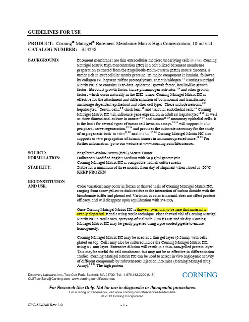
Discovery Labware, Inc ., Two Oak Park, Bedford, MA 01730, Tel: 1.978.442.2200 (U.S.)CLSTechServ@ /lifesciencesFor Research Use Only. Not for use in diagnostic or therapeutic procedures.GUIDELINES FOR USEPRODUCT: Corning ® Matrigel ® Basement Membrane Matrix High Concentration, 10 ml vial CATALOG NUMBER: 354248BACKGROUND: Basement membranes are thin extracellular matrices underlying cells in vivo . CorningMatrigel Matrix High Concentration (HC) is a solubilized basement membranepreparation extracted from the Engelbreth-Holm-Swarm (EHS) mouse sarcoma, atumor rich in extracellular matrix proteins. Its major component is laminin, followedby collagen IV, heparan sulfate proteoglycans, entactin/nidogen.1,2 Corning MatrigelMatrix HC also contains TGF-beta, epidermal growth factor, insulin-like growthfactor, fibroblast growth factor, tissue plasminogen activator,3,4 and other growthfactors which occur naturally in the EHS tumor. Corning Matrigel Matrix HC iseffective for the attachment and differentiation of both normal and transformedanchorage dependent epithelioid and other cell types. These include neurons,5,6hepatocytes,7 Sertoli cells,8,9 chick lens,10 and vascular endothelial cells.11 CorningMatrigel Matrix HC will influence gene expression in adult rat hepatocytes 12,13 as wellas three dimensional culture in mouse 14-17 and human 18,19 mammary epithelial cells. Itis the basis for several types of tumor cell invasion assays,20,21 will support in vivoperipheral nerve regeneration,22-24 and provides the substrate necessary for the studyof angiogenesis both in vitro 25,26 and in vivo .27-29 Corning Matrigel Matrix HC alsosupports in vivo propagation of human tumors in immunosupressed mice.30-32 Forfurther information, go to our website at /lifesciences.SOURCE:Engelbreth-Holm-Swarm (EHS) Mouse Tumor FORMULATION:Dulbecco's Modified Eagle's Medium with 50 g/ml gentamycinCorning Matrigel Matrix HC is compatible with all culture media STABILITY:Stable for a minimum of three months from day of shipment when stored at -20 CKEEP FROZENRECONSTITUTIONAND USE: Color variations may occur in frozen or thawed vials of Corning Matrigel Matrix HC,ranging from straw yellow to dark red due to the interaction of carbon dioxide with thebicarbonate buffer and phenol red. Variation in color is normal, does not affect productefficacy, and will disappear upon equilibration with 5% CO 2.Once Corning Matrigel Matrix HC is thawed, swirl vial to be sure that material isevenly dispersed. Handle using sterile technique. Place thawed vial of Corning MatrigelMatrix HC in sterile area, spray top of vial with 70% ETOH and air dry. CorningMatrigel Matrix HC may be gently pipetted using a pre-cooled pipette to ensurehomogeneity.Corning Matrigel Matrix HC may be used as a thin gel layer (0.5mm), with cellsplated on top. Cells may also be cultured inside the Corning Matrigel Matrix HC,using a 1 mm layer. Extensive dilution will result in a thin, non-gelled protein layer.This may be useful for cell attachment, but may not be as effective in differentiationstudies. Corning Matrigel Matrix HC can be used to assess in vivo angiogenic activityof different compounds by subcutaneous injection into mice (Corning Matrigel PlugAssay).2,8,25 The high proteinDiscovery Labware, Inc ., Two Oak Park, Bedford, MA 01730, Tel: 1.978.442.2200 (U.S.)CLSTechServ@ /lifesciencesFor Research Use Only. Not for use in diagnostic or therapeutic procedures.concentration augments the growth of tumors and also allows the Corning ® Matrigel ®Plug to maintain its integrity after injection. This keeps the injected tumor and/or angiogenic compounds localized for in situ analysis and/or future excision.Dispense remaining material into appropriate aliquots, using pre-cooled tubes, andrefreeze immediately. Avoid multiple freeze thaws. DO NOT STORE IN FROST-FREE FREEZER.CAUTION :Corning Matrigel Matrix HC will gel rapidly at 22o C to 35o C. Thaw overnight at 4o C on ice (Matrigel may gel at slightly elevated temperatures in a refrigerator). Keep product on ice before use, and use pre-cooled pipettes, tips, and tubes when preparing Corning Matrigel Matrix HC for use. Gelled Corning Matrigel Matrix HC may be re-liquified if placed at 4°C on ice for 24-48 hours.INJECTION PROTOCOL:1. It is critical to keep the Corning Matrigel Matrix HC and the Corning Matrigel/Cell suspension as cold aspossible, without freezing, prior to injecting into the mice. It is very important to keep the Corning Matrigel and the Corning Matrigel/Cell suspension as asceptic as possible throughout the procedure.2. For each recipient mouse, mix cells (2 x 10⁵ or greater) and Corning Matrigel Matrix HC together in a finalvolume of 0.5 ml on ice.3. The cells should be in as small a volume as possible. Typically, 250 l ice cold medium containing 2 x 10⁶cells/ml is mixed with 250 l ice cold Corning Matrigel Matrix HC.4. Inject the cells subcutaneously in athymic mice using a 19G needle for tissue samples and a 23G needle forcultured cells. The injections should be done quickly to prevent the Matrigel from solidifying.5. Rotate the syringe when withdrawing to prevent leakage. The needles will need to be changed frequently dueto blockage.NOTE: For more details on this application go to /lifesciences to access CLS-DL-CC-036 (Technical Bulletin 455: Methods for Implantation of Corning Matrigel Matrix into Mice and Tissue Fixation).CELL RECOVERY:Dispase (Catalog No. 354235), Corning Cell Recovery Solution (Catalog No. 354253)Most efficient recovery of cells growing on Corning Matrigel Matrix HC is accomplished using Corning CellRecovery Solution that depolymerizes the Matrigel Matrix within 7 hours on ice or with Dispase, a metalloenzyme which gently releases the cells allowing for continuous culture.REFERENCES:1.Kleinman, H.K., et al., Isolation and characterization of type IV procollagen, laminin, and heparan sulfate proteoglycan from the EHS sarcoma, Biochemistry, 21:6188 (1982).2. Kleinman, H.K., et al., Basement membrane complexes with biological activity, Biochemistry, 25:312 (1986).3. Vukicevic, S., et al., Identification of multiple active growth factors in basement membrane Matrigel suggests caution in interpretation ofcellular activity related to extracellular activity related to extracellular matrix components, Experimental Cell Research, 202:1 (1992).4. McGuire, P.G. and Seeds, N.W., The interaction of plasminogen activator with a reconstituted basement membrane matrix and extracellularmacromolecules produced by cultured epithelial cells, J. Cell. Biochem., 40:215 (1989).Discovery Labware, Inc ., Two Oak Park, Bedford, MA 01730, Tel: 1.978.442.2200 (U.S.)CLSTechServ@ /lifesciencesFor Research Use Only. Not for use in diagnostic or therapeutic procedures.5.Biederer, T. and Scheiffele, P., Mixed-culture assays for analyzing neuronal synapse formation, Nature Protocols, 2(3):670 (2007). 6. Li, Y., et al., Essential Role of TRPC channels in the guidance of nerve growth cones by brain-derived neurotrophic factor, Nature, 434:894(2005).7. Bi, Y., et al., Use of cryopreserved human hepatocytes in sandwich culture to measure hepatobiliary transport, Drug Metabo. and Dispos.,34(9):1658 (2006).8. Hadley, M.A., et al., Extracellular matrix regulates sertoli cell differentiation, testicular cord formation, and germ cell development in vitro, J.Cell Biol., 101:1511 (1985).9. Yu, X., et al., Essential role of extracellular matrix (ECM) overlay in establishing the functional integrity of primary neonatal rat sertolicell/gonocyte co-cultures: An improved in vitro model for assessment of male reproductive toxicity, Toxilogical Sciences, 84(2):378 (2005).10. Ireland, M.E., Quantification and regulation of mRNAs encoding beaded filament proteins in the chick lens, 16(8):838 (1997).11. McGuire, P.G., and Orkin, R.W., A simple procedure to culture and passage endothelial cells from large vessels of small animals,Biotechniques, 5(6):456 (1987).12. Bissel, D.M., et al., Support of cultured hepatocytes by a laminin-rich gel. Evidence for a functionally significant subendothelial matrix innormal rat liver, J. Clinical Invest., 79:801 (1987).13. Page, J.L., et al., Gene expression profiling of extracellular matrix as an effector of human hepatocyte phenotype in primary cell culture,Toxilogical Sciences, 97(2):384 (2007).14 Li, M.L., et al., Influence of a reconstituted basement membrane and its components on casein gene expression and secretion in mousemammary epithelial cells, Proc. Nat. Acad. Sci. USA, 84:136 (1987).15 Barcellof, M.H., et al., Functional differentiation and aveolar morphogenesis of primary mammary cultures on reconstituted basementmembrane, Development, 105:223 (1989).16. Roskelley, C.D., et al., Extracellular matrix-dependent tissue-specific gene expression in mammary epithelial cells requires both physical andbiochemical signal transduction, Proc. Nat. Acad. Sci. USA, 91(26):12378 (1994).17. Xu, R., et al., Extracellular matrix-regulated gene expression requires cooperation of SWI/SNF and transcription factors, J. Biol. Chem.,282(20):14992 (2007).18. Debnath, J., et al., Morphogenesis and oncogenesis of MCF-10A mammary epithelial acini grown in three-dimensional basement membranecultures, Methods, 30(3):256 (2003).19. Muthuswamy, S.K., et al., ErbB2, but not ErbB1, reinitiates proliferation and induces luminal repopulation in epithelial acini, Nat. Cell Biol.,3(9):785 (2001).20. Terranova, V.P., et al., Use of a reconstituted basement membrane to measure cell invasiveness and select for highly invasive tumor cells, Proc.Nat. Acad. Sci. USA, 83:465 (1986).21. Albini, A., et al., A rapid in vitro assay for quantitating the invasive potential of tumor cells, Cancer Research, 47:3239 (1987).22. Madison, R., et al., Increased rate of peripheral nerve regeneration using bioresorbable nerve guides and laminin containing gel, Exp. Neurology, 88:767 (1985).23. Xu, X.M., et al., Axonal regeneration into Schwann cell-seeded guidance channels grafted into transected adult rat spinal cord, J. Comp. Neurol., 351(1):145 (1994).24. Fouad, K., et al., Combining schwann cell bridges and olfactory-ensheathing glia grafts with chondroitinase promotes locomotor recovery aftercomplete transection of the spinal cord, The Journal of Neuroscience, 25(5):1169 (2005).25. Kubota, Y., et al., Role of laminin and basement membrane in the morphological differentiation of human endothelial cells into capillary-likestructures, J. Cell Biol., 107:1589 (1988).26. Maeshima, Y., et al., Identification of the anti-angiogenic site within vascular basement membrane-derived Tumstatin, J. Biol. Chem.,276(18):15240 (2001).27. Passaniti, A., et al., A simple, quantitative method for assessing angiogenesis and anti-angiogenic agents using reconstituted basementmembrane, heparin, and fibroblast growth factor , Lab Invest., 67:519 (1992).28. Isaji, M., et al., Tranilast inhibits the proliferation, chemotaxis and tube formation of human microvascular endothelial cells in vitro andangiogenesis in vivo, British Journal of Pharmacology, 122:1061 (1997).29. Kisucka, J., et al., Platelets and platelet adhesion support angiogenesis while preventing excessive hemorrhage, Proc. Nat. Acad. Sci. USA,103(4):855 (2006).30. Albini, A., et al., Matrigel promotes retinoblastoma cell growth in vitro and in vivo, Int. J. Cancer, 52(2):234 (1992).31. Yue, W., et al., MCF-7 human breast carcinomas in nude mice as a model for evaluating aromatase inhibitors, J. Steroid Biochem. Molec. Biol.,44(4-6):671 (1993).32. Angelucci, A., et al., Suppression of EGF-R signaling reduces the incidence of prostate cancer metastasis in nude mice, Endocrine-RelatedCancer, 13(1):197 (2006).CALIFORNIA PROPOSITION 65 NOTICEWARNING: This product contains a chemical known to the state of California to cause cancer.Component: Chloroform。
细胞电融合仪 ECM2001 安装操作手册

细胞电融合仪ECM2001安装/操作手册目录1.检查清点货物及安装2.技术规格3.操作细则4.产生杂交瘤的实验方法5.服务及保修6.附录:电穿孔技术在转基因及动物克隆中的应用第一节检查清点货物及安装1.拆封包装:ECM2001细胞电融合仪采用纸箱包装,收到货物后,请检查包装完好程度,如果有任何损伤请速与我公司联系。
请小心地拆开包装,将仪器及附件取出,并依据合同清点货物内容及数量,如果有任何外观损伤或者货物与合同有差异,请速与我公司联系。
请保留包装箱,以便满足将来一旦要运送该仪器的需求。
2.电源:该仪器采用220V电源,请确认您的电源为稳定的220V。
如果电源不稳定的话,有可能对仪器造成严重损害!请确认您的电源严格接地,我们提供给您的电源线为三芯带地线电源。
请不要改动该三芯电源线结构,否则有可能对仪器造成严重伤害!3.安装:如果确认仪器包装无问题,且与合同相符,则可以进行安装。
请将仪器安装在一个干燥,水平,常温环境中。
尽量避免灰尘和化学药品对仪器的损害。
仪器与其他物品的距离不少于15厘米,以保证仪器冷却的需求。
将电源线等附件拆包装,待用,依据后续章节继续操作。
第二节技术规格1.外观尺寸:宽:17"高:11"长:17.5"2.重量:47磅3.电气规格:电源:220V,单相,7A耐熔保险交流:频率固定在1MHZ电压:0–75V(从零到峰值)脉冲时间:0–99秒直流:高电压模式(HV)电压:10–3000V(峰值)脉冲时间:1–99毫秒低电压模式(LV)电压:10–500V(峰值)脉冲时间:1–99毫秒0.01–0.99毫秒脉冲次数:1–99第三节操作细则1.注意事项:请严格按照下述操作细则对ECM2001进行操作。
在没有连接打印机的情况下,严禁按“PRINTSETTINGS”按钮!否则仪器将会锁死。
如果打开仪器电源的时候,发现“MANUALSTART”按钮处于亮灯状态,请马上关闭电源!2.程序或步骤(操作过程)高电压警告:为了实际操作和安全的原因,在手动或自动操作模式中,ECM2001在施加交流电场或直流脉冲的过程中,请不要接触任何电缆或电极连接处,当需要连接或拆下电缆时,要检查系统不在操作中或处于备用状态。
夏米尔操作指南4
夏米尔操作指南4夏米尔ROBOFIL-XXX型操作指南状态信号行:GEO 路径转换激活时(MIR-ROT-INV)不亮,无功能激活时橙色 ALV 电极丝垂直校准时绿色ALP 电极丝工件校准时绿色GEN 脉冲电源接通时橙色CTC 电极丝短路时绿色ALM 数控在报警时红色UrG 紧急停止按钮按下时红色EXE 执行程序时黄色WIR 启动走丝时黄色显示当前使用的检查方式和时间MDI 键盘输入MEM 存储器JOG 手动Ready 准备就绪Start 执行Hold 中断用户参数SCF 缩放系数度 1 ROT 旋转角 0 MirX 对X轴镜像 0 MirY 对Y轴镜像 0 InvXY XY轴交换 0 TFE 考虑偏移 1 TER 考虑斜度角 1 ATH 自动重穿线ART 自动重启动SIM 空运行MLK 无移动模拟(程序校验)BLK 单段执行BLD 跳段有效,表示程序前有/的就不执行(如/M00) OSP 考虑可选停止制作:尹承效夏米尔ROBOFIL-XXX型操作指南BLK 打勾后,ISO程序单段执行OSP M01无效打勾后,M01有效(M01为暂停)BLD /开头的语句执行打勾后,/开头的语句跳过0、自动穿丝无效即断丝后不会自动穿丝ATH 1、断丝后回起割点自动穿丝再空走到断丝点切2、如果穿丝穿不进就直接加工下一个孔VSIM 空运行速度(6-900)1、机床坐标操做指令:SMA,X10 设制X轴机床坐标值为本10.00 SMA 设定机床坐标值X Y U V 同时为零MOV,X10 机床坐标系中的绝对移动将机床坐标X移到10处(注意无插补,各轴速度一样,路徑不是线性的) MOV XYUV都回零MOV,Z10 Z轴机床坐标移到10处绝对 Z轴不能同其他轴联动移动MVR 机床坐标的相对移动MVR,Z10 Z轴在现在的位置上向上移动10mm(不考虑转换、镜像、缩放,如果数控系统先前为绝对方式,移动后重新回到这一方式)制作:尹承效夏米尔ROBOFIL-XXX型操作指南CTR,X20 在此位置X轴向正方向加工20mm EDG,X-,X0.1252、工件坐标操作指令:SPA,X1 更新坐标SPA 同时更新XY的坐标为零MPA 绝对移动(不带值XY同时移动到零处) MPR 相对移动CPA 绝对加工CPR 相对加工3、预定义的移动GOH,Hh 按照工件高度H移动Z轴以定位喷嘴位 SEP,CPp 存储绝对坐标系中当前点的坐标p=点号,从0-5 GOP,p 移动到所定义的点位上,p=点号,从0-54、工艺和加工规准TEC<,表名> 激活含有准备使用的规准的工艺 WIR<,表名>激活准备使用的电极丝特性表HPA,h 修改当前高度h=新高度单位mm REX,Ee,(Hh)在所用工艺表中选择工艺规准Ee可选修改当高度(Hh) CLE(,c) 引入附加间隙(c=附加间隙,mm) CLE 设定附加间隙为零制作:尹承效夏米尔ROBOFIL-XXX型操作指南5、辅助功能AUX,m 辅助M功能(m=功能号)WTC 电极丝准备和切(拉)断WPR 断丝后的穿丝准备THR 自动穿丝(在WCT或WPR或人工丝准备之后) MDI模式常用指令:CT 相应的G\M代码SMA,XO,YO G74X0Y0 设定机床坐标的0,0点 SPA,XO,Y0 G92X0Y0 设定工件坐标的0,0点 MOV,X0,Y0 G75X0Y0 移动到机床坐标的0,0点 MVR,X2.0 X 方向相对移动2mm THD M60 穿丝WCT M50 剪丝SEP,CP1 G910 A1 设定当前点为1号点 GOP,1 G911 A1 从当前点移动到1号点EDG,Y,Y-0.125 Y方向对边,并把所对面设0CEN,R45 45度找孔中心 MID,R90 90度方向找中 CTR,X10 X方向相对切割10mm简单加工举例:TEC,LT25AREX,E2,H25制作:尹承效夏米尔ROBOFIL-XXX型操作指南CTR,X10程序执行:ZCL 加工计数器设回到零 SIM,I 激活(i=1) 不激活(i=0) EDG,s轴(,轴v) 找边CEN(,Xx)(,Yy)(,Rr) 找孔中心MID(,Xx)(,Yy)(,Rr) 在两个平行面之间找中GG00 快速移动G01 直线插补G02 顺时针圆弧插补G03 逆时针圆弧插补G04 暂停G09 准备停止一次移动到位 G10 指定偏移量/可选责程序跳步值 G11 自动加工顺序G17 编辑面选择G20 英制输入(英寸)G21 公制输入(mm)G22 存储行程极限功能ON(有效) G23 存储行程极限功能OFF G28 返回到第一参考点G29 从参考点返回到用户点 G30 返回到第二、第三、第四用户点 G32 存储当前点为用户第二、第三、第四参考点G33 设定用户点2,3,4G40 取消丝径补偿G41 电极丝左补偿G42 电极丝右补偿G46 最佳反向控制ONG47 最佳反向控制OFFG48 自动角部倒圆ONG49 自动角部倒圆OFFG50 取消斜度G51 左锥度G52 右锥度G53 在绝对坐标系中移动G61 恒定拐角半径制作:尹承效夏米尔ROBOFIL-XXX型操作指南G62 锥形拐角半径(最小) G63 锥形拐角半径(平均) G64 (最大) G65 宏调用(局部)G66 宏调用(全程)G67 取消宏调用(全程) G68 切割进给方式G69 准确停止方式G70 找边G71 找孔中心G72 找槽中心G73 电极丝校准和导向器设定循环 G74 设定测量点/机床坐标系设定 G75 在机床坐标系中定位 G76 定位-找边点类型1或2 G77 定位-测量点类型1、2或3 G78 定位-拐角点G79 计算工件倾斜角G86 取消支撑功能G87 “用支撑保留废芯”方式G88 “切除支撑”方式G90 绝对方式指令G91 相对G93 局部坐标系设定G94 恒速进给G95 伺服进给MM00 停止M01 可选停止(可用来建立支撑以保留废芯)M02 程序结束M06 穿丝水射流ON M07 上导电块退回ON M14 重穿丝块初始化 M15 锥度方式编成 M16 穿丝射水OFF M17 上导电块回退OFF M23 几何精度策略OFF M24 几何精度策略ON M27 保护策略OFF制作:尹承效夏米尔ROBOFIL-XXX型操作指南M28 一级粗加工策略ON M29 二级粗加工策略ON M30 程序结束和重绕 M31 预置计时器M32 检查水的离子度(电导率) M33 检查水的温度M34 工作液槽上水M35 放水M36 工作液槽位记忆+加载液位ON M37 加载液位OFF M40 空运行状态M42 走丝OFFM43 上、下冲液OFF M44 丝张力OFF M50 CUT 切丝M59 穿丝准备M60 穿丝M68 关闭循环泵M69 接通循环泵M70 轨迹返回功能M70 《执行有效》信号的管理 M71M73M74M80 加工状态M82 走丝开M83 下冲液开M87 存储功能1 ON M88 存储功能1 OFF M89 存储功能2 ON M90 存储功能2 OFF M91 遥控器报警有效 M92 遥控器报警无效 M93 遥控器报警接通ON M94 遥控器报警断开OFF M95 脉冲输出功能M96 完成加工M97 镜向反向加工M98 子程序调用M99 子程序结束制作:尹承效夏米尔ROBOFIL-XXX型操作指南穿丝射流水调整:EXE-service-calibration cyeles-wire calibration 激活卸下丝移动上加工头到相对于待加工工件所需的穿丝位置上接通穿丝射流水手动移动U/V轴以便使穿丝射流水对准下导向器的中心并在其周围分布均匀按memorize threading (记忆穿丝)ZUV 键,保存达到的位置存储UV位置;其值在穿丝操作中会自动用到。
- 1、下载文档前请自行甄别文档内容的完整性,平台不提供额外的编辑、内容补充、找答案等附加服务。
- 2、"仅部分预览"的文档,不可在线预览部分如存在完整性等问题,可反馈申请退款(可完整预览的文档不适用该条件!)。
- 3、如文档侵犯您的权益,请联系客服反馈,我们会尽快为您处理(人工客服工作时间:9:00-18:30)。
BD Bioscience Discovery Labware BD Biocoat TM产品使用指南BD Biocoat TM Matrigel TM 基质BD Biocoat TM细胞相关检测系统目录1BD Biocoat TM Matrigel TM 基质1.1 Matrigel基底膜基质胶 (1)1.2 高浓度HC Matrigel基底膜基质胶 (3)1.3 人来源细胞外基质 (4)1.4 Ⅲ型人重组胶原蛋白 (5)1.5 高浓度层粘连蛋白/巢蛋白混合物 (6)1.6 人胚胎干细胞专用的Matrigel基质 (7)2BD Biocoat TM细胞检测相关系统2.1 Biocoat肿瘤侵袭系统 (8)2.2 Biocoat血管生成系统:内皮细胞侵袭 (10)2.3 Biocoat血管生成系统:内皮细胞迁移 (12)2.4 Biocoat血管生成系统:内皮血管形成 (14)Caco-2检测系统 (15)2.5 BiocoatHTS附录:细胞培养系统快速使用指南 (17)Biocoat TM Matrigel TM常见问题 (17)1BD Biocoat TM Matrigel TM 基质1.1Matrigel基底膜基质胶BD产品货号:354230 354234 356230 356231 356234 356235 356237储存和运输:-20度储存,干冰运输。
操作指南BD采用专利技术,从富含胞外基质蛋白的EHS小鼠肿瘤中分离出BD Matrigel 基底膜基质,其主要成分由层粘连蛋白,Ⅳ型胶原,巢蛋白,硫酸肝素糖蛋白等组成,还包含生长因子和基质金属蛋白酶等。
BD Matrigel基底膜基质在室温条件下,聚合形成具有生物学活性的三维基质,模拟体内细胞基底膜的结构、组成、物理特性和功能,有利于体外细胞的培养和分化,以及对细胞形态、生化功能、迁移、侵染和基因表达的研究。
BD Matrigel 基底膜基质形成的三维培养基质,可促进上皮细胞、肝细胞、Sertoli细胞、黑色素瘤细胞、血管内皮细胞、甲状腺细胞及毛囊细胞等的贴壁与分化。
同时,Matrigel还能影响乳腺上皮细胞的蛋白表达,支持外周神经的新生和牛输卵管上皮细胞的分化。
高浓度的Matrigel适用于研究体内血管生成和肿瘤细胞迁移及肿瘤模型的建立等。
产品特性Matrigel基质会有色差变化(淡黄色到深红色),是由于酚红和碳酸氢盐与CO2作用引起的,但是与5%CO2平衡后色差即会减少。
冻融后,轻轻摇晃试剂瓶使Matrigel分散均匀。
所有操作均需在无菌环境下进行,试剂瓶瓶盖可用70%乙醇擦拭,并自然干燥。
应使用预冷的移液器以保证Matrigel呈匀浆状。
细胞可在0.5mm厚度的Matrigel基质层表面生长,也可在1mm厚度的Matrigel三维基质内生长。
过度稀释的Matrigel会形成非胶质的蛋白层,可以用于细胞贴壁,但不能用于细胞的分化研究。
注意:可将冻融后的Matrigel分装在多个小管,所有分装均需用预冷的冻存管,迅速冷冻并保存,避免多次冻融。
Matrigel在22-35℃温度环境下快速成胶,因此溶解时在4℃冰上过夜冻融(4度时会随着温度的上升部分成胶)。
所有用品在使用前需置于冰浴,必须使用预冷的移液管、吸头及小管操作Matrigel。
成胶后的Matrigel可以在4℃24-48小时后重新呈液态。
推荐包被与成胶方法注意:为了保证Matrigel基质膜的成胶性能与稳定性,稀释浓度不应低于1:3,可用无血清培养基稀释,Matrigel成胶后立即使用。
薄胶成胶方法:1、冻融后,用预冷的移液枪头混匀Matrigel基质成匀浆状。
2、将需要使用的培养板置于冰上,加入浓度为50μL/cm2生长面积的Matrigel基质。
3、在37℃放置30分钟,即可使用。
厚胶成胶方法:1、冻融后,用预冷的移液枪头混匀Matrigel基质成匀浆状。
2、将需要使用的培养板置于冰浴,将培养的细胞与Matrigel基质混合,用移液枪头使其悬浮于基质中。
加入浓度为150-200μL/cm2生长面积的Matrigel基质。
3、在37℃放置30分钟,可成胶。
可以加入细胞培养的基质,也可使细胞直接生长在胶表面。
薄层包被方法:1、冻融后,用预冷的移液枪头混匀Matrigel基质成匀浆状。
2、根据使用需要,采用无血清培养基稀释Matrigel基质。
根据实验需要确定最佳包被浓度。
3、将稀释的Matrigel基质包被于所需的培养器皿中,包被量至少覆盖整个器皿的生长表面。
室温下孵育1小时。
4、去除未结合的Matrigel,用无血清培养基轻轻地冲洗。
用BD细胞回收剂(354253)或离散酶(354235)可降解Matrigel基质,冰浴7小时后回收得到细胞。
1.2高浓度HC Matrigel基底膜基质胶BD 产品货号:354248 354262储存和运输:-20度储存,干冰运输。
产品特性Matrigel基质会有色差变化(淡黄色到深红色),是由于酚红和碳酸氢盐与CO2作用引起的,但是与5%CO2平衡后色差即会减少。
冻融后,轻轻摇晃试剂瓶使Matrigel分散均匀。
所有操作均需在无菌环境下进行,试剂瓶瓶盖可用70%乙醇擦拭,并自然干燥。
应使用预冷的移液器以保证Matrigel呈匀浆状。
细胞可在0.5mm厚度的Matrigel基质层表面生长,也可在1mm厚度的Matrigel三维基质内生长。
过度稀释的Matrigel会形成非胶质的蛋白层,可以用于细胞贴壁,但不能用于细胞的分化研究。
注意:可将冻融后的Matrigel分装在多个小管,所有分装均需用预冷的冻存管,迅速冷冻并保存,避免多次冻融。
Matrigel在22-35℃温度环境下快速成胶,因此溶解时在4℃冰上过夜冻融(4度时会随着温度的上升部分成胶)。
所有用品在使用前需置于冰浴,必须使用预冷的移液管、吸头及小管操作Matrigel。
成胶后的Matrigel可以在4℃24-48小时后重新呈液态。
注射步骤:1、注射前,必须保证Matrigel和细胞悬浮液在不冷冻的情况下,尽可能地保持低温,同时保证每一步都无菌操作。
2、对于每只注射小鼠,冰上混合0.5 mL细胞(2×10⁵或更多细胞)和Matrigel混合液。
3、细胞注射体积需尽可能小。
通常,用含有2×10⁶/mL细胞的250μL预冷培养基,混合250μL冰浴的Matrigel。
4、小鼠皮下注射,19G针用于组织样品,23G针用于培养的细胞。
注射动作必须快速,以避免Matrigel凝固。
5、抽出注射器时旋转以防止泄漏。
针头需要经常更换以避免堵塞。
用BD细胞回收剂(354253)或离散酶(354235)可降解Matrigel基质,冰浴7小时后回收得到细胞。
1.3人来源细胞外基质BD产品货号:354237储存和运输:-20度储存,干冰运输。
产品特性人源细胞外基质是来源于人体胚胎、经过层析纯化而得的细胞外基质提取物,由层粘连蛋白,IV型胶原,硫酸乙酰肝素蛋白多糖(HSPG)等成分组成。
人细胞外基质对贴壁依赖性上皮细胞特别是人源性细胞,有促进其贴壁、延展、代谢和分化的作用。
配方:0.02M磷酸钠溶液,pH值7.4包被方法:1用无血清培养基吸收细胞外基质到一定的浓度。
最终浓度应足以满足覆盖培养器皿表面的量。
例如:最终包被浓度为5.0 μg/cm2,应将基质稀释到50 μg /mL,在35 mm的培养皿中加入1mL基质,在60mm培养皿中加入3 mL基质。
2根据培养皿面积加入合适的已稀释基质。
3室温孵育2小时。
4吸走多余的基质。
5仔细清洗培养器皿,避免刮擦底部表面。
6包被后的培养器皿,应储存于2-8℃的潮湿环境中,或在无菌环境中自然干燥。
1.4Ⅲ型人重组胶原蛋白BD产品货号:354255储存和运输:-20度储存,干冰运输。
操作指南Ⅲ型胶原在皮肤、心血管系统和成年人的正常生理功能中发挥重要作用。
Ⅲ型胶原蛋白在正常人心血管纤维形成中起重要作用,还可用作促进细胞贴壁和调节细胞行为。
使用注意事项:Ⅲ型人重组胶原蛋白,可用于体外实验的薄胶包被,但也能优化用作胶。
如果一次不能使用完,可分装后储存于2-8℃。
推荐包被与成胶方法薄胶包被:1、选择合适的包被浓度。
推荐包被浓度为:0.2-2 μg/cm2,视细胞种类决定。
加入的包被体积必须足以覆盖细胞生长表面。
如有必要,可将胶原蛋白用10 mM醋酸稀释。
2、当生长表面完全覆盖后,室温孵育2小时。
倾斜培养皿至45度,使多余的胶原蛋白都聚集在培养皿的最低点。
3、用移液器去除多余的胶原蛋白。
4、在层流超净台上空气自然干燥或用灭菌的柔和气流干燥。
5、器皿包被完成,可备用。
成胶方法:1、加入9份的人重组胶原蛋白和1份的 10倍中性缓冲液或无菌组织培养基。
2、将混合液加入需要的培养器皿中。
3、室温孵育60分钟。
4、细胞可接种于胶的表面。
如需将胶从培养皿中转移或用于更多操作,在成胶前混合细胞和胶原蛋白。
1.5高浓度层粘连蛋白/巢蛋白混合物BD产品货号:354259储存和运输:-20度储存,干冰运输。
操作指南层粘连蛋白/巢蛋白混合物是EHS小鼠肿瘤基质膜的主要成分。
纯化后,成等摩尔浓度分布。
二价阳离子有利于混合物的形成。
在2 mg/mL浓度时,层粘连蛋白/巢蛋白可以形成三维胶,可用于细胞生长和分化的研究。
三维胶更接近体内微环境。
细胞的分化受到与单一或混合细胞外基质蛋白之间的相互作用的影响,因此在层粘连蛋白/巢蛋白混合物上培养细胞,可以用以观察并揭示细胞分化和功能性的特性机理。
混合蛋白的成胶过程对温度敏感。
此外,高浓度的层粘连蛋白/巢蛋白(2-6 mg/mL)也用于人下颌腺泡细胞分化和内皮细胞的血管形成研究。
注意事项:1、所有操作的工作温度不能高于4℃,蛋白需冰上操作保存,用冰块冷却的溶液或细胞悬浮液稀释。
2、由于混合蛋白溶液粘性较高,必须使用预冷的注射器型移液管,如Gilson M系列;避免使用气压式移液管或吸头。
推荐包被方法铺胶步骤:细胞在胶表面培养1、冰上溶解混合蛋白,所有操作均在冰浴上进行。
2、稀释到需要的浓度:稳定成胶浓度至少为3.5mg/mL。
3、用预冷的等压盐溶液或0.1mM钙离子培养基稀释4、将稀释的蛋白溶液加入到培养器皿,1-2小时,37℃成胶。
铺胶步骤:细胞在胶内部培养1、冰上溶解混合蛋白,所有操作均在冰浴上进行。
2、根据加样体积计算并稀释混合蛋白,成胶浓度至少为3.5mg/mL,稀释过程需在预冷的培养器皿或小管中进行。
3、加入预冷的细胞悬液,用等压盐溶液或培养基调节溶液浓度到第二步所需的浓度。
4、用Gilson M 系列移液管混匀混合蛋白和细胞悬液。
5、将混合后的细胞蛋白悬液加入到预冷的培养板中,37℃孵育1-2小时成胶。