
《隐球菌抗原检测试剂注册技术审查指导原则(征求意见稿)》
— 1 — 隐球菌抗原检测试剂 注册技术审查指导原则
(征求意见稿)
本指导原则旨在指导注册申请人对隐球菌抗原检测试剂注册申报资料的准备及撰写,同时也为技术审评部门对注册申报资料的技术审评提供参考。 本指导原则是对隐球菌抗原检测试剂的一般要求,申请人应依据产品的具体特性确定其中内容是否适用,若不适用,需具体阐述理由及相应的科学依据,并依据产品的具体特性对注册申报资料的内容进行充实和细化。 本指导原则是对申请人和审查人员的指导性文件,但不包括注册审批所涉及的行政事项,亦不作为法规强制执行,如果有能够满足相关法规要求的其他方法,也可以采用,但需要提供详细的研究资料和验证资料,相关人员应在遵循相关法规的前提下使用本指导原则。 本指导原则是在现行法规和标准体系以及当前认知水平下制定的,随着法规和标准的不断完善,以及科学技术的不断发展,本指导原则相关内容也将适时进行调整。 一、范围 隐球菌病是一种由隐球菌感染引起的深部真菌病,可累及全身多个系统。隐球菌属(Cryptococcus,有性期称线黑粉菌属)— 2 —
隶属于真菌界,双核亚界、担子菌门、伞菌亚门、银耳纲、银耳目、银耳科,有性期隶属于担子菌门、线黑粉菌目、线黑粉菌科。属内包括近70个种,其中对人致病的最主要病原菌是新生隐球菌(Cryptococcus neoformans)和格特隐球菌(Cryptococcus gattii)。根据新生隐球菌荚膜多糖成分和生化方面的差异,将
新生隐球菌分成2个变种(新生隐球菌新生变种C.neoformans var.neoformans和新生隐球菌格鲁比变种C.neoformans var.grubii),按血清学分为A、B、C、D和AD型5个型。其中新
生变种为血清D、AD型,欧洲、南美洲流行;格鲁比变种为血清A型,全球流行;新生隐球菌为血清B、C型,在热带\亚热带\温带流行;我国血清型主要为A型。此外,还发现了新生变种与格鲁比变种的杂合体(血清型AD)。隐球菌的基因型包括新生隐球菌的VNⅠ、Ⅱ、Ⅳ、B 和格特隐球菌的VGI-VGIV。两种隐球菌的无性繁殖体均为无菌丝的单芽孢酵母菌,在体外为无荚膜或仅有小荚膜, 进入人体内后很快形成厚荚膜, 有荚膜的隐球菌菌体直径明显增加,致病力明显增强。隐球菌荚膜的主要成分荚膜多糖是确定血清型特异性的抗原基础,并与其毒力、致病性及免疫性密切相关。隐球菌是一种机会感染性真菌,主要侵犯免疫力低下及免疫缺陷人群,也可发生于免疫正常者,可以感染人体的任何组织和脏器,最常见的部位是中枢神经系统,其次是肺部和皮肤。目前, 在免疫抑制患者中, 隐球菌感染的发病率约为5%~10%,— 3 —
在AIDS患者中, 隐球菌的感染率可以高达30%;而在免疫功能正常的人群中, 隐球菌的感染率约为十万分之一左右。 中枢神经系统隐球菌感染通常表现为脑膜炎或脑膜脑炎, 极少数表现为单个或多个局灶性肿块损害(隐球菌肉芽肿),中枢神经系统感染可伴发肺部或其他播散性感染, 但大多数不伴有其他感染的临床表现。通过对脑脊液样本进行培养、墨汁染色涂片可明确中枢神经系统感染诊断。 隐球菌感染中枢神经系统外的器官和部位,肺和皮肤是常见的两个部位。肺是隐球菌感染的主要门户,肺隐球菌感染的临床表现多种多样, 可从无症状的结节到严重的急性呼吸窘迫综合征(ARDS)。皮肤隐球菌感染根据感染来源, 可以分为原发性和继发性感染两种。继发性隐球菌感染一般预示已经发生播散性隐球菌感染, 主要来源于血行播散, 提示感染严重。确诊主要依靠组织病理检查和病原学涂片和培养。血清隐球菌荚膜多糖抗原检测阳性有临床疑似诊断价值。 隐球菌感染的病原学及实验室检测包括隐球菌的微生物学鉴定,隐球菌病的免疫学诊断(血清学实验),隐球菌的药物敏感试验和隐球菌病的组织病理学。其中隐球菌病的免疫学诊断(血清学实验)临床上常用的为隐球菌荚膜抗原检测,其方法有乳胶凝集试验(latex agglutination test ,LA) 、酶联免疫分析法(enzyme immunoassay ,EIA)及侧流免疫层析法(lateral — 4 —
flow immunoassay ,LFA)等。隐球菌荚膜抗原检测可辅助诊断隐球菌感染,隐球菌荚膜多糖抗原阳性提示隐球菌感染,脑脊液滴度的高低可提示疾病的严重程度。隐球菌荚膜抗原检测因其简单、快速已经是目前国内临床辅助诊断隐球菌感染最常用的方法之一。 本指导原则所指隐球菌抗原为隐球菌荚膜多糖抗原,适用于利用酶联免疫法、乳胶凝集试验和侧向免疫层析法等基于抗原抗体反应原理,对来源于血清和/或血浆和/或全血和/或脑脊液等人体样本中的隐球菌荚膜多糖抗原进行体外定性检测的试剂。结合临床和其他实验室指标,可用于隐球菌感染的辅助诊断。如涉及半定量检测试剂,或待测样本类型为血清、血浆、全血、脑脊液外其他样本类型的(如尿液、肺泡灌洗液),应根据申请的预期用途设计临床试验进行验证。可适量参考本指导原则相关要求。 隐球菌抗原检测的准确性至关重要,不正确的检测结果可能导致对患者管理决策失误,假阴性结果可能导致诊断不及时而延误治疗,假阳性结果可能导致不必要的干预措施。申请人应建立良好的产品性能,并对安全性和有效性进行科学合理的验证。 指导原则仅包括对隐球菌抗原检测试剂申报资料中部分项目的要求,适用于进行产品注册和相关许可事项变更的产品。其他未尽事宜,应当符合《体外诊断试剂注册管理办法》(国家食品药品监督管理总局令第5号)(以下简称《办法》)、《体外诊断试剂注册申报资料基本要求》(国家食品药品监督管理总局公告2014年第44号)等相关法规和文件的要求。 — 5 —
二、注册申报资料要求 (一)综述资料 综述资料主要包括产品预期用途、产品描述、有关生物安全性的说明、研究结果的总结评价以及同类产品上市情况介绍等内容。其中,需注意以下内容: 1.产品预期用途 详述检测试剂的预期用途,包括预期人群、适用的样本类型,检测结果的临床指导意义等。描述隐球菌的生物学特征、隐球菌感染疾病相关流行病学、潜伏期、易感人群、临床表现等。说明现有的临床或其他实验室诊断方法等。 2.同类产品上市情况 应着重从技术方法及预期用途(包括预期人群、样本类型、临床意义)等方面写明拟申报产品与现行临床诊断方法,以及目前市场上已获批准的同类产品之间的主要异同点。 (二)主要原材料研究资料 此产品的主要原材料包括抗体、固相载体、质控品(如适用)以及企业参考品等。应提供主要原材料的选择与来源、制备过程、质量标准等研究资料。如主要原材料为企业自制,应明确原材料的制备原理,简述制备过程,其生产工艺必须相对稳定;如主要原材料为外购,应提供的资料包括:原材料的筛选研究资料、供应商选择的研究资料、质量标准、供应商出具的主要原材料检验报告(质量证书)和入厂检验报告等。供应商为原材料的生产商,不得随意更换。 主要原材料的筛选研究资料,应包含试验方法、实验数据及— 6 —
分析。质量控制标准应包括相应试验方法及原材料应达到的客观评价标准。人源或其他生物源性原材料质量控制标准中还应包含生物安全指标。生产企业应依据质量控制标准对原材料进行检验,检验合格方可用于生产。 1. 抗体 应明确抗体名称、生物学来源、免疫原、供应商名称、质量标准(一般包括外观、蛋白纯度、浓度、效价、分子量、功能性试验)等。 如为自制抗体,还需明确抗体所针对的抗原表位、免疫原的具体信息,包括天然抗原的来源、重组或人工合成抗原的序列信息及制备方法。同时提交抗体制备、筛选、鉴定等详细试验资料。 2. 其他主要原辅料 包括基于酶联免疫技术的固相载体,如酶标板、微粒等;基于凝集反应的乳胶颗粒等;基于侧向免疫层析技术的金颗粒、硝酸纤维素膜等。申请人应提交上述原材料的选择依据及验证资料,明确供应商名称及原材料质量标准。 3.试剂盒质控品(如有) 应包括原料来源、质控品制备、阴阳性确认和质量标准等相关研究资料。质控品应至少包含阴性和阳性两个水平。阳性质控品可选择标准菌株或经鉴定、可溯源的临床分离株或荚膜多糖抗原。如荚膜多糖抗原为企业自制,请详述制备的方法。提交质量标准中各项指标,如浓度、纯度、活性测定的方法及结果。 4.企业参考品 如申报产品有相应的国家参考品,则企业参考品应参考国家— 7 —
参考品的项目设置,应不低于国家参考品要求。若尚无国家参考品,申请人应根据产品性能验证的实际情况自行设定企业参考品。 应提交企业参考品的原料选择、制备、阴阳性及浓度/滴度确认等相关验证资料。说明参考品阴阳性及浓度/滴度确认的方法或试剂(建议采用国内已上市的、临床上普遍认为质量较好的同类试剂)。参考品可采用临床真实样本,亦可采用标准菌株或临床分离株添加入待测样本基质的形式制备。企业参考品的基质应与待测样本相同。企业参考品的设置建议如下: 4.1阳性参考品 阳性参考品应考虑覆盖不同种及不同型别的隐球菌感染样本。建议包含新生隐球菌VNⅠ、Ⅱ、Ⅳ、B基因型和格特隐球菌VGI-VGIV基因型。参考品应设置不同浓度水平。 4.2 阴性参考品 阴性参考品应考虑检测特异性的评价,纳入正常临床样本、含类风湿因子等干扰因素的样本及其他病原体阳性样本,建议包含其他常见酵母菌和霉菌、HIV等。 4.3检测限参考品 可设置不同滴度的临床阳性样本/荚膜多糖抗原或临床阳性样本的系列梯度浓度样本,样本基质与检测样本一致,其中应包含检测限水平。 4.4精密度参考品 建议至少包含中阳性和弱阳性,至少两个浓度水平的参考品。
2020年新增和修订的免于进行临床试验体外诊断试剂目录(征求意见稿
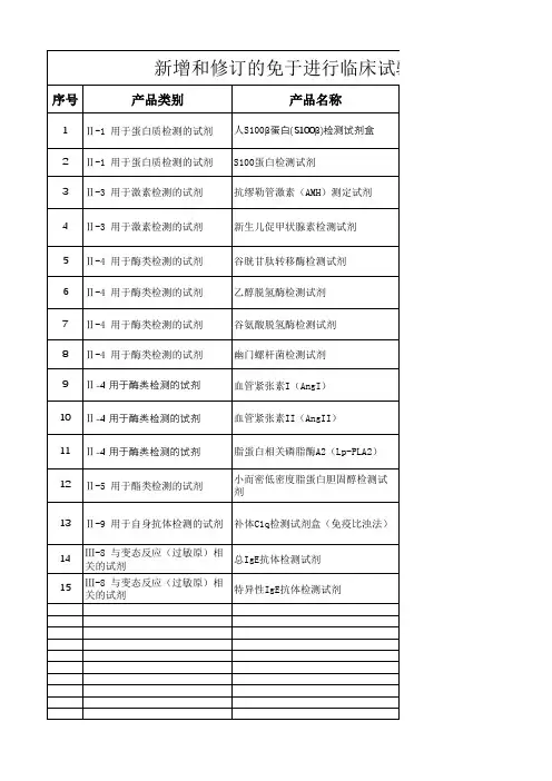
备注
新增 新增 新增 新增 新增 新增 新增 新增 新增 新增 新增 新增
新增 新增 新增
血管紧张素II(AngII)
11 Ⅱ-4 用于酶类检测的试剂 12 Ⅱ-5 用于酯类检测的试剂
脂蛋白相关磷脂酶A2(Lp-PLA2)
小而密低密度脂蛋白胆固醇检测试 剂
13 Ⅱ-9 用于自身抗体检测的试剂 补体C1q检测试剂盒(免疫比浊法)
14
Ⅲ-8 与变态反应(过敏原)相 关的试剂
总IgE抗体检测试剂
15
Ⅲ-8 与变态反应(过敏原)相 关的试剂
特异性IgE抗体检测试剂
临床试验体外诊断试剂目录(征求意见稿)
产品描述
用于检测人体样本中S100-β蛋白的含量,临床上通过检测 S100β 蛋 白的表达,能够对脑损伤程度进行判断,同时可对患者的预后情况进 行评估。 用于检测人体样本中S100蛋白的含量,临床上主要作为脑损伤的生化 标志物,用于脑组织损伤、脑缺血及脑中风的辅助诊断。
用于检测人体样本中谷氨酸脱氢酶的活性,临床上主要用于肝细胞损 伤的辅助诊断。
用尿素酶法定性检测胃粘膜或牙垢中的幽门螺杆菌,临床上主要用于 幽门螺杆菌感染的辅助诊断。 用于体外定量检测人血清或血浆中的紧张素I,临床上主要用于原发 性和继发性高血压分型诊断、治疗等评估,辅助诊断肾脏疾病的治疗 监测。 用于体外定量检测人血清或血浆中的紧张素Ⅱ,临床上主要用于原发 性和继发性高血压分型诊断、治疗等评估,辅助诊断肾脏疾病的治疗 监测。 用于检测人体样本中肾素的活性,临床上主要用于动脉粥样硬化炎症 程度,评估动脉粥样硬化引起的心脑血管疾病的辅助诊断。
国家食品药品监管总局公告2016年第98号关于批准注册医疗器械产品公告-国家规范性文件

国械注准20163400756
74
CYP2C19基因突变检测试剂盒(PCR-荧光探针法)
北京乐普医疗科技有限责任公司
国械注准20163400757
75
胃幽门螺旋杆菌抗原检测试剂盒(胶体金法)
杭州隆基生物技术有限公司
国械注准20163400758
76
肺炎衣原体IgM抗体检测试剂盒(胶体金法)
北京豪迈生物工程有限公司
国械注准20163400628
12
糖类抗原12-5(CA12-5)检测试剂盒(磁微粒化学发光法)
北京豪迈生物工程有限公司
国械注准20163400629
13
糖类抗原72-4(CA72-4)测定试剂盒(磁微粒化学发光法)
北京豪迈生物工程有限公司
国械注准20163400630
14
胃蛋白酶原I(PGI)测定试剂盒(磁微粒化学发光法)
厦门万泰凯瑞生物技术有限公司
国械注准20163400698
46
人乳头瘤病毒基因分型检测试剂盒(荧光PCR法)
厦门安普利生物工程有限公司
国械注准20163400699
47
肺炎衣原体核酸检测试剂盒(荧光PCR法)
厦门安普利生物工程有限公司
国械注准20163400700
48
乙型肝炎病毒基因分型检测试剂盒(荧光PCR法)
北京中检安泰诊断科技有限公司
国械注准20163400774
北京豪迈生物工程有限公司
国械注准20163400622
6
糖类抗原15-3(CA15-3)测定试剂盒(磁微粒化学发光法)
北京豪迈生物工程有限公司
国械注准20163400623
7
国产隐球菌荚膜多糖胶体金法检测试剂盒性能评价

DOI:10.13602/j.cnki.jcls.2021.02.04·临床检验技术研究·国产隐球菌荚膜多糖胶体金法检测试剂盒性能评价作者简介:陈东科,1961年生,男,副主任技师,从事临床微生物检验工作,E mail:c d k@263.net。
陈东科1,张洁2,董家丽3(1.北京医院检验科国家老年医学中心中国医学科学院老年医学研究院,北京100730;2.沂南县人民医院检验科,山东沂南061001;3.沧州市人民医院医学检验中心,河北沧州276300)摘要:目的 评价由丹娜(天津)生物科技股份有限公司生产的隐球菌荚膜多糖检测试剂盒(胶体金法,考核试剂)的检测性能。
方法 选取隐球菌和隐球菌近缘菌及肺部感染常见菌种共28属100株菌作为研究对象,配制0.5麦氏浊度单位(0.5M)菌液检测试剂的特异性,0.5M阳性的菌株以10倍倍比稀释液作为灵敏度检测菌液,以2M和4M浓度菌液作为钩状效应检测菌液,并以美国ImmunoMycologics公司生产的隐球菌抗原检测(胶体金免疫层析法)试剂盒作为对比试剂,比较两家试剂的一致性。
结果 31株隐球菌属菌株中,考核试剂检测出阳性25株(新型隐球菌和格特隐球菌均为阳性),阴性6株(维多利亚隐球菌、C.dimennae和大隐球菌);16株毛孢子菌属菌株中,检测出阳性12株,阴性4株;1株Cutaneotrichosporoncurvatum检测结果为阳性;52株其他菌株的检测结果均为阴性;灵敏度实验结果显示考核试剂隐球菌最低检出限为1.0×10²CFU/mL;2M和4M浓度菌液均未出现钩状效应。
结论 考核试剂隐球菌荚膜多糖检测试剂盒(胶体金法)检测隐球菌属的灵敏度高于比对试剂;特异性方面存在一定的属内和属外交叉反应,但与对比试剂的检测结果高度一致;考核试剂与比对试剂均无钩状效应产生。
关键词:隐球菌荚膜多糖抗原;胶体金免疫层析法;检测性能中图分类号:R446.5 文献标志码:A 隐球菌病是由新型隐球菌(Cryptococcusneoformans)感染引起的一种亚急性或慢性真菌病,主要有隐球菌性脑膜炎(cryptococcalmeningitis,CM)和肺隐球菌病(pulmonarycryptococcosis,PC)。
EB病毒核酸检测试剂注册技术审查指导原则

附件1EB病毒核酸检测试剂注册技术审查指导原则本指导原则旨在指导注册申请人对EB病毒核酸检测试剂注册申报资料的准备及撰写,同时也为技术审评部门对注册申报资料的技术审评提供参考。
本指导原则是针对EB病毒核酸检测试剂技术审查的一般要求,申请人应依据产品的具体特性确定其中内容是否适用,若不适用,需具体阐述理由及相应的科学依据,并依据产品的具体特性对注册申报资料的内容进行充实和细化。
本指导原则是对申请人和审查人员的指导性文件,但不包括注册审批所涉及的行政事项,亦不作为法规强制执行,如果有能够满足相关法规要求的其他方法,也可以采用,但需要详细阐明理由,并对其科学合理性进行验证,提供详细的研究资料和验证资料,相关人员应在遵循相关法规的前提下使用本指导原则。
本指导原则是在现行法规和标准体系以及当前认知水平下制定的,随着法规和标准的不断完善,以及科学技术的不断发展,本指导原则相关内容也将适时进行调整。
一、适用范围EB病毒(Epstein-Barr virus,EBV)为疱疹病毒科,疱疹病毒IV型,是一种嗜人类淋巴细胞的疱疹病毒。
EBV 是双链DNA病毒,基因组长约172kb,在病毒颗粒中呈线性分子,进入受感染细胞后,其DNA发生环化并能自我复制。
淋巴细胞中潜伏感染的EBV可表达2种不翻译成蛋白质的RNA(即EBV-encoded RNAs,EBERs),包括EBER1和EBER2,6种核抗原(EBNA1、EBNA2、EBNA3A、EBNA3B、EBNA3C和LP),2种潜伏期膜蛋白(潜伏膜蛋白,latent membrane protein,LMP),包括LMP1、LMP2A/B。
EBV在人群中感染非常普遍。
除原发性EBV感染可致传染性单核细胞增多症外,EBV还引起慢性活动性EBV感染和EBV相关噬血细胞性淋巴组织细胞增生症等非肿瘤性重症EBV相关疾病。
EBV还与许多肿瘤的诊疗相关。
针对不同的EBV感染相关疾病,选择适当的临床标本和实验室检测方法,对于EBV感染相关疾病的诊断和治疗十分重要。
2007-gdIgA1

see commentary on page 1089Patients with IgA nephropathy have increased serum galactose-deficient IgA1levelsZ Moldoveanu 1,RJ Wyatt 2,JY Lee 3,M Tomana 3,BA Julian 1,3,J Mestecky 1,3,W-Q Huang 1,SR Anreddy 1,5,S Hall 1,MC Hastings 2,KK Lau 2,6,WJ Cook 4and J Novak 11Department of Microbiology,University of Alabama at Birmingham,Birmingham,Alabama,USA;2Department of Pediatrics,University of Tennessee Health Sciences Center and Children’s Foundation Research Center,Memphis,Tennessee,USA;3Department of Medicine,University of Alabama at Birmingham,Birmingham,Alabama,USA and 4Department of Pathology,University of Alabama at Birmingham,Birmingham,Alabama,USAImmunoglobulin A (IgA)nephropathy is the most prevalent form of glomerulonephritis worldwide.A renal biopsy is required for an accurate diagnosis,as no convenientbiomarker is currently available.We developed a serological test based upon the observation that this nephropathy is characterized by undergalactosylated IgA1in the circulation and in mesangial immune deposits.In the absence ofgalactose,the terminal saccharide of O -linked chains in the hinge region of IgA1is terminal or sialylatedN -acetylgalactosamine.A lectin from Helix aspersa ,recognizing N -acetylgalactosamine,was used to develop an enzyme-linked immunosorbent assay that measures galactose-deficient IgA1in serum.The median serum lectin-binding IgA1level was significantly higher for 153Caucasian adult patients with IgA nephropathy without progression to end-stage renal disease as compared with that for 150healthy Caucasian adult controls.As thelectin-binding IgA1levels for the controls were not normally distributed,the 90th percentile was used for determination of significant ing a value of 1076U/ml as the upper limit of normal,117of the 153patients with IgA nephropathy had an elevated serum lectin-binding IgA1level.The sensitivity as a diagnostic test was 76.5%,with specificity 94%;the positive predictive value was 88.6%and the negative predictive value was 78.9%.We conclude that this lectin-binding assay may have potential as a noninvasive diagnostic test for IgA nephropathy.Kidney International (2007)71,1148–1154;doi:10.1038/sj.ki.5002185;published online 7March 2007KEYWORDS:IgA nephropathy;lectin;ELISA;IgA1;O -glycosylation;galactose deficiencySince its initial description in 1968,1immunoglobulin A (IgA)nephropathy (IgAN)has become recognized as the most common primary glomerulonephritis worldwide.2IgAN is characterized by mesangial deposits of IgA1with co-deposits of C3,and often also IgG or IgM or both.1,3–6Over 50%of IgAN patients exhibit increased serum levels of IgA and IgA-containing circulating immune complexes (CIC).7–9The mesangial immune deposits likely originate from these CIC.We have found that these complexes contain aberrantly glycosylated polymeric IgA1(galactose (Gal)-deficient in O -linked glycans).10–13In vitro studies have demonstrated that these CIC readily bind to mesangial cells and induce their proliferation.12–16Thus,aberrantly glyco-sylated polymeric IgA1may be involved in the pathogenesis of the glomerular injury in IgAN.Indeed,elution of glomerular immune deposits from renal tissue of patients with IgAN has demonstrated substantial quantities of Gal-deficient IgA1.17,18Several laboratories have localized the aberrant glycosyla-tion to the O -linked glycans in the hinge region of some IgA1molecules.5,15,19–21In the absence of Gal,the terminal N -acetylgalactosamine (GalNAc)residues,sialylated GalNAc,or the hinge-region glycopeptides are recognized by naturally occurring IgG or IgA1antibodies with anti-glycan or anti-hinge region peptide specificities,thus generating CIC.10Some of the resultant CIC escape normal clearance and deposit in the mesangium.These observations support the postulate that O -glycosylation abnormalities of IgA1predis-pose to mesangial IgA deposition.5,12,14,15,21,22For the diagnosis of IgAN,a renal biopsy is required because there is no established serological marker.To avoid this invasive procedure,particularly in individuals with mild disease,we sought to develop a noninvasive test to support the diagnosis of IgAN using a GalNAc-specific lectin.Based on previous work by our group and others,the lectin from Helix aspersa (HAA)is well suited for detecting Gal-deficient IgA1O -linked glycans.10,11,15–17,23–26In this study,we show that a quantitative lectin-based enzyme-linked immunosor-bent assay (ELISA)can be successfully used to measure serum levels of Gal-deficient IgA in sera of patients with IgAN.o r i g i n a l a r t i c l e &2007International Society of NephrologyReceived 12April 2006;revised 14January 2007;accepted 23January 2007;published online 7March 2007Correspondence:J Novak,Department of Microbiology,University of Alabama at Birmingham,84519th Street South,Room 734,Birmingham,AL 35294,USA.E-mail:jannovak@5Current address:Department of Internal Medicine,Thomas Jefferson University,Philadelphia,Pennsylvania,USA.6Current address:Department of Pediatrics,University of California,Davis,Sacramento,California,USA.RESULTSSerum IgA concentrationMedian serum IgA concentration was3943m g/ml(range 1171–11713m g/ml)for153Caucasian IgAN patients who had not progressed to end-stage renal disease as compared with2843m g/ml(range886–8185m g/ml)for150healthy controls(P o0.0001)(Table1).Fifty patients had a serum IgA concentration higher than4761m g/ml(90th percentile for controls).These data indicate a sensitivity of32.7%and specificity of90%with a positive predictive value of76.9% and a negative predictive value of56.7%(Table2).In agreement with a previous publication,22the diagnostic utility of total levels of serum IgA was not satisfactory. Serum HAA-IgA levelsSerum HAA-IgA levels for the patient and healthy control groups are shown in Figure1a.The individual levels for the control group were not distributed normally with a marked skewing towards elevated levels.The median serum HAA-IgA level for the patients of1731U/ml(range320–8317U/ml) was significantly higher than the median of615U/ml(range 264–1807U/ml)for the control group(P o0.0001).The serum HAA-IgA level was higher than1076U/ml(90th percentile for controls)for117of the153patients.These data indicate a sensitivity of76.5%and a specificity of90%with a positive predictive value of88.6%and a negative predictive value of78.9%.To illustrate the potential of this assay,the results are also presented as a receiver operating characteristic curve in Figure1b.The area under the curve is0.9021with a standard error0.0178,indicating that the true-positive rate was high and the false-positive rate was low.The95% confidence interval was0.8672–0.9370(P o0.0001).Reproducibility of the HAA-IgA ELISA was high(repeated the assays had an r¼0.970;P o0.0001).The serum HAA-IgA and IgA levels correlated significantly in IgAN patients(r2¼0.571, P o0.0001)and healthy controls(r2¼0.392,P o0.0001).The median value of the HAA-binding fraction of IgA was 445U/mg IgA(range129–879)for153Caucasian IgAN patients who had not progressed to end-stage renal disease as compared with248U/mg IgA(range56–666)for150healthy controls(P o0.0001).Ninety-four patients had a HAA-binding fraction of IgA higher than394U/mg IgA(90th percentile for controls).These data indicate a sensitivity of 61.4%and specificity of90%with a positive predictive value of86.2%and a negative predictive value of69.6%.Seven of 36patients with normal HAA-IgA levels had an HAA-binding fraction of IgA above the90th percentile for controls; among the135healthy controls with a HAA-IgA level belowTable1|Clinical features and levels of serum IgA and HAA-IgA for subgroups of patients with IgA nephropathyGroup N Genderratio(M/F)Age atdiagnosis(years)mean7SDInterval tostudy(years)mean7SDHAA-IgA(U/ml)median(range)IgA(l g/ml)median(range)HAA-IgA(U/mg IgA)median(range)UP/Crratiomedian(range)GFR a(ml/min/1.73m2)mean7SDAll patients153 1.6837.0714.5 4.976.91731(320–8317)3943(1171–11713)445(129–879)0.60(0.06–19.3)57.0731.4Male patients9639.8715.0 3.975.61731(373–8317)3846(1171–11713)455(129–834)0.65(0.06–5.35)53.3730.8Female patients5732.2727.8 6.578.41707(320–4366)4213(1409–7283)428(129–879)0.54(0.06–19.3)63.1732.0o8weeks after biopsy34 1.6237.5714.10.0670.041800(508–4366)3956(1418–6327)451(160–879)0.81(0.07–4.36)61.0739.045years after biopsy46 1.0930.2711.913.377.01974(504–4391)4440(2037–9148)425(129–812)0.19(0.06–19.3)60.7738.5GFR o30ml/min/1.73m235 3.3842.0715.5 4.577.21856(391–4391)3584(1418–8887)525(141–879)1.25(0.07–7.46)17.976.8GFR490ml/min/1.73m226 1.1727.579.5 3.274.91466(320–3579)3299(1409–7422)373(170–710)0.38(0.06–2.53)105.0716.3Normal urinalysis26 1.3630.0710.311.878.92030(504–3901)4571(2037–7350)417(208–820)0.09(0.06–0.19)45.5716.2GFR,glomerular filtration rate;HAA,lectin from Helix aspersa;IgA,immunoglobulin A;UP/Cr ratio,urinary protein/creatinine ratio.a Estimated GFR was calculated using the MDRD formula(53).Table2|Diagnostic utility of serum IgA and HAA-IgA assaysControl PatientsYes No Yes No Sensitivity Specificity Positive predictive Negative predictive IgA44761m g/ml151********.327(0.258,0.405)0.900(0.842,0.938)0.769(0.654,0.855)0.567(0.504,0.629) HAA-IgA41076U/ml151********.758(0.685,0.819)0.900(0.842,0.938)0.885(0.820,0.929)0.785(0.718,0.840) HAA-IgA4394U/mg IgA1513594590.614(0.535,0.685)0.900(0.842,0.938)0.785(0.628,0.756)0.696(0.628,0.756) HAA,lectin from Helix aspersa;IgA,immunoglobulin A.The numbers in the brackets are the95%confidence intervals.Z Moldoveanu et al.:Galactose-deficient IgA in IgA nephropathy o r i g i n a l a r t i c l ethe cutoff point,11had a similarly increased value for HAA-binding fraction of IgA.Western blot analysisTo verify the ELISA data,selected serum samples from four IgAN patients and two healthy controls representing a wide range of HAA-IgA values were analyzed by Western blotting for total and Gal-deficient IgA (see Materials and methods).The band intensity was measured by densitometry and the ratio of HAA-IgA to IgA heavy chain was calculated.This value was plotted against HAA-binding fraction of IgA1(U/mg IgA)determined by ELISA in the same sample.The data in Figure 2are representative for the IgAN patients and healthy controls over the wide range of HAA-IgA values.The results from the two assays showed good correlation (r 2¼0.818,P ¼0.046).Clinical and pathological associationsSerum HAA-IgA and IgA concentrations as well as values for the HAA-binding fraction of IgA (U/mg IgA)are shown for subgroups of patients in Table 2.There was no significant difference based upon gender.The median level for 26patients with a normal urinalysis at the time of study was 2030Units/ml (88%with an elevated level).Levels of HAA-IgA for subjects with an estimated glomerular filtration rate (GFR)X 90ml/min/1.73m 2were similar to those of patients with an estimated GFR o 30ml/min/1.73m 2.Furthermore,the HAA-IgA levels did not significantly differ between patients who had undergone renal biopsy within eight weeks of the blood sampling and patients whose blood was obtained more than five years after biopsy.Serum HAA-IgA levels did not significantly correlate with urinary protein/creatinine (UP/Cr)ratio (r ¼À0.128,P ¼0.211),estimated GFR (r ¼À0.147,P ¼0.069),or interval since biopsy (r ¼0.0844,P ¼0.300).Thus,the serum HAA-IgA level did not appear to be an indicator of disease activity in our cohort of Caucasian adults with IgAN.Renal biopsies from 34patients,who had blood obtained within eight weeks of diagnostic biopsy,were graded according to the Haas system 27by two investigators (WJC and BAJ)blinded to the clinical and laboratory data.Levels of HAA-IgA were above the cutoff value of healthy controls in five of eight (63%)patients with subclass 1,2,and 3histological features and in 20of 26(77%)patients with subclass 4and 5disease.DISCUSSIONIn this study,wedeveloped a quantitative assay to measure serum Gal-deficient IgA (HAA-IgA).Although the total IgA correlated with the HAA-IgA levels,only the serum HAA-IgA level differentiated IgAN patients from the healthy controls with high sensitivity (Table 2).This finding may be explained by a greater amount of Gal-deficient IgA recognized by naturally occurring anti-glycan antibodies,resulting in formation of CIC.10In our ELISA,serum IgA was captured by a polyclonal IgA heavy chain-specific antibody.As a result,both subclasses,IgA1that accounts for about 84%of serum IgA 28,29andControlsPatients10002000300040005000600070008000900090thpercentileU n i t s /m l1-SpecificityS e n s i t i v i t yabFigure 1|Serum levels of Gal-deficient IgA in IgAN patients and healthy controls.(a )HAA-IgA level (U/ml,see Materials and methods)in sera from IgAN patients and healthy controls.(b )Receiver operating characteristic curve for HAA-IgA levels.Area under the curve is 0.902.The arrows with numbers indicate key points:(1)HAA-IgA serum level 1076U/ml;sensitivity 0.765,specificity 0.900;(2)HAA-IgA serum level 1702U/ml;sensitivity 0.503,specificity 0.993;and (3)HAA-IgA serum level 1810U/ml;sensitivity 0.444,specificity 1.000.0.00.20.40.60.8HAA-IgA (ELISA; U/mg IgA)H A A -b i n d i n g f r a c t i o n o f I g A (W e s t e r n b l o t )Figure 2|Reactivity of HAA with serum IgA in ELISA and Western blot assays.ELISA data are expressed as HAA-IgA (U/mg IgA);Western blot data are the ratios of heavy-chain band densities for HAA-binding IgA and total IgA.Six samples with wide range of HAA-IgA reactivities were used.The inset shows the two extremes:a highly reactive sample and a modestly reactive sample.o r i g i n a l a r t i c l eZ Moldoveanu et al.:Galactose-deficient IgA in IgA nephropathyIgA2,are captured.However,only IgA1has O-linked glycans and,therefore,is the only IgA subclass that can bind to HAA, a GalNAc-specific lectin.Consequently,the HAA-IgA assay is a measure of serum Gal-deficient IgA1.Mesangial IgA deposition is incidentally encountered in asymptomatic individuals in frequencies that vary among studies,ranging from2to16%.30–34For example,in a study in Japan,biopsies from510newly procured living-donor renal allografts taken at the time of transplantation showed mesangial IgA deposition in82(16.1%)of them.33By contrast,in a study of necropsy examinations of756Finish adults who had committed suicide or met with a violent death,mesangial IgA deposits were found in18(2.4%)of the individuals.32However,these renal biopsies with IgA deposits rarely showed any significant pathological changes by light microscopy.Consequently,the clinical significance of these observations is not clear,but many otherwise normal healthy individuals may have mesangial deposits of IgA without clinical manifestation of nephritis or pathological changes of the glomeruli.It would have been interesting to have renal biopsy data on our cohort of healthy controls and to assess the frequency of IgA deposits with and without concurrent renal pathology by light microscopy.However,this could not have been carried out for ethical reasons.Several groups of investigators have shown significantly increased binding of GalNAc-specific lectins to serum IgA1 from patients with IgAN or Henoch–Scho¨nlein purpura nephritis.10,11,23,35–40Using HAA,Linossier et al.39showed increased binding for IgAN patients with normal thickness of the glomerular basement membrane on electron microscopy. Studies with other GalNAc-specific lectins,Vicia villosa lectin23,38and Glycine max lectin,40showed increased serum Gal-deficient IgA1in patients with Henoch–Scho¨nlein purpura nephritis and IgAN,respectively.However,these results did not distinguish the patients from healthy controls; thus,the test would not be adequate to diagnose either disorder.Several candidates for a serological diagnostic marker of IgAN,such as increased levels of serum IgA or of IgA-containing complexes withfibronectin,IgG,or C3,have been previously examined.7–9,39–45Although the serum IgA con-centration is significantly elevated in about half of the patients with IgAN,46,47this parameter is not satisfactory as a diagnostic test,as confirmed by our study(Table2).Jennette et al.42found that93%of30patients with IgAN or Henoch–Scho¨nlein purpura nephritis had serum IgA-fibro-nectin aggregates,compared with7%ter, the same group performed a larger study with63adult and 25pediatric subjects and found IgA-fibronectin aggregates in only40%of samples.43Furthermore,just48%of the patients ever showed IgA-fibronectin aggregates,despite sampling on more than one occasion.43Czerkinsky et al.7reported the presence of IgA1-and IgG-containing CIC in40%of30 patients with IgAN and none in the14normal subjects. Coppo et al.8,41found IgA-CIC in65%of IgAN patients using a conglutinin-binding assay.The levels of IgA-CIC correlated with clinical activity,as denoted by the magnitude of microscopic hematuria or a history of macroscopic hematuria.Mixed IgA1-IgG CICs were detected in68%of patients as reported by Schena et al.9Activated C3was detected in30–57%of patients with IgAN.44,45To date,none of these assays has been introduced as a diagnostic test.Using a panel of15lectins,our group has examined the reactivity of de-sialylated serum IgA1from10patients with IgAN and10healthy controls.11All of the GalNAc-specific lectins demonstrated higher reactivity with serum IgA1from the IgAN patients than with IgA1from healthy controls; however,the highest specificity was obtained with HAA.11 The specificity of many lectins(including those specific for hinge-region O-linked glycans)may vary by the geographical location of the source(animal or plant)and by the method of preparation.It is thus possible that the HAA from the source we chose had selectivity properties that were superior to those of the other GalNAc-specific lectins we tested.11,26 These results prompted us to select HAA to use in our lectin ELISA to measure the levels of Gal-deficient IgA1.Gal-deficient IgA1likely conveys nephritogenic properties to the IgA1-CIC.Gal-deficient IgA1has been isolated from the immune deposits in the renal mesangium of patients with IgAN.17,18Furthermore,immune complexes with Gal-deficient IgA1stimulate mesangial cells in culture to proliferate5,13,15,16,48and secrete components of the extra-cellular matrix and cytokines.49–51Although the size and biological activity of these CIC may be determined by various factors,including the antigen to antibody ratio,13the aberrant glycosylation seems to play a central role in inducing glomerular injury.52Thus,measurement of Gal-deficient IgA1in serum samples may be of diagnostic significance, despite the lack of an association between the serum HAA-IgA level and disease severity,as assessed by histological features,magnitude of proteinuria,or interval since diag-nosis.The elevated serum HAA-IgA levels in patients during a clinically quiescent phase many years after clinical onset suggest that defective galactosylation may serve as a biomarker for the disease.Although Gal-deficiency of IgA1O-linked glycans can be analyzed by Western blotting,gas-liquid chromatography, and mass spectrometry,these procedures are expensive and cumbersome.In contrast,an ELISA is a relatively rapid, inexpensive,and high-throughput assay with excellent reproducibility.We anticipate that the HAA-IgA ELISA could be easily adapted for the clinical laboratory.If the sensitivity of this assay can be improved while maintaining its high specificity,it may be developed into a noninvasive diagnostic test for IgAN.MATERIALS AND METHODSPatients and healthy controlsThe study included153Caucasian patients with IgAN who had undergone biopsy for the evaluation of proteinuria,hematuria,or renal dysfunction.Thirteen patients were younger than age18years at the time of biopsy.All patients were older than age18at time ofZ Moldoveanu et al.:Galactose-deficient IgA in IgA nephropathy o r i g i n a l a r t i c l eblood sampling for the study.Ninety-three were male,yielding a male to female ratio of1.68.Blood and urine samples were obtained on one occasion.Mean age at biopsy was37.0years(range10–75 years).The mean interval between the date of diagnosis by biopsy and the date of blood sampling for this study was4.9years(range 0–29years).Thirty-four(22%)patients were studied within eight weeks of biopsy and47(31%)had blood drawn more thanfive years after biopsy.Serum creatinine concentration,urinalysis and random spot UP/Cr ratio were obtained at the time of study.Estimated GFR was calculated using the Modification of Diet in Renal Disease formula.53Estimated GFR was X90ml/min/1.73m2for26(17%) patients,X60but o90ml/min/1.73m2for40(26%)patients,X30 but o60ml/min/1.73m2for52(34%)patients,and o30ml/min/ 1.73m2for35(23%)patients.The biopsy-proven diagnosis of IgAN was based upon the demonstration by direct immunofluorescence of IgA as the dominant or co-dominant Ig in a predominantly mesangial distribution and the lack of clinical or serological evidence for systemic lupus erythematosus,vasculitis,or Henoch–Scho¨nlein purpura3and the absence of X2þmesangial C1q deposition.54 The healthy controls consisted of153Caucasian residents of Alabama,Kentucky,or Tennessee.All were at least18years of age (range18–80years).The mean age was27.071.9years.The group consisted of74males and76females.Serum creatinine,UP/Cr ratio and blood pressure were measured for each healthy control. Individuals with UP/Cr ratio40.20or estimated GFR o60ml/ min/1.73m2were excluded.Estimated GFR was o90ml/min/ 1.73m2for78(50.9%)of the healthy controls.The Institutional Review Boards at the University of Alabama at Birmingham and the University of Tennessee Health Sciences Center approved this study.Written informed consent was obtained from all subjects.Determination of serum IgA and Gal-deficient IgA by ELISA For measurement of serum total and Gal-deficient IgA,high-adsorption polystyrene96-microwell plates(Nalge Nunc Interna-tional,Rochester,NY,USA)were coated overnight with3m g/ml F(ab0)2fragment of goat IgG anti-human IgA(Jackson Immuno-Research Labs.,West Grove,PA,USA)in phosphate-buffered saline. Coated plates were blocked with1%bovine serum albumin(Sigma Chemical Company,St Louis,MO,USA)in phosphate-buffered saline-0.05%-Tween20,and serial twofold dilutions of samples and standards in blocking solution(in duplicate)were incubated overnight at room temperature.The standard for IgA consisted of a pool of normal human sera(Binding Site,Birmingham,UK), previously calibrated for Ig isotype concentration by ELISA and radial immunodiffusion.The bound IgA was detected by incubation with biotin-labeled F(ab0)2fragment of goat IgG anti-human IgA (BioSource,Camarillo,CA,USA)for3h at371C followed by1h incubation with horseradish peroxidase-conjugated ExtrAvidin (Sigma).For quantitation of Gal-deficient IgA,the standard consisted of a polymeric Gal-deficient IgA1isolated from serum of a patient with IgA1myeloma.This Ig has been previously shown to mimic the aberrantly glycosylated IgA1from IgAN patients.10To remove terminal sialic acid from O-linked GalNAc on IgA,the samples and the IgA1myeloma protein standard were incubated with100m l(1mU)/well neuraminidase(Roche Diagnostic Corp., Indianapolis,IN,USA)in0.01M acetate buffer pH5for3h at 371C.11After washing,100m l biotin-labeled HAA,a GalNAc-specific lectin,(1:500dilution;Sigma)was added to each well.Following3h incubation at371C,the plates were washed and horseradish peroxidase-ExtrAvidin(Sigma)was added and incubated for1h at 371C.For both IgA and Gal-deficient IgA,the wells were developed with the peroxidase chromogenic substrate O-phenylenediamine-H2O2(Sigma).The color reaction was stopped with1M sulfuric acid and the absorbance was measured at490nm with an EL312Bio-Kinetics microplate reader(Bio-Tek Instruments Inc.,Winooski,VT, USA).The amounts of total IgA and Gal-deficient IgA in the tested samples were calculated with the DeltaSoft II program(BioMettalics Inc.,Princeton,NJ,USA)by interpolating the optical densities on calibration curves,constructed using serum IgA and a Gal-deficient IgA1myeloma protein.As the standard IgA1myeloma protein is not entirely devoid of galactose,the expression of results in m g/ml does not precisely reflect the concentration of Gal-deficient IgA1in the sera.Therefore,we expressed the results in U/ml.One unit of HAA-IgA was defined as1m g of this standard.The results were also normalized to total IgA and expressed in U/mg IgA.Duplicate serum samples from21healthy controls were analyzed to assess the degree of intra-assay variation.The results showed excellent reproducibility(r¼0.970;P o0.0001).To verify the accuracy of the ELISA measurements of total IgA level in serum,five serum samples and the calibration serum were analyzed in the clinical laboratory of the University of Alabama Hospital.The differences in the measurements by ELISA and nephelometry ranged between2and17%,the mean difference was975%.Western blot analysisSerum samples from six individuals(four IgAN patients and two controls)representing a wide range of HAA-IgA values were normalized for IgA content(1.0or0.1m g IgA/well for blots to be developed with HAA or anti-IgA,respectively),then reduced with 5%b-mercaptoethanol,and electrophoreticaly separated on a 4–15%gradient sodium dodecyl sulfate-polyacrylamide gel(Bio-Rad,Hercules,CA,USA).The gels were then electroblotted to polyvinylidine difluoride membrane(0.45m m,Millipore,Billerica, MA,USA).After the transfer,polyvinylidine difluoride membranes were blocked with SuperBlock buffer(Pierce,Rockford,IL,USA) containing0.05%Tween20,washed and treated for3h at371C with 10mU/ml neuraminidase(Roche).Membranes were then washed and incubated overnight at41C with1:500dilution biotin-labeled HAA(Sigma)in blocking buffer.To detect the heavy chain of IgA,a duplicate membrane was incubated overnight at41C with biotinylated goat F(ab’)2IgG anti-human IgA(Biosource).The biotin-labeled probes bound to IgA were detected,after addition of streptavidin-horseradish peroxidase conjugate,with SuperSignal West Pico Solution(Pierce).The positive bands were visualized using X-rayfilm(Kodak,Rochester,NY,USA)and thefilms were analyzed by densitometry to calculate the ratios of HAA-IgA to a-chain.Statistical analysisWilcoxon rank sum test was used to compare subject and control groups for serum concentrations of IgA,HAA-IgA levels,and relative HAA-binding to IgA.The90th percentile for healthy controls was the cutoff point for calculation of sensitivity and specificity.The receiver operating characteristic curve for HAA-IgA levels in patients and controls was constructed using GraphPad Prism version4.00for Windows(GraphPad Software,San Diego,CA,USA).The Spearman correlation coefficients were used to assess the relationship between HAA-IgA levels and UP/Cr ratio,estimated GFR and the interval since diagnosis by biopsy.SAS9.1(SAS Institute,Cary,NC,USA)was used for descriptive statistics and calculation of correlation coefficients.o r i g i n a l a r t i c l e Z Moldoveanu et al.:Galactose-deficient IgA in IgA nephropathyACKNOWLEDGMENTSThis work was supported by National Institutes of Health grants DK57750and DK61525and by the General Clinical Research Centers of the University of Alabama at Birmingham M01RR00032and the University of Tennessee Health Sciences Center M01RR00211.JN was also supported by NIH Grants DK71802and DK64400.We express our appreciation to Catherine Barker and Sue Woodford(University of Alabama at Birmingham)and Dr Kimberly Fisher and Sandra Grimes (University of Tennessee)for help with collecting clinical samples and to Rose Kulhavy(University of Alabama at Birmingham)for providing the IgA1myeloma protein.We appreciate the technical assistance of Rhubell Brown,Candace Kirksey,and Claretha Nichols in the processing of the serum samples.REFERENCES1.Berger J,Hinglais N.Les depots intercapillaires d’IgA-IgG(Intercapillarydeposits of IgA-IgG).J Urol Nephrol1968;74:694–695.2.Julian BA,Waldo FB,Rifai A et al.IgA nephropathy,the most commonglomerulonephritis worldwide.A neglected disease in the United States?Am J Med1988;84:129–132.3.Emancipator SN.IgA nephropathy and Henoch–Scho¨nlein syndrome.In:Jennette JC,Olson JL,Schwartz MM,Silva FG(eds).Heptinstall’sPathology of the Kidney.Lippincott-Raven Publishers:Philadelphia,1998, pp479–539.4.Conley ME,Cooper MD,Michael AF.Selective deposition ofimmunoglobulin A1in immunoglobulin A nephropathy,anaphylactoid purpura nephritis,and systemic lupus erythematosus.J Clin Invest1980;66:1432–1436.5.Julian BA,Novak J.IgA nephropathy:an update.Current Opin NephrolHypertens2004;13:171–179.6.Novak J,Julian BA,Tomana M et al.Progress in molecular and geneticstudies of IgA nephropathy.J Clin Immunol2001;21:310–327.7.Czerkinsky C,Koopman WJ,Jackson S et al.Circulating immunecomplexes and immunoglobulin A rheumatoid factor in patients with mesangial immunoglobulin A nephropathies.J Clin Invest1986;77:1931–1938.8.Coppo R,Basolo B,Piccoli G et al.IgA1and IgA2immune complexes inprimary IgA nephropathy and Henoch–Scho¨nlein nephritis.Clin ExpImmunol1984;57:583–590.9.Schena FP,Pastore A,Ludovico N et al.Increased serum levels of IgA1-IgGimmune complexes and anti-F(ab’)2antibodies in patients with primary IgA nephropathy.Clin Exp Immunol1989;77:15–20.10.Tomana M,Novak J,Julian BA et al.Circulating immune complexes in IgAnephropathy consist of IgA1with galactose-deficient hinge region and antiglycan antibodies.J Clin Invest1999;104:73–81.11.Tomana M,Matousovic K,Julian BA et al.Galactose-deficient IgA1in seraof IgA nephropathy patients is present in complexes with IgG.Kidney Int 1997;52:509–516.12.Novak J,Vu HL,Novak L et al.Interactions of human mesangial cells withIgA and IgA-containing circulating immune complexes.Kidney Int2002;62:465–475.13.Novak J,Tomana M,Matousovic K et al.IgA1-containing immunecomplexes in IgA nephropathy differentially affect proliferation ofmesangial cells.Kidney Int2005;67:504–513.14.Moura IC,Arcos-Fajardo M,Sadaka C et al.Glycosylation and size of IgA1are essential for interaction with mesangial transferrin receptor in IgA nephropathy.J Am Soc Nephrol2004;15:622–634.15.Coppo R,Amore A.Aberrant glycosylation in IgA nephropathy(IgAN).Kidney Int2004;65:1544–1547.16.Amore A,Cirina P,Conti G et al.Glycosylation of circulating IgA inpatients with IgA nephropathy modulates proliferation and apoptosis of mesangial cells.J Am Soc Nephrol2001;12:1862–1871.17.Allen AC,Bailey EM,Brenchley PEC et al.Mesangial IgA1in IgAnephropathy exhibits aberrant O-glycosylation:observations in threepatients.Kidney Int2001;60:969–973.18.Hiki Y,Odani H,Takahashi M et al.Mass spectrometry provesunder-O-glycosylation of glomerular IgA1in IgA nephropathy.Kidney Int 2001;59:1077–1085.19.Floege J,Feehally J.IgA nephropathy:recent developments.J Am SocNephrol2000;11:2395–2403.20.Smith AC,Feehally J.New insights into the pathogenesis of IgAnephropathy.Pathogenesis of IgA nephropathy.Springer SeminImmunopathol2003;24:477–493.21.Barratt J,Feehally J.IgA Nephropathy.J Am Soc Nephrol2005;16:2088–2097.22.van der Boog PJ,van Kooten C,van Seggelen A et al.An increasedpolymeric IgA level is not a prognostic marker for progressive IgAnephropathy.Nephrol Dial Transplant2004;19:2487–2493.23.Allen AC,Harper SJ,Feehally J.Galactosylation of N-and O-linkedcarbohydrate moieties of IgA1and IgG in IgA nephropathy.Clin ExpImmunol1995;100:470–474.24.Carpenter GH,Proctor GB,Shori DK.O-glycosylation of salivary IgA asdetermined by lectin analysis.Biochem Soc Trans1997;25:S659.25.Hiki Y,Kokubo T,Iwase H et al.Underglycosylation of IgA1hinge plays acertain role for its glomerular deposition in IgA nephropathy.J Am Soc Nephrol1999;10:760–769.26.Moore JS,Kulhavy R,Tomana M et al.Reactivities ofN-acetylgalactosamine-specific lectins with human IgA1proteins.Mol Immunol2007;44:2598–2604.27.Haas M.Histologic subclassification of IgA nephropathy:aclinicopathologic study of244cases.Am J Kidney Dis1997;29:829–842.28.Mestecky J,Russell MW.IgA subclasses.Monogr Allergy1986;19:277–301.29.Mestecky J,Russell MW,Jackson S et al.The human IgA system:areassessment.Clin Immunol Immunopathol1986;40:105–114.30.Waldherr R,Rambousek M,Duncker WD et al.Frequency of mesangial IgAdeposits in non-selected autopsy series.Nephrol Dial Transplant1989;4: 943–946.31.Sinniah R.Occurence of mesangial IgA and IgM deposits in a controlnecropsy population.J Clin Pathol1983;36:276–279.32.Varis J,Rantala I,Pasternack A et al.Immunoglobulin and complementdeposition in glomeruli of756subjects who had committed suicide or met with a violent death.J Clin Pathol1993;46:607–610.33.Suzuki K,Honda K,Tanabe K et al.Incidence of latent mesangial IgAdeposition in renal allograft donors in Japan.Kidney Int2003;63:2286–2294.34.Rosenberg HG,Martinez PS,Vaccarezza AS et al.Morphological findingsin70kidneys of living donors for renal transplant.Pathol Res Pract1990;186:619–624.35.Andre PM,Le Pogamp P,Chevet D.Impairment of jacalin binding toserum IgA in IgA nephropathy.J Clin Lab Anal1990;4:115–119.36.Hiki Y,Horii A,Iwase H et al.O-linked oligosaccharide on IgA1hingeregion in IgA nephropathy.Fundamental study for precise structure and possible role.Contrib Nephrol1995;111:73–84.37.Mestecky J,Tomana M,Crowley-Nowick PA et al.Defectivegalactosylation and clearance of IgA1molecules as a possibleetiopathogenic factor in IgA nephropathy.Contrib Nephrol1993;104: 172–182.38.Allen AC,Willis FR,Beattie TJ et al.Abnormal IgA glycosylation inHenoch–Scho¨nlein purpura restricted to patients with clinical nephritis.Nephrol Dial Transplant1998;13:930–934.39.Linossier MT,Palle S,Berthoux F.Different glycosylation profile of serumIgA1in IgA nephropathy according to the glomerular basementmembrane thickness:normal versus thin.Am J Kidney Dis2003;41:558–564.40.Hashim OH,Shuib AS,Chua CT.The interaction of selective plant lectinswith neuraminidase-treated and untreated IgA1from the sera of IgAnephropathy patients.Immunol Invest2001;30:21–31.41.Coppo R,Basolo B,Martina G et al.Circulating immune complexescontaining IgA,IgG and IgM in patients with primary IgA nephropathy and with Henoch–Scho¨nlein nephritis.Correlation with clinical andhistologic signs of activity.Clin Nephrol1982;18:230–239.42.Jennette JC,Wieslander J,Tuttle R et al.Serum IgA-fibronectinaggregates in patients with IgA nephropathy and Henoch–Scho¨nleinpurpura:diagnostic value and pathogenic implications.The Glomerular Disease Collaborative Network.Am J Kidney Dis1991;18:466–471. 43.Baldree LA,Wyatt RJ,Julian BA et al.Immunoglobulin A-fibronectinaggregate levels in children and adults with immunoglobulin Anephropathy.Am J Kidney Dis1993;22:1–4.44.Wyatt RJ,Kanayama Y,Julian BA et plement activation in IgAnephropathy.Kidney Int1987;31:1019–1023.45.Zwirner J,Burg M,Schulze M et al.Activated complement C3:Apotentially novel predictor of progressive IgA nephropathy.Kidney Int 1997;51:1257–1264.46.Clarkson AR,Seymour AE,Thompson AJ et al.IgA nephropathy:asyndrome of uniform morphology,diverse clinical features and uncertain prognosis.Clin Nephrol1977;8:459–471.47.Droz D.Natural history of primary glomerulonephritis with mesangialdeposits of IgA.Contrib Nephrol1976;2:150–157.Z Moldoveanu et al.:Galactose-deficient IgA in IgA nephropathy o r i g i n a l a r t i c l e。
微生物限度检查知识指导

精心整理微生物限度检查知识培训1、微生物方法验证,要求菌株回收率≧70%,是否有上限要求?(如不得高于100%或者110%)。
答:没有上限。
但一般不会高于100%,因为加进去的菌量是一样的,如果超过100%可看成是操作上的误差。
按微生物的特性,如果不超过太多,是可以接受的,但没有具体上限。
(本所控制在110%以内)。
2、在微生物方法验证时,霉菌、酵母菌计数验证只用黑曲霉及白色念珠菌做方法学验证,在操作似?答:3答:计数,超过204阴性对照是检查整个检验系统是否被微生物污染,实际上也包括了培养基的无菌性检查。
5、微生物限度检查法中所用的培养基如:营养琼脂、EMB 、Macl 等培养基其分装容器需要预先灭菌吗?答:不用。
只要是洁净的就行,因为分装后还要灭菌。
6、具有抑菌性的供试品,细菌、霉菌、都是用薄膜过滤法来检查的,那么沙门菌可以用常规法来检查吗?就是可以不经过过滤直接接种到营养肉汤中吗?答:这要看你验证的结果。
如果通过验证,你的品种是对沙门菌有抑制作用,且用培养基稀释法不能消除其抑菌性,则就必需考虑用薄膜过滤法。
7、微生物限度检查中,稀释剂对照组,是在稀释液中加入50~100cfu的菌,还是把稀释液制成含量50~100cfu的浓度呢?答:是把稀释液制成含50~100cfu/ml菌的浓度。
(在这里,我对上次讲课时关于稀释剂对照组操作的PPT进行纠正,对于离心薄膜过滤法的稀释剂对照组的操作应该是:含50~100cfu/ml菌的稀释89、10答:11、答:报告书不需要显示第一、第二天的结果,只需报告最终结果。
理由请见第9题答复。
或者是在营养琼脂中加入TTC试剂(每100ml加入0.1%)12、由于微生物检验的特殊性,对于样品的微生物限度检查计数数据是否可登记在表格内(此表格仅含样品名称、批号及检查结果)。
之后再将检测结果移至详细记录中?答:原始记录应该只有1份。
如果你说的详细记录是属于一般记录,是没有必要的,如果是指报告书,就应该没问题。
组织工程医疗产品第25部分动物源性生物材料DNA残留量测定法
组织工程医疗产品第25部分动物源性生物材料DNA残留量测定法ICSC中华人民共和国医药行业标准YY/T XXXX.3,20XX组织工程医疗产品第25部分动物源性生物材料DNA残留量测定法:荧光染色法Quantification of Residues DNA in Biological Materials:Fluorescence Method(送审稿)20XX-XX-XX 发布 20XX-XX-XX 实施国家食品药品监督管理局发布YY/T XXXX.3,20XX目次前言------------------------------------------------------------------------------------------------------------------------2 引言------------------------------------------------------------------------------------------------------------------------3 1 范围--------------------------------------------------------------------------------------------------------------- -----4 2 规范性引用文献------------------------------------------------------------------------------------------------------4 3术语、定义及缩略词------------------------------------------------------------------------------------------------4 4 实施标准概要---------------------------------------------------------------------------------------------------------5 5 意义和使用------------------------------------------------------------------------------------------------------------6 6 实验方案和技术依据------------------------------------------------------------------------------------------------6 7 试剂和仪器------------------------------------------------------------------------------------------------------------7 7.1 试剂------------------------------------------------------------------------------------------ 7.2 仪器和器材----------------------------------------------------------------------------------------------- 8 试验过程--------------------------------------8.1蛋白酶K消化-------------------------8.2 DNA纯化-----------------------------------8.3 DNA含量测定(荧光染色法)---------------------------9 结果计算----------------------------------10 结果判定--------------------11 生物材料残留DNA限量的讨论--------------------------------------参考文献---------------------------------------------------------------------------------------------------1YY/T XXXX.3,20XX前言本标准的编写格式贯彻了GB/T 1.1-2009《标准化工作导则第1部分:标准的结构和编写》。
13 血清淀粉样蛋白A测定试剂注册技术审查指导原则 (征求意见稿)
血清淀粉样蛋白A测定试剂(盒)注册技术审查指导原则(征求意见稿)本指导原则旨在指导注册申请人对血清淀粉样蛋白A 测定试剂注册申报资料的准备及撰写,同时也为技术审评部门审评注册申报资料提供参考。
本指导原则是对血清淀粉样蛋白A测定试剂的一般要求,申请人应依据产品的具体特性确定其中的具体内容是否适用,若不适用,需具体阐述理由及相应的科学依据,并依据产品的具体特征对注册申报资料的内容进行充实和细化。
本指导原则是供申请人和审查人员适用的指导文件,不涉及注册审批等行政事项,亦不作为法规强制执行,如有能够满足相关法规要求的其他方法,也可以采用,但应提供详细的研究资料和验证资料。
应在遵循相关法规、国家标准、行业标准的前提下使用本指导原则。
本指导原则是在现行法规、标准体系及当前认知水平下制定的,随着法规、标准体系的不断完善和科学技术的不断发展,本指导原则相关内容也将适时进行调整。
一、适用范围血清淀粉样蛋白A测定试剂是指用于体外定量测定人全血、血清或血浆中血清淀粉样蛋白A(serum amyloid A protein, SAA)含量的试剂。
本指导原则适用于以胶乳免疫比浊法为基本原理对血清淀粉样蛋白A进行定量检测的体外诊断试剂。
本法采用的原理是包被有血清淀粉样蛋白A(SAA)抗体的胶乳颗粒,当与含有SAA抗原的样品混合时,会发生凝集反应,从而引起吸光度或光强度的变化,其大小与样品中SAA抗原含量成比例。
将吸光度或光强度变化与已知浓度的校准品比较,可以定量得出样品中血清淀粉样蛋白A的含量。
基于胶乳免疫比浊法原理的诊断试剂由于适用仪器的不同分为透射免疫比浊法和散射免疫比浊法:1、透射免疫比浊法:一定波长的光束通过反应溶液时,可被抗原抗体复合物吸收,从而使透射光减少,用吸光度表示,复合物的浓度与吸光度呈正比。
2、散射免疫比浊法:一定波长的光束通过反应溶液时,由于抗原抗体复合物的形成而被散射,复合物的形成速率或终浓度与散射光强度呈正比。
隐球菌荚膜抗原检测在隐球菌病中的诊断价值
·综述·基金项目:“十三五”国家科技重大专项(2018ZX10302104);重庆市医学科研重点项目(2019ZDXM012)通信作者:陈耀凯,E-mail: yaokaichen @引用格式:陈力,何小庆,陈耀凯.隐球菌荚膜抗原检测在隐球菌病中的诊断价值[J].新发传染病电子杂志,2021,6(1):58-61. Chen Li, He Xiaoqing, Chen Yaokai.The value of detecting cryptococcal antigen in the diagnosis of cryptococcosis[J].Electronic Journal of Emerging Infectious Diseases,2021,6(1):58-61.陈力1,何小庆2,陈耀凯2(1.重庆市公共卫生医疗救治中心急诊科,重庆 400036;2.重庆市公共卫生医疗救治中心感染科,重庆 400036)隐球菌荚膜抗原检测在隐球菌病中的诊断价值 【摘要】隐球菌病是由隐球菌感染人体引起的一种侵袭性真菌病,是艾滋病患者及实体器官移植者中常见的感染和致死原因。
10年来,隐球菌病的诊断方法已从真菌培养、墨汁染色,到目前的隐球菌荚膜抗原(cryptococcal capsular antigen,CrAg)检测。
CrAg检测操作简便快速,花费少,敏感性及特异性高,因此在世界范围内得到广泛应用。
世界卫生组织推荐对高危人群进行CrAg筛查,对CrAg阳性患者进行抢先治疗,从而降低病死率。
本文就CrAg检测在隐球菌病中的诊断价值展开综述。
【关键词】隐球菌荚膜抗原;隐球菌病;诊断价值 DOI:10.19871/ki.xfcrbzz.2021.01.013The va l ue of detect i ng cryptococca l ant i gen i n the d i agnos i s of cryptococcos i sChen Li 1, He Xiaoqing 2, Chen Yaokai 2(1. Department of Emergency, Chongqing Public Health MedicalCenter, Chongqing 400036, China; 2. Department of Infectious Diseases, Chongqing Public Health Medical Center, Chongqing 400036, China) 【Abstract】 Cryptococcosis is an invasive fungal disease caused by cryptococcosis,it is a common cause of infection and death among AIDS patients and solid organ transplant recipients.In the past ten years, the diagnostic methods for cryptococcosis have ranged from fungal culture and ink staining to the current detection of cryptococcal capsular antigen (CrAg).CrAg detection is simple and fast, inexpensive, high sensitivity and specificity, so it is widely used worldwide.The World Health Organization recommends CrAg screening for high-risk groups and preemptive treatment of CrAg-positive patients to reduce the mortality rate.This article reviews the value of cryptococcal capsularantigendetecting in the diagnosis of cryptococcosis. 【Key words】 Cryptococcal capsular antigen; Cryptococcosis; Diagnosis 隐球菌是一种溶组织酵母菌,为条件致病菌,主要通过呼吸道传播,人因吸入鸽粪污染的空气而感染。
ICHQ1稳定性学习
ICHQ1稳定性学习ICHQ1稳定性Q1指导原则Q1A(R2)新原料药和制剂的稳定性试验Q1B稳定性试验:新原料药和制剂的光稳定性试验Q1C新剂型的稳定性试验Q1D新原料药和制剂稳定性试验的括号法和矩阵法设计Q1E稳定性数据的评价Q1F气候带III和IV注册申请的稳定性数据包附:ExplanatoryNoteontheWithdrawalofICHQ1FfortheICHWebsiteICHQ1FStabilityDataPackageforRegistrationApplicationsinClimaticZonesIII andIVdefinedstorageconditionsforstabilitytestingincountrieslocatedinClimaticZonesIII(hotanddry)andIV(hotandhumid),i.e.countriesnotlocatedintheICHregionsandnotcoveredbyICHQ1A(R2)StabilityTestingforNewDrugSubstancesa ndDrugProducts.ICHQ1Fdescribedharmonisedglobalstabilit ytestingrequirementsinordertofacilitateaccesstomedicinesb yreducingthenumberofdifferentstorageconditions.Inthecou rseofthediscussionswhichledtothedevelopmentoftheguidel ine,WHOconductedasurveyamongsttheirmemberstatestofin dconsensuson30°C/65%RHasthelong-termstorageconditionsforhotandhumidregions.Asnosignificantobjectionswereraisedin thissurvey,30°C/65%RHwasdefinedasthelong-termstorageconditionforClimaticZoneIII/IVcountriesinICHQ1F.Thedocume ntwasadoptedbytheICHSteeringCommitteeinFebruary2003a ndsubsequentlyimplementedintheICHregions.However,basedonnewcalculationsanddiscussions,somecountriesinClimaticZon eIVhaveexpressedtheirwishtoincludealargersafetymarginformedicinalproductstobemarketedintheirregionthanforesee ninICHQ1F.Asaconsequence,severalcountriesandregionshav erevisedtheirownstabilitytestingguidelines,definingupto30°C/75%RHasthelong-termstorageconditionsforhotandhumidregions.Duetothisdivergenceinglobalstabilitytestingrequirements,theICHSteeringCommitteehasdecidedtowithdrawI CHQ1FandtoleavedefinitionofstorageconditionsinClimaticZonesIIIandIVtotherespectiveregionsandWHO.InassessingtheimpactofthewithdrawalofICHQ1Fonintermediatetestingc onditionsdefinedinICHQ1A(R2),thedecisionwasreachedtore tain30°C/65%RH.However,regulatoryauthoritiesintheICHregi onshaveagreedthattheuseofmorestringenthumiditycondition ssuchas30°C/75%RHwillbeacceptableshouldtheapplica ntdeci detousethem.附:关于ICHQ1F退出ICH网站的说明我Q1F稳定性数据包注册应用程序在第三和第四气候区域定义存储条件稳定性测试的国家位于气候区域III(炎热和干燥)和第四(湿热),即国家不位于我地区和未被我Q1(R2)稳定测试新的药物物质和药物产品。