
常用荧光染料探针列表
这是来自于Salk的一个比较全的荧光染料列表,这些荧光染料可广泛用于流式细胞术以及荧光显微镜技术,汇集了各种荧光染料的特性,方便大家查找。可根据实际所用的检测平台、染料的最大激发光波长和最大发射光波长来选择合适的荧光染料用于实验。请注意这上面所显示的颜色可能会由于所用浏览器不同而有所不同,他们只是一个与实际颜色的近似值。 染料名称 Excitation(nm) Emission(nm) 分子量 备注信息 Reactive and conjugated probes Hydroxycoumarin 325 386 331 Succinimidyl ester Aminocoumarin 350 445 330 Succinimidyl ester Methoxycoumarin 360 410 317 Succinimidyl ester Cascade Blue (375);401 423 596 Hydrazide Pacific Blue 403 455 406 Maleimide Pacific Orange 403 551 Lucifer yellow 425 528 NBD 466 539 294 NBD-X R-Phycoerythrin (PE) 480;565 578 240 k
PE-Cy5 conjugates 480;565;650 670 aka Cychrome, R670, Tri-Color, Quantum Red
PE-Cy7 conjugates 480;565;743 767 Red 613 480;565 613 PE-Texas Red PerCP 490 675 Peridinin chlorphyll protein TruRed 490,675 695 conjugate FluorX 494 520 587 GE Healthcare Fluorescein 495 519 389 FITC; pH sensitive BODIPY-FL 503 512 TRITC 547 572 444 TRITC X-Rhodamine 570 576 548 XRITC Lissamine Rhodamine B 570 590
Texas Red 589 615 625 Sulfonyl chloride Allophycocyanin (APC) 650 660 104 k
APC-Cy7 conjugates 650;755 767 PharRed Alexa Fluor系列荧光染料 Alexa Fluor 350 343 442 410 Alexa Fluor 405 401 421 1028 Alexa Fluor 430 434 540 702 Alexa Fluor 488 499 519 643 QY Alexa Fluor 500 503 525 700 Alexa Fluor 514 517 542 714 Alexa Fluor 532 530 555 724 QY Alexa Fluor 546 561 572 1079 QY Alexa Fluor 555 553 568 1250 QY Alexa Fluor 568 579 603 792 QY Alexa Fluor 594 591 618 820 QY Alexa Fluor 610 610 629 1285 Alexa Fluor 633 632 648 1200 Alexa Fluor 647 652 668 1300 QY Alexa Fluor 660 663 691 1100 Alexa Fluor 680 680 702 1150 Alexa Fluor 700 696 719 1400 Alexa Fluor 750 752 776 1300 Alexa Fluor 790 782 804 1750 Cy系列荧光染料 Cy2 489 506 714 QY
Cy3 (512);550 570;(615) 767 QY
Cy3B 558 572;(620) 658 QY 581 594;(640) 1102 QY Cy5 (625);650 670 792 QY 675 694 1128 QY Cy7 743 767 818 QY 核酸相关探针染料 Hoechst 33342 343 483 616 AT-selective DAPI 345 455 AT-selective Hoechst 33258 345 478 624 AT-selective SYTOX Blue 431 480 ~400 DNA Chromomycin A3 445 575 CG-selective Mithramycin 445 575 YOYO-1 491 509 1271 Ethidium Bromide 493 620 394 Acridine Orange 503 530/640 DNA/RNA SYTOX Green 504 523 ~600 DNA TOTO-1, TO-PRO-1 509 533 Vital stain, TOTO: Cyanine Dimer TO-PRO: Cyanine Monomer Thiazole Orange 510 530 Propidium Iodide (PI) 536 617
LDS 751 543;590 712;607 472 DNA (543ex/712em), RNA (590ex/607em)
7-AAD 546 647 7-aminoactinomycin D, CG-selective SYTOX Orange 547 570 ~500 DNA TOTO-3, TO-PRO-3 642 661
DRAQ5 647 681,697 413 usable excitation down to 488 细胞功能探针 Indo-1 361/330 490/405 1010 AM ester. Low/High Ca++, Fluo-3 506 526 855 AM ester. pH > 6
DCFH 505 535 529 2'7'Dichorodihydrofluorescein, oxidized form
DHR 505 534 346 Dihydrorhodamine 123, oxidized form, light catalyzes oxidation SNARF 548/579 587/635 pH 6/9 荧光蛋白 QY BR PS Y66H 360 442 Y66F 360 508 EBFP 380 440 monomer EBFP2 383 448 20 monomer Azurite 383 447 15 monomer GFPuv 385 508
T-Sapphire 399 511 26 25 weak dimer
Cerulean 433 475 27 36 weak dimer mCFP 433 475 13 64 monomer ECFP 434 477 3
CyPet 435 477 18 59 weak dimer Y66W 436 485 mKeima-Red 440 620 3 monomer TagCFP 458 480 29 dimer AmCyan1 458 489 29 tetramer mTFP1 462 492 54 dimer S65A 471 504 Midoriishi Cyan 472 495 25 dimer Wild Type GFP 396,475 508 26k S65C 479 507 TurboGFP 482 502 26 k 37 dimer TagGFP 482 505 34 monomer S65L 484 510
Emerald 487 509 39 weak dimer; (Invitrogen) S65T 488 511
EGFP 488 507 26k 34 174 weak dimer Azami Green 492 505 41 monomer ZsGreen1 493 505 105k 40 tetramer TagYFP 508 524 47 monomer
EYFP 514 527 26k 51 60 weak dimer Topaz 514 527 57 monomer
Venus 515 528 53 15 weak dimer mCitrine 516 529 59 49 monomer
YPet 517 530 80 49 weak dimer TurboYFP 525 538 26 k dimer ZsYellow1 529 539 13 tetramer Kusabira Orange 548 559 31 monomer mOrange 548 562 49 9 monomer
脂质过氧化荧光探针
脂质过氧化荧光探针
脂质过氧化荧光探针是一种用于检测脂质过氧化反应的荧光探针。
脂质过氧化是指脂质分子在氧气存在下发生的一种氧化反应,会导致细胞膜的损伤和细胞死亡。
因此,对脂质过氧化的检测具有重要的生物学意义。
脂质过氧化荧光探针的原理是利用荧光分子的特性,当荧光分子与脂质过氧化产生反应时,荧光分子的荧光强度会发生变化,从而可以检测脂质过氧化的程度。
目前常用的脂质过氧化荧光探针有BODIPY、DCFH-DA、DHE等。
BODIPY是一种荧光染料,具有高荧光强度和良好的稳定性,可以用于检测脂质过氧化的程度。
DCFH-DA是一种非极性荧光探针,可以通过细胞膜进入细胞内,被细胞内的酯酶水解成DCFH,然后与脂质过氧化反应产生荧光。
DHE是一种脂质过氧化荧光探针,可以通过细胞膜进入细胞内,被细胞内的脂肪酸酯酶水解成DHE,然后与脂质过氧化反应产生荧光。
脂质过氧化荧光探针的应用非常广泛,可以用于检测细胞膜的脂质过氧化程度,评估细胞的氧化应激水平,研究脂质过氧化与疾病的关系等。
例如,在肝脏疾病中,脂质过氧化是导致肝细胞损伤和肝纤维化的重要因素之一,因此可以利用脂质过氧化荧光探针来评估肝细胞的脂质过氧化程度,为肝脏疾病的诊断和治疗提供参考。
脂质过氧化荧光探针是一种重要的生物学工具,可以用于检测脂质过氧化反应的程度,为研究脂质过氧化与疾病的关系提供重要的实验手段。
线粒体荧光探针信息大全

线粒体荧光探针信息大全(Probes for Mitochondria)包括各种常用探针,如JC-1,JC-9,TMRM,TMRE等Mitochondria are found in eukaryotic cells, where they make up as much as 10% of the cell volume. They are pleomorphic organelles with structural variations depending on cell type, cell-cycle stage and intracellular metabolic state. The key function of mitochondria is energy production through oxidative phosphorylation (OxPhos) and lipid oxidation.1,2Several other metabolic functions are performed by mitochondria, including urea production and heme, non-heme iron and steroid biogenesis, as well as intracellular Ca2+ homeostasis. Mitochondria also play a pivotal role in apoptosis —a process by which unneeded cells are removed during development, and defective cells are selectively destroyed without surrounding organelle damage in somatic tissues 3–5 (Section 15.5). For many of these mitochondrial functions, there is only a partial understanding of the components involved, with even less information on mechanism and regulation.Visualizing Mitochondria in Cells and TissuesThe morphology of mitochondria is highly variable. In dividing cells, the organelle can switch between a fragmented morphology with many ovoid-shaped mitochondria, as often shown in textbooks, and a reticulum in which the organelle is a single, many-branched structure. The cell cycle– and metabolic state–dependent changes in mitochondrial morphology are controlled by a set of proteins that cause fission and fusion of the organelle mass. Mutations in these proteins are the cause of several human diseases, indicating the importance of overall morphology for cell functioning (see Note 12.2 "Technical Focus: Mitochondria in Diseases"). Organelle morphology is also controlled by cytoskeletal elements, including actin filaments and microtubules. In nondividing tissue, overall mitochondrial morphology is very cell dependent, with mitochondria spiraling around the axoneme in spermatozoa, and ovoid bands of mitochondria intercalating between actomyosin filaments. There is emerging evidence of functionally significant heterogeneity of mitochondrial forms within individual cells.The abundance of mitochondria varies with cellular energy level and is a function of cell type, cell-cycle stage and proliferative state. For example, brown adipose tissue cells,6 hepatocytes 7 and certain renal epithelial cells8tend to be rich in active mitochondria, whereas quiescent immune-system progenitor or precursor cells show little staining with mitochondrion-selective dyes.9 The number of mitochondria is reduced in Alzheimer's disease and their protein and nucleic acids are affected by reactive oxygen species, including nitric oxide 10 (Chapter 18).Molecular Probes has a range of mitochondrion-selective dyes with which to monitor mitochondrial morphology and organelle functioning. The uptake of most mitochondrion-selective dyes is dependent on the mitochondrial membrane potential; nonyl acridine orange and possibly our MitoTracker Green FM, MitoFluor Green and MitoFluor Red 589 probes are notable exceptions, although their membrane potential–independent uptake and fluorescence has been questioned in some cell types.11,12Mitochondrion-selective reagents enable researchers to probe mitochondrial activity, localization and abundance,13,14 as well as to monitor the effects of some pharmacological agents, such as anesthetics that alter mitochondrial function.15 Molecular Probesoffers a variety of cell-permeant stains for mitochondria, as well as subunit-specific monoclonal antibodies directed against proteins in the oxidative phosphorylation (OxPhos) system, all of which are discussed below.MitoTracker Probes: Fixable Mitochondrion-Selective ProbesAlthough conventional fluorescent stains for mitochondria, such as rhodamine 123 and tetramethylrosamine, are readily sequestered by functioning mitochondria, they are subsequently washed out of the cells once the mitochondrion's membrane potential is lost. This characteristic limits their use in experiments in which cells must be treated with aldehyde-based fixatives or other agents that affect the energetic state of the mitochondria. To overcome this limitation, Molecular Probes has developed MitoTracker probes — a series of patented mitochondrion-selective stains that are concentrated by active mitochondria and well retained during cell fixation.16 Because the MitoTracker Orange, MitoTracker Red and MitoTracker Deep Red probes are also retained following permeabilization, the sample retains the fluorescent staining pattern characteristic of live cells during subsequent processing steps for immunocytochemistry, in situ hybridization or electron microscopy. In addition, MitoTracker reagents eliminate some of the difficulties of working with pathogenic cells because, once the mitochondria are stained, the cells can be treated with fixatives before the sample is analyzed.Properties of MitoTracker ProbesMitoTracker probes are cell-permeant mitochondrion-selective dyes that contain a mildly thiol-reactive chloromethyl moiety. The chloromethyl group appears to be responsible for keeping the dye associated with the mitochondria after fixation. To label mitochondria, cells are simply incubated in submicromolar concentrations of the MitoTracker probe, which passively diffuses across the plasma membrane and accumulates in active mitochondria. Once their mitochondria are labeled, the cells can be treated with aldehyde-based fixatives to allow further processing of the sample; with the exception of MitoTracker Green FM, subsequent permeabilization with cold acetone does not appear to disturb the staining pattern of the MitoTracker dyes.Molecular Probes offers seven MitoTracker reagents that differ in spectral characteristics, oxidation state and fixability (Table 12.2). MitoTracker probes are provided in specially packaged sets of 20 vials, each containing 50 µg for reconstitution as required.Orange-, Red- and Infrared-Fluorescent MitoTracker DyesWe offer MitoTracker derivatives of the orange-fluorescent tetramethylrosamine (MitoTracker Orange CMTMRos, M7510; Figure 12.3) and the red-fluorescent X-rosamine (MitoTracker Red CMXRos, M7512; Figure 12.4), as well as our newest derivatives, the MitoTracker Red 580 and MitoTracker Deep Red 633 probes (M22425, M22426; Figure 12.5, Figure 12.6). Because the MitoTracker Red CMXRos, MitoTracker Red 580 and MitoTracker Deep Red 633 probes produce longer-wavelength fluorescence that is well resolved from the fluorescence of green-fluorescent dyes, they are suitable for multicolor labeling experiments (Figure 1.45, Figure 8.7, Figure 12.7, Figure 12.8, Figure 12.9), including those that employ image deconvolution techniques (see Note12.3 "Technical Focus: Wide-Field Deconvolution Microscopy"). Also available are chemicallyreduced forms of the tetramethylrosamine (MitoTracker Orange CM-H2TMRos, M7511; Figure12.10) and X-rosamine (MitoTracker Red CM-H2XRos, M7513; Figure 12.11) MitoTrackerprobes. Unlike MitoTracker Orange CMTMRos and MitoTracker Red CMXRos, the reduced versions of these probes do not fluoresce until they enter an actively respiring cell, where they are oxidized to the fluorescent mitochondrion-selective probe and then sequestered in the mitochondria (Figure 12.12, Figure 12.50, Figure 15.13). The MitoTracker probes have proven useful for:•Assaying the role of a kinesin-like protein on germ plasm aggregation in Xenopus oocytes 17•Detecting early apoptosis (Section 15.5), which is marked by a disruption of mitochondrial transmembrane potential in all cell types studied 18–20•Determining the mechanism by which mitochondrial shape is established and maintained in yeast 21•Examining the time course of cell swelling in a human collecting-duct cell line using total internal reflection (TIR) microfluorimetry 22•Localizing a novel kinesin motor protein involved in transport of mitochondria along microtubules 23•Simultaneously observing fluorescent signals from a green-fluorescent protein (GFP) chimera and from the MitoTracker dye24–27(see Note 12.1 "Product Highlight: Fluorescent Probes for Use with GFP" in Section 12.1)•Studying the localization of mitochondria in fibroblasts transformed with cDNA of wild-type and mutant kinesin heavy chains 28•Visualizing mitochondria while characterizing the subcellular distribution of calcium channel subtypes in Aplysia californica bag cell neurons 29 and of the verotoxin B subunit in Vero cells 30Our Vybrant Apoptosis Assay Kit #11 (V35116, Section 15.5) utilizes MitoTracker CMXRos in combination with Alexa Fluor 488 annexin V in a two-color assay of apoptotic cells (Figure15.95). MitoTracker Orange CMTMRos and its reduced form CM-H2TMRos have also been usedto investigate the metabolic state of Pneumocystis carinii mitochondria.31 Following fixation, the oxidized forms of the tetramethylrosamine and X-rosamine MitoTracker dyes can be detected directly by fluorescence or indirectly with either anti-tetramethylrhodamine or anti–Texas Red dye antibodies (A6397, A6399; Section 7.4).MitoTracker Green FM ProbeMitochondria in cells stained with nanomolar concentrations of our patented MitoTracker Green FM dye (M7514) exhibit bright green, fluorescein-like fluorescence (Figure 12.13, Figure 12.33, Figure 14.68, Figure 16.21). The MitoTracker Green FM probe has the added advantage that it is essentially nonfluorescent in aqueous solutions and only becomes fluorescent once it accumulates in the lipid environment of mitochondria. Hence, background fluorescence is negligible, enabling researchers to clearly visualize mitochondria in live cells immediately following addition of the stain, without a wash step.Unlike MitoTracker Orange CMTMRos and MitoTracker Red CMXRos, the MitoTracker Green FM probe appears to preferentially accumulate in mitochondria regardless of mitochondrial membrane potential in certain cell types, making it a possible tool for determining mitochondrial mass 32,33 (see Note 12.4 "Product Highlight: Estimating Mitochondrial Mass"). Furthermore, the MitoTracker Green FM dye is substantially more photostable than the widely used rhodamine 123 fluorescent dye and produces a brighter, more mitochondrion-selective signal at lower concentrations. Because its emission maximum is blue-shifted approximately 10 nm relative to the emission maximum of rhodamine 123, the MitoTracker Green FM dye produces a fluorescent staining pattern that should be better resolved from that of red-fluorescent probes in double-labeling experiments. The MitoTracker Green FM probe has been used to:•Assay the differentiation state of Trypanosoma brucei bloodstream forms 34•Demonstrate mitochondrion-selective labeling by avidin, streptavidin and anti-biotin antibodies 35•Identify mitochondria in immunolocalization experiments in CHO cells 36•Label sperm in order to determine the fate of sperm mitochondria during fertilization and subsequent embryo development 37–39 (Figure 12.13, Figure 12.14)•Monitor mitochondrial distribution and transport in Tau-expressing CHO cells 40•Study the regulation of calcium signaling by mitochondria in T lymphocytes 41The mitochondrial proteins that are selectively labeled by the MitoTracker Green FM reagent have been separated by capillary electrophoresis.42MitoFluor Probes: Nonfixable Mitochondrion-Selective ProbesMitoFluor Green ProbeAs a companion to the MitoTracker Green FM derivative, we have developed the MitoFluor Green probe 11 (M7502), which has a structure similar to MitoTracker Green FM (Figure 12.15) but lacks its reactive chloromethyl moieties (Figure 12.16) and is not as well retained following fixation. As with MitoTracker Green FM, the MitoFluor Green probe can selectively stain mitochondria in live cells.11,43 The MitoFluor Green probe is also substantially more photostable than rhodamine 123, produces a brighter, more mitochondrion-selective signal at lower concentrations, and exhibits a blue-shifted emission maximum relative to that of rhodamine 123 that is better resolved from that of red-fluorescent probes in double-labeling experiments. Neither MitoTracker Green FM, nor the MitoFluor Green probe, appears to be retained after cell permeabilization.Long-Wavelength MitoFluor Red ProbesWe offer two mitochondria markers with long-wavelength fluorescence emission: MitoFluor Red 589 (M22424, Figure 12.17) and MitoFluor Red 594 44 (M22422, Figure 12.17). The MitoFluor Red 589 probe appears to accumulate in mitochondria regardless of the mitochondria's membrane potential, making it a potentially useful stain for estimating mitochondrial mass. This probe has absorption and emission peaks at 588 nm and 622 nm, respectively, and can be viewed with filter sets appropriate for the Texas Red dye. The MitoFluor Red 594 probe is a mitochondrial membrane potential–sensing dye that has been designed for optimal excitation by the 594 nmspectral line of the He–Ne laser. Both of these MitoFluor Red dyes provide a clear spectral window below 600 nm for dual labeling with green-fluorescent probes, including other site-selective probes or GFP chimeras.MitoSOX Red Mitochondrial Superoxide IndicatorMitochondrial superoxide is generated as a by-product of oxidative phosphorylation. In an otherwise tightly coupled electron transport chain, approximately 1–3% of mitochondrial oxygen consumed is incompletely reduced; those "leaky" electrons can quickly interact with molecular oxygen to form superoxide anion, the predominant ROS in mitochondria. Increases in cellular superoxide production have been implicated in cardiovascular diseases, including hypertension, atherosclerosis and diabetes-associated vascular injuries, as well as in neurodegenerative diseases such as Parkinson's, Alzheimer's and amyotrophic lateral sclerosis (ALS). The assumption that mitochondria serve as the major intracellular source of ROS has been based largely on experiments with isolated mitochondria rather than direct measurements in living cells.MitoSOX Red mitochondrial superoxide indicator (M36008) is a novel fluorogenic dye for highly selective detection of superoxide in the mitochondria of live cells (Figure 12.18). MitoSOX Red reagent is live-cell permeant and is rapidly and selectively targeted to the mitochondria. Once in the mitochondria, MitoSOX Red reagent is oxidized by superoxide and exhibits bright red fluorescence upon binding to nucleic acids (excitation/emission maxima = 510/580 nm). MitoSOX Red reagent is readily oxidized by superoxide but not by other ROS- or reactive nitrogen species (RNS)–generating systems, and oxidation of the probe is prevented by superoxide dismutase. This reagent may enable researchers to distinguish artifacts of isolated mitochondrial preparations from direct measurements of superoxide generated in the mitochondria of live cells. It may also provide a valuable tool in the discovery of agents that modulate oxidative stress in various pathologies.RedoxSensor Red CC-1 StainRedoxSensor Red CC-1 (2,3,4,5,6-pentafluorotetramethyldihydrorosamine, R14060; Figure 12.19) stain is a unique probe whose fluorescence localization appears to be based on a cell's cytosolic redox potential. Once it passively enters live cells, the RedoxSensor Red CC-1 stain may be oxidized in the cytosol to a red-fluorescent product (excitation/emission maxima ~540/600 nm), which then accumulates in the mitochondria. Alternatively, this nonfluorescent probe may be transported to the lysosomes where it is oxidized. The differential distribution of the oxidized product between mitochondria and lysosomes appears to depend on the redox potential of the cytosol.45 In proliferating cells, mitochondrial staining predominates; whereas in contact-inhibited cells, the staining is primarily lysosomal (Figure 18.15). The best method we have found to quantitate the distribution of the oxidized product is to use the mitochondrion-selective MitoTracker Green FM stain (M7514) in conjunction with the RedoxSensor Red CC-1 stain.45JC-1 and JC-9: Dual-Emission Potential-Sensitive ProbesThe green-fluorescent JC-1 probe (5,5',6,6'-tetrachloro-1,1',3,3'-tetraethylbenzimidazolylcarbocyanine iodide, T3168; Figure 22.13) exists as a monomer at low concentrations or at low membrane potential. However, at higher concentrations (aqueous solutions above 0.1 µM) or higher potentials, JC-1 forms red-fluorescent "J-aggregates" that exhibit a broad excitation spectrum and an emission maximum at ~590 nm (Figure 12.20, Figure 12.21, Figure 22.14). Thus, the emission of this cyanine dye can be used as a sensitive measure of mitochondrial membrane potential. Various types of ratio measurements are possible by combining signals from the green-fluorescent JC-1 monomer (absorption/emission maxima ~514/529 nm in water) and the J-aggregate (emission maximum 590 nm), which can be effectively excited anywhere between 485 nm and its absorption maximum at 585 nm (Figure22.15). The ratio of red-to-green JC-1 fluorescence is dependent only on the membrane potentialand not on other factors that may influence single-component fluorescence signals, such as mitochondrial size, shape and density. Optical filters designed for fluorescein and tetramethylrhodamine (Table 23.12) can be used to separately visualize the monomer and J-aggregate forms, respectively. Alternatively, both forms can be observed simultaneously using a standard fluorescein longpass optical filter set. Chen and colleagues have used JC-1 to investigate mitochondrial potentials in live cells by ratiometric techniques 46–48 (Figure 22.16). JC-1 has also been used to:•Analyze the effects of drugs by flow cytometry 49•Detect human encephalomyopathy 50•Follow mitochondrial changes during apoptosis 51,52•Investigate mitochondrial poisoning, uncoupling and anoxia 53•Monitor effects of ellipticine on mitochondrial potential 54JC-1 has been combined with the reagents in our LIVE/DEAD Sperm Viability Kit (L7011, Section 15.3) to permit simultaneous assessment of cellular integrity and mitochondrial function by flow cytometry.55We also offer JC-1 as part of the MitoProbe JC-1 Assay Kit for flow cytometry (M34152, Section 22.3). We have discovered another mitochondrial marker, JC-9 (3,3'-dimethyl--naphthoxazolium iodide, D22421; Figure 22.18), with a very different chemical structure (Figure 22.17) but similar potential-dependent spectroscopic properties. However, the green fluorescence of JC-9 is essentially invariant with membrane potential, whereas the red fluorescence is significantly increased at hyperpolarized membrane potentials.Mitochondrion-Selective Rhodamines and RosaminesRhodamine 123Rhodamine 123 (R302; FluoroPure Grade, R22420; Figure 12.22) is a cell-permeant, cationic, fluorescent dye that is readily sequestered by active mitochondria without inducing cytotoxic effects.56 Uptake and equilibration of rhodamine 123 is rapid (a few minutes) compared with dyes such as DASPMI (4-Di-1-ASP, D288), which may take 30 minutes or longer.14 Viewed through a fluorescein longpass optical filter (Table 23.12), fluorescence of the mitochondria of cells stained by rhodamine 123 appears yellow-green. Viewed through a tetramethylrhodamine longpass optical filter, however, these same mitochondria appear red. Unlike the lipophilic rhodamine and carbocyanine dyes, rhodamine 123 apparently does not stain the endoplasmic reticulum.Rhodamine 123 has been used with a variety of cell types such as presynaptic nerve terminals,57 live bacteria,58,59plants60,61and human spermatozoa.62Using flow cytometry, researchers employed rhodamine 123 to sort respiratory-deficient yeast cells63,64and to isolate those lymphocytes that are responsive to mitogen stimulation.65 Rhodamine 123 has also been used to study:•Apoptosis 52,66•Axoplasmic transport of mitochondria 67•Bacterial viability and vitality 58•Mitochondrial enzymatic activities 68,69•Mitochondrial transmembrane potential and other membrane activities 15,60,70–73•Multidrug resistance 74–81 (Section 15.6)•Mycobacterial drug susceptibility 82,83•Oocyte maturation 84Although rhodamine 123 is usually not retained by cells when they are washed, a variety of human carcinoma cell lines (but not sarcomas or leukemic cells) retain the dye for unusually long periods85(>24 hours), making rhodamine 123 a potential anticancer agent for photodynamic therapy.86–91 Rhodamine 123 is known to be preferentially taken up and retained by mitochondria of carcinoma cells92and to inhibit their proliferation;93,94cardiac muscle cells also retain rhodamine 123 for days.95Rosamines and Other Rhodamine Derivatives, Including TMRM and TMREOther mitochondrion-selective dyes include tetramethylrosamine (T639, Figure 12.23), whose fluorescence contrasts well with that of fluorescein for multicolor applications, and rhodamine 6G 89,96–98 (R634, Figure 12.24), which has an absorption maximum between that of rhodamine 123 and tetramethylrosamine. Tetramethylrosamine and rhodamine 6G have both been used to examine the efficiency of P-glycoprotein–mediated exclusion from multidrug-resistant cells74 (Section 15.6). Rhodamine 6G has been employed to study microvascular reperfusion injury 99 and the stimulation and inhibition of F1-ATPase from the thermophilic bacterium PS3.100At low concentrations, certain lipophilic rhodamine dyes selectively stain mitochondria in live cells.101 Molecular Probes' researchers have observed that low concentrations of the hexyl ester of rhodamine B (R 6, R648MP) accumulate selectively in mitochondria (Figure 12.25) and appear to be relatively nontoxic. We have included this probe in our Yeast Mitochondrial Stain Sampler Kit (Y7530, see below for description). At higher concentrations, rhodamine B hexyl ester and rhodamine 6G stain the endoplasmic reticulum of animal cells 101 (Section 12.4).The accumulation of tetramethylrhodamine methyl and ethyl esters (TMRM, T668; TMRE, T669) in mitochondria and the endoplasmic reticulum has also been shown to be driven by their membrane potential102,103(Section 22.3). Moreover, because of their reduced hydrophobic character, these probes exhibit potential-independent binding to cells that is 10 to 20 times lower than that seen with rhodamine 6G.104 Tetramethylrhodamine ethyl ester has been described as one of the best fluorescent dyes for dynamic and in situ quantitative measurements —better thanrhodamine 123 — because it is rapidly and reversibly taken up by live cells.105–107TMRM and TMRE have been used to measure mitochondrial depolarization related to cytosolic Ca2+ transients108and to image time-dependent mitochondrial membrane potentials.106 A high-throughput assay utilizes TMRE and our low-affinity Ca2+ indicator fluo-5N AM (F14204, Section 19.3) to screen inhibitors of the opening of the mitochondrial transition pore.109 Researchers have also taken advantage of the red shift exhibited by TMRM, TMRE and rhodamine 123 upon membrane potential–driven mitochondrial uptake to develop a ratiometric method for quantitating membrane potential.70Reduced Rhodamines and RosaminesInside live cells, the colorless dihydrorhodamines and dihydrotetramethylrosamine are oxidized to fluorescent products that stain mitochondria.110 However, the oxidation may occur in organelles other than the mitochondria. Dihydrorhodamine 123 (D632, D23806; Figure 12.26) reacts with hydrogen peroxide in the presence of peroxidases,111 iron or cytochrome c 112 to form rhodamine 123. This reduced rhodamine has been used to monitor reactive oxygen intermediates in rat mast cells 113 and to measure hydrogen peroxide in endothelial cells.112 Dihydrorhodamine 6G (D633, Figure 12.27) is another reduced rhodamine that has been shown to be taken up and oxidized by live cells.114–116Chloromethyl derivatives of reduced rosamines (MitoTracker Orange CM-H2TMRos, M7511; MitoTracker Red CM-H2XRos, M7513), which can be fixed in cells by aldehyde-based fixatives, have been described above. The acetoxymethyl (AM) ester of dihydrorhod-2, which is prepared by chemical reduction of the calcium indicator rhod-2 AM (R1244, R1245MP; Section 19.3) has been extensively used to measure the relatively slow changes in intramitochondrial Ca2+ (Figure 19.33, Figure 19.39).Other Mitochondrion-Selective ProbesCarbocyaninesMost carbocyanine dyes with short (C1–C6) alkyl chains (Section 22.3) stain mitochondria of live cells when used at low concentrations (~0.5 µM or ~0.1 µg/mL); those with pentyl or hexyl substituents also stain the endoplasmic reticulum when used at higher concentrations (~5–50 µM or ~1–10 µg/mL). DiOC6(3) (D273) stains mitochondria in live yeast 21,117–119 and other eukaryotic cells,98,120as well as sarcoplasmic reticulum in beating heart cells.121It has also been used to demonstrate mitochondria moving along microtubules.23Photolysis of mitochondrion- or endoplasmic reticulum–bound DiOC6(3) specifically destroys the microtubules of cells without affecting actin stress fibers, producing a highly localized inhibition of intracellular organelle motility.122 We have included DiIC1(5) and DiOC2(3) in two of our MitoProbe Assay Kits for flow cytometry (M34151, M34150; Section 22.3). Several other potential-sensitive carbocyanine probes described in Section 22.3 also stain mitochondria in live cultured cells.98The carbocyanine DiOC7(3) (D378), which exhibits spectra similar to those of fluorescein, is a versatile dye that has been reported to be a sensitive probe for mitochondria in plant cells.123 Its other uses include:•Distinguishing cycling and noncycling fibroblasts 124 and viable and nonviable bacteria 125•Following the reorganization of the endoplasmic reticulum during fertilization in the ascidian egg 126•Identifying functional vasculature in murine tumors 127,128•Studying multidrug resistance 129 (Section 15.6)•Visualizing the detailed morphology of neurites of Alzheimer's disease neurons 130 Styryl DyesThe styryl dyes DASPMI (4-Di-1-ASP, D288) and DASPEI (D426) can be used to stain mitochondria in live cells.14These dyes have large fluorescence Stokes shifts and are taken up relatively slowly as a function of membrane potential. The kinetics of mitochondrial staining with styrylpyridinium dyes has been investigated using the concentration jump method.131DASPMI and DASPEI have been shown to be useful for:•Determining the distribution of mitochondria in yeast mutants 63•Long-term imaging of live mammalian nerve cells and their connections 132–134•Monitoring the metabolic state of Pneumocystis carinii mitochondria 31•Screening aberrant mitochondrial distribution and morphology in yeast 135Nonyl Acridine OrangeNonyl acridine orange (A1372) is well retained in the mitochondria of live HeLa cells for up to 10 days, making it a useful probe for following mitochondria during isolation and after cell fusion.136–138The mitochondrial uptake of this metachromatic dye is reported not to depend on membrane potential. It is toxic at high concentrations 139 and apparently binds to cardiolipin in all mitochondria, regardless of their energetic state.140–143This derivative has been used to analyze mitochondria by flow cytometry,144 to characterize multidrug resistance 145 (Section 15.6) and to measure changes in mitochondrial mass during apoptosis in rat thymocytes.52Carboxy SNARF-1 pH IndicatorA special cell-loading technique permits ratiometric measurement of intramitochondrial pH withour SNARF dyes. Cell loading with 10 µM 5-(and 6-)carboxy SNARF-1, acetoxymethyl ester, acetate (C1271, C1272; Section 20.2), followed by 4 hours of incubation at room temperature leads to highly selective localization of the carboxy SNARF-1 dye in mitochondria (Figure 20.13), where it responds to changes in mitochondrial pH.146CoroNa Red ChlorideAs shown by colocalization with MitoTracker Green FM, the CoroNa Red Na+ indicator (C24430, C24431; Section 21.1) spontaneously localizes in the mitochondria (Figure 21.14) and may be useful for measuring intramitochondrial Na+ transients.LucigeninThe well-known chemiluminescent probe lucigenin (L6868) accumulates in mitochondria of alveolar macrophages.147Relatively high concentrations of the dye (~100 µM) are required to。
oligogreen荧光染料结构

OligoGreen荧光染料是一种用于生物学和生物医学研究的常见荧光探针。
它的发展受益于近年来对DNA和RNA分子结构和功能的深入研究,尤其是在基因组学和生物医学领域的突破性发现和应用。
OligoGreen荧光染料的独特结构使其在DNA和RNA的染色和检测方面具有显著的优势,成为科研工作者进行生物分子定量和可视化研究的重要工具。
1. OligoGreen荧光染料的结构特点OligoGreen荧光染料是一种高度特异的DNA/RNA染色剂,其分子结构具有一系列独特的特点,使其在染色和检测方面具有显著的优势。
OligoGreen荧光染料的分子结构主要包括荧光基团、连接基团和靶向基团。
荧光基团使其能够在激发光源的作用下发出特定波长的荧光信号,从而实现对DNA/RNA的定量检测和可视化观察。
连接基团使其能够与DNA/RNA分子特定部位的化学基团发生特异性反应,从而实现对DNA/RNA的高效染色。
靶向基团使其能够与DNA/RNA分子特定序列发生特异性结合,进一步提高了染色和检测的特异性和灵敏性。
2. OligoGreen荧光染料在生物学和生物医学研究中的应用OligoGreen荧光染料在生物学和生物医学研究中具有广泛的应用,主要包括DNA/RNA染色和检测、DNA/RNA分子定量和可视化、基因组学研究和生物医学诊断等方面。
在DNA/RNA染色和检测方面,OligoGreen荧光染料具有高度特异性和灵敏性,能够对DNA/RNA 进行高效染色,并能够实现对DNA/RNA的定量检测和可视化观察。
在DNA/RNA分子定量和可视化方面,OligoGreen荧光染料能够对DNA/RNA进行定量检测,并能够实现对DNA/RNA分子的高分辨率可视化观察。
在基因组学研究和生物医学诊断方面,OligoGreen荧光染料能够对DNA/RNA进行定量检测和定位,从而为研究人员和临床医生提供了重要的实验数据和临床诊断依据。
3. OligoGreen荧光染料的发展趋势和应用前景随着生物学和生物医学研究的不断深入和发展,OligoGreen荧光染料的应用前景将更加广阔。
干货满满!荧光染料大总结!

干货满满!荧光染料大总结!荧光显微镜技术的基本原理是借助荧光剂让细胞成分呈现高度具体的可视化效果,比如在目的蛋白后面连一个通用的荧光蛋白—GFP。
在组织样本中,目的基因无法进行克隆,则需要用免疫荧光染色等其他技术手段来观察目的蛋白。
为此,就需要利用抗体,这些抗体连接各种不同的荧光染料,直接或间接地与相应的靶结构相结合。
此外,借助荧光染料,荧光显微镜技术不只局限于蛋白质,它还可以对核酸、聚糖等其他结构进行染色,即便钙离子等非生物物质也可以检测出来。
本文就对几种常用的荧光剂进行了具体的介绍。
免疫荧光 (IF)在荧光显微镜技术中,可以通过两种方式观察到你的目的蛋白:利用内源荧光信号,即通过克隆手段,用遗传学方法将荧光蛋白与目的蛋白相连;或利用荧光标记的抗体特异性结合目的蛋白。
有些生物学问题采用第二种方法会更有用或更有必要。
比如,组织学样品无法使用荧光蛋白,因为通常来说,标本都是从无法保存荧光蛋白的生物体中获取。
此外,当有一个有功能的抗体可用时,免疫荧光法会比荧光蛋白技术快很多,因为后者必须先克隆目的基因再将DNA转染到适当的细胞中。
荧光蛋白的另一项劣势在于其本身属于蛋白质。
因此,细胞内的这些荧光蛋白具有特定的蛋白质特性,其会导致附着的目的蛋白质发生功能紊乱或出现误释的情况。
然而,荧光蛋白技术仍然是观察活细胞的首选方法。
免疫荧光法利用了抗体可以和相应抗原特异性结合的这个特性,对此它还有两种不同的表现形式。
最简单的方式是使用可与目的蛋白相结合的荧光标记抗体。
这种方法被称为“直接免疫荧光法”。
在很多情况下,我们可以利用两种不同特性的抗体。
第一种抗体可以结合目的蛋白,但其本身并未进行荧光标记(一抗)。
第二种抗体本身就携带荧光染料(二抗),并且可以特异性结合一抗。
这种方法被称为“间接免疫荧光法”。
这种方法存在诸多优势。
一方面,它会产生放大效应,因为不只一个二抗可以与一抗相结合。
另一方面,没有必要始终用荧光染料标记目的蛋白的每个抗体,但可以使用市售荧光标记的二抗。
解析-常用荧光染料的激发及发射波长及荧光产品
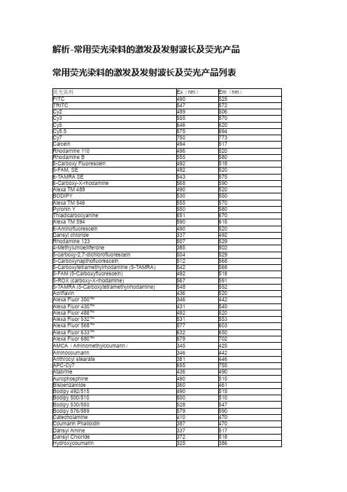
解析-常用荧光染料的激发及发射波长及荧光产品常用荧光染料的激发及发射波长及荧光产品列表西安瑞禧生物科技有限公司部分荧光相关产品列表:CY3CY5CY7CY3-NHS Cy3 NHS EsterCY5-NHS Cy5 NHS EsterCY7-NHS Cy7 NHS EsterCY3-N3CY5-N3CY7-N3CY3-MALCY5-MALCY7-MALCY3-COOHCY5-COOHCY7-COOHDSPE-PEG-CY5DSPE-PEG-CY7FITC-PEG-NH2FITC-PEG-COOHFITC-PEG-MALFITC-PEG-NHSFITC-PEG-BiotinRB-PEG-NH2RB-PEG-COOHRB-PEG-MALRB-PEG-NHSRB-PEG-BiotinCY3-PEG-NH2CY3-PEG-COOHCY3-PEG-MALCY3-PEG-NHSCY3-PEG-BiotinCY5-PEG-NH2CY5-PEG-COOHCY5-PEG-MALCY5-PEG-NHSCY5-PEG-BiotinFITC-Poly-L-lysineFITC-PLLFITC-dextranFITC-CM-dextranTRITC-dextranBSA-FITCBSA-TRITCFITC-Hyaluronic acidRhodamine B-Hyaluronic acid6-Carboxy Fluorescein; 6-FAM5(6)Carboxy fluorescein; 5(6)-FAM5-Carboxyfluorescein succinimidyl ester; 5-FAM, SE5-Carboxytetramethylrhodamine; 5-TAMRA5(6)-Carboxytetramethylrhodamine succinimidyl ester;5(6)-TAMRA, SE5-Carboxy-X-rhodamine; 5-ROX6-Carboxy-X-rhodamine; 6-ROX5(6)-Carboxy-X-rhodamine; 5(6)-ROX5(6)-Aminofluorescein5(6)-Carboxyfluorescein diacetate5-AminofluoresceinFluorescein isothiocyanateAMCA AlkyneAMCA AzideDBCO-Cy3DBCO-Cy55-FAM Alkyne5-FAM Azide6-Carboxy-X-Rhodamine,SE (NHS) 6-羧基-X-罗丹明琥珀酰亚胺酯5-TAMRA-Osu 5-羧基四甲基罗丹明琥珀酰亚胺酯Streptavidin-CY5.5Streptavidin-CY5Streptavidin-CY3Streptavidin-TRITCStreptavidin-FITCStreptavidin-thiolAvidin-CY3Avidin-CY5Avidin-CY5.5Avidin (without modification)Avidin-SepharoseAvidin-thiolAvidin-FITCAvidin-TRITCHuman serum albumin, Sepharose beads (HAS- Sepharose beads)Human serum albumin thiol (HSA-SH)Human serum albumin Biotin (HSA-Biotin) Human serum albumin TRITC (HSA-TRITC) Human serum albumin Fluorescein (HSA-FITC) Human serum albumin CY5 (HSA-CY5)Human serum albumin CY3 (HSA-CY3)Human serum albumin CY5.5 (HSA-CY5.5) Human serum albumin (HSA, without modification)小编YQ2020.12。
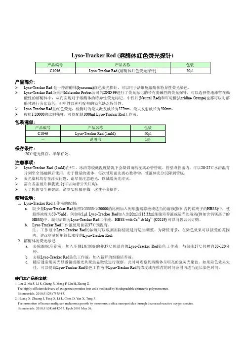
C1046 Lyso-Tracker Red 溶酶体红色荧光探针
包装 50μl
产品简介:
Lyso-Tracker Red 是一种溶酶体(lysosome)红色荧光探针,可以用于活细胞溶酶体特异性荧光染色。 Lyso-Tracker Red为采用Molecular Probes公司的DND 99进行了荧光标记的带有弱碱性的荧光探针,可以选择性地滞留在偏
酸性的溶酶体中,从而实现对于溶酶体的特异性荧光标记。中性红(Neutral Red)和吖啶橙(Acridine Orange)也都可以对溶 酶体进行荧光染色,但中性红和吖啶橙的染色缺乏特异性。 Lyso-Tracker Red呈红色荧光,检测时的最大激发波长为577nm,最大发射波长为590nm。 按照1:20000的比例稀释,可以配制1000ml Lyso-Tracker Red工作液。
佳,可以提高Lyso-Tracker Red染色工作液中Lyso-Tracker Red的浓度或在推荐的时间范围内适当延长染色时间。
使用本产品的文献:
1. Liu G, Ma S, Li S, Cheng R, Meng F, Liu H, Zhong Z. The highly efficient delivery of exogenous proteins into cells mediated by biodegradable chimaeric polymersomes. Biomaterials. 2010;31(29):7575-85.
?lysotrackerred为采用molecularprobes公司的dnd99进行了荧光标记的带有弱碱性的荧光探针可以选择性地滞留在偏酸性的溶酶体中从而实现对于溶酶体的特异性荧光标记
Lyso-Tracker Red (溶酶体红色荧光探针)
产品编号 C1046
荧光探针——精选推荐
荧光探针荧光探针(fluorescent probe)在化学传感、光学材料及⽣物检测和识别等领域得到了⼴泛的应⽤,并成为实现上述功能的⼀种主要的技术⼿段。
但以传统的有机荧光染料为主的荧光探针在应⽤中也存在⼀些难以克服的缺陷。
最近,⽆机发光量⼦点、荧光聚合物纳⽶微球、复合荧光⼆氧化硅纳⽶粒⼦等荧光纳⽶探针的相继出现,在⼀定程度上克服了传统有机荧光试剂的缺陷,为⽣物分析提供了新的发展领域,成为了近年来研究的热点,在此我想作⼀简单介绍,希望能起到抛砖引⽟的作⽤,如果⼤家觉得我有什么地⽅说错的话,欢迎批评指正!让我也从中受益!1、荧光纳⽶粒⼦的分类荧光纳⽶粒⼦是指可以发荧光的半导体纳⽶微晶体(量⼦点)或将荧光团(Fluorophore)通过包埋、共价键连接以及超分⼦组装等⽅式引⼊有机或⽆机纳⽶粒⼦中,并让纳⽶粒⼦承担有机⼩分⼦荧光染料的检测、标记等功能。
与传统的荧光染料相⽐,荧光纳⽶粒⼦具有更⾼的亮度和光稳定性,也能更加容易地实现⽔分散性和⽣物相容性。
另外,随着纳⽶制备技术的进⼀步提⾼,对纳⽶粒⼦的尺度的精确控制及对粒⼦功能化⼿段的⽇臻完善,这在很⼤程度上使荧光纳⽶粒⼦满⾜了化学传感器、⽣物探针等领域的要求。
⽬前荧光纳⽶粒⼦主要有⽆机发光量⼦点、荧光⾼分⼦纳⽶微球、复合荧光⼆氧化硅纳⽶粒⼦三⼤类。
1.1.量⼦点量⼦点(quantum dot, QD)⼜可称为半导体纳⽶微晶体,是由数百到数千个原⼦组成的⽆机纳⽶粒⼦,是⼀种由II-VI 族或者III-V 族元素组成的纳⽶颗粒。
⽬前研究较多的主要是CdX(X = S、Se、Te)。
量⼦点粒径很⼩,它们的电⼦和空⽳被量⼦限域,连续能带变成具有分⼦特性的分⽴能级结构,因此光学⾏为与⼀些⼤分⼦很相似,可以发射荧光。
量⼦点的体积⼤⼩严格控制着它的光谱特征。
量⼦点的晶体颗粒越⼩,⽐表⾯积越⼤,分布于表⾯的原⼦就越多,⽽表⾯的光激发的正电⼦或负电⼦受钝化表⾯的束缚作⽤就越⼤,其表⾯束缚能就越⾼,吸收的光能也越⾼,即存在量⼦尺⼨效应,从⽽使其吸收带蓝移,荧光发射峰也相应蓝移。
星锋生物-荧光染料总表
/product3.asp
2010-4-7
Cy 7-N-羟基琥珀酰亚胺酯
Dihydrorhodamine 123 二氢罗丹明123 Dihydrorhodamine 123 *5 mM solution in DMSO* 二氢罗丹明123(5 mM DMSO溶液) DAPI [4,6-Diamidino-2-phenylindole, dihydrochloride] 4,6-联脒-2-
28718-90-3 苯基吲哚二盐酸盐
C16H15N5 · 2HCl
/product3.asp
Байду номын сангаас
2010-4-7
星锋生物,专业的生化试剂供应商
页码,2/2
PI [Propidium iodide] 碘化丙啶 Hoechst 33258 荧光染料33258
25535-16-4 23491-45-4
Hoechst 33342 荧光染料33342
BAPTA, AM 胞内钙荧光探针BAPTA, AM BAPTA, tetrapotassium salt BAPTA四钾盐 BAPTA, tetrasodium salt BAPTA四钠盐
Fluo-3, AM 钙荧光探针Fluo-3, AM
23491-52-3 126150-97-8
SYBR Green II RNA *10,000X concentrate in DMSO* SYBR Green II 172827-25-7
RNA胶体染料(10,000×DMSO溶液)
5(6)-FAM [5-(and-6)-Carboxyfluorescein] *Mixed isomers* 5(6)-羧 72088-94-9
基荧光素(混合物)
【干货】LAMP检测各种常见染料一览
【干货】LAMP检测各种常见染料一览在过去的十年中,等温扩增(如HCA、MDA和环介导等温扩增LAMP)在即时护理(POC)诊断中发挥了越来越重要的作用。
等温扩增具有与qPCR相似的灵敏度和特异性,但是在快速、低成本和便携性方面具有优势,因此成为在资源有限环境下可用来进行现场检测的理想选择。
LAMP反应产物的检测方法包括:电泳法、浊度法和染料法,其中电泳法和浊度法是终点检测法,而染料法现在更为常用,可实现实时检测。
LAMP染料法中用的染料包括两种:•荧光染料:自发光荧光染料和嵌合型荧光染料•比色染料(colorimetric dye):pH指示剂本文整理了近十年来常用的几种染料,供各位同仁开发LAMP检测试时参考选择。
荧光染料1自发光荧光染料常见的自发光荧光染料有:羟基萘酚蓝HNB和钙黄绿素Calcein。
•羟基萘酚蓝(HNB)一种金属离子指示剂, 变色原理为:HNB与镁离子结合使得反应体系初始颜色为紫罗兰色,随着反应的进行,Mg2+与析出的焦磷酸根离子反应生成焦磷酸镁沉淀,羟基萘酚蓝失去了镁离子使得体系颜色变为天蓝色,而未反应的体系则仍保持着紫罗兰色。
•钙黄绿素(Calcein)使用钙黄绿素的时候体系中必须要添加Mn2+,钙黄绿素作为螯合剂会跟Mn2+结合处于猝灭状态,不显示荧光,LAMP反应过程中产生焦磷酸会与Mn2+结合解除淬灭状态,钙黄绿素与Mg2+结合发出黄绿荧光。
钙黄素的使用对反应体系当中的Mg2+的浓度要求很高,所以体系中Mg2+需要优化。
2嵌合型荧光染料常见的嵌合型荧光染料有:Sybr Green,EvaGreen,Syto。
这一类染料会渗入到DNA双链的小沟中,与双链DNA结合以后,荧光信号会增强 800~1000倍。
↓常见的荧光染料整理对比↓(点击看大图)2019年科研人员使用包括Sybr Green,EvaGreen和多种Syto 在内多达23种荧光染料进行LAMP检测平行测试,研究显示,Syto82 和Syto9是相对灵敏度最高、对检测没有抑制作用的荧光染料之一(1)。