
大脑的解剖结构和功能——布鲁德曼分区
. 精品 大脑的解剖结构和功能 ——布罗德曼分区系统
布罗德曼分区是一个根据细胞结构将大脑皮层划分为一系列解剖区域的系统。神经解剖学中所谓细胞结构(Cytoarchitecture),是指在染色的脑组织中观察到的神经元的组织方式。 布罗德曼分区1909年由德国神经科医生科比尼安·布洛德曼(Korbinian Brodmann)提出。根据皮质细胞的类型及纤维的疏密把大脑皮质分为52个区,并用数字给予表示。 Brodmann Area 1, BA1 Brodmann Area 2, BA2 Brodmann Area 3, BA3 位置:位于中央后回 (postcentral gyrus) 和前顶叶区 。 功能:分别为体感皮层内侧、末尾和前端区,BA1、BA2、BA3共同组成体感皮层 ; 具备基本体感功能 (first somatic sensory area) 接受对侧肢体的感觉传入。 Brodmann Area 4, BA4 位置: 位于中央前回(precentral gyrus),中央沟(central sulcus)的内侧面 功能:初级运动皮层(first somatic motor area),包含“运动小人”(motor homunculus )。 控制行为运动,与BA6 (前)和BA3 、BA2 、BA1、(后)相连,同时与丘脑腹外侧核相连。 体感小人(Somatosensory Homunculus ) 传入体感信息较多的身体区域获得的皮层代表区域较大。比如手部在初级体感皮层中的代表区域比背部的大。体感皮质定位可用“体感小人”(Somatosensory homunculus)来表示。 Brodmann Area 5, BA5 位置:位于顶叶前梨状皮质区(梨状皮质piriform cortex为下边缘皮质的组成部分)。 功能:与BA7形成体感联合皮层。 Brodmann Area 7, BA7 位置:位于顶叶皮质顶部,体感皮层后方,视觉皮层(visual area)上方。 功能:将视觉和运动信息联合起来;与BA5形成体感联合皮层;视觉-运动协调功能。 Sensory Areas ---------Somatosensory Association Area. 精品 位置:位于初级躯体感觉皮层后方(BA5、BA7) 功能:整合各种感觉传入触压觉、其它感觉;利用以往储存的感觉体验。 Brodmann Area 6, BA6 位置:位于额叶(frontal lobe),中央前回(precentral gyrus)前端区 功能 :与BA8共同构成前运动皮层;指导感官运动;辅助运动区SMA(控制身体的近端和躯干肌肉)
8 9 46 44 45 lateral 47 orbital 47 11 10 dorsolateral orbitofrontal, ventromedial, basal, orbital frontopolar, anterior, rostral dorsolateral ventrolateral posterior dorsolateral mid-dors
olateral . 精品 Brodmann Area 8, BA8 位置:位于前额叶(prefrontal cortex, PFC) 功能:与BA6共同构成前运动皮层(premotor cortex),包括额叶眼动区(frontal eye field, FEF ),控制眼球的随意运动,尤其与眼球的追随运动有关;运动规划 Brodmann Area 9, BA9 位置:位于前额叶(prefrontal cortex, PFC) Brodmann Area 10, BA10 位置:位于前额叶(prefrontal cortex, PFC),额下回(inferior frontal gyrus)顶部的延髓区 (上额回superior frontal gyrus和中额回middle frontal gyrus最前侧的部分) Brodmann Area 11, BA11 位置: 位于额叶内侧腹面(ventral intrafrontal cortex,前额皮层区 (眶回orbital frontal gyrus ,直回和上额回前侧的一部分) Association Areas – Prefrontal Cortex BA9、BA10、BA11共同构成前额叶皮层(prefrontal cortex)执行认知功能 功能: 思维thought和知觉Perception的所有方面; 信息的记忆memory和回忆 memories; 解决问题 problem solving ;情绪 emotion ;与前脑的边缘部分有密切的联系。
Brodmann Area 12, BA12 位置: 位于上额回和下前回之间的区域(额眶区) Brodmann Area 13, BA13 Brodmann Area 14, BA14 Brodmann Area 15, BA15 Brodmann Area 16, BA16.
精品 位置:位于岛叶(insular lobe) 功能: 处理收敛信息产生的一种感官经验(如厌恶、不安的感觉),与情感相关; 前岛叶有嗅觉(smell)、味觉(gustation)、边缘功能,同时后岛与听觉(auditory)、自主感觉-运动功能有关 ;还与上瘾(addiction)有关 Brodmann Area 17, BA17 位置: 位于枕叶内侧面(初级视觉皮层,纹状皮质striate cortex 、V1) 功能: 视觉信息加工的初步场所 ;方位柱、眼优势柱组织; 接收来自外侧膝状体(lateral geniculate body、丘脑后外侧的下方)的信息 沿背、腹途径将信息发送到其他视觉区 Brodmann Area 18, BA18 位置:位于枕叶,包括部分楔回(cuneate gyrus)、舌回(lingualis gyrus)和外侧枕叶回,部分纹外皮层(二级视觉皮层,V2,纹外视觉皮层) 功能:视觉处理(视觉联合皮层) Brodmann Area 19, BA19 位置:位于枕叶 功能:视觉处理; 与BA18构成视觉联合皮层 (Visual association cortices) Brodmann Area 20, BA20 位置:位于颞下回( inferior temporal gyrus) 功能:更高水平的信息处理; 视觉腹侧通路(ventral visual pathway) Sensory Areas – Ventral and Dorsal Streams 腹侧通路– 进入颞叶下部(BA20、BA37) › 负责物体、文字、面孔的识别,即“是什么” 背侧通路– 延伸至中央后回 (BA39、BA40) › 感知空间关系,即“在哪里” Brodmann Area 21, BA21 位置: 位于颞叶侧面,在BA20上方、BA40和BA41下方,颞中回区 功能:语言和听觉处理 Brodmann Area 22, BA22 位置:位于颞叶(temporal lobe) . 精品 功能 :后一部分包含威尔尼克区(包括颞上回、颞中回后部、缘上回supramarginal gyrus以及角回angular gyrus。是大脑书写中枢、视觉性语言中枢。威尔尼克区的损伤将产生严重的感觉性失语症。 ) ;负责语言理解。 Brodmann Area 23, BA23 位置:位于枕叶(occipital lobe) , 扣带皮层后端(posterior cingulate cortex) , 末端极限到达顶枕沟( parietooccipital sulcus ) 功能: 作为边缘系统的一部分,与杏仁核(amygdala)和眶额皮层和海马(hippocampus)相连接;参与情绪系统。 Brodmann Area 24, BA24 位置:位于扣带皮层腹节 功能:作为边缘系统的一部分,与杏仁核和眶额皮层和海马相连接;参与情绪系统 Brodmann Area 25, BA25 位置:位于膝下皮层 功能:与嗅觉有关 Brodmann Area 26, BA26 位置:压外区,位于扣带回“峡谷”处 功能:记忆系统的一部分;响应偶发事件 Brodmann Area 29, BA29 位置:压后扣带皮层 功能:记忆系统的一部分;响应偶发事件 Brodmann Area 30, BA30 位置:压部后颗粒皮质,扣带回的一部分 功能:记忆系统的一部分;响应偶发事件 Brodmann Area 27, BA27 位置: 海马旁回(parahippocampal gyrus)的延髓(medulla oblongata)部分 功能:记忆系统的一部分 Brodmann Area 28, BA28 位置:后内嗅皮层 Brodmann Area 31, BA31 . 精品 位置:上后扣带皮层 功能:情感处理和识别 Brodmann Area 32, BA32 位置:上前扣带皮层,位于顶叶 功能:负责决策制定的过程 Brodmann Area 34, BA34 位置: 前扣带皮层,位于海马旁回 Brodmann Area 35, BA35 位置:旁嗅皮层,位于海马旁回 Brodmann Area 36, BA36 位置:海马旁皮层 Brodmann Area 37, BA37 位置:梭状回,位于颞叶,颞下沟和侧副沟之间 功能:多模态联合;高阶对象和人脸识别(Fusiform face area, FFA) Brodmann Area 38, BA38 位置:颞极区,颞上回、颞中回的延髓部分 功能:与记忆和情感有关 Brodmann Area 39, BA39 位置: 角回,韦尼克区的一部分,位于顶叶 功能:语义加工 Brodmann Area 40, BA40 位置:缘上回,威尔尼克区的一部分,外侧沟上端末 功能:次级体感皮层;响应躯体刺激,完成结构区分任务 Brodmann Area 41, BA41 位置: 初级听觉皮层,位于颞叶,外侧沟内侧的前颞横回区 功能:早期听觉信息处理;低频率多集中在喙侧;高频率多集中在尾/内侧 Brodmann Area 42, BA42 位置:听觉联合皮层,位于颞叶,在外侧沟的边沿处 功能:早期听觉信息处理;低频率多集中在喙侧;高频率多集中在尾/内侧
端脑
内囊前肢
内囊
内囊膝 内囊后肢
内囊前肢
额桥束 丘脑前辐射 皮质核束 皮质脊髓束 皮质红核束 顶桥束 丘脑中央辐射 视辐射 枕桥束 听辐射 颞桥束
内囊膝
内囊肢
边缘系统 limbic system
海马和齿状回 三大主要结构 杏仁体 隔区
端脑的主体结构
大脑皮质 cerebral cortex 基底核basal nuclei
髓质
侧脑室 lateral ventricle
形端 和脑 分的 叶外
第一躯体运动区 第一躯体感觉区 听觉型语言中枢 视觉性语言中枢 视觉区
大 脑 皮 质 功 能 定 位
运动性语言中枢 书写中枢 听觉区 嗅觉区
武汉大学医学院人体解剖学教研室
Department of Anatomy Wuhan University School of Medicine
制作:戴冀斌
端脑 telencephalon
端脑的外形和分叶 大脑皮质的功能定位 端脑的内部结构
侧脑室 基底核 大脑皮质Brodmann分区 端脑的髓质
边缘系统
联系两侧半球的额、顶、枕、颞叶
连合纤维
2) 前连合 anterior commissure 3)穹窿 fornix 穹窿连合 fornical commissure
联络纤维 association fiber: 钩束 上纵束 下纵束 扣带
投射纤维 projection fibers
内囊 internal capsule
旧纹状体
壳 putamen
苍 白 球 globus pallidus
尾状核 背侧丘脑
豆状核
内囊
尾状核 背侧丘脑 壳 苍白球
大脑Brodmann分区
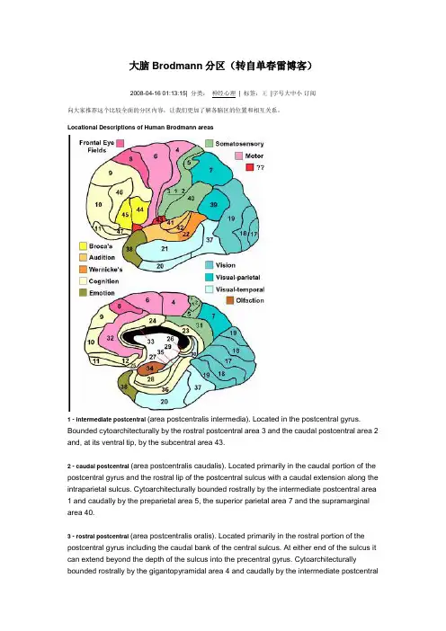
大脑Brodmann分区(转自单春雷博客)2008-04-16 01:13:15| 分类:神经心理| 标签:无|字号大中小订阅向大家推荐这个比较全面的分区内容,让我们更加了解各脑区的位置和相互关系。
Locational Descriptions of Human Brodmann areas1 - intermediate postcentral(area postcentralis intermedia). Located in the postcentral gyrus. Bounded cytoarchitecturally by the rostral postcentral area 3 and the caudal postcentral area2 and, at its ventral tip, by the subcentral area 43.2 - caudal postcentral(area postcentralis caudalis). Located primarily in the caudal portion of the postcentral gyrus and the rostral lip of the postcentral sulcus with a caudal extension along the intraparietal sulcus. Cytoarchitecturally bounded rostrally by the intermediate postcentral area 1 and caudally by the preparietal area 5, the superior parietal area 7 and the supramarginal area 40.3 - rostral postcentral(area postcentralis oralis). Located primarily in the rostral portion of the postcentral gyrus including the caudal bank of the central sulcus. At either end of the sulcus it can extend beyond the depth of the sulcus into the precentral gyrus. Cytoarchitecturally bounded rostrally by the gigantopyramidal area4 and caudally by the intermediate postcentralarea 1.4 - gigantopyramidal(area gigantopyramidalis). Located in the precentral gyrus. Cytoarchitecturally the caudal boundary with the rostral postcentral area 3 does not coincide precisely with the floor of the central sulcus but lies variably in the banks of the postcentral gyrus and the precentral gyrus. The area also does not extend in all cases to the cingulate sulcus medially or to the end of the central sulcus ventro-laterally. Bounded rostrally by the frontal agranular area 6.5 - preparietal(area praeparietalis). Occupies the superior parietal lobule and a portion of the postcentral gyrus, particularly on the medial aspect of the hemisphere. Bounded approximately by the cingulate sulcus on the medial aspect of the hemisphere and by the superior postcentral sulcus on the lateral aspect. Cytoarchitecturally bounded by the caudal postcentral area 2, the superior parietal area 7 and on the medial bank of the hemisphere by the gigantopyramidal area 4 and the dorsal posterior cingulate area 31.6 - agranular frontal(area frontalis agranularis). Located primarily in the caudal portions of the superior frontal gyrus and the middle frontal gyrus and the rostral portions of the precentral gyrus not occupied by the gigantopyramidal area 4. It extends from the cingulate sulcus on the medial aspect of the hemisphere to the lateral sulcus on the lateral aspect. Cytoarchitecturally bounded rostrally by the frontal region and caudally by the gigantopyramidal area 4.7 - superior parietal(area parietalis superior). Occupies much of the superior parietal lobule and some of the precuneus. Bounded approximately by the superior postcentral sulcus rostrally, the intraparietal sulcus laterally, the parieto-occipital sulcus caudally and, on the medial bank of the hemisphere, the subparietal sulcus. Cytoarchitecturally bounded rostrally by the preparietal area 5 and the caudal postcentral area 2; caudally by the peristriate area 19; and medially by the dorsal posterior cingulate area 31.8 - intermediate frontal(area frontalis intermedia). Located primarily in the superior frontal gyrus extending from the cingulate sulcus on the medial surface over the margin of the hemisphere to the middle frontal gyrus. Cytoarchitecturally bounded caudally by the agranular frontal area 6 and ventrally by the granular frontal area 9.9 - granular frontal(area frontalis granularis). Occupies portions of the superior frontal gyrus and the middle frontal gyrus. Its approximate boundary on the medial aspect of the hemisphere is the cingulate sulcus and, on the lateral aspect, the inferior frontal sulcus. Cytoarchitecturally bounded dorsocaudally by the intermediate frontal area 8, caudally by the agranular frontal area 6, and ventrally by the frontopolar area 10, the middle frontal area 46 and the opercular area 44.10 - frontopolar(area frontopolaris). Occupies the most rostral portions of the superior frontal gyrus and the middle frontal gyrus. On the medial aspect of the hemisphere it is bounded ventrally by the superior rostral sulcus. It does not extend as far as the cingulate sulcus.Cytoarchitecturally bounded dorsally by the granular frontal area 9, caudally by the middle frontal area 46, and ventrally by the orbital area 47 and by the frontopolar area 12.11 - prefrontal(area praefrontalis). Constitutes most of the orbital gyri, gyrus rectus and the most rostral portion of the superior frontal gyrus. Bounded medially by the inferior rostral sulcus and laterally approximately by the frontomarginal sulcus. Cytoarchitecturally bounded on the rostral and lateral aspects of the hemisphere by the frontopolar area 10, the orbital area 47, and the triangular area 45; on the medial surface it is bounded dorsally by the area 12 and caudally by the subgenual area 25.12 - prefrontal(area praefrontalis). Occupies the area between the superior rostral sulcus and the inferior rostral sulcus. Cytoarchitecturally bounded dorsally by the frontopolar area 10 and the dorsal anterior cingulate area 32; caudally, ventrally and rostrally it is bounded by the prefrontal area 11. (Originally described as part of prefrontal area 11 but not shown in the map, subsequently it was labeled as an independent area 12.)17 - striate(area striata). Part of the occipital lobe of the cerebral cortex that is defined on the basis of cyto- and myeloarchitecture, primarily by the band/stripe of Gennari.18 - parastriate(area parastriata). Located in parts of the cuneus, the lingual gyrus and the lateral occipital gyrus of the occipital lobe. Cytoarchitecturally bounded on one side by the striate area 17, from which it is distinguished by absence of a band/stripe of Gennari, and on the other by the peristriate area 19.19 - peristriate(area peristriata). Located in parts of the lingual gyrus, the cuneus, the lateral occipital gyrus and the superior occipital gyrus of the occipital lobe where it is bounded approximately by the parieto-occipital sulcus. Cytoarchitecturally bounded on one side by the parastriate area 18 which it surrounds. Rostrally it is bounded by the angular area 39 and the occipitotemporal area 37.20 - inferior temporal(area temporalis inferior). Corresponds approximately to the inferior temporal gyrus. Cytoarchitecturally bounded medially by the ectorhinal area 36, laterally by the middle temporal area 21, rostrally by the temporopolar area 38 and caudally by the occipitotemporal area 37.21 - middle temporal(area temporalis media). Corresponds approximately to the middle temporal gyrus. Bounded rostrally by the temporopolar area 38, ventrally by the inferior temporal area 20, caudally by the occipitotemporal area 37, and dorsally by the superior temporal area 22.22 - superior temporal(area temporalis superior). Corresponds approximately to the lateral and caudal two thirds of the superior temporal gyrus. Bounded rostrally by the temporopolar area 38, medially by the posterior transverse temporal area 42, ventrocaudally by the middle temporal area 21 and dorsocaudally by the supramarginal area 39.23 - ventral posterior cingulate(area cingularis posterior ventralis). Occupies most of the posterior cingulate gyrus adjacent to the corpus callosum. At the caudal extreme it is bounded approximately by the parieto-occipital sulcus. Cytoarchitecturally bounded dorsally by the dorsal posterior cingulate area 31, rostrally by the ventral anterior cingulate area 24, and ventrorostrally in its caudal half by the retrosplenial region.24 - ventral anterior cingulate(area cingularis anterior ventralis). Occupies most of the anterior cingulate gyrus in an arc around the genu of corpus callosum. Its outer border corresponds approximately to the cingulate sulcus. Cytoarchitecturally bounded internally by the pregenual area 33, externally by the dorsal anterior cingulate area 32, and caudally by the ventral posterior cingulate area 23 and the dorsal posterior cingulate area 31.25 - subgenual(area subgenualis). A narrow band located in the caudal portion of the subcallosal area adjacent to the paraterminal gyrus from which it is separated by the posterior parolfactory sulcus. It is bounded by the prefrontal area 11rostrally and by the paraterminal gyrus caudally.26 - ectosplenial(area ectosplenialis). A narrow band located in the isthmus of cingulate gyrus adjacent to the fasciolar gyrus internally. It is bounded externally by the granular retrolimbic area 29.28 - entorhinal(area entorhinalis). Located in the entorhinal area on the medial aspect of the temporal lobe. It and the dorsal entorhinal area 34 together constitute approximately the entorhinal area.29 - granular retrolimbic(area retrolimbica granularis). A narrow band located in the isthmus of cingulate gyrus. Cytoarchitecturally bounded internally by the ectosplenial area 26 and externally by the agranular retrolimbic area 30.30 - agranular retrolimbic(area retrolimbica agranularis). Located in the isthmus of cingulate gyrus. Cytoarchitecturally bounded internally by the granular retrolimbic area 29, dorsally by the ventral posterior cingulate area 23 and ventrolaterally by the ectorhinal area 36.31 - dorsal posterior cingulate(area cingularis posterior dorsalis). Occupies portions of the posterior cingulate gyrus and medial aspect of the parietal lobe. Approximate boundaries are the cingulate sulcus dorsally and the parieto-occipital sulcus caudally. It partially surrounds the subparietal sulcus. Cytoarchitecturally bounded rostrally by the ventral anterior cingulate area 24, ventrally by the ventral posterior cingulate area 23, dorsally by the gigantopyramidal area 4 and preparietal area 5 and caudally by the superior parietal area 7.32 - dorsal anterior cingulate(area cingularis anterior dorsalis). Forms an outer arc around the anterior cingulate gyrus. The cingulate sulcus defines approximately its inner boundary and the superior rostral sulcus its ventral boundary; rostrally it extends almost to the margin of the frontal lobe. Cytoarchitecturally bounded internally by the ventral anterior cingulate area 24,externally by medial margins of the agranular frontal area 6, intermediate frontal area 8, granular frontal area 9, frontopolar area 10, and prefrontal area 11.33 - pregenual(area praegenualis). A narrow band located in the anterior cingulate gyrus adjacent to the supracallosal gyrus in the depth of the callosal sulcus. Cytoarchitecturally bounded by the ventral anterior cingulate area 24 and the supracallosal gyrus.34 - dorsal entorhinal(area entorhinalis dorsalis). Located in the entorhinal area on the medial aspect of the temporal lobe. It and the entorhinal area 28 together constitute approximately the entorhinal area.35 - perirhinal(area perirhinalis). Located along the rhinal sulcus. Cytoarchitectually bounded medially by the entorhinal area 28 and laterally by the ectorhinal area 36.36 - ectorhinal(area ectorhinalis). Located primarily in the fusiform gyrus, with its medial boundary corresponding approximately to the rhinal sulcus. Cytoarchitecturally bounded laterally and caudally by the inferior temporal area 20, medially by the perirhinal area 35 and rostrally by the temporopolar area 3837 - occipitotemporal (area occipitotemporalis). Located primarily in the caudal portions of the fusiform gyrus and inferior temporal gyrus on the mediobasal and lateral surfaces at the caudal extreme of the temporal lobe. Cytoarchitecturally bounded caudally by the peristriate area 19, rostrally by the inferior temporal area 20 and middle temporal area 21 and dorsally on the lateral aspect of the hemisphere by the angular area 39.38 - temporopolar (area temporopolaris). Located primarily in the most rostral portions of the superior temporal gyrus and the middle temporal gyrus. Cytoarchitecturally bounded caudally by the inferior temporal area 20, the middle temporal area 21, the superior temporal area 22 and the ectorhinal area 36.39 - angular (area angularis). Corresponds to the angular gyrus surrounding the caudal tip of the superior temporal sulcus. Dorsally it is bounded approximately by the intraparietal sulcus. Cytoarchitecturally bounded rostrally by the supramarginal area 40, dorsally and caudally by the peristriate area 19, and ventrally by the occipitotemporal area 37.40 - supramarginal (area supramarginalis). Located primarily in the supramarginal gyrus surrounding the posterior ascending limb of lateral sulcus. Bounded approximately by the intraparietal sulcus, the inferior postcentral sulcus the posterior subcentral sulcus and the lateral sulcus. Cytoarchitecturally bounded caudally by the angular area 39, rostrally and dorsally by the caudal postcentral area 2, and ventrally by the subcentral area 43 and the superior temporal area 22.41 - anterior transverse temporal (area temporalis transversa anterior). Occupies the anterior transverse temporal gyrus in the bank of the lateral sulcus on the dorsal surface of the temporal lobe. Cytoarchitecturally bounded medially by the parainsular area 52 and laterally by the posterior transverse temporal area 42.42 - posterior transverse temporal (area temporalis transversa posterior). Located in the bank of the lateral sulcus on the dorsal surface of the temporal lobe. Cytoarchitecturally bounded medially by the anterior transverse temporal area 41 and laterally by the superior temporal area 22.43 - subcentral (area subcentralis). Occupies the postcentral gyrus and the precentral gyrus between the ventrolateral extreme of the central sulcus and the depth of the lateral sulcus at the insula. Its rostral and caudal borders are approximated by the anterior subcentral sulcus and the posterior subcentral sulcus respectively. Cytoarchitecturally bounded rostrally by the agranular frontal area 6 and caudally, for the most part, by the caudal postcentral area 2 and the supramarginal area 40.44 - opercular (area opercularis). Corresponds approximately to the opercular part of the inferior frontal gyrus. Bounded caudally by the inferior precentral sulcus and rostrally by the anterior ascending limb of lateral sulcus. It surrounds the diagonal sulcus. In the depth of the lateral sulcus it borders on the insula. Cytoarchitectonically bounded caudally and dorsally by the agranular frontal area 6, dorsally by the granular frontal area 9 and rostrally by the triangular area 45.45 - triangular (area triangularis). Occupies the triangular part of the inferior frontal gyrus and, surrounding the anterior horizontal limb of lateral sulcus, a portion of the orbital part of inferior frontal gyrus. Bounded caudally by the anterior ascending limb of lateral sulcus, it borders on the insula in the depth of the lateral sulcus . Cytoarchitectonically bounded caudally by the opercular area 44, rostrodorsally by the middle frontal area 46 and ventrally by the orbital area 47.46 - middle frontal (area frontalis media). Occupies approximately the middle third of the middle frontal gyrus and the most rostral portion of the inferior frontal gyrus. Cytoarchitecturally bounded dorsally by the granular frontal area 9, rostroventrally by the frontopolar area 10 and caudally by the triangular area 45.47 - orbital (area orbitalis). Surrounds the caudal portion of the orbital sulcus from which it extends laterally into the orbital part of inferior frontal gyrus. Cytoarchitectonically bounded caudally by the triangular area 45, medially by the prefrontal area 11, and rostrally by the area frontopolaris 10.48 - retrosubicular (area retrosubicularis). Located on the medial surface of the temporal lobe. Cytoarchitectually bounded rostrally by the perirhinal area 35 and medially by the presubiculum. (While described by Brodmann, it was not included in his areal maps of human cortex.)52 - parainsular (area parainsularis) Located in the bank of the lateral sulcus on the dorsal surface of the temporal lobe. Its medial boundary corresponds approximately to the junction between the temporal lobe and the insula. Cytoarchitecturally bounded laterally by the anterior transverse temporal area 41.13, 14, 15, 16, 27, 49, 50, 51 - monkey only。
大脑皮质结构与功能分区

★优势半球: 左半球---语言、意识、数学分析,抽象思维等; 右半球---非语言信息、音乐、图形、时空概念,形象
思维, 情感性 信息处理。
一些人沉着稳重,善于思考,言语逻辑性 强,但缺乏生动性,可能与其左半球功能 优势有关;另一些人情感丰富,言语形象 生动,富有感染力,可能与其右半球功能 优势有关。
△海马结构:海马(Ammon horn)齿状回、下托复合 体和内嗅皮质。
△边缘叶:
隔区(胼胝体下区+终板 旁回),扣带回,海马 结构,海马旁回,钩, 岛叶前部
多形层 锥体层 分子层
CA1 CA3
齿状回
★边缘系统组成:
边缘叶+与之相关皮质下结构(如杏仁核、隔核、下 丘脑、丘脑前核、缰核、中脑被盖等)
大脑皮质的神经元——
大脑皮质的神经元都是多极神经元,按其细胞的形 态分为锥体细胞、颗粒细胞和梭形细胞三大类。
锥体细胞(pyramidal cell):数量较多,可分大、 中、小三型。胞体呈锥形或三角形,直径约15120um,属于GolgiⅠ型神经元,尖端发出一条较 粗的主树突,伸向皮质表面,沿途发出许多小分支, 胞体还向四周发出一些水平走向的树突。轴突自胞 体底部发出,长短不一,短者不越出所在皮质范围, 长者离开皮质,进入髓质(白质),组成投射纤维 (下行至脑干或脊髓)或联合纤维(到同侧或对侧 的另一皮质区)。因而,锥体细胞是大脑皮质的主 要投射(传出)神经元。
大脑半球外侧面观 Brodmann分区
大脑半球内侧面观 Brodmann分区
★躯体运动区:位于中央前回及中央旁小叶前部 (4区),是支配对侧躯体随意运动的中枢。它 主要接受来自对侧骨骼肌、肌腱和关节的本体感 觉冲动,以感受身体的位置、姿势和运动感觉, 并发出纤维,即锥体束控制对侧骨骼肌的随意运 动。
brodmann大脑皮层分区
Brodmann大脑皮层分区是德国神经解剖学家Korbinian Brodmann 于1909年提出的一种关于大脑皮层分区的理论。
这一分区方案被广泛应用于解剖学、神经科学和临床医学领域,并对了解大脑结构和功能起着重要作用。
本文将详细介绍Brodmann大脑皮层分区的背景、原理和应用,旨在帮助读者全面了解这一重要的神经科学理论。
一、背景Korbinian Brodmann是20世纪初期的神经解剖学家,他在研究大脑皮层时发现不同的脑区在细胞结构和组织类型上存在巨大的差异。
为了更好地描述和理解这些差异,Brodmann开始系统地研究大脑皮层的细胞组织结构,并最终提出了大脑皮层分区的理论。
这一理论基于对大量人类和动物大脑的显微镜观察和测量,被视为解剖学上关于大脑皮层最具权威的分类系统之一。
二、原理Brodmann大脑皮层分区的原理基于神经元的细胞结构和分布。
根据Brodmann的研究,大脑皮层中的神经元可以根据其形态、密度和分布分为不同的类型,不同类型的神经元聚集在一起形成具有特定功能的脑区。
Brodmann通过对大脑组织进行显微镜观察和测量,将大脑皮层分为了52个不同的区域,每个区域有其独特的细胞结构和功能特征。
这一分区方式为后续的大脑研究奠定了重要的基础,也为神经科学领域的发展做出了重要贡献。
三、应用Brodmann大脑皮层分区的理论在解剖学、神经科学和临床医学领域有着广泛的应用。
它为解剖学和神经科学研究提供了重要的工具和参考标准。
研究人员可以利用Brodmann的分区方式对大脑进行定量和定性的分析,揭示不同区域的功能和相互通联,从而深入了解大脑的结构和功能。
Brodmann的分区方案对于临床诊断和治疗也具有重要意义。
医生可以根据大脑分区的特定功能特征,判断患者的脑损伤位置和程度,为临床治疗提供重要参考。
现代的神经影像技术也常常结合Brodmann的分区方式,帮助医生观察和分析大脑结构,对各种疾病进行诊断和治疗。
大脑功能分区(整理部分)()
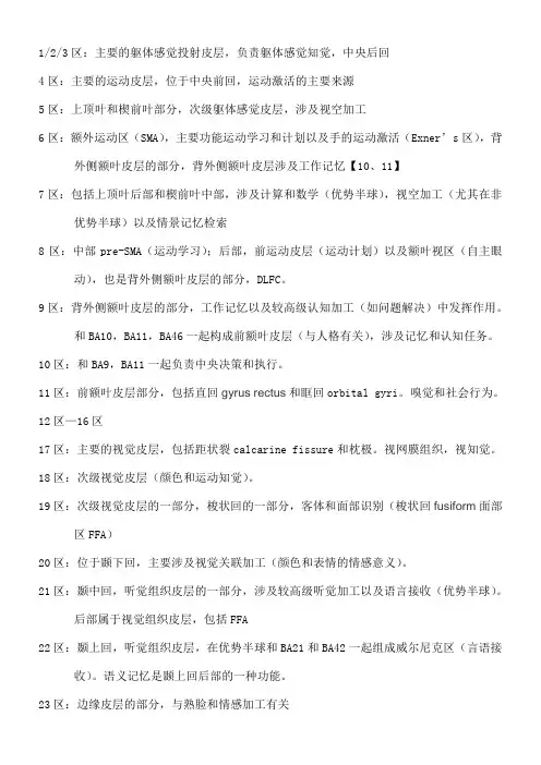
1/2/3区:主要的躯体感觉投射皮层,负责躯体感觉知觉,中央后回4区:主要的运动皮层,位于中央前回,运动激活的主要来源5区:上顶叶和楔前叶部分,次级躯体感觉皮层,涉及视空加工6区:额外运动区(SMA),主要功能运动学习和计划以及手的运动激活(Exner’s区),背外侧额叶皮层的部分,背外侧额叶皮层涉及工作记忆【10、11】7区:包括上顶叶后部和楔前叶中部,涉及计算和数学(优势半球),视空加工(尤其在非优势半球)以及情景记忆检索8区:中部pre-SMA(运动学习);后部,前运动皮层(运动计划)以及额叶视区(自主眼动),也是背外侧额叶皮层的部分,DLFC。
9区:背外侧额叶皮层的部分,工作记忆以及较高级认知加工(如问题解决)中发挥作用。
和BA10,BA11,BA46一起构成前额叶皮层(与人格有关),涉及记忆和认知任务。
10区:和BA9,BA11一起负责中央决策和执行。
11区:前额叶皮层部分,包括直回gyrus rectus和眶回orbital gyri。
嗅觉和社会行为。
12区—16区17区:主要的视觉皮层,包括距状裂calcarine fissure和枕极。
视网膜组织,视知觉。
18区:次级视觉皮层(颜色和运动知觉)。
19区:次级视觉皮层的一部分,梭状回的一部分,客体和面部识别(梭状回fusiform面部区FFA)20区:位于颞下回,主要涉及视觉关联加工(颜色和表情的情感意义)。
21区:颞中回,听觉组织皮层的一部分,涉及较高级听觉加工以及语言接收(优势半球)。
后部属于视觉组织皮层,包括FFA22区:颞上回,听觉组织皮层,在优势半球和BA21和BA42一起组成威尔尼克区(言语接收)。
语义记忆是颞上回后部的一种功能。
23区:边缘皮层的部分,与熟脸和情感加工有关24区:边缘叶的另一部分,涉及躯体感觉的情感评价(痛)25区:嗅觉和工作记忆加工是该区多种可能的功能的两种,包括终板lamina terminalis 和前穿孔。
大脑皮质功能定位

➢书写中枢:优势侧额中回后部/ 8、9区
➢运动性语言中枢:Broca运动语言中枢,优势侧额下回后部(岛盖 部)/44和45区,管理语言运动。
➢额叶前部(联络区):额极,三个额回大部及眶回和额内侧回/912区,与记忆、判断、抽象思维、情感和冲动行为有关。
大体说来,大脑左半部分主要处理语言、逻辑思维、 分析综合和计算功能,即所谓的学术学习部分。大脑右 半部分主要在音乐、美术、综合能力、空间、几何图形 和人物面容的识别占优势,即所谓的创造性活动。
•额叶:与躯体运动、发音、语言及高级思维活动有关。 •顶叶:与躯体感觉、味觉、语言等有关。 •颞叶:与听觉、语言和记忆功能有关。 •枕叶:与视觉信息的整合有关。 •岛叶:与内脏感觉和运动有关。 •边缘系统:高级神经、精神和内脏的活动 。
Brodmann分区法 (52区)
大脑皮质Brodmann 分区的名称、部位和主要功能
Brodmann分区 (BA)
1
大脑皮质部位
顶叶中央后回,又名中央后回中间部
机能定位
第一躯体感觉区
2
顶叶中央后回,又名中央后回尾侧部 第一躯体感觉区
3
顶叶中央后回,又名中央后回吻侧部 第一躯体感觉区
4
额叶后部,中央前回
颞叶后部,梭状回一部
参与视觉的认知
颞叶前极
参与行为、情绪、决定等过程
颞、枕、顶叶交界处的角回
参与语言及空间定位,理解看到的文字 符号意义
顶叶下部的缘上回
参与空间定位及语言功能,运用中枢
颞 叶 颞 上 回 后 部 的 颞 横 回 , 又 名 听觉初级中枢 Heschl‘s 回
颞叶后部围绕41区的部分
大脑功能分区简介
大脑功能分区简介大脑功能分区简介人的大脑分为左右两半球,体积占中枢神经系统总体积的一半以上,重量约为脑的总重量的60%左右。
从进化的观点看,大脑比脑干出现的晚,是各种心理活动的中枢。
大脑的表面布满深浅不同的沟或裂。
沟裂简隆起的部分称为脑回(gyrus)。
有三条大的沟裂,即中央沟(central sulcus)、外侧裂和顶枕裂,这些沟裂将大脑分为额叶、顶叶、枕叶和颞叶几个区域。
在每一叶内,一些较细小的沟裂又将大脑表面分成许多回。
如额叶的额上回、额中回、额下回、中央前回;颞叶的颞上回、颞中回和颞下回;顶叶的中央后回等大脑皮层可以分为不同的区域,分别负责不同的功能,根据前人的研究成果,我们可以把大脑皮层分为几个功能区域。
而与我们的皮纹有关的则有精神功能区、视觉区、听觉区、机体感觉区、语言区等。
视觉区(visual area)位于顶枕裂后面的枕叶内。
属布鲁德曼的第十七区,他接受在光刺激的作用下由眼睛输入的神经冲动,喊声初级形式的视觉,如对光的察觉等。
若大脑两半球的视觉区受到破坏,即使眼睛的功能正常,人也将完全丧失视觉而成为全盲。
听觉区(auditory area)位于颞叶的颞横回处,属布鲁德曼的第四十一、四十二区。
它接受在声音的作用下由耳朵传传入的神经冲动,产生初级形式的听觉,如对声音的察觉等。
若破坏了大脑两半球的听觉区,即使双耳功能正常,人叶将完全丧失听觉而成为全聋。
机体感觉区(somato-sensory area)位于中央沟后面的一条狭长区域内,属布鲁德曼的第一、二、三区。
它接受由皮肤、肌肉和内脏器官传入的感觉信号,产生触压觉、温度觉、痛觉、运动觉和内脏感觉等。
躯干四肢在体感区的投射言语区:对大多数人来数,言语区(speech area)主要定位在大脑左半球,它由较广大的区域组成。
若损坏了这些区域将引起各种形式的失语症。
在做半球额叶的后下方,靠近外侧裂处,由一个言语运动区,即布鲁德曼第四十四、四十五区,亦称之为布洛卡区(Broca’s area),它通过邻近的运动区控制说话时的舌头和颚的运动。
【影像视界】值得收藏的大脑分区及其功能
【影像视界】值得收藏的大脑分区及其功能大脑各区及功能额叶运动中枢、言运(布)、视觉(眶回)、嗅觉(嗅三角)、听觉、注意、问题解决、动机、行为程序制定(计划)、人格、记忆、情绪(左积右消)等高级认知活动、顶叶初级感觉区(疼痛、触压、品尝、温度)枕叶视中枢、言视中枢(角回)、视-听跨通道联合(角回)、记忆(近期,与视觉表象有关)、运动知觉(与视觉有关)颞叶听觉区、言听(威-语言的接受)、嗅觉中枢、味觉中枢、记忆、情绪间脑丘脑-痛觉、睡眠觉醒、除嗅觉外的外界感官输入中转(视听触味)下丘脑-调节交感、副交感、进食饮水、情绪小脑身体平衡、协调动作边缘系动物本能活动、注意(注意神经元)统(大脑内侧面最深处边缘一些结构)脑干延脑-生命中枢中脑-视听反射中枢、身体姿势、随意运动(红核-舞蹈症、黑质-手脚动作不协调、面部表情呆板)桥脑-中枢外周的桥梁、睡眠扣带回:(前下部)情绪加工、(后上部)认知功能海马:记忆(LTP)、嗅觉、情绪背景调节(损害不该出现情绪时闹情绪)杏仁核:情绪、厌恶学习、危险线索识别脑干网状结构:情绪产生基础、上行网状结构-维持觉醒、注意、下行-调节肌肉紧张弓形束:连接布和威,理解语义(因为不能接受来自威的信息了);左半球顶-枕部及颞-顶部:同时性空间图式-语义性失语症大脑半球内侧面大脑半球的上外侧面观Npt影像园Npt影像园大脑半球底部内面人的大脑分为左右两半球,体积占中枢神经系统总体积的一半以上,重量约为脑的总重量的60%左右。
从进化的观点看,大脑比脑干出现的晚,是各种心理活动的中枢。
大脑的表面布满深浅不同的沟或裂。
沟裂简隆起的部分称为脑回(gyrus)。
有三条大的沟裂,即中央沟(central sulcus)、外侧裂和顶枕裂,这些沟裂将大脑分为额叶、顶叶、枕叶和颞叶几个区域。
在每一叶内,一些较细小的沟裂又将大脑表面分成许多回。
如额叶的额上回、额中回、额下回、中央前回;颞叶的颞上回、颞中回和颞下回;顶叶的中央后回等大脑皮层可以分为不同的区域,分别负责不同的功能,根据前人的研究成果,我们可以把大脑皮层分为几个功能区域。
大脑皮质的分区及机能
大脑皮质的分区及机能大脑的机能主要是接受下级中枢的神经冲动,引起大脑皮质有关区域神经细胞的兴奋,产生对特定刺激的感觉及支配躯体和四肢的运动。
大脑皮质具有整合的功能,机体能对事物的作用产生知觉、联想、估价及发出行动的信息,而这种行动信息可以通过神经冲动经传出神经到达有关的效应器(如肌肉、肌腱组织等),产生特定的行为反应;大脑皮质具有分析与综合的功能,各种感官接收的信息传到中枢,中枢即依据信息的意义去主动调节感受器,以便有选择性和更有效地去了解这些信息。
此外,从中枢传出的指令引起效应器的运动后,这种动态也会反馈给中枢,以便中枢对运动做进一步的调节。
1909年,德国解剖学家布鲁德曼通过对大脑皮质各区域的研究,将大脑皮质分为不同的机能区。
(1)视觉区。
视觉区位于顶枕裂后面的枕叶内,属布鲁德曼的第十七区。
视觉区接受在光线的作用下由眼睛输入的神经冲动,产生初级形式的视觉,如对光的察觉等。
若两侧大脑半球的视觉区均受到破坏,即使患者眼睛的功能并无异常,也将完全丧失视觉,即全盲。
(2)听觉区。
听觉区位于颞叶的颞横回处,属布鲁德曼的第四十一、四十二区。
听觉区接受在声音的作用下由耳朵传入的神经冲动,产生初级形式的听觉,如对声音的察觉等。
若破坏了两侧大脑半球的听觉区,即使双耳功能正常,患者也将完全丧失听觉,甚至全聋。
(3)机体感觉区。
机体感觉区位于中央沟后面的一条狭长区域内,属布鲁德曼的第一、二、三区。
机体感觉区接受由皮肤、肌肉和内脏器官传入的感觉信号,产生触压觉、温度觉、痛觉、运动觉和内脏感觉等。
(4)言语区。
对大多数人来说,言语区主要定位在大脑左半球。
言语区有较广大的区域组成,若损坏了这些区域,将引起各种形式的失语症。
①左侧大脑半球额叶的后下方,靠近外侧沟处为运动性语言中枢,即布鲁德曼第四十四、四十五区,又称布洛卡区,该区域通过邻近的运动区控制说话时舌头和上颚的运动。
布洛卡区受到损伤会引发运动性失语症,导致患者说话不流利,话语中常常遗漏功能,因而形成“电极式”语言。
大脑皮质结构与功能分区4
大脑皮质结构与功能分区4大脑皮质结构与功能分区4大脑皮质是大脑最外层的一层薄壳,其结构和功能分区的研究对于理解大脑的功能及其相关疾病具有重要意义。
现今学界普遍认同的大脑皮质结构和功能分区有多种,包括布罗德曼分区法、Jülich-Brain分区法、Desikan-Killiany分区法等。
以下将以布罗德曼分区法为例,介绍大脑皮质结构和功能分区的一些基本概念和内容。
布罗德曼分区法是一种基于脑图像数据和神经系统表型数据库的大脑皮质结构和功能分区方法。
根据这个方法,人类大脑皮质被划分为多个区域,每个区域都具有相对独特的功能。
这些区域的边界是根据大脑皮质的结构和功能特征来确定的。
根据布罗德曼分区法,大脑皮质可以分为两个大的区域:大脑前额叶和大脑后颞叶。
大脑前额叶主要与决策、情绪调节、动机控制等高级认知功能相关。
大脑后颞叶主要参与听觉、语言、视觉和空间导航等感知和认知功能的处理。
在大脑前额叶区域中,主要包括背外侧前额叶、背内侧前额叶和顶外侧前额叶。
背外侧前额叶与注意力调控、工作记忆和认知控制相关。
背内侧前额叶在认知控制和冲动控制中起着重要作用。
顶外侧前额叶主要参与语言、社交情绪和认知跳跃等高级认知功能。
大脑后颞叶主要分为先上后内侧颞叶、先下后内侧颞叶和后外侧颞叶。
先上后内侧颞叶主要与听觉信息的处理和认知控制相关。
先下后内侧颞叶参与语言、记忆和情感调节等功能。
后外侧颞叶在语言和视觉信息处理中发挥重要作用。
除了前额叶和后颞叶,布罗德曼分区法还将大脑皮质划分为顶叶、枕叶和扣带回。
顶叶主要与视觉注意力、工作记忆和情绪调节等认知功能相关。
枕叶参与视觉信息处理和空间导航等功能。
扣带回主要与视觉和运动等感知和运动控制有关。
除了上述大的区域划分外,布罗德曼分区法还对大脑皮质进行了更细致的分区,将大脑皮质划分为数十个不同的细分区域,每个区域都有其独特的结构和功能。
这些细分区域的研究为我们理解大脑的功能及其相关疾病提供了更详细的信息。