neural stem cell self-renewal
悬浮培养与贴壁培养方式神经干细胞分化表型特点

悬浮培养与贴壁培养方式神经干细胞分化表型特点刘友;刘希光;李爱民【摘要】目的观察神经干细胞在悬浮培养与贴壁培养方式下的形态学特点及分化表型.方法新生6 h内的大鼠皮质应用无血清悬浮培养与贴壁培养神经干细胞(NSCs)技术培养,观察神经干细胞的形态学特点.体外胎牛血清(FBS)诱导NSCs分化,通过巢蛋白(Nestin)和GFAP免疫荧光染色鉴定分化表型.台盼蓝活细胞计数法比较细胞增殖能力.结果新生大鼠皮质应用无血清悬浮培养可获得稳定的NSCs细胞球,应用贴壁培养形成长梭状NSCs,NSCs巢蛋白免疫荧光染色结果呈阳性.体外FBS诱导分化后,神经球内开始出现放射状的神经纤维丝,免疫荧光染色可见GFAP 阳性细胞向外伸展以及Nestin阳性细胞边集.贴壁的NSCs分化成星形胶质细胞,免疫荧光染色可见Nestin和GFAP染色阳性细胞.在相同条件下,应用悬浮培养在第2~3d时增殖速度最大,而以贴壁培养方式培养出的神经干细胞在第4~5 d时增殖速度最大.结论应用无血清培养物B27与bFGF、EGF组合的NSCs完全培养基通过悬浮培养法与贴壁培养法,都可以成功在体外培养出NSCs;悬浮培养法培养NSCs在早期增殖速率上优于贴壁培养法.%Objective To observe the morphological characteristics and differentiation phenotype of neural stem cells in suspension culture and adherent culture .Methods The neural stem cells ( NSCs) of rat cortex was cultured by serum-free suspension culture and adherent culture . The morphological characteristics of neural stem cells were observed .In vitro fetal bovine serum (FBS) induced differentiation of NSCs,and differentiated phenotypes were identified by Nestin and GFAP immunofluorescence staining .Proven blue viable cell count method compared cell proliferation ability .Results The neonatal ratcortex was cultured in serum-free suspension to obtain stable NSCs cell pellets .Adherent culture was used to form long fusiform NSCs .NSCs were positive for nestin immunofluorescence staining .In vitro, FBS induced differentiation , radial nerve fibers began to appear in the nerve ball , immunofluorescence staining showed GFAP positive cells outward and Nestin positive cells set .NSCs were stained into astrocytes , and Nestin and GFAP staining positive cells were observed by immunofluorescence staining .The proliferation rate of the neural stem cells cultured in the same way was the highest at the 4th ~3rd day,and the proliferation rate was the highest at the 4th ~5th day.Conclusions The neural stem cells can be successfully cultured in vitro by suspension culture and adherent culture using the serum -free culture medium B27 and bFGF,EGF combined neural stem cells .Cultivation of neural stem cells by suspension culture was superior to adherent culture in early proliferation rate .【期刊名称】《临床神经外科杂志》【年(卷),期】2017(014)005【总页数】5页(P385-389)【关键词】神经干细胞;悬浮培养;贴壁培养【作者】刘友;刘希光;李爱民【作者单位】222002 连云港,徐州医科大学附属连云港医院神经外科;222002 连云港,徐州医科大学附属连云港医院神经外科;222002 连云港,徐州医科大学附属连云港医院神经外科【正文语种】中文【中图分类】R741.05成年哺乳动物大脑的急性或慢性损伤通常与持续功能性缺陷相关,其再生潜力和重建缺失神经结构的能力有限[1]。
常用英文及缩写

AABB American association of blood bank 美国血库协会G-csf Granulocyte colony-stimulating factor 人类粒细胞集落刺激因子PBMC Peripheral blood mononuclear cell 外周血单个核细胞PBSC Peripheral blood stem cell 外周造血干细胞BMT Bone marrow transplantation 骨髓移植GTP Good tissue practice 人体细胞组织优良操作规范GLP Good laboratory practice 优良实验室规范GMP Good manufacturing practice 优质生产规范HPC Hematopoietic progenitor cell定向造血干细胞(造血祖细胞)CB Cord blood 脐带血HLA Human leukocyte antigen 人类白细胞抗原SAB Sciene advisory board 科学咨询委员会SOP Standard operating procedure 标准作业程序IPS Induced pluripotent stem cell 诱导多能干细胞ips cellBDC Blood donor centers 献血中心BB Blood bands 血库ASH American society of hematology美国血液病学会AAHHPBM Patient blood management certification患者血液管理认证CAP College of American pathologists 美国病理学会IACT International society of cell therapy国际细胞治疗学会NMDP National marrow donor program 美国国家骨髓库WMDA World marrow donor association 世界骨髓捐赠协会FACT F 美国细胞治疗基金会NIH National Institutes of Health美国国立卫生研究院ISSCR International Society for Stem Cell Research国际干细胞研究协会ISCF International Stem Cell Forum国际干细胞论坛WMDA World Marrow Donor Association世界骨髓捐赠者协会NMDP National Marrow Donor Program美国骨髓库CRISPR/case9Clustered regularly interspaced short palindromic repeats/case9基因编辑技术IVF In vitro fertilization体外受精CAR-TChimeric Antigen Receptor T cell嵌合抗原受体T细胞GVHD graft-versus-host-disease移植物抗宿主病ASHAmerican Society of Hematology美国血液学会CRS cytokine-release syndrome细胞因子释放综合征PD-1 Programmed Cell Death Protein 1-Ligand细胞程序性死亡蛋白配体1NIPTNon-invasive Prenatal Testing无创胎儿产前检测NGS Next Generation Sequencing下一代测序High-Throughput Sequencing高通量测序HGP Human Genome Project人类基因组计划JPM JP Morgan Healthcare Conference JP摩根医疗健康年会(john.pierpont.morgan)MSC Mesenchymal stem cells 骨髓间充质干细胞MRT Mitochondrial-replacement techniques线粒体替换技术PrPPlatelet Rich Plasma富(高浓度)血小板血浆ABI Autologous Blood Injection 自体血注射ACP Autologous Conditioned Plasma自体血离子化SCI Scientific Citation Index科学文献索引CD Cluster differentiation分化群集CD Cluster of Differentiation白细胞分化抗原MHC Major Histocompatibility Complex主要组织相容性复合体常用单词Accredited认证的Facilities机构Molecular testing 分子检测Cellular therapies细胞疗法Somatic cell 体细胞Immunohematology reference免疫细胞学参考Molecular testing 分子检测Perioperative 围手术期AABB价值观The pursuit of excellence 追求完美Focus on the patient and donor关注患者和捐赠人Integrity诚信Transparency透明Consensus building建立共识Innovation创新CRISPR-Cas9 基因敲除Genes-edited 基因编辑Stem cellsStem cells are cells that have the capacity to self-renew by dividing and to develop into more mature, specialised cells. Stem cells can be unipotent, multipotent, pluripotent or totipotent, depending on the number of cell types to which they can give rise.Related SubjectsAdult stem cellsAdult stem cells are cells in the adult that can both self-renew by dividing and give rise to more specialised cell types present in the tissue in which they reside. They serve to maintain and repair the tissue.AgeingAgeing is the process during which structural and functional changes accumulate in an organism as a result of the passage of time. The changes manifest as a decline from the organism’s peak fertility and physiological functions until death.Cancer stem cellsCancer stem cells are rare immortal cells within a tumour that can both self-renew by dividing and give rise to many cell types that constitute the tumour, and can therefore form tumours.Such cells have been found in various types of human tumours and might be attractive targets for cancer treatment.Embryonic germ cellsEmbryonic germ cells are the cells in the embryo that give rise to the reproductive cells –gametes –of sexually reproducing organisms. In animals, male gametes are sperm cells and female gametes are egg cells, also known as ova.Embryonic stem cellsEmbryonic stem cells are pluripotent cells isolated from the inner cell mass of a blastocyst, the early mammalian embryo that implants into the uterus. Embryonic stem cells self-renew by dividing and can differentiate into any specialised cell of the body, but not extra-embryonic tissues such as the placenta.Epigenetic memoryThe epigenetic memory of a cell defines the set of modifications to the cell's deoxyribonucleic acid (DNA) that do not alter the DNA sequence, and have been inherited from the cell from which it descends. Such modifications can alter gene expression and therefore the properties and behaviour of the cell.Haematopoietic stem cellsHaematopoietic stem cells are progenitor cells that have the ability to both generate all types of blood cells, including those of the myeloid and lymphoid lineages, and to replace themselves. In adults, they mainly reside in the bone marrow.Heart stem cellsHeart stem or progenitor cells are multipotent cells residing in the adult mammalian heart that are capable of self-renewing and generating coronary vessels and heart muscle cells called cardiomyocytes. Heart stem cells can contribute to new cardiomyocyte formation following experimental myocardial infarction in mice.Intestinal stem cellsIntestinal stem cells are multipotent adult stem cells, which in mammals reside in the base of the crypts of the adult intestine. Intestinal stem cells continuously self-renew by dividing and differentiate into the specialised cells of the intestinal epithelium, which renews throughout life. Mammary stem cellsMammary stem cells are multipotent adult stem cells that reside in the mammary gland, can self-renew by dividing and can differentiate into all specialised mammary epithelial cells. Mammary stem cells drive the development of the mammary gland during puberty and are responsible for its expansion during pregnancy.Mesenchymal stem cellsMesenchymal stem cells are multipotent adult stem cells that are present in multiple tissues, including umbilical cord, bone marrow and fat tissue. Mesenchymal stem cells can self-renew by dividing and can differentiate into multiple tissues including bone, cartilage, muscle and fat cells, and connective tissue.Multipotent stem cellsMultipotent stem cells are cells that have the capacity to self-renew by dividing and to develop into multiple specialised cell types present in a specific tissue or organ. Most adult stem cells are multipotent stem cells.Muscle stem cellsMuscle stem cells are adult stem cells, present in skeletal muscle tissue, which can self-renew and are capable of giving rise to skeletal muscle cells. These stem cells are activated in response to muscle injury to regenerate damaged muscle tissue.Neural stem cellsNeural stem cells are multipotent adult stem cells present in the adult central nervous system that can self-renew, and give rise to new neurons and supporting cells called glial cells. Activation of neural stem cells or their transplantation into areas of central nervous system injury can lead to regeneration in animal models.Pluripotent stem cellsPluripotent stem cells are cells that have the capacity to self-renew by dividing and to develop into the three primary germ cell layers of the early embryo and therefore into all cells of the adult body, but not extra-embryonic tissues such as the placenta. Embryonic stem cells and induced pluripotent stem cells are pluripotent stem cells.QuiescenceQuiescence is the reversible state of a cell in which it does not divide but retains the ability to re-enter cell proliferation. Some adult stem cells are maintained in a quiescent state and can be rapidly activated when stimulated, for example by injury to the tissue in which they reside. RegenerationRegeneration is the process by which lost or damaged tissues, organs or limbs are re-formed from the remaining tissue. During regeneration, adult stem cells and/or progenitor cells differentiate to replace the missing tissue, although in some instances differentiated cells can also participate in the regeneration by proliferation or transdifferentiation.ReprogrammingCell reprogramming is the process of reverting mature, specialised cells into induced pluripotent stem cells. Reprogramming also refers to the erasure and re-establishment of epigenetic marks during mammalian germ cell development.Self-renewalSelf-renewal is the process of giving rise to indefinitely more cells of the same cell type. All stem cells have the capacity to self-renew by dividing.Skin stem cellsSkin stem cells are multipotent adult stem cells present in the adult skin, which can self-renew and differentiate into different cell lineages of the skin. Skin stem cells are active during skin renewal, which occurs throughout life, and in skin repair after injury.Stem-cell differentiationStem-cell differentiation is the process by which a more specialised cell is formed from a stem cell, leading to loss of some of the stem cell's developmental potential. Stem-cell differentiation occurs during development of an organism to produce new specialised cells, and also in adults to replenish cells that are lost.Stem-cell nicheA stem-cell niche is an area of a tissue that provides a specific microenvironment, in which stem cells are present in an undifferentiated and self-renewable state. Cells of the stem-cell niche interact with the stem cells to maintain them or promote their differentiation.Totipotent stem cellsTotipotent stem cells are cells that have the capacity to self-renew by dividing and to develop intothe three primary germ cell layers of the early embryo and into extra-embryonic tissues such as the placenta. A fertilised egg is a totipotent stem cell and as such can develop into any specialised cell found in the organism.TransdifferentiationTransdifferentiation is the conversion of a cell type present in one tissue or organ into a cell type from another tissue or organ without going through a pluripotent cell state. Transdifferentiation between some cell types can occur naturally in response to injury and can be induced experimentally.华南医院完成首例基因敲除技术CRISPR-Cas9应用于肺癌的临床实验。
neural stem cell

• Control medium.DMEM–F-12 medium(1/1 v/v)containing L-glutamine(2 mM),glucose(0.6%),putrescine(9.6 g/ml),insulin (0.025 mg/ml),progesterone(6.3 ng/ml),apotransferrin(0.1 mg/ml), and sodium selenite(5.2 ng/ml).This is the basal medium used to prepare the experimental media(growth and differentiation media). • Growth medium:Control medium containing mitogens(EGF,20 ng/ml;FGF-2,10 ng/ml).
1.Three-month-old or postnatal day 15(P15)CD-1 albino mice(Charles River)were anesthetized 2. Tissue dissected out Dissected tissue from each region was transferred to Earl’s Balanced SaltSolution(Invitrogen,SanDiego,CA)containing 1 mg/ml papain(27 U/mg;Sigma,St.L ouis,MO),0.2 mg/ml cysteine(Sigma) ),and 0.2 mg/ml EDTA(Sigma)and incubated for 45 min at 37°C on a rocking platform. 3. transferred to DM EM–F-12 medium(1/1 v/v;Invitrogen)containing 0.7 mg/ml ovomucoid(Sigma)and mechanically dissociated 4.Transfer to medium
神经干细胞体外培养技术

神经干细胞体外培养与鉴定一前言神经干细胞(Neural stem cells, NSC S)是神经系统内能产生神经元和胶质细胞的未分化原始细胞【1-2】。
文献报道,NSC具有无限增殖、自我更新和多向分化能力,在适宜的环境条件下,能分化形成各种靶组织细胞【3-5】。
此外,NSC 还可用作为是细胞移植治疗的有效载体,被广泛用于神经疾病细胞或载体治疗。
根据目前的研究结果,其主要作用包括下列三方面:①NSC分化成宿主细胞来替代丢失细胞的作用;②分泌多种细胞因子发挥生物学功能;③桥接作用【6-8】。
基于NSC的上述特性,从而奠定了NSC在细胞替代治疗中有广泛应用前景,而如何获取NSC则是科学研究和临床实践中首先面临的关键问题。
本实验拟建立绿色荧光蛋白(Green fluorescent protein,GFP)转基因小鼠NSCs原代和传代培养技术,为后面的实验提供方法支持。
二实验所需仪器、试剂及其配制(一)实验仪器光学显微镜(Olympus,Japan)倒置荧光显微镜(LEICA DMIRB)冰冻切片机(Leica CM1900,Germany)光明DZKW型电热恒温水浴锅(北京市光明医疗仪器厂)XK96-A快速混匀器(姜堰市新康医疗器械有限公司)HT-200 电子天平(成都普瑞逊电子有限公司川制)FA1004型精密电子天平(上海良平仪器仪表有限公司)MP 8001单臂脑立体定位仪(深圳市瑞沃德科技有限公司)10μl微量进样器(Pressure-Lok,PRECISION SAMPLINGCORP,BATON ROUGE,LOUISIANA,Made in USA)0.5ml、1ml Eppendorff管(Ependorff)手术器械:眼科剪、解剖剪、显微剪、显微有齿镊及无齿镊、蚊式钳、刀柄刀片等。
(二)试剂1. DMEM/F12,Hank's液(Hyclone),N2(Gibico),bFGF(Invitrogen)。
梯度有序支架材料诱导神经干细胞的定向迁移

梯度有序支架材料诱导神经干细胞的定向迁移 李梦媛;李晓然;戴建武;文铁桥 【摘 要】基质细胞衍生因子-1α(stromal-cell-derived factor-1α,SDF1α)在诱导神经干细胞(neural stem cells,NSCs)迁移中发挥重要作用.采用静电纺丝技术制备放射状梯度有序纤维支架材料负载胶原特异结合的SDF1α(collagen-binding domain SDF1α,CBD-SDF1α),针对神经干细胞及神经干细胞球(neural stem cell sphere,NSCS)迁移效果的影响进行了研究.实验结果表明:梯度支架可诱导神经干细胞以及神经干细胞球沿着有序纤维支架从CBD-SDF1α 低浓度区域向高浓度区域迁移;神经干细胞球在CBD-SDF1α 高浓度区域扩散出的神经干细胞数量和扩散面积比CBD-SDF1α 低浓度区域显著增加;神经干细胞球沿着梯度有序纤维方向上呈现极化及定向延伸.
【期刊名称】《上海大学学报(自然科学版)》 【年(卷),期】2019(025)001 【总页数】7页(P164-170) 【关键词】神经干细胞;神经干细胞球;细胞迁移 【作 者】李梦媛;李晓然;戴建武;文铁桥 【作者单位】上海大学 生命科学学院, 上海 200444;中国科学院苏州纳米技术与纳米仿生研究所 纳米-生物界面研究重点实验室, 江苏 苏州 215123;宁夏医科大学 回医药现代化教育部重点实验室, 银川 750004;中国科学院苏州纳米技术与纳米仿生研究所 纳米-生物界面研究重点实验室, 江苏 苏州 215123;中国科学院苏州纳米技术与纳米仿生研究所 纳米-生物界面研究重点实验室, 江苏 苏州 215123;上海大学 生命科学学院, 上海 200444
【正文语种】中 文 【中图分类】Q42
脊髓损伤是中枢神经系统的严重创伤, 脊髓损伤后脊髓神经元大量缺失, 星形胶质细胞增生形成新的微环境, 分泌出基质细胞衍生因子-1α(stromal-cell-derived factor-1α, SDF1α)形成浓度梯度, 可长距离募集神经干细胞(neural stem cells, NSCs) 迁移到损伤部位[1]. 然而, SDF1α 在损伤部位浓度低且保留时间短, 导致神经干细胞不能形成足够的群落实现神经网络连接, 这是脊髓损伤修复效果不佳的原因之一[2]. 近年来, 已有学者通过注射、移植包载SDF1α 的活性材料到骨损伤[3]、心肌损伤[4]、软骨损伤[5]等的损伤部位, 实现SDF1α 局部梯度浓度释放, 可诱导自体干细胞迁移到损伤部位, 促进组织再生. 本工作采用静电纺丝法制备放射状胶原/聚己内酯(polycaprolactone, PCL)电纺纤维支架, 担载具有胶原特异结合区的SDF1α, 制备得到胶原特异结合的SDF1α(collagen-binding domain SDF1α, CBD-SDF1α)梯度有序支架, 在进行神经干细胞培养后, 研究出此梯度有序支架材料对神经干细胞及神经干细胞球迁移能力的影响. 1 材料与仪器 1.1 材料和试剂 PCL (Sigma); 1, 1, 1, 3, 3, 3-六氟-2-异丙醇(Sigma); 胰酶替代物(Gibco); 表皮生长因子(epidermal growth factor, EGF)(Peprotech); 碱性成纤维细胞生长因子(basic fibroblast growth factor, bFGF)(Peprotech); B27(Gibco)等. 1.2 仪器 激光共聚焦扫描显微镜(A1RSi, 尼康); 场发射环境扫描电子显微镜(Quanta 400 FEG,美国FEI); CO2恒温细胞培养箱(HERA cell i150, 赛默飞); 微量注射泵(KDS100, 美国KDS)等. 2 实验方法 2.1 梯度有序纤维支架的制备 纤维支架溶质为胶原与PCL, 以3∶7 比例混合溶于六氟异丙醇(浓度比为20%). 固定静电纺丝装置, 接收装置采用点电极与环形电极的组合[6]. 使用微量注射泵泵出胶原和PCL 的混合溶液, 控制其流速为0.15 mL·h−1, 控制高压直流电源电压为10 kV; 溶液喷头距接收装置的距离约为10 cm. 2.2 CBD-SDF1α 的制备 制备负载CBD-SDF1α, 大肠杆菌BL12(DE3)负载pET-CBD-SDF1α 质粒以表达CBDSDF1α 蛋白, 并用6×HIS 纯化标签纯化和检测[3]. 2.3 支架材料孵育CBD-SDF1α 将胶原/PCL 纤维支架材料用1-(3-二甲氨基丙基)-3-乙基碳二亚胺盐酸盐(1-(3-Dimethylaminopropyl)-3-ethylcarbodiimide hydrochloride,EDC)和N-羟基硫代琥珀酰亚胺(N-Hydroxysuccinimide; 1-hydroxypyrrolidine-2, 5-dione, NHS) 交联后, 磷酸缓冲盐溶液(phosphate buffer saline, PBS) 清洗3 遍. 支架材料在75% 的乙醇中浸泡2 h 消毒, PBS 清洗3 遍. 将500 µL 的CBD-SDF1α(0.5×10⁻⁶M)滴加在交联后的支架材料上, 在37 ◦C 培养箱中孵育2 h. 2.4 NSCs的分离培养 出生24 h 的美国癌症研究所(Institute of Cancer Research, ICR)饲养小鼠断颈处死后放入75%的乙醇中浸泡消毒, 在超净工作台中提取其大脑海马区. 使用胰酶替代物在37 ◦C 恒温下消化分离的海马区10 min, 然后将获得的细胞重悬培养在无血清的DMEM(dulbecco’s modified eagle medium)/F12培养基中,同时添加20 ng·mL−1 bFGF、20 ng·mL−1 EGF、B27、丙酮酸盐和非必需氨基酸[7]进行NSCs 培养. 2.5 NSCs 的迁移 将NSCs 接种在材料外周, 使用贴壁培养基培养2 h (贴壁培养基:高糖DMEM/F12、胎牛血清(fetal bovine serum, FBS) 和B27) 后, 更换分化培养基培养1 d (分化培养基:DMEM/F12 和B27). 在培养1 d 后, 用4% 多聚甲醛固定NSCs 20 min, 然后再用0.08%triton X-100 透膜5 min, 用5% 牛血清白蛋白(albumin from bovine serum, BSA) 封闭30 min. 将处理后的NSCs 样品4 ◦C 恒温下孵育一抗12 h. 最后, 在室温孵育二抗和4’, 6-二脒基-2-苯基吲哚(4’, 6-diamidino-2-phenylindole, DAPI)40 min 后, 使用激光共聚焦扫描荧光显微镜观察拍照. 2.6 数据处理 数据的处理和计算采用Microsoft Office Excel 2007 软件, 数据以“平均值± 标准差(X±SD)”的形式表示, 各指标组间差异采用t 检验法进行统计. 3 实验结果 3.1 梯度有序纤维支架 图1 为CBD-SDF1α 梯度支架材料图. 如图1(b)中的梯度有序纤维支架扫描电子显微镜图(scanning electron microscope, SEM)所示, 利用静电纺丝技术制备一种可同时提供有序纤维结构和梯度浓度生物信号分子的新型梯度材料. 该材料中心密度高, 外周密度低, 由此形成外周向中心逐渐增大的连续性密度梯度, 这种有序纤维结构可促进NSCs 迁移的接触引导. CBD-SDF1α 孵育在制备好的支架材料上, 形成稳定结合的CBD-SDF1α 浓度梯度.CBD-SDF1α 是具有胶原特异结合性的基质衍生因子[8], 选用制备的材料中含有30%的胶原成分, 此支架材料可以与CBD-SDF1α 特异结合, 增大结合稳定性并且使CBD-SDF1α 存留时间更长, 可使其缓慢释放[9]. 趋化因子受体4(chemokine receptor-4, CXCR4)是CBD-SDF1α的特异受体, 其强烈的趋化作用可促使NSCs 以及NSCS 迁移运动, 这将有利于NSCs 从CBD-SDF1α 低浓度区域向高浓度区域迁移. 图1 CBD-SDF1α 梯度支架材料Fig.1 CBD-SDF1α embedded in the radially aligned electrospun fiber scaffolds 3.2 NSCs在梯度有序支架上的迁移 图2 为有序梯度支架材料上NSCs 和NSCS 的迁移图. 图2(a)中, 将NSCs 种植在支架材料外周(红色虚线右侧)[10], 在贴壁培养2 h 后, 更换分化培养基培养1 d, 激光共聚焦扫描荧光显微镜拍照发现NSCs 沿着CBD-SDF1α 浓度梯度由低(材料外周)向高(材料中心)方向定向迁移. DAPI 染色观察到部分NSCs 迁移至材料中心区域[11]. 将体外增殖培养4 d 的NSCS 如上所述种植在支架材料外周, 在贴壁培养2 h 后, 更换分化培养基培养1 d 后观察如图2(b)中的DAPI 染色所示, NSCS 在迁移过程中由团聚状态分散成单个细胞继而沿支架梯度由CBD-SDF1α 低浓度区域向高浓度区域迁移, 部分迁移出来的单细胞迁移至材料中心区域. 图2 有序梯度支架材料上NSCs 和NSCS 的迁移Fig.2 Migration of NSCs and NSCS on orderly gradient fiber scaffolds 图3 为NSCS 在有序梯度支架上的迁移图. 图中(b)∼(e)为NSCS 种植在放射状有序支架外周后迁移1 d 的局部放大图, 其中(b)为支架材料中心的部位即支架材料CBD-SDF1α 最高浓度区域, 可观察到有少量NSCs 迁移到此处. NSCS 扩散出的NSCs 沿着CBD-SDF1α 浓度低的外周迁移至浓度高的中心(约5 mm). 图3 NSCS 在有序梯度支架上的迁移Fig.3 Migration of NSCS on aligned gradient fiber scaffolds 图4 显示了NSCS 的扩散情况. 图中(a)为种植在梯度有序支架材料外周的NSCS
stem cell treatments

Stem Cell Trts
Mouse iPSCs demonstrate important characteristics of pluripotent stem cells, including expressing stem cell markers, forming tumors containing cells from all three germ layers, and being able to contribute to many different tissues when injected into mouse embryos at a very early stage in development. Although additional research is needed, iPSCs are already useful tools for drug development and modeling of diseases, and scientists hope to use them in transplantation medicine.
Current Treatments
Potential Treatments
Discussion 11
胶质瘤免疫诊疗技术的创新与推广应用-中华医学会

中华医学科技奖形式审查结果公布年份2018推荐奖种医学科学技术奖项目名称胶质瘤免疫诊疗技术的创新与推广应用推荐单位推荐单位:中国医科大学推荐意见:我单位认真审阅了该项目提名书及附件材料,确认全部材料真实有效,相关栏目符合填写要求。
按照要求,我单位和完成人所在单位都已对该项目进行了公示,目前无异议。
该项目研究的胶质瘤是中枢神经系统最常见的原发性肿瘤,是当今肿瘤诊疗领域的热点与难点。
吴安华团队专注胶质瘤免疫诊疗研究,完成如下工作。
1)创建基于免疫评估的胶质瘤综合评价体系,使胶质瘤临床评估达到精准、全面:提出了胶质瘤领域首个免疫反应评价方法,开发了首个针对中国人群的胶质瘤生存预测系统。
2)提出胶质瘤免疫细胞拟态学说,创建有效的综合治疗体系:提出由NFAT1介导的胶质瘤免疫细胞拟态学说,并有针对性的建立锂剂联合替莫唑胺的协同治疗策略。
3)创建特色胶质瘤体内实验平台及大样本多组学数据库,为促进胶质瘤诊疗技术创新奠定基础:首创基于基因组不稳定理论的胶质瘤自发动物模型,共同发起亚洲最大的脑质瘤生物样本信息库“中国胶质瘤基因组图谱计划”。
项目发表代表性SCI论文20篇,总IF 82.02,申请发明专利2项,获得实用新型专利授权4项。
研究成果入选“美国神经病学学会”(AAN)推荐名录,被23部国外专著引用。
项目组参与制定胶质瘤领域国际指南1部、国内指南3部。
上述成果在20家单位推广应用,效果显著。
综上,本单位推荐该项目参选中华医学科技奖。
项目简介胶质瘤是中枢神经系统最常见的原发性肿瘤,具有异质性强、死亡率高等特点。
免疫治疗作为精准医学的重要突破口,已经在多种肿瘤疾病中取得了令人瞩目的成效,但是在胶质瘤中应用仍面临以下难题:(1)胶质瘤领域仍缺乏相应的免疫评估方法;(2)胶质瘤免疫治疗理论仍不完善,难以驱动研究成果临床转化;(3)国内胶质瘤领域尚缺少集免疫诊疗、基础研究、临床转化为一体的综合创新平台。
针对上述领域难题,项目组在“国家自然科学基金”等项目资助下,坚持以临床转化为导向,完成了以下研究工作。
干细胞ppt课件
any cell of the extraembryonic membranes (e.g., placenta).
ppt精选版Lawrence M. Hinman
18
多潜能( Pluripotent)干细胞—是具有自我更新 能力和高度增殖以及多向分化潜能,并可以定向 诱导分化为三个胚层中所有种类的去了发育成完整个体的能力,发育潜能 受到一定限制。
• 在体外培养扩增时,经遗传操作、选择和 冻存,均不失其全能性;在不同生长条件 下具有不同的功能状态。
ppt精选版Lawrence M. Hinman
14
成体干细胞横向分化的普遍性
n 预测并阐明横向分化机制,就有可能利用 病人自身健康组织的干细胞诱导分化成 病损组织的功能来治疗疾病。
n 这样既可解决免疫排斥问题,又可避免胚 胎细胞来源不足和社会伦理问题。
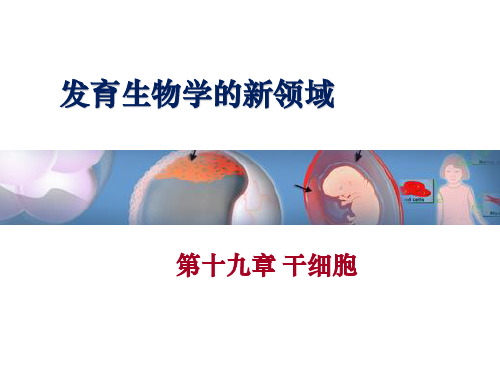
发育生物学的新领域
第十九章 干细胞
近年来在干细胞理论和技术研究方面取得 了长足的进展。 1999 年12 月,美国《科学》杂志把干细胞生物 学和以干细胞临床应用为内容的干细胞生物 工程评为20 世纪世界科学进展的最重要领域 。 已经成为继人类基因组大规模测序之后最具 活力、最有影响和最有应用前景的生命科学 研究领域之一。
2004-2005 韩国研究者黄禹锡(Hwang Woo-suk)声称利用体细胞核 转移技术培育出几种人类胚胎干细胞系,后来被证实是伪造的
2006 英国科学家用脐带血干细胞分化出第一个人造肝细胞
ppt精选版Lawrence M. Hinman
6
干细胞的起源和进展
2007.1 科学家发现在羊水中存在一种新型干细胞,它可以在研究与治疗 中替代胚胎干细胞
2008.3 自体间充质干细胞成功治疗关节变性病
- 1、下载文档前请自行甄别文档内容的完整性,平台不提供额外的编辑、内容补充、找答案等附加服务。
- 2、"仅部分预览"的文档,不可在线预览部分如存在完整性等问题,可反馈申请退款(可完整预览的文档不适用该条件!)。
- 3、如文档侵犯您的权益,请联系客服反馈,我们会尽快为您处理(人工客服工作时间:9:00-18:30)。
Behaviorally, adult TLX mutants exhibit increased aggressiveness, decreased copulation,progressively violent behavior, late onset epilepsy and reduced learning abilities [10,11,16,17].We have shown that TLX is an essential regulator of neural stem cell self-renewal [5]. TLX maintains adult neural stem cells in an undifferentiated and self-renewable state (Fig. 1). The TLX-expressing cells isolated from adult TLX-heterozygote brains can proliferate, self-renew and differentiate into all neural cell types in vitro . By contrast, TLX-null cells isolated from the brains of adult TLX-mutant mice fail to proliferate. Reintroducing TLX into TLX-null cells rescues their ability to proliferate and self-renew [5]. In vivo , TLX mutant mice show a loss of cell proliferation and reduced neural precursors in the neurogenic areas of adult brains. TLX represses the expression of astrocyte markers, such as GFAP, and tumor suppressor gene, pten,in neural stem cells, suggesting that transcriptional repression is crucial in maintaining the undifferentiated state of neural stem cells [5,14]. Similarly, the Drosophila tailless acts as a dedicated repressor in the early Drosophila embryo to support normal embryonic development and establish accurate patterns of gene expression [18]. TLX could be a key regulator that acts by controlling the expression of a network of target genes to establish the undifferentiated and self-renewable state of neural stem cells. Elucidation of the network regulated by TLX in producing these outcomes would be a significant advance in understanding neural stem cell self-renewal and neurogenesis.Recently, other nuclear receptors such as estrogen receptors (ER), thyroid hormone receptors (TR), and peroxisome proliferator-activated receptor γ (PPAR γ), have also been shown to regulate neural stem cell proliferation and differentiation [19-24]. Significant neuronal loss has been detected in adult brains of ER β knockout mice, suggesting an important role of ER β in neuronal maintenance in the central nervous system [25]. Knockout of TR α inhibits progression of neural stem cells through cell cycles, suggesting a critical role of TR α in neurogenesis of mammalian adult brains [26]. Neural stem cells prepared from heterozygous PPAR γ-deficientmouse brains have significantly reduced cell growth, which is also seen in PPAR γ short hairpinRNA silenced or dominant negative PPAR γ-treated neural stem cells, suggesting thatPPAR γ plays a role in neural stem cell proliferation control [27]. N-CoR, a nuclear receptorco-repressor, also plays a role in neural stem cell self-renewal. Deficiency in N-CoR leads toreduced neural stem cell self-renewal and premature differentiation into astrocytes [28].Bmi1 is a polycomb family transcriptional repressor that has been shown to be required forpost-natal maintenance of neural stem cells in the peripheral and central nervous system [29].Deficiency in Bmi1 leads to progressive postnatal growth retardation and neurological defects[30]. Bmi1-null mice exhibit a post-natal self-renewal defect that leads to the depletion of stemcells by early adulthood [29]. One way in which Bmi1 promotes the maintenance of adult stemcells is by repressing the cyclin-dependent kinase inhibitors, p16Ink4a and p19Arf [31,32].The Sox family of high-mobility-group (HMG) DNA binding proteins plays a role inmaintaining the undifferentiated state of neural stem cells in a context-dependent manner. Invertebrates, SoxB1 factors (Sox1, Sox2, and Sox3) are widely expressed in proliferating neuralstem/progenitor cells, throughout development and adulthood [33-35]. SoxB1 factors havebeen shown to play a role in maintaining the undifferentiated state of embryonic neuralprogenitors [36]. Overexpression of Sox2 and/or Sox3 inhibits neuronal differentiation ofneural progenitors and causes them to retain undifferentiated properties. In contrast, expressionof a dominant negative form of Sox2 and/or Sox3 results in premature exit of neural progenitorsfrom cell cycle and onset of neuronal differentiation [37,38]. In addition to its function in earlybrain development [37-40], Sox2 is also necessary for the maintenance of neural stem cells inadult neurogenic areas [41,42]. Regulatory mutations of Sox2 cause neurodegeneration andNIH-PA Author ManuscriptNIH-PA Author ManuscriptNIH-PA Author Manuscriptimpaired adult neurogenesis [41]. In parallel, Sox10, a member of the Sox E subfamily,maintains the multipotency of neural crest stem cells in the peripheral nervous system [43].Multiple basic helix-loop-helix (bHLH) genes also play a critical role in regulation of neural stem cell maintenance and differentiation [44-46]. Hes genes, homologs of Drosophila hairy and enhancer of split, are repressor-type bHLH genes. Among seven members of the Hes family, Hes1 and Hes5 are essential effectors of Notch signaling, the expression of which is upregulated by Notch activation [47,48]. Hes1 and Hes5 are highly expressed by neural stem cells [49-51], mis-expression of which inhibits neuronal differentiation and maintains neural stem cells in the embryonic brain [52-54]. In contrast, neural progenitors underwent premature neuronal differentiation in Hes1 and Hes5 double knockout mice [48,55-57], suggesting that Hes1 and Hes5 are essential for the maintenance and self-renewal of neural stem cells. Hes genes regulate neural stem cell self-renewal by repressing premature onset of the activator type bHLH genes such as Mash1, Math, and Neurogenin [46]. Hes-related bHLH genes, Hesr1 and Hesr2 are also expressed by neural stem cells in the embryonic brains and act as Notch signaling effectors. Hesrs regulate neural stem cell maintenance and self-renewal too, possibly through cooperative action with Hes [46]. Together, these data suggest that intrinsic transcription factors can work together to coordinate neural stem cell maintenance and self-renewal.3. Epigenetic Control Stem cell self-renewal and differentiation are the result of transcription control in concert with chromatin remodeling and epigenetic modifications. During central nervous system development in vertebrates, neural stem cell fate are strictly controlled under regional and temporal manners [58], accompanied by precise epigenetic control, including covalent histone modification and DNA methylation of CpG dinucleotides.Histone modification includes histone acetylation, methylation, phosphorylation ubiquitylation, sumolyation, and ADP-ribosylation [59]. Histone acetylation, which isregulated by histone acetylases (HATs) and histone deacetylases (HDACs) (Fig. 2), has beenbest studied. HDACs can deacetylate the conserved acetylated lysine residues in histone tail,resulting in local condensation in chromatin and block access of transcriptional factors to theirtarget genes. Mammalian HDACs can be classified based on their structure and homology toyeast HDACs. Class I HDACs (HDAC1, 2, 3, 8, and 11) are ubiquitously expressed, whereasClass II HDACs (HDAC4, 5, 7, and 9) display tissue-specific expression. Class II HDACScontain an amino-terminal extension that interacts with other transcriptional cofactors andconfers responsiveness to extracellular signals. We have shown that HDAC-mediatedtranscriptional repression is essential for the maintenance and self-renewal of neural stem cells(Sun et al. unpublished results). In addition to self-renewal, HDACs also regulate neural celldifferentiation. Treatment of adult neural stem cells with HDAC inhibitors induced neuronaldifferentiation [60], due in part to upregulating REST (RE1 silencing transcription factor, orNRSF)-regulated neuronal-specific genes. REST is a key transcriptional regulator of manyneuronal genes through binding to a conserved 21 bp RE1 binding site [61,62]. In non-neuronalcells, REST interacts with its co-factors, including Co-REST, N-CoR, and mSin3A, whichthen recruit HDAC complexes to repress neuronal gene expression through epigeneticregulation [61,63,64].More recently, histone methylation has gained attention as an epigenetic marker [65,66].Unlike histone acetylation, which only occurs on lysine (K) residues and is generally relatedwith active transcription, methylation was detected on both lysine and arginine (R) residuesand is linked to both transcriptional activation and repression [66]. For instance, histone H3K9 methylation is associated with transcriptional silencing (Fig. 2). In contrast, methylationof histone H3 K4 and arginine residues of H3 and H4 leads to transcriptional activation [67].NIH-PA Author ManuscriptNIH-PA Author ManuscriptNIH-PA Author ManuscriptRecent identification of LSD1 as a histone demethylase [68] indicates that similar to histoneacetylation, histone methylation is also a dynamic process subject to regulation by both methyltransferases and demethylases. The degree of lysine methylation (mono-, di-, or trimethylhistones), as well as the residues modified, is tightly associated with neural cell differentiation[69]. For example, histones H3 trimethyl K9 and H4 monomethyl K20 were detected inproliferating neural stem cells, whereas histone H4 trimethyl K20 was enriched indifferentiating neurons (Fig. 2).The epigenetic state can also be regulated at the DNA level by DNA methylation. The mostprominent form of DNA methylation in mammals is the symmetric methylation of cytosine inthe 5’ position in CpG dinucleotides. DNA methylation and its related chromatin remodelingplay critical roles in regulating gene transcription in response to neuronal activity [70]. Forexample, DNA methylation can repress astrocytic GFAP expression by preventing the bindingof a transcription factor, signal transducer and activator of transcription 3 (STAT3), to theGFAP gene promoter at early stages of brain development [71]. The gene silencing effect ofDNA methylation is mediated by a family of methyl-cytosine-binding proteins, includingMeCP2, which is abundantly expressed in the central nervous system [72,73]. MeCP2 is amember of a group of methylated-CpG binding proteins and is expressed at high levels in thepostnatal brain. Mutations in the Mecp2 gene have been linked to a neurodevelopmentaldisorder, Rett syndrome [74], suggesting that MeCP2 may play a role in regulating neuronalfunction. MECP2 has been shown to be critical for embryonic neurogenesis for Xenopus[75]. Recent studies demonstrated that Mecp2 is involved in the maturation and maintenanceof neurons at late stages of neuronal differentiation but not critical for embryonic neurogenesisin mammalian brains [76], consistent with the postnatal onset of symptoms in Rett Syndromepatients. Methyl-CpG binding protein MBD1 is also crucial for normal neural stem cell andbrain functions. MBD1-/- neural stem cells exhibited reduced neuronal differentiation andincreased genomic instability [77]. DNA methyltransferases are also expressed in the centralnervous system and have a role in neurogenesis [78,79]. DNA methyltransferase 1 (Dnmt1)deficiency mice displayed decreased neurogenesis. Conditional knockout of Dnmt1 in neuralprogenitor cells results in DNA hypomethylation and precocious astroglial differentiation[80]. Multiple layers of epigenetic modifications, therefore, regulate key transitions in theregulation of neural stem cell self-renewal and their differentiation.4. miRNA RegulatorsMany different classes of small non-coding RNAs are present in the brain, with diverse rolesincluding RNA modification and chromatin remodeling [81]. Small double-strandedmodulatory RNAs have been proposed to regulate the generation of neurons from adult neuralstem cells by binding to REST [82]. miRNA is another recently identified large family of smallnon-coding RNAs, which are likely key post-transcriptional players in stem cell self-renewaland differentiation (Fig. 3). miRNAs are short 20-22 nucleotide RNA molecules that areexpressed in a tissue-specific and developmentally-regulated manner and function as negativeregulators of gene expression in a variety of eukaryotes. miRNAs are involved in numerouscellular processes including development, proliferation, and differentiation [83,84].miRNA genes belong to class II genes, which are transcribed by RNA polymerase II. A majorityof miRNA loci are found in intronic regions of protein-coding or non-coding transcriptionunits; others are found in exonic regions of non-coding transcription units [85]. The primarytranscripts of miRNAs (normally > 1kb), called pri-miRNAs, are processed in the nucleus byRNase III endonuclease, Drosha, into 60 to 75 neocleotide hairpin-like precursors (pre-miRNAs). Pre-miRNAs are subsequently exported to the cytoplasm by exportin-5, a memberof the Ran-dependent nuclear transport receptor family. In the cytoplasm, the hairpin precursorsare cleaved into mature miRNAs by Dicer, a cytoplasmic RNase III-type protein. MatureNIH-PA Author Manuscript NIH-PA Author ManuscriptNIH-PA Author ManuscriptmiRNAs are then transferred to Argonaute proteins in the RNA-induced silencing complex (RISC), which direct the miRNAs to their target transcripts [86]. By base pairing with the target mRNA, miRNA functions as a guide molecule in post-transcriptional gene silencing, leading to mRNA cleavage or translational repression (Fig. 3).miRNAs are especially attractive candidates for regulating stem cell self-renewal and cell fate decisions because of their ability to simultaneously regulate many target genes. Studies based on expression patterns, predicted targets, and overexpression analyses suggest that miRNAs are key regulators in stem cell biology. Distinct sets of miRNAs have been shown to be specifically expressed in embryonic stem cells [87,88]. Loss of Dicer1 causes embryonic lethality and loss of stem cell populations [89,90]. Argonaute family members, key components of RISC complexes, are required for maintaining germline stem cells in various species [91].These observations together support a role for miRNAs in stem cell self-renewal.Among miRNAs identified, around 70% of them are found to be expressed in mammalian brains [92,93], suggesting possible roles of these miRNAs in neural function [94-97]. Indeed,miRNAs have been shown to regulate the development of chemosensory system of C.elegans [98] and brain morphogenesis of zebrafish [99]. Study of a set of highly expressed neural miRNA during mammalian brain development revealed significant differences in the onset and magnitude of induction for individual miRNAs and marked lineage specificity of the miRNAs [100]. During differentiation, progenitor cells express families of miRNAs sequentially, resulting in expression of lineage-specific genes. The most highly expressed miRNAs in adult brain, miR-124 and miR-128, were preferentially expressed in neurons; while miR-23 was restricted to astrocytes; miR-26 and miR-29 have stronger expression in astrocytes than neurons; whereas miR-9 and miR-125 were fairly evenly distributed [100].Overexpression of miR-124, miR-128, and miR-9 in neural precursors decreased astrocyte differentiation. In contrast, inhibition of miR-9 expression alone or in combination with miR-124 led to reduced neurogenesis [101]. The regulation mediated by miR-9 and miR-124is at least partly through modulating the STAT3 signaling pathway [101]. Let-7 familymembers are also highly represented in libraries of brain miRNAs, although not restricted tothe nervous system [102]. Let-7 family members are highly expressed in nervous tissues ofzebrafish [92] and in the developing mouse brains [103]. Both transcriptional activation andincreased precursor processing activity led to significant induction of mature forms of let-7family members during neural differentiation, suggesting a role of let-7 in neural cellspecification [103].Target prediction of miRNA indicates that more than one-third of animal genes may beregulated by miRNAs [104]. Each miRNA may suppress multiple targets, and one mRNA canbe targeted by many miRNAs [84]. So far, some of mRNA targets have been validated andshown to control over a broad spectrum of cellular process [83]. MiR-124, the most abundantmiRNA in adult mammalian brains [102], has its binding motif in more than 1100 genes aspredicted computationally [104]. Introduction of miR-124 into HeLa cells down-regulatedmore than 100 genes [105] and promoted a neuronal-like mRNA profile [106]. Recentlylaminin gamma 1 and integrin beta1 were identified as targets of miR-124 in chicken neuraltube, both of which are highly expressed in neural precursor cells and repressed upon neuronaldifferentiation [107]. In addition, miR-124 binds to 3’UTR of small C-terminal domainphosphatase 1 (SCP1), a phosphatase that plays a role in neural developement, to suppress itsexpression, further supporting a role of miR-124 in neurogenesis [108]. S enseless, a binaryswitch during sensory organ precursor selection in flies [109], has been identified as a targetgene of miR-9 [110]. Loss of miR-9 in flies induced extra sense organs, whereas overexpressionof miR-9 led to massive loss of sensory organ precursors [110]. Hunchback, a gene regulatesthe temporal identity of neuroblasts [111], is regulated by let-7 miRNA as a target gene [112,113]. Identification of comprehensive miRNA targets in the neural system remains anNIH-PA Author ManuscriptNIH-PA Author ManuscriptNIH-PA Author Manuscriptimportant and non-trivial task that will help us to better understand the regulation of neuralstem cell self-renewal and differentiation.5. Cell-extrinsic signaling Stem cell self-renewal and differentiation is also regulated by the specialized microenvironment, or niche, in which these cells reside [114-117]. Direct physical interactions between stem cells and their niches are critical for maintaining stem cell characters. Signaling molecules in the niche are composed of soluble factors, membrane bound molecules, and extracellular matrix, including Wnt, Notch, and Sonic hedgehog (Shh) [114]. Receptor tyrosine kinase (RTK) signaling has also been implicated in regulation of neural stem cell proliferation and self-renewal.Genetic studies have implicated the Wnt/β-catenin pathway in neural stem cell self-renewal.In mice that express a stabilized β-catenin, the central nervous system is greatly enlarged.Progenitors exit the cell cycle less frequently and continue to proliferate in the brain [118]. In contrast, ablation of β-catenin results in a marked decrease of the overall size of the nervous system [119]. Mice with null alleles of LRP6, a required co-receptor for Wnt signaling, also showed reduced dentate granule cell production and failed expansion of a defined dentate granule precursor cell pool [120]. These studies indicate that the Wnt/β-catenin signaling plays an important role in the proliferation and self-renewal of neural precursors, presumably through its downstream target genes, such as cyclin D1 [121]. On the other hand, Wnt proteins have been shown to promote neuronal differentiation in neural stem cell culture and in adult hippocampus [122-124], suggesting that signaling by the canonical Wnt pathway has multiple functions in stem cells. Indeed, recently it has been shown that Wnt3a and Wnt5a could both increase proliferation and stimulate neuronal differentiation of neural progenitor cells isolated from postnatal mouse brains [125]. Thus, Wnt pathways are critical regulators of both neural stem cell self-renewal and neurogenesis.Notch signaling is activated when a Notch receptor on one cell interacts with Notch ligands,such as Delta, on an adjacent cell. This interaction triggers proteolytic release of the Notchintracellular domain and its translocation to the nucleus, where it binds to transcriptionalregulator CSL and induces downstream effector expression [126]. The best known Notcheffectors are members of the Hes families, including Hes1 and Hes5 [47,48,127]. Studies usingmouse models of the Notch pathway led to a prevailing view that Notch maintains the self-renewable state of neural stem cells [128]. One of the first Notch pathway genes to be knocked-out was Notch1 [129,130]. Consistent with the view that Notch activity is needed for stem cellmaintenance, increased neuronal differentiation was detected in Notch1 mutant brains.Conditional deletion of Notch1 in the neural progenitor pool was also found to result inprecocious neuronal differentiation and earlier neural progenitor pool depletion [131,132]. Inaddition to the receptor mutations, many Notch ligand mutations have been examined. Onesuch study found that deleting Delta-like 1 (Dll1) led to a decrease in the radial glia progenitormarker RC2 and an increase in neuronal marker expression in embryonic brains, supportingthe traditional view that Notch signaling inhibits neuronal differentiation in the developingcentral nervous system [128]. Notch pathway components are also expressed in postnatal andadult mouse brains, both in germinal zones and in neurons [133]. Notch signaling can becontext-dependent. In addition to maintaining stem cell state in germinal regions, Notch canalso promote terminal glial differentiation [134].Shh is an important morphogen in development. In Shh null mice, the telencephalon is greatlydysmorphic and much reduced in size. Both dorsoventral patterning and general brainproliferation are significantly affected [135,136]. Shh is also a potent mitogen for neuralprogenitor cells of adult brains [137]. Over-expression of Shh near the dentate gyrus increasesNIH-PA Author ManuscriptNIH-PA Author ManuscriptNIH-PA Author Manuscriptproliferation and neurogenesis of hippocampal subgranular zone cells [137]. In vitro , Shhmaintains proliferation of adult hippocampal neuronal progenitors [137] and increasessubventricular zone cell proliferation [138]. The hedgehog signaling is mediated by the zincfinger-containing transcription factor Gli1, a direct transcriptional target of Shh signaling[139,140], acting to both promote proliferation and maintain populations of neural progenitorsin postnatal brains [141].Epidermal growth factor (EGF), transforming growth factor α (TGF α), and fibroblast growthfactor (FGF) are all extracellular ligands of RTKs and play critical roles in the proliferation ofneural stem cells [142-145]. EGF and TGF α bind preferentially to the EGFR [146,147], amember of the RTK family. EGFR is expressed in neurogenic regions such as the adultsubventricular zone (SVZ) [148-150]. Targeted disruption of EGFR causes forebrain corticaldysgenesis at late embryonic and postnatal ages [151-153]. TGF α is considered thepredominant endogenous EGFR ligand [150], the expression of which is temporally andspatially coordinated with EGFR expression. TGF α null mice exhibit decreased proliferationwithin the SVZ [154]. In vivo administration of EGF and TGF α via intraventricular infusionincreased neural stem cell proliferation in the adult brain. In vitro experiments have also shownthat SVZ-derived progenitor cells can be expanded by EGF and FGF administration [143].FGF acts primarily through FGF receptors (FGFR), members of the RTK family also (Johnsonand Williams 1993). Targeted deletion of FGFR1 causes defects in cell proliferation andembryonic lethality [155-157]. FGF2, the first member of the FGF family, is expressed in thedorsolateral cortical neuroepithelium of the forebrain and acts primarily through FGFR1. FGF2null mice show significant reduction in cortical progenitor cell proliferation beforeneurogenesis begins [158]. Intraventricular delivery of FGF2 increased cell proliferation withinthe adult SVZ [159,160]. Cyclin D2 has been recently suggested to be an effector of the FGFsignaling [161], which promotes early G1 cell cycle progression, a function relevant to neuralstem cell proliferation.Neural stem cells in the brain are likely to be influenced by a convergence of extracellularsignals, such as Wnt, Notch, and Shh, from many neighboring cell types, including astrocytes,neuroblasts, ependymal cells, and endothelial cells [117,162,163]. These extrinsic factors mayinteract with intrinsic regulators by various signaling cascades, including Wnt/β-catenin-cyclinD1 [121], Notch-Hes1/5 [47,48,127], and Shh-Gli1 [139,140]. Understanding more about thesesignaling pathways will help reveal how the niches influence neural stem cell self-renewal andtissue development [164].6. ConclusionAn emerging regulatory network controlling stem cell self-renewal and differentiation isdefined by integration of cell-intrinsic regulators, including transcription factors, epigeneticcontrols, and small RNA regulators, with cell-extrinsic signals from stem cell niches. Thesemechanisms are coordinated to regulate the development, maintenance, self-renewal, anddifferentiation of stem cells. Unraveling how individual signaling cascades integrate into theglobal regulatory networks will be essential to better understand stem cell biology. It will alsofacilitate the development of new and targeted therapies using neural stem cells for a host ofneurological disorders, including brain injuries, brain tumors, and neurodegenerative diseasessuch as Huntington’s, Alzheimer’s and Parkinson’s disease.AcknowledgementsWe apologize to colleagues whose work could not be cited due to space limitations and thank Rose Chavarin and Dr.Kamil Alzayady for proof-reading of the manuscript. GS is a Herbert Horvitz Fellow and RS is a NIH NIMH DPNFellow. YS is a Kimmel Scholar. This work is supported by Whitehall Foundation, the Margaret E. Early MedicalResearch Trust, the Sidney Kimmel Foundation, and NIH NINDS.NIH-PA Author Manuscript NIH-PA Author ManuscriptNIH-PA Author ManuscriptReferences1. McKay R. Stem cells in the central nervous system. Science Apr 4;1997 276(5309):66–71. [PubMed:9082987]2. Alvarez-Buylla A, Temple S. Stem cells in the developing and adult nervous system. J Neurobiol Aug;1998 36(2):105–110. [PubMed: 9712298]3. Gage FH, Kempermann G, Palmer TD, Peterson DA, Ray J. Multipotent progenitor cells in the adult dentate gyrus. J Neurobiol Aug;1998 36(2):249–266. [PubMed: 9712308]4. Weiss S, van der Kooy D. CNS stem cells: where’s the biology (a.k.a. beef)? J Neurobiol Aug;199836(2):307–314. [PubMed: 9712311]5. Shi Y, Chichung Lie D, Taupin P, et al. Expression and function of orphan nuclear receptor TLX in adult neural stem cells. Nature Jan 1;2004 427(6969):78–83. [PubMed: 14702088]6. Land PW, Monaghan AP. Expression of the transcription factor, tailless, is required for formation of superficial cortical layers. Cereb Cortex Sep;2003 13(9):921–931. [PubMed: 12902391]7. Land PW, Monaghan AP. Abnormal development of zinc-containing cortical circuits in the absence of the transcription factor Tailless. Brain Res Dev Brain Res Aug 8;2005 158(12):97–101.8. Roy K, Kuznicki K, Wu Q, et al. The Tlx gene regulates the timing of neurogenesis in the cortex. J Neurosci Sep 22;2004 24(38):8333–8345. [PubMed: 15385616]9. Stenman JM, Wang B, Campbell K. Tlx controls proliferation and patterning of lateral telencephalic progenitor domains. J Neurosci Nov 19;2003 23(33):10568–10576. [PubMed: 14627641]10. Monaghan AP, Bock D, Gass P, et al. Defective limbic system in mice lacking the tailless gene.Nature Dec 4;1997 390(6659):515–517. [PubMed: 9394001]11. Chiang, MY.; Evans, RM. Dissertation. Univ of California San Diego; La Jolla, CA: 1997. Reverse Genetic Analysis of Nuclear Receptors, RXR γ, RAR β, and TLX in Mice.12. Yu RT, Chiang MY, Tanabe T, et al. The orphan nuclear receptor Tlx regulates Pax2 and is essential for vision. Proc Natl Acad Sci U S A Mar 14;2000 97(6):2621–2625. [PubMed: 10706625]13. Miyawaki T, Uemura A, Dezawa M, et al. Tlx, an orphan nuclear receptor, regulates cell numbers and astrocyte development in the developing retina. J Neurosci Sep 15;2004 24(37):8124–8134.[PubMed: 15371513]14. Zhang CL, Zou Y, Yu RT, Gage FH, Evans RM. Nuclear receptor TLX prevents retinal dystrophyand recruits the corepressor atrophin1. Genes Dev May 15;2006 20(10):1308–1320. [PubMed:16702404]15. Uemura A, Kusuhara S, Wiegand SJ, Yu RT, Nishikawa S. Tlx acts as a proangiogenic switch byregulating extracellular assembly of fibronectin matrices in retinal astrocytes. J Clin Invest Feb;2006116(2):369–377. [PubMed: 16424942]16. Roy K, Thiels E, Monaghan AP. Loss of the tailless gene affects forebrain development and emotionalbehavior. Physiol Behav Dec;2002 77(45):595–600. [PubMed: 12527005]17. Young KA, Berry ML, Mahaffey CL, et al. Fierce: a new mouse deletion of Nr2e1; violent behaviourand ocular abnormalities are background-dependent. Behav Brain Res May 14;2002 132(2):145–158. [PubMed: 11997145]18. Moran E, Jimenez G. The tailless nuclear receptor acts as a dedicated repressor in the early Drosophilaembryo. Mol Cell Biol May;2006 26(9):3446–3454. [PubMed: 16611987]19. Brannvall K, Korhonen L, Lindholm D. Estrogen-receptor-dependent regulation of neural stem cellproliferation and differentiation. Mol Cell Neurosci Nov;2002 21(3):512–520. [PubMed: 12498791]20. Ambrogini P, Cuppini R, Ferri P, et al. Thyroid hormones affect neurogenesis in the dentate gyrusof adult rat. Neuroendocrinology 2005;81(4):244–253. [PubMed: 16113586]21. Fowler CD, Johnson F, Wang Z. Estrogen regulation of cell proliferation and distribution of estrogenreceptor-alpha in the brains of adult female prairie and meadow voles. J Comp Neurol Aug 22;2005489(2):166–179. [PubMed: 15984004]22. Katayama K, Wada K, Nakajima A, Kamisaki Y, Mayumi T. Nuclear receptors as targets for drugdevelopment: the role of nuclear receptors during neural stem cell proliferation and differentiation.J Pharmacol Sci Feb;2005 97(2):171–176. [PubMed: 15725702]NIH-PA Author ManuscriptNIH-PA Author ManuscriptNIH-PA Author Manuscript。