CHOP基NshRNA载体构建及沉默效应
SHH-N基因腺相关病毒表达载体的构建及其对神经干细胞增殖相关基因的影响

SHH-N基因腺相关病毒表达载体的构建及其对神经干细胞增殖相关基因的影响刘东升;崔岩;申仑;杜延平;李桂林;王任直;王岩;张波【摘要】Objective To construct and identify SHH-N gene adeno-associated virus vector and to detect its effect on genes controlling to proliferation in neural stem cells(NSCs). Methods The neural stem cells in the subventricular zone of postnatal rat brain were used to collect SHH-N. pSNAV2. 0-CMV-SHH-N-IRES-EGFP was established by enzyme cutting and ligation , and then transfected packaging cell line 293T. Real time PCR was performed after SHH-N transfection of neural stem cells by rAAV-SHH-N-EGFP vector for 48 hours, SHH-N gene expression of infected cells after 2 week examined by fluorescence microscopy. Results ( 1 ) SHH-N gene was coincident with NCBI report. (2) pSNAV2. 0-CMV-SHH-N-IRES-EGFP expression vector and rAAV-SHH-N-EGFP vector was successful established and packaged. (3)Real time PCR was performed after SHH-N infection for 48 hour, induction of Nmyc and Glil in rAAV-SHH-N-EGFP-treated group was enhanced compared to control group. Conclusion rAAV-SHH-N-EGFP vector has been successfully established and it can stably express in neural stem cells. Forced expression of SHH-N in NSCs resulted in enhanced induction of Nmyc and Glil.%目的构建rAAV-SHH-N-EGFP载体并检测其对神经干细胞增殖相关基因影响.方法分离并培养大鼠脑室下区神经干细胞,提取RNA、反转录、PCR得到SHH-N的cDNA,克隆入pSNAV2.0-CMV-IRES-EGFP,包装得到腺相关病毒rAAV-SHH-N-EGFP,感染神经干细胞48 h后,采用实时定量PCR检测SHH信号通路下游相关基因的mRNA水平变化,并观察rAAV-SHH-N-EGFP载体在神经干细胞内的表达情况.结果 (1)克隆得到SHH-N基因,测序结果与NCBI报道序列一致.(2)成功构建pSNAV2.0-CMV-SHH-N-EGFP表达载体并鉴定,包装得到rAAV-SHH-N-EGFP.(3)实时定量PCR 分析rAAV-SHH-N-EGFP感染神经干细胞48 h后,rAAV-SHH-N-EGFP处理组较感染rAAV-EGFP的对照组,Glil和Nmyc的mRNA水平上调.(4)rAAV-SHH-N-EGFP可有效感染神经干细胞,在感染14 d后稳定表达SHH-N.结论成功构建rAAV-SHH-N-EGFP过表达载体并使其在神经干细胞内稳定表达.过表达SHH-N 可使神经干细胞内SHH信号通路相关基因Glil转录水平上调,并使细胞增殖相关基因Nmyc转录水平上调.【期刊名称】《基础医学与临床》【年(卷),期】2011(031)003【总页数】6页(P291-296)【关键词】神经干细胞;SHH信号通路;Nmyc基因【作者】刘东升;崔岩;申仑;杜延平;李桂林;王任直;王岩;张波【作者单位】大连医科大学附属第一医院,神经外科,辽宁,大连,116011;大连医科大学附属第一医院,神经外科,辽宁,大连,116011;大连医科大学附属第一医院,神经外科,辽宁,大连,116011;大连医科大学附属第一医院,神经外科,辽宁,大连,116011;中国医学科学院北京协和医院,神经外科,北京,100730;中国医学科学院北京协和医院,神经外科,北京,100730;清华大学玉泉医院,神经外科,疼痛病区,北京,100730;大连医科大学附属第一医院,神经外科,辽宁,大连,116011【正文语种】中文【中图分类】R394.3音猬蛋白(sonic hedgehog,SHH)分泌蛋白在神经系统的发育时期中调节神经管腹侧的发育和脑背侧部神经管前体的增殖发育[1]。
RNAi沉默和载体构建

近年来的研究表明 ,一些短片断的双链 RNA 可以通过促使特定基因的mRNA 降解来高效、特异的阻断体内特定基因表达 ,诱使细胞表现出特定基因缺失的表型 ,称为 RNA 干扰( RNAinterference,RNAi ).siRNA (smallinteferingRNA9就是这种短片断双链 RNA分子,能够以序列同源互补的mRNA为靶目标,降解特定的mRNA.RNA的发现具有划时代的意义 ,它不仅深入揭示了细胞内基因沉默的机制 ,而且它还是后基因组时代基因功能分析的有力工具 ,极大地促进了人类揭示生命奥秘的进程•现在越来越多的研究人员开始采用 RNAi来研究生物体的基因表达.RNAi技术可广泛应用到包括功能基因组学,药物靶点筛选,细胞信号传导通路分析,疾病治疗等等.目前为止较为常用的5种制备siRNAs的方法包括:化学合成体外转录长片断 dsRNAs经 RNaseIII类降解(e.g.Dicer,E.coli,RNaseHI) siRNA表达载体或者病毒载体在细胞中表达 siRNAsPCR制备的siRNA表达框在细胞中表达获得高纯度的siRNA产物是进行实验的第一步,而转染的效率则是非常关键的因素 .一、基本概念RNA: (RNAinteferenee) RNA干扰内源性或外源性双链RNA(dsRNA介导的能诱导细胞内与其序列同源的特异基因表达沉默或抑制的效应 ,诱使细胞表现出特定基因缺失的表型,称为RNA干扰,它也是体内抵御外在感染的一种重要保护机制.siRNA (smallinteferingRNAs小干扰 RNA由长dsRNA裂解而成的一种19-25nt的短片断双链RNA分子,能够以同源互补序列的RNA为靶目标降解特定的 mRNA,RNA的关键效应分子. shRNAs:(smallhairpinRNA小发夹 RNA是设计为能够形成发夹结构的非编码小 RNA分子,shRNA需通过载体导入细胞后,然后利用细胞内的酶切机制得到 siRNA而最终发挥RNA 干扰作用 .Dicer:属于RNaseffi家族,是dsRNA的特异性核酸内切酶RISC(RNA-inducingsilencingcomplex)RNA诱导的沉默复合体,具有核酸内切、外切以及解旋酶活性二、机制目前普遍认为,共抑制、基因压制和RNAi很可能具有相同的分子机制都是通过dsRNA的介导而特异地降解靶mRNA抑制相应基因的表达. 即RNA、共抑制、quelling均属于PTGS现已初步阐明dsRNA介导的同源性靶mRNA降解过程主要分为两步.第一步(起始阶段)是较长dsRNA在 ATP参与下被RNaseffi样的特异核酸酶切割加工成21~23nt的由正义和反义链组成的小干扰 RNAsmallinterferingRNA,siRNA) 第二步(效应阶段)是siRNA在ATP参与下被RNA解旋酶解旋成单链,并由其中反义链指导形成 RNA诱导的沉默复合体(RNA-inducedsilencingcomplex,RISC).RNAi途径主要存在于细胞浆中,但是siRNA产生、靶mRNA降解的亚细胞位置尚未明确•外源性(注射或喂养)的dsRNA和病毒性dsRNA 可能可以直接进入细胞浆中的RNAi途径,仅在细胞浆中复制的RNA病毒可被dsRNA 介导的沉默机制所抑制.外源性dsRNA则还可导致细胞核中的同源早期RNA转录产物减少.在许多机体中反向重复转基因序列在细胞核内转录成发夹dsRNA进而可介导RNAi这种dsRNA可能需要转移至胞浆中才可有效地沉默同源靶mRNA.RNAi的放大效应机制siRNA不仅可引导RISC切割靶RNA而且可作为引物在RNA依赖的RNA 聚合酶(RdRP作用下以靶mRNA为模板合成新的dsRNA.新合成的长链dsRNA同样可被RNasen样核酸酶切割、降解而生成大量的次级siRNA次级siRNA又可进入合成-切割的循环过程,进一步放大RNAi 作用这种合成-切割的循环过程称为随机降解性 PCR( randomdegradativePCR) .三、RNAi表达载体的构建1.目的基因的确定(1)、检索文献获取有实验证明有效的靶点序列(核对)3)、or 2、设计siRNA靶序列在制备siRNA前都需要单独设计siRNA序列.研究发现对哺乳动物细胞最有效的siRNAs是 21-23个碱基大小、3’端有两个突出碱基的双链 RNA;而对非哺乳动物,比较有效的是长片段dsRNA.siRNA勺序列专一性要求非常严谨,与靶mRNA之间一个碱基错配都会显着削弱基因沉默的效果.(1)、选择siRNA靶位点:从转录AUG起始密码子开始,搜寻下游AA序列,记录跟每个AA3端相邻的19个核苷酸作为候选的siRNA靶位点.有研究结果显示GC含量在30%—50%左右的siRNA要比那些GC含量偏高的更为有效.Tuschl 等建议在设计siRNA时不要针对5'和3'端的非编码区(un tra nslatedregio ns,UTRs)原因是这些地方有丰富的调控蛋白结合区域,而这些UTR结合蛋白或者翻译起始复合物可能会影响siRNP核酸内切酶复合物结合mRNA从而影响siRNA的效果.( 2)、序列同源性分析 : 将潜在的序列和相应的基因组数据库(人 ,或者小鼠 ,大鼠等等)进行比较,排除那些和其他编码序列/EST同源的序列.例如使用BLAST() 选出合适的目标序列进行合成.并非所有符合条件的siRNA都一样有效,其原因还不清楚 ,可能是位置效应的结果 ,因此对于一个目的基因 ,般要选择 3-5 个靶位点来设计 siRNA. 通常来说,每个目标序列设计3—4对siRNAs选择最有效的进行后续研究.(3)、设计阴性对照:一个完整的siRNA实验应该有阴性对照,作为阴性对照的siRNA应该和选中的siRNA序列有相同的组成,但是和mRNA没有明显的同源性.通常的做法是将选中的siRNA中的碱基序列打乱•当然,同样要保证它和其他基因没有同源性 .3、RNAi表达载体的选用化学合成与体外转录方法都是在体外得到 siRNA后再导入细胞内,但是这两种方法主要有两方面无法克服的缺点: siRNA进入细胞后容易被降解;进入细胞siRNA在细胞内的RNAi效应持续时间短.针对这种情况,出现了质粒、病毒类载体介导的siRNA体内表达.该方法的基本思路是:将siRNA对应的DNA双链模板序列克隆入载体的 RNA聚合酶III的启动子后,这样就能在体内表达所需的siRNA分子.这种方法总体的优点在于不需要直接操作 RNA能达到较长时间的基因沉默效果. 通过质粒表达siRNAs大都是用PolIII启动子启动编码shRNA(smallhairpinRNA的序列选用PolIII启动子的原因在于这个启动子总是在离启动子一个固定距离的位置开始转录合成RNA遇到4—5 个连续的U即终止,非常精确.当这种带有PolIII启动子和shRNA模板序列的质粒转染哺乳动物细胞时,这种能表达siRNA的质粒确实能够下调特定基因的表达 ,可抑制外源基因和内源基因 .采用质粒的优点在于,通过siRNA表达质粒的选择标记,siRNA载体能够更长时间地抑制目的基因表达 .当然还有一点 ,那就是由于质粒可以复制扩增 ,相比起其它合成方法来说,这就能够显着降低制备siRNA的成本.此外,带有抗生素标记的siRNA表达载体可用于长期抑制研究,通过抗性辅助筛选 ,该质粒可以在细胞中持续抑制靶基因的表达数星期甚至更久.同时RNAi-Ready表达载体还能与逆转录病毒和腺病毒表达系统整合(BDKnockoutRNAiSystem),大大提高siRNA表达载体对宿主细胞的侵染性 ,彻底克服某些细胞转染效率低的障碍 ,是实现哺乳动物细胞siRNAs 瞬时表达与稳定表达的理想工具.4、合成模板合成编码shRNA的DNA模板的两条单链,模板链后面接有RNAPolyIII 聚合酶转录中止位点,同时两端分别设计BamHI和HindHI酶切位点,可以克隆到siRNA载体多克隆位点的BamHI和HindHI酶切位点之间.95C ,5min,缓慢退火,DNA单链得到shRNA的DNA双链模板5、连接与转化基本步骤:(1)将100卩感受态细胞于冰上解冻.(2)取5卩连接产物加入到感受态细胞中,轻轻旋转几次以混匀内容物.在冰上放置 30 分钟 .(3)将管放入预加温到42C的水浴中,热激90秒•快速将管转移到冰浴中,使细胞冷却 1~2分钟.(4)每管中加700卩IL弗养基,37C振荡培养1小时,进行复苏.(5)室温4,000rpm离心5分钟,弃去上清后,用剩余100卩培养基重悬细胞并涂布到含抗性的LB琼脂平板表面.注意:细胞用量应根据连接效率和感受态细胞的效率进行调整 .( 6)将平板置于室温直至液体被吸收 .(7)倒置平皿,于37C培养,12~16小时后可出现菌落.6、P CR鉴定和测序鉴定在插入编码shRNA的DNA双链模板两侧设计鉴定PCR引物,扩增片段在 100-200bp 之间 .7、特点siRNA表达载体的优点在于这是众多方法中唯一可以进行长期研究的方法——带有抗生素标记的载体可以在细胞中持续抑制靶基因的表达,持续数星期甚至更久 .即使是对转染带有筛选标记质粒的细胞进行瞬时筛选 ,也有助于富集带质粒的细胞 .这也可以帮助解决一些难转染的细胞由于转染效率低造成的问题 .载体上的抗性标记有助于快速筛选出阳性克隆 ,而且可以在细胞中持续抑制靶基因的表达 ,持续数星期甚至更久 ,可以进行较长期研究 .四、RNAi的前景展望1 、研究基因功能的新工具已有研究表明RNAi能够在哺乳动物中灭活或降低特异性基因的表达制作多种表型 ,而且抑制基因表达的时间可以随意控制在发育的任何阶段,产生类似基因敲除的效应 .线虫和果蝇的全部基因组序列已测试完毕,发现大量未知功能的新基因,RNA i将大大促进对这些新基因功能的研究 .与传统的基因敲除技术相比 ,这一技术具有投入少 ,周期短 ,操作简单等优势,近来RNAi成功用于构建转基因动物模型的报道日益增多,标志着RNAi将成为研究基因功能不可或缺的工具.2、研究信号传导通路的新途径联合利用传统的缺失突变技术和 RNAi技术可以很容易地确定复杂的信号传导途径中不同基因的上下游关系 Qemensy等应用RNAi研究了果蝇细胞系中胰岛素信息传导途径 ,取得了与已知胰岛素信息传导通路完全一致的结果,在此基础上分析了 DSH3PX1与DACK之间的关系,证实了DACK是位于DSH3PX1磷酸化的上游激酶只NAi技术较传统的转染实验简单、快速、重复性好 ,克服了转染实验中重组蛋白特异性聚集和转染效率不高的缺点 ,因此认为 RNAi 技术将可能成为研究细胞信号传导通路的新途径 .3、开展基因治疗的新策略RNAi具有抵抗病毒入侵,抑制转座子活动,防止自私基因序列过量增殖等作用,因此可以利用RNAi现象产生抗病毒的植物和动物,并可利用不同病毒转录序列中高度同源区段相应的 dsRNA抵抗多种病毒. 肿瘤是多个基因相互作用的基因网络调控的结果 ,传统技术诱发的单一癌基因的阻断不可能完全抑制或逆转肿瘤的生长,而RNAi可以利用同一基因家族的多个基因具有一段同源性很高的保守序列这一特性 , 设计针对这一区段序列的dsRNA分子,只注射一种dsRNA即可以产生多个基因同时剔除的表现,也可以同时注射多种dsRNA而将多个序列不相关的基因同时剔除 .尽管目前RNAi技术在哺乳动物中的应用还处于探索阶段,但它在斑马鱼和老鼠等脊椎动物中的成功应用预示着 RNAi将成为基因治疗中重要的组成部分,人工合成的dsRNA寡聚药物的开发将可能成为极具发展前途的新兴产业 .。
基因沉默与RNAi技术

基因沉默与RNAi技术定义:基因沉默双是指链RNA被特异的核酸酶降解,产生干扰小RNA(siRNA),这些siRNA与同源的靶RNA互补结合,特异性酶降解靶RNA,从而抑制、下调基因表达。
RNA干扰是指在进化过程中高度保守的、由双链RNA诱发的、同源mRNA高效特异性降解的现象。
由双链引发的植物RNA沉默,主要有转录水平的基因沉默(TGS)和转录后水平的基因沉默(PTGS)两类:TGS是指由于DNA修饰或染色体异染色质化等原因使基因不能正常转录;PTGS是启动了细胞质内靶mRNA序列特异性的降解机制。
有时转基因会同时导致TGS和PTGS。
基因沉默是一种RNA干扰技术。
RNA干扰是由双链RNA 引发的转录后基因静默机制。
其原理是:RNaseIII核酶家族的Dicer,与双链RNA结合,将其剪切成21 - 25nt及3'端突出的小干扰RNA (small interfering RNA,siRNA),随后siRNA与RNA诱导沉默复合物(RNA - induced silencing complex,RISC结合,解旋成单链,活化的RISC受已成单链的siRNA引导,序列特异性地结合在靶mRNA上并将其切断,引发靶mRNA的特异性分解,从而阻断相应基因表达的转录后基因沉默机制.一、基因沉默的分类及其机制(一)转录水平基因沉默转录水平基因沉默是指对基因专一的细胞核RNA合成的失活,它的发生主要是由于基因无法被顺利转录成相应的RNA而导致基因沉默。
转录水平基因沉默可以通过有性世代传递,表现为减数分裂的可遗传性。
引起转录水平基因沉默的机制主要有以下几种:1.基因及其启动子甲基化甲基化是活体细胞中最常见的一种DNA共价修饰形式,通常发生在DNA的CG序列的碱基上,该区碱基甲基化往往导致转录受抑制,该区甲基化的频率在人类及高等植物中分别可达4%和36%。
[4]近来的研究表明,发生在转基因启动子5’端的甲基化是造成转录水平基因沉默的主要原因。
构建shRNA表达质粒

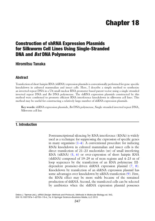
C hapter 18 C onstruction of shRNA Expression Plasmidsfor Silkworm Cell Lines Using Single-StrandedDNA and B st DNA PolymeraseHiromitsu TanakaAbstractT ransfection of short hairpin RNA (shRNA) expression plasmids is conventionally performed for gene-speci fic knockdown in cultured mammalian and insect cells. Here, I describe a simple method to synthesize an inverted repeat DNA in a U6 small nuclear RNA promoter-based parent vector using a single-stranded inverted repeat DNA and B st DNA polymerase. The shRNA expression plasmids constructed by this method were con fir med to promote ef fic ient RNA interference knockdown in silkworm cell lines. This method may be useful for constructing a relatively large number of shRNA expression plasmids.K ey words:s hRNA expression plasmids ,B st DNA polymerase ,S ingle-stranded inverted repeat DNA , S ilkworm cell line1.IntroductionP osttranscriptional silencing by RNA interference (RNAi) is widelyused as a technique for suppressing the expression of speci fic genesin many organisms (1–4). A conventional procedure for inducingRNAi knockdown in cultured mammalian and insect cells is thedirect transfection of 21–23 nucleotides (nt) of small interferingRNA (siRNA) (5, 6)or over-expression of short hairpin RNA(shRNA) composed of 19–29 nt of stem regions and 4–23 nt ofloop sequences by the transfection of an RNA polymerase III-dependent promoter-driven shRNA expression plasmid (7, 8).Knockdown by transfection of an shRNA expression plasmid hassome advantages over knockdown by siRNA transfection (9). First,the RNAi effect may be more stable because of the sustainedproduction of shRNA. Second, the transfected cells can be selectedby antibiotics when the shRNA expression plasmid possesses Debra J. Taxman (ed.), siRNA Design: Methods and Protocols, Methods in Molecular Biology,vol. 942,DOI 10.1007/978-1-62703-119-6_18, © Springer Science+Business Media, LLC 2013347348H. Tanakaantibiotic-resistance genes. Furthermore, inducible shRNAexpression is available. In general, shRNA expression plasmids canbe generated by two methods. One method is the insertion of adouble-stranded inverted repeat (IR) DNA that is obtained byannealing of two complementary oligonucleotides into a parentvector ( 9) . The second method is a polymerase chain reaction (PCR)-based strategy in which the promoter sequence serves as the template( 10) . We developed another simple method to create IR DNA in the parent vector using a single-stranded DNA possessing a short hairpinstructure and B st DNA polymerase, which has strand displacementactivity (11 ) . This method comprises the following steps: (a) linear-ization of the plasmid with the 5 ¢ end of one terminus dephosphory-lated by treating stepwise with one restriction enzyme, alkalinephosphatase, and then a second restriction enzyme; (b) ligation of ahairpin oligonucleotide to one end of the linear plasmid; (c) execu-tion of the strand displacement reaction by B st DNA polymerase; and(d) self-ligation of the linear plasmid (Fig.1 ). This method reduces the cost of unique oligonucleotides compared with the conventionalmethod. Therefore, it is useful for constructing relatively large num-bers of shRNA expression plasmids. We further demonstrated thatthe shRNA expression plasmid constructed by this method effec-tively induces target-speci fi c RNAi a silkworm cell line ( 11) . 1. S ynthesized oligonucleotides: 53 mer; 21 mer of IR structure separated by 11 mer of spacer DNA. These oligonucleotides maybe obtained from most custom oligonucleotide-synthesizingfacilities and companies. For a discussion of the oligonucleotidedesign, see N otes 1 – 5 . Store at −20°C.2. 10× M buffer: 100 mM Tris–HCl (pH 7.9), 100 mM MgCl 2 , 500 mM NaCl, and 10 mM DTT. Store at −20°C.3. U ltrapure water: Milli Q grade; sterilized by autoclaving.1. P arent plasmid for constructing an shRNA expression plasmid: We used pIEx-4-BmU6M, which contains the enhancer and promoter region between S ma I and N co I . Multicloning sitesbetween the N co I and D ra I II sites of pIEx-4 were substitutedby 467 bp of the promoter region of the B ombyx mori U6-2small nuclear RNA gene (12 ) and the sequence “5 ¢ - C CATGG C TGCAG A GGCCT T TTTCACTAAGTG-3 ¢ ” (underliningindicates the N co I site; bold letters indicate the S tu I site),respectively.2. R estriction endonucleases: N co I and S tu I at 10 U/ m L . Storeat −20°C.2.Materials2.1.OligonucleotideAnnealing 2.2.Constructionof shRNA ExpressionPlasmids34918 shRNA Construction for Silkworms Using Bst DNA Polymerase 3. 10× H buffer: 500 mM Tris–HCl (pH 7.9), 100 mM MgCl 2 , 1 M NaCl, and 10 mM DTT. Store at −20°C.4. A lkaline phosphatase: 10 U/ m L . Store at −20°C.5. C IA: 24:1 (v/v) mixture of chloroform and isoamyl alcohol.6.P henol/chloroform: 1:1 (v/v) mixture of Tris–HCl (pH 8.0) buffered phenol and CIA. Store at 4°C.7. E thanol: 100% and 70% (v/v) solution.5’ P 3’ OHNco I digestion Alkaline phosphatase treatmentStuOH 3’OH 5’LigationOH 5’Bst DNA polymerase treatment5’ OH3’ OH T4 polynucleotide kinase treatmentSelf-ligationa b F ig. 1. T he structure of pIEx-4-BmU6M and the procedure for construction of an shRNA expression plasmid. ( a ) Diagram of pIEx-4-BmU6M. The nucleotide sequences possessing the S tu I recognition site ( S tu I ) and a T cluster were inserted into the N co I and D ra I II sites of pIEx-4 (Novagen); the enhancer and promoter region between the S ma I and N co I sites of pIEx-4 was replaced by 467 bp of a promoter region of B ombyx mori U6-2 small nuclear RNA gene ( b lack box ). Gray box indicates the terminator region from the A utographa californica nucleopolyhedrovirus-derived immediate early 1 gene. ( b )Strategy to create the IR DNA in pIEx-4-BmU6M. A short hairpin oligonucleotide is ligated with the S tu I -digested blunt end of linear pIEx-4-BmU6M. B st DNA polymerase extends the 3 ¢ end of the N co I -digested terminus and 3 ¢ end at the nick followed by the displacement of the 5 ¢ portion of the hairpin oligonucleotide. Kinase reaction and self-ligation yield a circular shRNA expression plasmid.350H. Tanaka8. 3 M Sodium acetate (pH 5.2): Sterilized by autoclaving. 9. T E buffer: 10 mM Tris–HCl (pH 8.0) and 1 mM EDTA. Sterilized by autoclaving. 10. 10× M buffer: 100 mM Tris–HCl (pH 7.9), 100 mM MgCl 2 , 500 mM NaCl, and 10 mM DTT. 11. D NA Ligation Kit Mighty Mix: Available from Takara Bio. Store at −20°C. 12. 50× TAE: 2 M Tris–acetate, 50 mM EDTA. 13. A garose gels: Electrophoresis grade agarose in 1× TAE. 14. W izard SV Gel and PCR Clean-Up System: Available from Promega. 15. B st DNA polymerase large fragment and 10× ThermoPol Reaction Buffer: B st DNA polymerase at 8 U/ m L and 10× ThermoPol Reaction Buffer at 200 mM Tris–HCl (pH 8.8), 100 mM KCl, 100 mM (NH 4 ) 2 S O 4 , 20 mM MgSO 4 , and 1% Triton X-100. Available from New England Biolabs. Store at −20°C. 16. 10 mM dNTP mixture: A mixture in water that contains 10 mM of each deoxyribonucleoside triphosphate. Store at −20°C. 17. T 4 polynucleotide kinase and 5× kinase buffer: T4 polynucle-otide kinase at 10 U/ m L and 5× buffer at 50 mmol/L Imidazole–HCl (pH 6.4), 18 mM MgCl 2 , 5 mM DTT, 6% (w/v) PEG6000. Store at −20°C. 18. 2 mM ATP: Store at −20°C. 19. C ompetent E scherichia coli : We successfully used both the Sure2 Supercompetent Cells (Stratagene) and DH5 a (Takara Co. Ltd) strains. Store at −80°C. 20. 2× YT agar plate: To make 1 L, add 16 g of polypeptone, 10 g of yeast extract, 5 g of NaCl, and 15 g of agar to 900 mL of water. Fill to 1 L with water and autoclave. After cooling, add ampicillin to a fi n al concentration of 100 m g /mL. Pour into plates and store the plates at 4°C. 1. F orward and reverse primers: Dilute each synthetic oligonucle-otide to 10 m M with water. Store at −20°C. 2. 10× PCR buffer: 100 mM Tris–HCl (pH 8.3), 500 mM KCl,and 15 mM MgCl 2 . Store at −20°C.3. T aq polymerase (5 U/m L): Store at −20°C.4. 2× YT medium: 1.6% polypeptone, 1.0% yeast extract, and85 mM NaCl. Sterilize by autoclaving.2.3.Con firmationof Insert Sizeby Colony PCR35118 shRNA Construction for Silkworms Using Bst DNA Polymerase 1. O ligonucleotides were suspended in water to a concentration of 100 pmol/ m L . 2. M ix 32 m L of oligonucleotide solution (100 pmol/ m L ), 32 m L of 10× M buffer, and 40 m L of water in a 0.2 mL tube. 3. H eat at 95°C for 5 min and gradually cool to 30°C (1–2°C/min). Annealed oligonucleotides should form a hairpin structure. 4. S tore at −20°C if the annealed oligonucleotides are not to be used immediately. T he construction method using pIEx-4-BmU6M ( 11 ) is as follows: 1. D igest 10 m g of pIEx-4-BmU6M with 25 units of N co I at37°C for 1–12 h in a 400 m L reaction volume containing 40 m L of 10×H buffer. Heat DNA at 65°C for 5 min to inactivate the enzymes.2. A dd 2 m L of alkaline phosphatase and incubate the solution at37°C for 30 min.3. E xtract the reaction solution with phenol/chloroform andthen CIA. Add 1 mL of absolute ethanol and 40 m L of 3 M sodium acetate to the upper phase solution. Centrifuge for 12,000 × g at 4°C for 10 min. Discard the supernatant, wash the pellet in 70% ethanol, and recentrifuge for 5 min. Dissolve the pellet with 357 m L of water and then add 40 m L of 10× M buffer and 2 m L of S tu I . Incubate at 37°C for 1–12 h. Extract the reaction solution with phenol/chloroform and then CIA. Precipitate the reactant DNA with ethanol. The pellet is dis-solved with TE buffer at a concentration of 0.25 m g / m L . The product can be stored at −20°C.4. M ix 1.5 m L of linear plasmid, 1 m L of annealed oligonucle-otide, 5 m L of DNA Ligation Kit Mighty Mix, and 2.5 m L of water. Incubate at 16°C for 30 min.5. L oad 10 m L of ligated DNA solution onto a 1% agarose gel in1× TAE gel running buffer. After electrophoresis is performed, remove the desired bands from the gel. See N ote 6 .6. R ecover DNA from the gel slice using a Wizard SV Gel andPCR Clean-Up System according to the instruction manual. Finally, elute DNA with 50 m L of water.7. M ix 43 m L of recovered DNA, 5 m L of 10× ThermoPolReaction Buffer, 1 m L of 10 mM dNTP mixture, and 1 m L ofB st DNA polymerase. Incubate at 50°C for 2 min, and then move to 62.5°C for 30 min.8. E xtract the reaction solution with phenol/chloroform and then CIA. Precipitate the reactant DNA with ethanol. Dissolve3.Methods3.1.OligonucleotideAnnealing3.2.Constructionof shRNA ExpressionPlasmids352H. Tanakathe pellet with 43 m L of water. Add 11.3 m L of 5× kinase buffer, 1 m L of 2 mM ATP, and 1 m L of T4 polymerase kinase. Incubate at 37°C for 30 min.9.E xtract the reaction solution with phenol/chloroform and then CIA. Precipitate the reactant DNA with ethanol. Dissolve the pellet with 20 m L of water.10. M ix 5 m L of reactant solution and 5 m L of DNA Ligation KitMighty Mix. Incubate at 16°C for 30 min.11. T ransform 10 m L of ligation reaction into 100 m L of a compe-tent strain of E . coli . Plate the appropriate amount of cells onto 2× YT agar plates. Incubate at 37°C overnight.1. P repare the PCR reaction mixture. For 500 m L , mix 50 m L of 10× PCR buffer, 10 m L of 10 mM dNTP mixture, 10 m L of each of forward and reverse primers (10 m M ), and 5 m L of Taqpolymerase. Then, bring to 500 m L with water. Dispense 10 m L of the PCR reaction mixture to each 0.2 mL PCR tube.2.P ick each colony with a sterile toothpick, and swirl it into the PCR reaction mixture in a tube.3.P lace each PCR tube in a thermal cycler. Heat at 95°C for 2 min, and then subject to 35 cycles as follows:4.R un on a 1.5–2% agarose gel to analyze the insert size (See N otes 7 and 8 ).5.C ulture positive colonies in 2 mL of 2× YT medium at 37°C overnight.6. P repare a plasmid from the cultured E . coli , and con fi r m thatthe nucleotide sequence of the inserted DNA is correct.1.G C contents in the stem region should be less than 55%; the extension by B st polymerase may not be completed if the GC contents are higher.2.S tretches of four or more T nt should not be included in the stem region because RNA polymerase III may terminate transcription by recognizing it as a terminator in the transfected cells.3.F or the spacer sequence of oligonucleotides, we used “5-GTGT GCTGTCC-3 ¢ ,” which was derived from human microRNA 3.3.Con firmationof Insert Size by Colony PCR 4.Notes35318 shRNA Construction for Silkworms Using Bst DNA Polymerasemir26b and has been reported to be an effective spacer sequencein mammalian and D rosophila cell lines (13, 14). We con fir medthat the shRNA possessing this spacer sequence also effectivelyknocked down the target gene in silkworm cells. Furthermore,fir med that a randomly designed spacerwe con“5 ¢-AGTCCAACAGG-3 ¢” functioned ef fic iently in the silk-worm cell lines.4. s hRNA with 19 or 17 nt stem regions were inef fic ient in silk-worm cell lines (11). Therefore, the length of the stem regionshould be at least 21 nt.5. I n general, RNAi ef fic iency in the cells is known to depend onthe sequence of the stem region, and only approximately 30%of random siRNAs have been reported to show highly effectiveRNAi in cultured mammalian cells (15). However, in ourexperiment, six of eight shRNA expression plasmids—eachhaving randomly designed nucleotide sequences at the stemregion—suppressed the expression of the reporter gene bymore than 95% in silkworm cells (11). Another two constructsalso showed 75–80% reductions. These results suggest thatsequence preference in silkworm cell lines is much lower thanthat in mammalian cell lines.6. A garose gel electrophoresis should be performed after ligationof a hairpin oligonucleotide to the linear plasmid to removefree hairpin oligonucleotides (Fig. 2).7. I R DNA-inserted plasmids can be easily distinguished fromempty plasmids by colony PCR (Fig. 3). However, incompleteIR DNA is sometimes inserted into the vector. Therefore,con fir mation of the nucleotide sequence of each plasmid isnecessary.8. A n examination of nine constructions using oligonucleotideswith 21 nt of stem regions and 11 nt of the spacer sequence“5 ¢-GTGTGCTGTCC-3 ¢” revealed that 20–70% of the trans-formed clones contained correctly sized inserts by colony PCR.The ef fic iency of creating an expected DNA insert in the plas-mid would be dependent on the nt sequences of the stemregion. Analysis of nt sequences revealed that 68% of therecombinant clones possessing correctly sized inserts had cor-rect nt sequences, and that one additional nt was created in80% of the clones at the junction between the N co I site and theoligonucleotide (11).AcknowledgmentsT his work was supported by a grant from Promotion of BasicResearch Activities for Innovative Biosciences (PRO-BRAIN).354H. TanakaReferencesF ig. 2. A garose gel electrophoresis to separate the short hairpin oligonucletotide-ligated plasmid and free oligonucleotides. The short hairpin oligonucleotide-ligated plasmid ( a rrow) is recovered by Wizard SV Gel and the PCR Clean-Up System. M; 1 kb DNA ladder.F ig. 3. C on fir mation of insert size in pIEx-4-BmU6M by colony PCR. Colony PCR was per-formed in a total volume of 10 m L that contained 200 nM of each forward “5 ¢-TGTAAAGTCGAGTGTTGTTGTA-3¢” and reverse “5 ¢-CAAAACCCCACACCAACAAC-3¢”primer. In this experiment, an oligonucleotide, “5 ¢-TCATTCCTGAAGACAGCTGAG¢,” was used for construction of an shRNA expres-sion plasmid. Two different sizes of bands were detected. The band that was ampli fie d from empty plasmids (Lines 3 and 5) was 221 bp long, and the band that was ampli fie d from shRNA plasmids was 275 bp long (Lines 1, 2, 4, 6, 7, 8, 9, 10, 11). Nucleotide sequences of these plasmids were con fir med. C; pIEx-4-BmU6M. M; 100 bp DNA ladder.1. A grawal N, Dasaradhi PV, Mohmmed A,Malhotra P, Bhatnagar RK, Mukherjee SK (2003) RNA interference: biology, mechanism, and applications. Microbiol Mol Biol Rev 67: 657–6852. D awe RK (2003) RNA interference, transposons,and the centromere. Plant Cell 15:297–301 3. F ire A, Xu S, Montgomery MK, Kostas SA,Driver SE, Mello CC (1998) Potent and speci fic genetic interference by double-stranded RNA inC aenorhabditis elegans. Nature 391:806–8114. H annon GJ (2002) RNA interference. Nature418:244–251355 18 shRNA Construction for Silkworms Using Bst DNA Polymerase5. M cManus MT, Sharp PA (2002) Gene silencingin mammals by small interfering RNAs. Nat Rev Genet 3:737–7476. H ammond SM, Bernstein E, Beach D, HannonGJ (2000) An RNA-directed nuclease mediatespost-transcriptional gene silencing in D rosophilacells. Nature 404:293–2967. B rummelkamp TR, Bernards R, Agami R(2002) A system for stable expression of short interfering RNAs in mammalian cells. Science 296:550–5538. D ykxhoorn DM, Novina CD, Sharp PA (2003)Killing the messenger: short RNAs that silencegene expression. Nat Rev Mol Cell Biol 4:457–4679. S ano M, Kato Y, Akashi H, Miyagishi M, Taira K(2005) Novel methods for expressing RNA interference in human cells. Methods Enzymol392:97–11210. C astanotto D, Scherer L (2005) Targeting cel-lular genes with PCR cassettes expressing shortinterfering RNAs. Methods Enzymol 392:173–18511. T anaka H, Fujita K, Sagisaka A, Tomimoto K,Imanishi S, Yamakawa M (2009) shRNAexpression plasmids generated by a novel methodef fic iently induce gene-speci fic knockdown in asilkworm cell line. Mol Biotechnol 41:173–179 12. H ernandez G Jr, Valafar F, Stumph WE (2007)Insect small nuclear RNA gene promoters evolve rapidly yet retain conserved features involved in determining promoter activity and RNA polymerase speci fic ity. Nucleic Acids Res 35:21–3413. M iyagishi M, Sumimoto H, Miyoshi H,Kawakami Y, Taira K (2004) Optimization of an siRNA-expression system with an improved hairpin and its signi fic ant suppres-sive effects in mammalian cells. J Gene Med 6:715–72314. W akiyama M, Matsumoto T, Yokoyama S(2005) D rosophila U6 promoter-driven short hairpin RNAs effectively induce RNA interfer-ence in Schneider 2 cells. Biochem Biophys ResCommun 331:1163–117015. U i-Tei K, Naito Y, Takahashi F, Haraguchi T,Ohki-Hamazaki H, Juni A, Ueda R, Saigo K (2004) Guidelines for the selection of highly effective siRNA sequences for mammalian and chick RNA interference. Nucleic Acids Res 32:936–948。
shRNA沉默CBS基因的慢病毒载体构建及稳定转染PC12细胞系的建立

shRNA沉默CBS基因的慢病毒载体构建及稳定转染PC12细胞系的建立尹涵予;廖志强;尹蔚兰【摘要】目的:硫化氢是一种重要的气体信号分子,作为一种神经调质在神经系统中起重要作用。
胱硫醚-β-合成酶(CBS)是脑内硫化氢合成的主要酶。
构建针对大鼠 CBS 基因的 shRNA 干扰载体,稳定转染 PC12细胞,观察该载体对PC12细胞CBS 基因的沉默效应。
方法:构建三条针对大鼠CBS 基因的shRNA,经前期实验筛序一条最有效靶点与载体 GV248(hU6-MCS-Ubiquitin-EGFP-IRES-puromycin)连接,经转化及 PCR 阳性克隆筛选及测序鉴定。
将 LV-CBS-ShRNA 慢病毒载体连同包装载体经脂质体2000共转染到293T 细胞,慢病毒包装后用荧光法进行滴度测定。
将包装好的慢病毒转染到 PC12细胞,用嘌呤霉素进行筛选,得到稳定转染 LV-CBS ShRNA的 PC12细胞。
实时荧光定量 PCR 检测CBSmRNA 的表达,Western-blot 检测 CBS 蛋白的表达。
结果:PCR 扩增和测序结果证明,成功构建大鼠 LV-CBS ShRNA 慢病毒载体,经包装产生的慢病毒滴度为1×10⁹ TU /mL。
与转染阴性对照慢病毒(LV-NC-ShRNA)的细胞比较,LV-CBS ShRNA 慢病毒转染可使 PC12的 CBSmRNA 和 CBS 蛋白表达分别下降51.2%和48%。
成功构建 CBS 基因 ShRNA 干扰的 PC12细胞株,为后续研究CBS 在神经系统中的作用奠定基础。
%Objective:Hydrogen sulfide gas is an important signaling molecule.As a neuromodulator,it plays an impor-tant role in the nervous system.Cystathionine-beta-synthase (CBS)is the main synthesis enzyme of hydrogen sulfide in the brain.To construct the lentivirus with shRNA interference vector for CBS gene by RNA interference technology,lenti-virus was packaged,then transduced intoPC12 cells.Method:Three shRNA sequences targeting CBS gene were de-signed and synthesized,and the best one was cloned into GV248 (hU6-MCS-Ubiquitin-EGFP-IRES-puromycin)vector to construct the lentiviral expression vector containing CBS ShRNA (LV-CBS-ShRNA).Positive clones confirmed by PCR and DNA sequencing.Subsequently LV-CBS-ShRNA and packing plasmids were contransfected into 293T cells by Lipofectamin 2000.The titer of virus was detected according to the expression of enhanced green fluoreseen protein (EGEP).The packaged lentivirus transfected into PC12 cells were filtered with puromycin,then the PC12 cells with CBS ShRNA transfected were constructed.Real-time PCR detected the expression of CBS mRNA.Western-blot detected the expression of protein CBS.Results:PCR analysis and DNA sequencing proved that LV-CBS-ShRNA was constructed successfuly.The lentivirus titer is,1 ×109 TU /pared with negative control lentivirus(LV-NC ShRNA)the level of CBS mRNA and CBS protein in LV-CBS-ShRNA decreased 51.2% and 48% respectively.Conclusion:PC12 cells with CBS ShRNA strain was successfully constructed by using lentiviral vector,and provided a stable cell lines to study the effect of H2 S on nervous system.【期刊名称】《激光生物学报》【年(卷),期】2016(025)005【总页数】5页(P451-455)【关键词】RNA干扰;PC12;胱硫醚-β-合成酶;转染;慢病毒【作者】尹涵予;廖志强;尹蔚兰【作者单位】湖南省衡阳市第八中学,湖南衡阳 421008;南华大学医学院临床医学系,湖南衡阳 421001;南华大学医学院生理学教研室 /神经生物学研究所,湖南衡阳 421001【正文语种】中文【中图分类】Q78;R318H2S 是近年来发现的被称为第三种气体信号分子[1-2],它的作用仅次于CO和NO,在人体内广泛发挥作用。
siRNA和shRNA:通过基因沉默抑制蛋白表达的工具

siRNA和shRNA:通过基因沉默抑制蛋白表达的工具RNA干扰(RNAi)是有效沉默或抑制目标基因表达的过程,该过程通过双链RNA(dsRNA)使得目标基因相应的mRNA选择性失活来实现的。
RNA干扰由转运到细胞细胞质中的双链RNA激活。
沉默机制可导致由小干扰RNA(siRNA)或短发夹RNA(shRNA)诱导实现靶mRNA的降解,或者通过小RNA(miRNA)诱导特定mRNA翻译的抑制。
这篇综述将重点介绍shRNA和siRNA是如何导致蛋白质表达抑制的。
通过几种蛋白的活性(下面讨论),通过短反义核酸(siRNA和shRNA序列)锁定细胞mRNA,从而实现其随后的降解。
这反过来阻断了该蛋白的进一步表达/聚集,导致其水平的下降,最终实现抑制作用。
[放大]图1.siRNA和shRNA结构。
(A)siRNAs是短的RNA双链,在3‘端有两个碱基的游离。
(B)shRNA由正义链和反义链通过环状序列隔开共同组成。
(C) shRNA构建用于插入表达载体。
源自 [1, 2] 。
背景调控途径的发现和组成元件早在1984年人们就发现反义RNA能够抑制基因的表达。
1993年,Nellen和Lichtenstein提出了一个模型来解释这个观察。
然而,直到1998年,Fire等人发表了在线虫RNA干扰的结果,他们发现双链RNA在抑制基因表达方面实际上比单链RNA更有效。
最终确定小RNA 途径涉及的蛋白质组分有许多与RNA 干扰途径一样。
表一总结了RNA 干扰机制的主要元件。
它们包括锁定靶基因的双链RNA (siRNA 或shRNA )、Dicer 酶,Argonaute 蛋白家族的蛋白质(具体来说是Ago-2)、Drosha 、RISC 、TRBP 和PACT 。
术语描述 siRNA 小干扰(siRNA ),有在3’端有两个碱基的游离,可激活RNA 干扰,通过与目标mRNA 互补结合序列特异性地实现mRNA降解。
shRNA 短发夹RNA (shRNA ) ,包含一个环结构,可加工成siRNA ,也可通过与目标mRNA 互补结合序列特异性地实现靶mRNA 降解.Drosha 是一种核糖核酸酶III 的酶,可加工细胞核中的前体-miRNA和shRNA 。
基因沉默和RNAi技术
基因沉默与RNAi技术定义:基因沉默双是指链RNA被特异的核酸酶降解,产生干扰小RNA(siRNA),这些siRNA 与同源的靶RNA互补结合,特异性酶降解靶RNA,从而抑制、下调基因表达。
RNA干扰是指在进化过程中高度保守的、由双链RNA诱发的、同源mRNA高效特异性降解的现象。
由双链引发的植物RNA沉默,主要有转录水平的基因沉默(TGS)和转录后水平的基因沉默(PTGS)两类:TGS是指由于DNA修饰或染色体异染色质化等原因使基因不能正常转录;PTGS是启动了细胞质内靶mRNA序列特异性的降解机制。
有时转基因会同时导致TGS和PTGS。
基因沉默是一种RNA干扰技术。
RNA干扰是由双链RNA 引发的转录后基因静默机制。
其原理是:RNaseIII核酶家族的Dicer,与双链RNA结合,将其剪切成21 - 25nt及3'端突出的小干扰RNA (small interfering RNA ,siRNA),随后siRNA与RNA诱导沉默复合物(RNA - induced silencing complex,RISC结合,解旋成单链,活化的RISC受已成单链的siRNA引导,序列特异性地结合在靶mRNA上并将其切断,引发靶mRNA的特异性分解,从而阻断相应基因表达的转录后基因沉默机制.一、基因沉默的分类及其机制(一)转录水平基因沉默转录水平基因沉默是指对基因专一的细胞核RNA合成的失活,它的发生主要是由于基因无法被顺利转录成相应的RNA而导致基因沉默。
转录水平基因沉默可以通过有性世代传递,表现为减数分裂的可遗传性。
引起转录水平基因沉默的机制主要有以下几种:1.基因及其启动子甲基化甲基化是活体细胞中最常见的一种DNA共价修饰形式,通常发生在DNA的CG序列的碱基上,该区碱基甲基化往往导致转录受抑制,该区甲基化的频率在人类及高等植物中分别可达4%和36%。
[4]近来的研究表明,发生在转基因启动子5'端的甲基化是造成转录水平基因沉默的主要原因。
Per2_shRNA慢病毒构建及嘌呤霉素筛选Per2低表达的细胞株
·论著·P e r2-shRN A 慢病毒构建及嘌呤霉素筛选P e r2低表达的细胞株尹磊1 ,王成泉1 ,王珺1 ,夏鹤春2( 1〃宁夏医科大学,银川750004;2〃宁夏医科大学总医院神经外科,银川750004)摘要: 目的构建人Peri o d2( Per2)基因shRNA 慢病毒。
方法设计并合成3 对特异性人Per2 基因shRNA 靶序列,构建到pENTRT M / U6( GV112)慢病毒载体中,包装慢病毒,用real - time PCR筛选取最佳片段。
对感染的U343 细胞,经嘌呤霉素初步筛选,Western bl o t 鉴定Per2 蛋白表达,得到Per2 低表达的细胞。
结果成功构建了具有Per2 沉默效应的shRNA 慢病毒。
结论此次包装的慢病毒可以成功抑制Per2 基因在胶质瘤U343 细胞中的表达,并可用嘌呤霉素筛选低表达细胞株。
关键词: Peri o d2 基因; shRNA; U343 细胞;慢病毒中图分类号: Q78 文献标识码: APeriod2 ( Per2 ) 基因是生物钟必不可少的组成部分,不仅具有调节生理节律的作用,还与动物和人的癌症发生有关[1]。
当Per2过表达时,可以显著抑制乳腺癌、结肠癌等肿瘤组织的生长,促进细胞发生凋亡反应[2 - 3]。
Per2在胶质瘤瘤组织和周围非瘤组织中,存在着差异性表达[4]。
为深入研究Per2对胶质瘤细胞的作用机制,需要筛选Per2长期稳定低表达的细胞系,因此我们设计并构建s hRNA慢病毒,并用嘌呤霉素筛选,以期得到稳定沉默Per2的胶质瘤细胞株。
1材料与方法1〃1主要材料与试剂人胶质瘤细胞株U343 购自广州吉妮欧生物公司; 慢病毒载体及慢病毒包装系统L entivector Packaging K it购自上海吉凯基因化学公司; 限制性内切酶Agel/E coRI和连接试剂盒购自NE B 公司; 质粒提取试剂盒、逆转录试剂盒、切胶回收试剂盒购自Qiagen公司; 转染试剂L i pof ectamine2000、RNA抽提试剂T ri z ol 1〃2方法1〃2〃1Per2-s hRNA慢病毒干扰载体的构建根据Genebank中人Per2基因mRNA序列(G e- neID: N M_022817),借助siRNA设计软件设计3 条Per2-RNAi( a~c) s iRNA干扰序列。
RNA沉默
21~ 23nt RNA 是RNA沉默的特异性决定 因子
Only the transgenic N. benthamiana infiltrated with the A. tumefaciens accumulated 23-nt GFP anti-sense RNA.
一篇发表在Cell杂志上的论文则证明了23-nt mRNAs在 动物的RNA干涉中也存在,是其RNA干涉所必须的,并 且决定这其RNA干涉的特异性。
特异性与高效性
特异性:基因沉默是由于转基因与内源基因之间 (或者多拷贝基因之间)的序列同源性造成的。 高效性:siRNA除了上述模型中作用之外,还可以 作为引物,以靶标mRNA为模板合成dsRNA, dsRNA又被分解为siRNA,参与上述循环。这种 持续产生大量的siRNA的过程,不单导致沉默信 号的放大,同时使靶标mRNA不断减少,呈现基 因沉默的现象。
RNA沉默的发现
现象最早在转查尔酮合酶基因( CHS) 的矮 牵牛( Petunia) 中发现
The mechanism of RNA silencing
1.外源RNA超过一定的量时RdRp被激 活,这些外源RNA被转化成 dsRNA ; 2.这些dsRNA在Dicer酶或其同系物的 作用下,被切割成小的2l~23 bp的 核苷酸片段,并被修饰成siRNA。 3.siRNA与RISC结合,后经过一系列 的反应,在siRNA的反义链的指导 下,降解外源RNA的同源mRNA。 从而抑制外源基因的表达。 RdRp:RNA复制酶 dsRNA:双链RNA siRNA:小的干扰性RNA RISC:沉默复合体
RNA沉默
101140012 程伟
RNA沉默(RNA silencing)机制及其在抗 病毒中的应用
RNA 沉默技术及其在研究中的应用
RNA 沉默技术及其在研究中的应用RNA 沉默技术(RNA interference, RNAi)是一种通过特定的RNA分子抑制基因表达的技术。
这项技术的重要性在于,它可以用来研究基因的功能。
RNA 沉默技术首先是在线虫中被发现的,随后被证明可以应用于很多生物体。
RNA 沉默技术的基本原理是:RNAi通过RNA酶切割特定的mRNA分子来阻止它们被翻译成蛋白质。
这个过程通常分为两步,首先,RNAi酶切割一个长的RNA分子,或者特定的基因,将它分解成较短的RNA分子,这个过程被称为核酸酶溶解(nuclease digestion)。
然后,RNAi复合物会在完全或部分匹配对应RNA序列的mRNA上识别并与之结合,使mRNA无法被翻译成蛋白质,从而达到了抑制基因表达的目的。
这个过程被称作片段介导的RNA 沉默(siRNA-mediated RNAi)。
RNA 沉默技术在研究中的应用非常广泛。
其中最重要的一个应用就是帮助识别基因的功能。
在研究人员未知基因的功能的时候,他们可以通过RNA 沉默技术来验证这个基因是否是真正的功能基因,而不是一个不重要的垃圾DNA。
在这种情况下,RNAi技术就可以将这个基因在目标细胞中沉默,从而通过比较有基因和无基因实验组的表型来分辨出这个基因的功能。
此外,RNA 沉默技术还可以用于快速测试活性分子以及模拟基因缺失状态。
研究人员可以使用小分子化合物或GMOi(一种基于CRISPR-Cas系统的RNAi替代品)来局部打断基因表达。
这些实验可以在非常短的时间内得出结论,这对于新药开发和生物医学研究尤为重要。
除了在科学研究中的应用之外,RNA 沉默技术还可以用于肿瘤治疗和免疫调节。
肿瘤治疗中,RNAi技术可以有选择性地打断肿瘤细胞的生长和分裂,从而将其切断。
在免疫调节方面,RNAi技术可以通过沉默免疫激活基因来抑制免疫反应,从而治疗过度免疫反应性的疾病。
RNA 沉默技术在科学研究中的应用越来越广泛,尤其是在基因治疗和药物开发上。
