
刘祖洞遗传学第三版答案-第13章-细胞质和遗传
刘祖洞遗传学第三版答案-第13章-细胞质和遗传第十三章细胞质和遗传1.母性影响和细胞质遗传有什么不同?答:1)母性影响是亲代核基因的某些产物或者某种因子积累在卵细胞的细胞质中,对子代某些性状的表现产生影响的现象。
这种效应只能影响子代的性状,不能遗传。
因此F1代表型受母亲的基因型控制,属于细胞核遗传体系;细胞质遗传是细胞质中的DNA或基因对遗传性状的决定作用。
由于精卵结合时,精子的细胞质往往不进入受精卵中,因此,细胞质遗传性状只能通过母体或卵细胞传递给子代,子代总是表现为母本性状,属于细胞质遗传体系,2)母性影响符合孟德尔遗传规律;细胞质遗传是非孟德尔式遗传。
3)母性遗传杂交后代有一定的分离比, 只不过是要推迟一个世代而已;细胞质遗传杂交后代一般不出现一定的分离比。
2.细胞质基因和核基因有什么相同的地方,的细胞器如线粒体、叶绿体等)没有均分机制,是随机分配的。
D. 细胞核遗传时,正反交相同,即子一代均表现显性亲本的性状;细胞质遗传时,正反交不同,子一代性状均与母本相同,即母系遗传。
3.在玉米中,利用细胞质雄性不育和育性恢复基因,制造双交种,有一个方式是这样的:先把雄性不育自交系A【(S)rfrf】与雄性可育自交系B【(N)rfrf】杂交,得单交种AB,把雄性不育自交系C【(S)rfrf】与雄性可育自交系D【(N)RfRf】杂交,得单交种CD。
然后再把两个单交种杂交,得双交种ABCD,问双交种的基因型和表型有哪几种,它们的比例怎样?解:A【(S)rfrf】⨯B【(N)rfrf】C【(S)rfrf】⨯ D【(N)RfRf】↓↓AB【(S)rfrf】⨯CD【(S)Rfrf】基因型:1/2【(S)rfrf】1/2【(S)Rfrf】表型:雄性不育雄性可育4.“遗传上分离的”小菌落酵母菌在表型上跟我们讲过的“细胞质”小菌落酵母菌相似。
当一个遗传上分离的小菌落酵母菌与一个正常酵母菌杂交,二倍体细胞是正常的,以后形成子囊孢子时,每个子囊中两个孢子是正常的,两个孢子产生小菌落酵母菌。
用图说明这些结果,并注明相应的基因型。
解:“遗传上分离的”小菌落是受单个核基因控制的,按孟德尔方式遗传,小菌落为隐性,因此一个遗传上分离的小菌落酵母菌与一个正常酵母菌杂交,二倍体为杂合体,菌落是正常的;减数分裂形成子囊孢子,子囊中4个孢子出现1∶1的分离比率,两个孢子是正常的,两个孢子产生小菌落。
图解如下:5.“遗传上分离的”小菌落酵母菌与细胞质小菌落酵母菌杂交,形成的二倍体是正常的。
这些二倍体细胞形成的子囊,正常细胞与突变细胞各有两个。
解释这些结果,作图概括你的说明。
解:“遗传上分离的”小菌落是受单个核基因控制的,按孟德尔方式遗传,小菌落为隐性;细胞质小菌落是线粒体遗传因子决定的,表现为细胞质遗传。
“遗传上分离”的小菌落酵母菌与细胞质小菌落酵母菌杂交,形成的二倍体是杂合体,又具有正常的线粒体,因此菌落是正常的;减数分裂形成子囊孢子时,由于核基因的分离,子囊中4个孢子出现1∶1的分离比率,两个孢子是正常的,两个孢子产生小菌落。
图解如下:6.一个雄性不育植株,用对育性恢复基因Rf 是纯合的花粉授粉,F1的基因型怎样?表型怎样?解析:雄性不育植株的基因型为S(rfrf),恢复系的基因型为N(RfRf),因此F1的基因型为S(Rfrf),表现型为雄性可育。
雄性不育植株⨯恢复系S(rfrf)N(RfRf)↓F1 S(Rfrf) 雄性可育7.上题的F1植株作为母本,用一正常植株(rfrf)的花粉测交,测交的结果应该怎样?写出基因型和表型,注明细胞质种类。
解:F1的基因型为S(Rfrf),正常植株(rfrf)的基因型为N(rfrf),实际上就是保持系,因此测交的后代的基因型和表现型为:F1 ⨯保持系S(Rfrf)N(rfrf)F1 S(Rfrf) 雄性可育S(rfrf) 雄性不育8.举一个经典的孟德尔式遗传的例子,正交和反交的结果是不同的。
解:胚乳性状是核基因控制的,但是由于胚乳细胞的染色体数目是3N,母方提供两个极核,父方提供一个精子,故正交与反交结果不一致:正交:♀AA×♂aa →胚(Aa)、胚乳(AAa)反交:♀aa×♂AA →胚(Aa)、胚乳(Aaa)9.一个Dd的椎实螺自交。
子代的基因型和表型怎样?如子代个体自交,它们的下一代表型又怎样?解:椎实螺外壳的螺旋方向受母亲的基因型控制,外壳的右旋和左旋,是由一对基因控制,右旋(D)对左旋(d)是显性。
其基因型的分离完全可以按照孟德尔的分离法则来分析。
但是后代个体的表现型不是由它本身的基因型决定,而是由它的母亲的基因型决定。
因此一个Dd的椎实螺自交,子代的基因型为1/4DD : 1/2 Dd : 1/4 dd,而表现型都为右旋。
子代个体自交,下一代有3/4的个体为右旋,1/4的个体为左旋。
Dd ⨯Dd↓1/4 DD(右旋)1/2 Dd(右旋)1/4 dd(右旋)↓↓↓1/4 DD(右旋)1/2{1/4 DD(右旋)1/2 Dd(右旋)1/4 dd(右旋)}1/4 dd(左旋)10.上题中开始做实验所用的杂合体椎实螺的表型是怎样的?说明。
解:上题中开始做实验所用的杂合体椎实螺的表型可能是左旋,也可能是右旋。
如果其母本是DD或Dd基因型,则为右旋;如果其母本是dd基因型,则为左旋。
11.正反交在F1往往得出不同的结果。
这可以由(1)伴性遗传,(2)细胞质遗传,(3)母性影响。
如果你得到这样的一种结果,怎样用实验方法来决定是属于哪一种范畴?解析:根据伴性遗传、细胞质遗传和母性影响的遗传特点来设计实验。
答案:伴性遗传是由细胞核基因控制的,子代性状与性别有关。
在细胞质遗传中,正交、反交时F1总表现出母本性状,正反交中母本性状不同,因而后代的性状不同。
母性影响依赖于母方基因的作用,而这些基因是以经典方式传递的,它的特点是显性基因延迟一代表现和分离。
如果发现正反交的F1具有不同的结果,则首先考察F1雌雄个体性状是否一致,若正交反交子代雌雄个体性状不一致者,为伴性遗传;若子代性状与性别无关,并且子代性状与母本性状不同者,则为母性影响;若子代性状与性别无关,并且子代性状与母本性状相同者,则可能为母性影响,也可能为细胞质遗传。
在这种情况下,令F1自交,若正反交F1的自交后代F2结果相同,则为母性影响,否则就是细胞质遗传。
12.从现有科学事实,怎样正确理解在遗传中细胞核与细胞质之间的关系。
解析:强调核基因的主导地位,注意质、核之间的相互依存和相互制约。
答案:细胞核遗传和细胞质遗传各自都有相对独立性,但并不意味着没有关系:核基因是主要的遗传物质,但要在细胞质中表达,细胞质虽然控制一些性状,但还要受到细胞核的影响。
所以细胞质基因与核基因是相互依存,相互制约的。
细胞核之所以在遗传中起主导作用,是因为细胞中的绝大部分基因位于细胞核内的染色体上,通过转录产生的mRNA进入细胞质,翻译成各种蛋白质,从而决定细胞的新陈代谢类型和个体发育方向。
但是,细胞核的主导作用不是绝对的,核的活动不仅要受到细胞质中一些物质的调节和制约,而且还要对细胞质的不同状态作出不同的反应。
作为核外的一个遗传系统,线粒体有其自主性,表现为除含有DNA外,还含有自身的蛋白质合成系统(tRNA、rRNA和核糖体等),并由自己的聚合酶完成。
类似于细菌,线粒体的蛋白质合成可受红霉素,氯霉素等一类抗生素的抑制。
而不同于真核生物的蛋白质合成(受放线菌酮的影响)。
但是,线粒体除自身的少数成分外,大部分蛋白质是由核编码的,如参与呼吸作用的一些酶某部分亚基也是由核基因编码。
这说明线粒体这类细胞器是受核内和核外基因组的双重控制,这反映了作为整体的细胞,其核与质之间的密切相互作用。
叶绿体基因组也有其自主复制的遗传特性,但同时还需要核遗传系统提供编码信息。
叶绿体基因只对组成叶绿体的部分多肽具有控制作用,而整个叶绿体的发育、增殖以及其机能的正常发挥却是由核DNA和叶绿体DNA共同控制的。
所以,和线粒体一样,叶绿体也是半自主性细胞器。
叶绿体机能的正确发挥,要有二套的作用,一套在细胞核内,另一套在细胞器内。
13.衣澡的一个链霉素抗性品系,在细胞核和细胞质中都有抗性因子。
它与链霉素敏感品系杂交,如果抗性品系是“+”亲本,敏感品系是“-”亲本,预期的结果是什么?(b)如果做的是反交呢?答案:(a)如果抗性品系是“+”亲本,敏感品系是“-”亲本,预期的结果为:抗性品系⨯敏感品系配子smr (smr mt+) sms (sms mt-)↓合子smr (smr mt+ /sms mt-)↓减数分裂F1 smr (smr mt+) smr (smr mt+) smr (sms mt-) smr (sms mt-)抗性抗性抗性抗性由于所有的后代都表现mt+ 亲本对抗菌素反应的表型,因此上述杂交的后代都是抗链霉素的。
(b)如果做的是反交,则预期的结果为:敏感品系⨯抗性品系配子sms (sms mt+) smr (smr mt-)↓合子sms (sms mt+ /smr mt-)↓减数分裂F1 sms (sms mt+) sms (sms mt+) sms (smr mt-) sms (smr mt-)敏感敏感抗性抗性所有的后代细胞质中都没有抗性因子,但是一半后代的细胞核中有抗性因子,因此抗性个体与敏感个体的比为2:2。
遗传学课后习题及答案-刘祖洞

第二章孟德尔定律1、为什么分离现象比显、隐性现象有更重要得意义?答:因为1、分离规律就是生物界普遍存在得一种遗传现象,而显性现象得表现就是相对得、有条件得;2、只有遗传因子得分离与重组,才能表现出性状得显隐性。
可以说无分离现象得存在,也就无显性现象得发生。
2、在番茄中,红果色(R)对黄果色(r)就是显性,问下列杂交可以产生哪些基因型,哪些表现型,它们得比例如何(1)RR×rr(2)Rr×rr(3)Rr×Rr(4) Rr×RR(5)rr×rr3、下面就是紫茉莉得几组杂交,基因型与表型已写明。
问它们产生哪些配子?杂种后代得基因型与表型怎白色粉红粉红粉红样?(1)Rr × RR(2)rr × Rr(3)Rr × Rr 粉红红色4、在南瓜中,果实得白色(W)对黄色(w)就是显性,果实盘状(D)对球状(d)就是显性,这两对基因就是自由组合得。
问下列杂交可以产生哪些基因型,哪些表型,它们得比例如何?(1)WWDD×wwdd (2)XwDd×wwdd(3)Wwdd×wwDd(4)Wwdd×WwDd5、在豌豆中,蔓茎(T)对矮茎(t)就是显性,绿豆荚(G)对黄豆荚(g)就是显性,圆种子(R)对皱种子(r)就是显性。
现在有下列两种杂交组合,问它们后代得表型如何?(1)TTGgRr×ttGgrr (2)TtGgrr×ttGgrr解:杂交组合TTGgRr × ttGgrr:即蔓茎绿豆荚圆种子3/8,蔓茎绿豆荚皱种子3/8,蔓茎黄豆荚圆种子1/8,蔓茎黄豆荚皱种子1/8。
杂交组合TtGgrr ×ttGgrr:即蔓茎绿豆荚皱种子3/8,蔓茎黄豆荚皱种子1/8,矮茎绿豆荚皱种子3/8,矮茎黄豆荚皱种子1/8。
6、在番茄中,缺刻叶与马铃薯叶就是一对相对性状,显性基因C控制缺刻叶,基因型cc就是马铃薯叶。
遗传学(第3版) 刘祖洞、乔守怡、吴燕华、 赵寿元 高等教育出版社 (2013-01)课后习题答案6

Chapter 6 Circulating Methylated DNA as Biomarkers for Cancer DetectionHongchuan Jin, Yanning Ma, Qi Shen andXian WangAdditional information is available at the end of the chapter/10.5772/514191. IntroductionIn addition to genetic alterations including deletion or point mutations, epigenetic changes such as DNA methylation play an important role in silencing tumor suppressor genes dur‐ing cancer development. By adding a methyl group from S-adenosyl-L-methionine to the cy‐tosine pyrimidine or adenine purine ring, DNA methylation is important to maintain genome structure and regulate gene expression. In mammalian adult tissues, DNA methyla‐tion occurs in CpG dinucleotides that often cluster in the genome as CpG islands in the 5’regulatory regions of the genes. Through recruiting transcriptional co-repressors including methyl-CpG-binding domain proteins (MBDs) and chromatin remodeling proteins like his‐tone deacetylases (HDACs) or impeding the binding of transcriptional activators, DNA methylation could suppress the transcription of many tumor suppressor genes critical to cancer initiation and progression [1-3].More and more results confirmed that cancer is a multi-stage process fuelled by many epige‐netic changes in addition to genetic changes in DNA sequence [4]. Chemical molecules like Trichostatin A (TSA) and 5-aza-2'-deoxycytidine (5-Aza-CdR) targeting epigenetic regula‐tors such as histone modifications and DNMTs (DNA methyltransferases) have been found to inhibit tumor growth both in vitro and in vivo. By reversing the epigenetic silencing of important tumor suppressor genes, an increasing number of epigenetic drugs such as 5-Aza-CdR, 5-Aza-CR and Vorinostat (SAHA) are currently investigated in the clinical trials for cancer treatment as a single drug or in combination with other epigenetic drugs or other ap‐proaches such as chemotherapy and showed very promising activities by offering signifi‐cant clinical benefits to cancer patients [5-13].© 2013 Jin et al.; licensee InTech. This is an open access article distributed under the terms of the CreativeCommons Attribution License (/licenses/by/3.0), which permits unrestricted use,distribution, and reproduction in any medium, provided the original work is properly cited.As one of the major epigenetic changes to inactivate tumor suppressor genes critical to hu‐man cancer development, DNA methylation was recognized as the biomarker for cancer de‐tection or outcome prediction in addition to the identification of novel tumor suppressor genes. DNA mutations will occur randomly in any nucleotides of one particular gene and the comprehensive determination of DNA mutations is thus very difficult and time-consum‐ing. In contrast, aberrant DNA hypermethylation usually takes place in defined CpG Islands within the regulatory region of the genes and it is much more convenient to detect DNA methylation in a quantitatively manner. In addition, DNA methylation can be amplified and is thus easily detectable using PCR-based approaches even when the DNA concentration af‐ter sample extraction is relatively low. Due to such advantages over DNA mutation- or pro‐tein-based biomarkers, DNA methylation-based biomarkers have been intensively investigated in the recent years. A large body of research reports has proved the value of DNA methylations in the prognosis prediction and detection of various cancers. DNAs used for such methylation analyses are usually extracted from tumor tissues harvested after sur‐gical operation or biopsy, thus limiting its wide application as the biomarkers for the early detection or screening of human cancers. Recently, it has been reported that there are certain amount of circulating DNAs in the peripheral blood of cancer patients, providing an ideal source to identify novel biomarkers for non-invasive detection of cancers. Both genetic and epigenetic changes found in the genomic DNAs extracted from primary tumor cells could be detected in the circulating DNAs, indicating that the detection of methylated DNAs in the circulation represents a new direction to develop novel biomarkers for cancer detection or screening in a non-invasive manner.2. Cell free DNA in the circulationAccording to the origin of circulating tumor-related DNA, it could be grouped into circulat‐ing cell free DNA or DNA from cells in the blood such as circulating tumor cells (CTC) in cancer patients (Figure 1).In 1869, the Australian physician Thomas Ashworth observed CTCs in the blood of a cancer patient. Therefore, it was postulated that CTCs were responsible for the tumor metastases in distal sites and should have important prognostic and therapeutic implications [14-16].However, the number of CTCs is very small compared with blood cells. Usually around 1-10CTCs together with several million blood cells could be found in 1 ml of whole blood, mak‐ing the specific and sensitive detection of CTCs very difficult [17-18]. Until recently, technol‐ogies with the requisite sensitivity and reproducibility for CTC detection have been developed to precisely analyze its biological and clinical relevance. The US Food and Drug Administration (FDA) approved the test for determining CTC levels in patients with meta‐static breast cancer in 2004. Currently, it has been expanded to other cancer types such as advanced colorectal cancer and prostate cancer. Although CTCs-counting based test have proven its value in predicting prognosis and monitoring therapeutic effects, the number of CTCs per ml of blood limited its sensitivity greatly [19]. With the development of high-sen‐sitive PCR-based methods, the detection of gene mutations or epigenetic changes such asMethylation - From DNA, RNA and Histones to Diseases and Treatment138DNA methylation within small amount of CTCs could be the next generation of CTC-based test for cancer detection. However, the cost of such tests will be greatly exacerbated, thuslimiting its wide application in the clinic [20-22].Figure 1. Circulating tumor cells and cell free DNA. Circulating Tumor cells (CTC) escape from primary sites and spread into the vessel to form metastases in the distal organs with. Cell free DNAs (cf-DNAs) are released into the circulation from dead cancer cells or proliferating tumor cells. RBC: red blood cell; WBC: white blood cell.Although its origin and biological relevance remains unknown, circulating cell free DNA (cf-DNA) is supposed to be valuable source to identify cancer markers with ideal sensitivity and specificity for non-invasive detection of cancer [23-24]. Early in 1948, two French scientists Mandel and Metais firstly reported the presence of cf-DNAs in human plasma [25]. Such an important discovery has been unnoticed for a long time until cell-free circulating nucleic acid was found to promote the spread and metastasis of crown gall tumor in plants [26]. Subse‐quently, increased level of cf-DNAs was found in patients with various diseases such as lupus erythematosus and rheumatoid arthritis cancer [27-28]. In 1977, Leon et al. reported that higher level of circulating DNA in the plasma of cancer patients when compared to healthy con‐trols. Moreover, greater amounts of cf-DNA were found in the peripheral blood of cancer patients with tumor metastases and cf-DNA levels decreased dramatically after radiothera‐py while persistently high or increasing DNA concentrations were associated with a lack of response to treatment [29], clearly revealing the potential value of cf-DNA as biomarker for cancer detection. Following studies confirmed that cf-DNAs in the plasma contains genetic and epigenetic changes specific to DNAs within the tumor cells from primary tissues, indicat‐ing that tumor specific cf-DNAs are originated from tumor cells rather than lymphocytes reacting towards the disease [30-31]. For example, K-Ras mutation was found in cf-DNA from 17 out of 21 patients with pancreatic adenocarcinoma and mutations were similar in corre‐sponding plasma and tissues samples. Importantly, such DNA alterations were found inCirculating Methylated DNA as Biomarkers for Cancer Detection/10.5772/51419139patients with pancreatitis who were diagnosed as pancreatic cancer 5-14 months later, indi‐cating that release of tumor-specific DNA into the circulation is an early event in cancer development and cf-DNA could be used as the biomarkers for early cancer detection [32].Treatment resulted in disappearance of K-Ras mutations in plasma DNA in six of nine pa‐tients. Three patients with a persistently positive K-Ras gene mutation in plasma samples from patients before and after treatment showed early recurrence or progression and pancreatic carcinoma patients with the mutant-type K-ras gene in plasma DNA exhibited a shorter survival time than patients with the wild-type gene, indicating the cf-DNA could be of value in monitoring disease progression or evaluating treatment response [31, 33].Through quantitatively analyzing plasma DNAs from patients with organ transplantation,Lo et al found that the majority of plasma DNAs was released from the hematopoietic sys‐tem. However, donor DNA could be detected in the plasma of recipients suffering from the graft rejection because of the large amount of cell death which promotes the release of donor DNAs into the peripheral blood of the recipients [34]. Therefore, it was postulated that cell-free tumor related DNA could originate from the apoptotic tumor cells since high-rate of apoptosis indeed occurs in primary and metastatic tumor tissues. However, cf-DNA quanti‐ties are significantly reduced in cancer patients after radiotherapy when a great number of tumor cells were believed to undergo apoptotic cell death and cf-DNAs in supernatants of cultured cancer cells increases with cell proliferation rather than apoptosis or necrosis, indi‐cating that proliferating tumor cells could actively release cf-DNA into the tumor microen‐vironment and circulation.In contrast to labile RNAs that were included into the actively secreted exosomes, the nature of cf-DNAs remains to be clarified. As negatively charged molecules, cf-DNA was bound by plasma proteins to escape from endonuclease-mediated degradation. Unfortunately, plasma proteins bound to cf-DNAs was not well characterized yet. Meanwhile, secreted exosomes could remodel microenviroments and promote tumor metastasis since RNAs within exo‐somes especially microRNA with high stability may influence gene expression in neighbor cells. The biological relevance of cf-DNAs remains unknown. DNA was believed to be more structural rather than functional. However, it was supposed that cf-DNA could play a role as vaccine in tumor microenvironment.3. Methods for the detection of methylated DNAIt is unclear so far whether serum or plasma is better for cf-DNA extraction. Although the DNA amount is significantly higher in the serum, the majority of the increase was due to the release of nuclear acids from destroyed blood cells during blood clotting [35]. In addition,the time gap between blooding drawing and DNA extraction as well as the methodologies used for DNA isolation contribute greatly to the amount of cf-DNA harvested. On an aver‐age, around 30 ng cf-DNA could be extracted from one ml of blood sample [36]. Therefore,in order to determine the quantity of potential cf-DNA-based biomarkers precisely and pro‐mote its wide application for cancer detection, it is very important to unify the source asMethylation - From DNA, RNA and Histones to Diseases and Treatment140well as the methodologies for cf-DNA extraction and use various internal controls to adjustpossible inter-laboratory variations.Figure 2. Schematic introductions of various methods for methylation analyses. MSP, BGS and COBRA are based on bisulfite-mediated conversion of unmethylated cytosines into uracils. CpG methylation could block DNA digestion by some restriction enzymes, making it possible to determine methylation status independent of bisulfite treatment by analyzing digestion products. Alternatively, DNA fragments containing methylated CpG sites could be enriched by an‐ti-methylcytosine antibody or methylation binding proteins. Advances in next generation genome sequencing tech‐nology led to the development of noel techniques such as SMRT which can specially analyze 5-methylcytosines with genome wide coverage.In general, the detection of DNA methylation could be bisulfite-dependent or -independent (Figure 2).The chemical reaction of sodium bisulfite with DNA could convert unmethylated cytosine of CpG into uracil or UpG but leave methylated cytosine of CpG unchanged. The following analyses such as methylation-and unmethylation specific polymerase chain reaction (M- and U-SP), bisulfite genome sequencing (BGS) or combined bisulfite restriction analysis (CO‐BRA) could determine the conversion of CpG sites of interest, thus reflecting their methyla‐tion status as methylated or unmethylated [37]. With varied resolution levels, different bisulfite-dependent DNA methylation analysis methods detect the conversion after bisulfite treatment of genomic DNA, which could have certain artificial effects such as incomplete conversion of unmethylated CpG into UpG, leading to high rate of false negative conclusion of DNA methylation status.Recently, some new modifications of cytosine in CpG dinucleotides have been discovered such as 5-hydoxymethylcytosine which was called the sixth base since 5-methylcytosine was named as the fifth base [38]. Generated from the oxidation of 5-methylcytosine by the Tet family of enzymes, 5-hydoxymethylcytosine was first found in bacteriophages and recentlyCirculating Methylated DNA as Biomarkers for Cancer Detection/10.5772/51419141shown to be abundant in human and mouse brains as well as in embryonic stem cells [39-40]. Although the exact relevance of 5-hydoxymethylcytosine in the genome is still not fully clarified, it has been found to regulate gene expression or promote DNA demethyla‐tion. The in vitro synthesized artificial oligonucleotides containing 5-hydoxymethylcyto‐sines can be converted into unmodified cytosines when introduced into mammalian cells,indicating that 5-hydoxymethylcytosine might be one of intermediate products during ac‐tive DNA demethylation [41]. Therefore, the increase of 5-hydoxymethylcytosine might re‐flect the demethylation of CpG dinucleotides. Unfortunately, 5-hydoxymethylcytosines,similar to 5-methylcytosines, appear to be resistant to bisulfite-mediated conversion and PCR could amplify DNA fragments containing 5-hydoxymethylcytosines or 5-methylcyto‐sines with similar efficiency [42-43]. Therefore, bisulfite-dependent methylation analyses could produce false positive results by counting 5-hydoxymethylcytosines into 5-methylcy‐tosines. In addition to 5-hydroxymethylcytosines, some forms of DNA modifications such as the seventh base, 5-formylcytosine and the eighth base, 5-carboxylcytosine, have been found in mammalian cells recently [44-47]. As the products of 5-hydoxymethylcytosine oxidation through TET hydroxylases, both 5-formylcytosine and 5-carboxylcytosine will be read as the uracil after bisulfite conversion, thus making it impossible for bisulfite-dependent analyses to distinguish unmodified cytosines from 5-formylcytosines and 5-carboxylcytosines.Bisulfite independent analyses such as MedIP (methylated DNA immunoprecipitation)could more or less detect DNA methylation specifically. In bisulfite independent analyses, 5-methylcytosines are differentiated from unmethylated cytosine by either enzyme digestion or affinity enrichment. DNA methylation analysis using restriction enzyme digestion is based on the property of some methylation-sensitive and -resistant restriction enzymes such as HpaII and MspI that target CCGG for digestion. HpaII fails to cut it once the second cyto‐sine was methylated while MspI-mediated digestion is not affected by DNA methylation,thus making it possible to determine the methylation status of CpG in the context of CCGG tetranucleotides by analyzing the products of DNAs digested by HpaII and MspI respective‐ly. As a primary method to analyze DNA methylation, it can only determine the methyla‐tion of CpG in the context of CCGG tetranucleotides and will overlook the majority of CpG dinucleotides in the genome.The development of monoclonal antibody specific to 5-methylcytosines revolutionized the analyses of DNA methylation [48-49]. Immunoprecipitated DNA by this antibody could be subject to DNA microarray or even deep sequencing to reveal novel sequences or sites con‐taining 5-methylcytosines [50]. This antibody specifically recognizes 5-methylcytosines but not 5-hydoxymethylcytosines. However, 5-methylcytosines could present not only in CpG dinucleotides but also in CHH or CHG trinucleotides, especially in plants, human embryon‐ic stem cells and probably cancer cells as well. CHH methylation indicates a 5-methylcyto‐sine followed by two nucleotides that may not be guanine and CHG methylation refers to a 5-methylcytosine preceding an adenine, thymine or cytosine base followed by guanine. Such non-CpG DNA methylations were enriched at transposons and repetitive regions, although the exact biological relevance remains unknown. However, antibody against 5-methylcyto‐Methylation - From DNA, RNA and Histones to Diseases and Treatment142sine may precipitate methylated CHH and CHG trinucleotide containing DNA fragments in addition to DNA sequences with methylated CpG sites.DNA methylation functions as the signal for DNA-interacting proteins to maintain genome structure or regulate gene expression. The proteins such as MBD1 (methyl-CpG binding do‐main protein 1), MeCP2 (methyl CpG binding protein 2) and MBD4 (methyl-CpG binding domain protein 4) bind methylated CpG specifically to regulate gene expression [51-52].Therefore, methyl-CpG binding domain could specifically enrich differentially methylated regions (DMRs) of physiological relevance [53]. Similar to MeDIP, MBD capture specifically enrich methylated CpG sites rather than hydroxymethlated CpG sites. The detailed analysis to compare MeDIP and MBD capture revealed that both enrichment techniques are sensitive enough to identify DMRs in human cancer cells. However, MeDIP enriched more methylat‐ed regions with low CpG densities while MBD capture favors regions of high CpG densities and identifies the greater proportion of CpG islands [49].Recently, the advance of next generation sequencing led to the development of several novel techniques, making it possible to quantitatively analyze DNA methylation at single nucleo‐tide resolution with genome wide coverage. Both the single molecule real time sequencing technology (SMRT) and the single-molecule nanopore DNA sequencing platform could dis‐criminate 5-methylcytosines from other DNA bases including 5-hydroxymethylcytosines even methyladenine independent of bisulfite conversion [54-55]. With many advantages such as less bias during template preparation, lower cost and better accuracy, such new techniques could offer more methods to detect DNA methylation with high specificity and sensitivity in addition to more potential DNA methylation based biomarkers for cancer de‐tection and screening.4. Potential DNA methylation biomarkers for cancer detectionIt has been questioned whether the methylated DNA in the circulation is sensitive to detect cancers early enough for curative resection. However, the development of sensitive detection methods confirmed the potential value of DNA methylation in cancer detection (Table 1).Most of DNA methylation biomarkers are well-known tumor suppressor genes silenced in primary tumor tissues. However, the biomarks do not have to be functional relevant. For ex‐ample, currently well-used biomarkers such as AFP (Alpha-Fetal Protein), PSA (Prostate-specific antigen) and CEA (Carcinoembryonic antigen) are not tumor suppressor genes with important biological functions. Profiling of methylated DNA in the circulation instead of primary tumor tissues with MeDIP or MBD capture or other methylation specific analyses methods would identify more potential biomarks rather than functional important tumor suppressor genes.Circulating Methylated DNA as Biomarkers for Cancer Detection/10.5772/51419143Cancer Markers Sensitivity Specificity Methods Ref.Bladder cancer CDKN2A (ARF) CDKN2A(INK4A)CDKN2A (INK4A)13/27 (48%)2/27 (7%)19/86 (22%)N/AN/A31/31 (100%)MSPMSPMSP[58][59]Breast cancer CDKN2A (INK4A)CDKN2A (INK4A)5/35 (14%)6/43 (14%)N/AN/AMS-AP-PCRMS-AP-PCR[56][57]Colorectal cancerMLH1CDKN2A (INK4A) CDKN2A(INK4A) CDKN2A (INK4A)ALX4CDH4NGFRRUNX3SEPT9TMEFF23/18 (17%)14/52 (27%)13/94 (11%)21/58 (36%)25/30 (83%)32/46 (70%)68/133 (51%)11/17 (65%)92/133 (69%)87/133 (65%)N/A44/44 (100%)N/AN/A36/52 (70%)17/17 (100%)150/179 (84%)10/10 (100%)154/179 (86%)123/179 (69%)MSPMSPMSPMSPMSPMSPMSPMSPMSPMSP[60][61][62][63][64][65][66][67][66]Esophageal cancer APCAPCCDKN2A (INK4A)13/52 (25%)2/32 (6%)7/38 (18%)54/54 (100%)54/54 (100%)N/AMSPMSPMSP[68][69]Gastric cancer CDH1CDKN2A (INK4A)CDKN2B (INK4B)DAPK1GSTP1Panel of five 31/54 (57%)28/54 (52%)30/54 (56%)26/54 (48%)18/54 (15%)45/54 (83%)30/30 (100%)30/30 (100%)30/30 (100%)30/30 (100%)30/30 (100%)30/30 (100%)MSPMSPMSPMSPMSPMSP[70]Head and neck cancer CDKN2A (INK4A)DAPK1MGMTPanel of threeDAPK18/95 (8%)3/95 (3%)14/95 (15%)21/95 (22%)N/AN/AN/AN/AN/AN/AMSPMSPMSPMSPMSP[71][72]Liver cancer CDKN2A (INK4A) CDKN2B(INK4B)13/22 (45%)4/25 (16%)48/48 (100%)35/35 (100%)MSPMSP[73][74]Lung cancer CDKN2A (INK4A)DAPK1GSTP1MGMTPanel of fourCDKN2A (INK4A)APC 3/22 (14%)4/22 (18%)1/22 (5%)4/22 (18%)11/22 (50%)N/A42/89 (47%)N/AN/AN/AN/AN/AN/A50/50 (100%)MSPMSPMSPMSPMSPMSPMSP[75][76][77]Methylation - From DNA, RNA and Histones to Diseases and Treatment 144Cancer Markers Sensitivity Specificity Methods Ref.CDKN2A (INK4A)CDKN2A (INK4A)77/105 (73%)12/35 (34%)N/A15/15 (100%)MSP MSP [78][79]Prostate cancer GSTP1GSTP123/33 (70%)25/69 (36%)22/22 (100%)31/31 (100%)MSP MSP[80][81]Table 1. Methylated DNA biomarkers in the literature.Most of the methods used for methylation biomarkers analyses are still bisulfite dependent.Few reports used MS-AP-PCR (methylation-sensitive arbitrarily primed PCR) which takes the advantage of methylation sensitive restriction endonucleases to distinguish methylated CpG from unmethylated form, although the sensitivity seems to be lower than MSP [56-57].Interestingly, combination of more than one methylated DNA as a methylation panel could great increase the sensitivity for cancer detection without significant reduction of specificity.Unfortunately, most of studies were performed in a retrospective manner. More prospective studies with large sample sizes will be warranted to compare different approaches especial‐ly bisulfite-independent methods in addition to confirm the value of DNA methylation for cancer detection.5. Conclusion and PerspectivesWith the development of the next generation genome sequencing as well as single molecular PCR, it became possible to analyze trace amount of DNAs including circulating cell-free DNA. Circulating tumor cells have been proven its value in prognosis predication even ear‐ly detection of various cancers. The analyses of methylated DNAs in the circulating will be the next promising epigenetic biomarkers for cancer detection. As one of the intermediate products of DNA demethylation, 5-hydroxymethlcytosines are resistant to bisulfite conver‐sion. Therefore, it should be carefully to interpret the data of methylation analyses based on bisulfite treatment due to potentially high rate of false positive results. Although some me‐thylated DNAs were found to valuable as a single biomarker for cancer detection, more po‐tential DNA methylations will be found after the wide application of SMRT and other sequencing platforms with high speed, depth and accuracy. DNA methylation signatures in‐cluding a panel of methylated DNAs will show the potential in the early diagnosis or screening and prognosis or therapy response prediction of many cancers. In addition, such DNA methylation biomarkers could be more sensitive and specific for cancer detection when combined with well-used biochemical biomarkers. However, unified methods with gold standards will be warranted to promote the development and clinical application of DNA methylation biomarkers.Circulating Methylated DNA as Biomarkers for Cancer Detection/10.5772/51419145AcknowledgementsThis work was supported by the National Natural Science Foundation of China (81071963;81071652), Program for Innovative Research Team in Science and technology of Zhejiang Province (2010R50046) and Program for Qianjiang Scholarship in Zhejiang Province (2011R10061; 2011R10073).Author detailsHongchuan Jin, Yanning Ma, Qi Shen and Xian Wang **Address all correspondence to: wangx118@Department of Medical Oncology, Laboratory of Cancer Epigenetics, Biomedical Research Center, Sir Runrun Shaw Hospital, Zhejiang University, ChinaReferences[1]Jones, P. A., & Baylin, S. B. (2007). The epigenomics of cancer. Cell , 128, 683-692.[2]Jones, P. A., & Baylin, S. B. (2002). The fundamental role of epigenetic events in can‐cer. Nat Rev Genet , 3, 415-428.[3]Baylin, S. B., Esteller, M., Rountree, M. R., Bachman, K. E., Schuebel, K., & Herman, J.G. (2001). Aberrant patterns of DNA methylation, chromatin formation and gene ex‐pression in cancer. Hum Mol Genet , 10, 687-692.[4]Baylin, S. B., & Herman, J. G. (2000). DNA hypermethylation in tumorigenesis: epige‐netics joins genetics. Trends Genet , 16, 168-174.[5]Oki, Y., & Issa, J. P. (2006). Review: recent clinical trials in epigenetic therapy. Rev Re‐cent Clin Trials , 1, 169-182.[6]Kelly, T. K., De Carvalho, D. D., & Jones, P. A. (2010). Epigenetic modifications astherapeutic targets. Nat Biotechnol , 28, 1069-1078.[7]Ramalingam, S. S., Maitland, M. L., Frankel, P., Argiris, A. E., Koczywas, M., Gitlitz,B., Thomas, S., Espinoza-Delgado, I., Vokes, E. E, Gandara, D. R., & Belani,C. P.(2010). Carboplatin and Paclitaxel in combination with either vorinostat or placebo for first-line therapy of advanced non-small-cell lung cancer. J Clin Oncol , 28, 56-62.[8]Braiteh, F., Soriano, A. O., Garcia-Manero, G., Hong,D., Johnson, MM, Silva Lde, P.,Yang, H., Alexander, S., Wolff, J., & Kurzrock, R. (2008). Phase I study of epigenetic modulation with 5-azacytidine and valproic acid in patients with advanced cancers.Clin Cancer Res , 14, 6296-6301.Methylation - From DNA, RNA and Histones to Diseases and Treatment146/10.5772/51419 [9]Font, P. (2011). Azacitidine for the treatment of patients with acute myeloid leukemiawith 20%-30% blasts and multilineage dysplasia. Adv Ther, 3(28), 1-9.[10]Fu, S., Hu, W., Iyer, R., Kavanagh, J. J., Coleman, R. L., Levenback, C. F., Sood, A. K.,Wolf, J. K., Gershenson, D. M., Markman, M., Hennessy, B. T., Kurzrock, R., & Bast, R. C., Jr. (2011). Phase 1b-2a study to reverse platinum resistance through use of a hypomethylating agent, azacitidine, in patients with platinum-resistant or platinum-refractory epithelial ovarian cancer. Cancer, 117, 1661-1669.[11]Silverman, L. R., Fenaux, P., Mufti, G. J., Santini, V., Hellstrom-Lindberg, E., Gatter‐mann, N., Sanz, G., List, A. F., Gore, S. D., & Seymour, J. F. (2011). Continued azaciti‐dine therapy beyond time of first response improves quality of response in patients with higher-risk myelodysplastic syndromes. Cancer.[12]Sonpavde, G., Aparicio, A. M., Zhan, F., North, B., Delaune, R., Garbo, L. E., Rousey,S. R., Weinstein, R. E., Xiao, L., Boehm, K. A., Asmar, L., Fleming, M. T., Galsky, M.D., Berry, W. R., & Von Hoff, D. D. (2011). Azacitidine favorably modulates PSA ki‐netics correlating with plasma DNA LINE-1 hypomethylation in men with chemo‐naive castration-resistant prostate cancer. Urol Oncol, 29, 682-689.[13]Keating, G. M. (2012). Azacitidine: a review of its use in the management of myelo‐dysplastic syndromes/acute myeloid leukaemia. Drugs, 72, 1111-1136.[14]Alix-Panabieres, C., Schwarzenbach, H., & Pantel, K. (2012). Circulating tumor cellsand circulating tumor DNA. Annu Rev Med, 63, 199-215.[15]Zhe, X., Cher, M. L., & Bonfil, R. D. (2011). Circulating tumor cells: finding the needlein the haystack. Am J Cancer Res, 1, 740-751.[16]Fidler, I. J. (2003). The pathogenesis of cancer metastasis: the ‘seed and soil’ hypothe‐sis revisited. Nat Rev Cancer, 3, 453-458.[17]Ghossein, RA, Bhattacharya, S, & Rosai, J. (1999). Molecular detection of micrometa‐stases and circulating tumor cells in solid tumors. Clin Cancer Res, 5, 1950-1960. [18]Pelkey, TJ, Frierson, H. F., Jr, & Bruns, D. E. (1996). Molecular and immunological de‐tection of circulating tumor cells and micrometastases from solid tumors. Clin Chem, 42, 1369-1381.[19]Mocellin, S., Keilholz, U., Rossi, C. R., & Nitti, D. (2006). Circulating tumor cells: the‘leukemic phase’ of solid cancers. Trends Mol Med, 12, 130-139.[20]Chimonidou, M., Strati, A., Tzitzira, A., Sotiropoulou, G., Malamos, N., Georgoulias,V., & Lianidou, E. S. (2011). DNA methylation of tumor suppressor and metastasis suppressor genes in circulating tumor cells. Clin Chem, 57, 1169-1177.[21]Garcia-Olmo, D. C., Gutierrez-Gonzalez, L., Ruiz-Piqueras, R., Picazo, M. G., & Gar‐cia-Olmo, D. (2005). Detection of circulating tumor cells and of tumor DNA in plas‐ma during tumor progression in rats. Cancer Lett, 217, 115-123.。
最新遗传学第二版课后题答案-刘祖洞

遗传学第二版课后题答案-刘祖洞P42 第二章孟德尔定律1、答:因为(1)分离规律是生物界普遍存在的一种遗传现象,而显性现象的表现是相对的、有条件的;(2)只有遗传因子的分离和重组,才能表现出性状的显隐性。
可以说无分离现象的存在,也就无显性现象的发生。
2、(1)RR×rr → Rr 红果色(2)Rr×rr → 1/2Rr,1/2rr 1/2红果色,1/2黄果色(3)Rr×Rr → 1/4RR,2/4Rr,1/4rr 3/4红果色,1/4黄果色(4)Rr×RR → 1/2RR,1/2Rr 红果色(5)rr×rr → rr 黄果色3、(1)Rr × RR → R,r;R →1/2RR,1/2Rr 1/2红色,1/2粉红(2)rr × Rr → r;R,r →1/2Rr,1/2rr 1/2粉红,1/2白色(3)Rr × Rr → R,r;R,r →1/4RR,2/4Rr,1/4rr 1/4红色,2/4粉色,1/4白色4、(1)WWDD×wwdd → WwDd 白色、盘状果实(2)WwDd×wwdd → 1/4WwD d,1/4Wwdd,1/4wwDd,1/4wwdd, 1/4白色、盘状,1/ 4白色、球状,1/4黄色、盘状,1/4黄色、球状(3)Wwdd×wwDd → 1/4WwDd,1/4Wwdd,1/4wwDd,1/4wwdd, 1/4白色、盘状,1/ 4白色、球状,1/4黄色、盘状,1/4黄色、球状(4)Wwdd×WwDd → 1/8WWDd,1/8WWdd,2/8WwDd,2/8Wwdd,1/8wwDd,1/8w wdd 3/8白色、盘状,3/8白色、球状,1/8黄色、盘状,1/8黄色、球状5.(1)TTGgRr × ttGgrr:即蔓茎绿豆荚圆种子3/8,蔓茎绿豆荚皱种子3/8,蔓茎黄豆荚圆种子1/8,蔓茎黄豆荚皱种子1/8。
刘祖洞遗传学习题答案

1、在番茄中,圆形(O )对长形(o )是显性,单一花序(S )对复状花序(s )是显性。
这两对基因是连锁的,现有一杂交得到下面4种植株:圆形、单一花序(OS )23 长形、单一花序(oS )83 圆形、复状花序(Os )85 长形、复状花序(os )19 问O —s 间的交换值是多少?解:在这一杂交中,圆形、单一花序(OS )和长形、复状花序(os )为重组型,故O —s 间的交换值为:%20%100198583231923=⨯++++=r2、根据上一题求得的O —S 间的交换值,你预期杂交结果,下一代4种表型的比例如何?O_S_ :O_ss :ooS_ :ooss = 51% :24% :24% :1%, 即4种表型的比例为:圆形、单一花序(51%), 圆形、复状花序(24%), 长形、单一花序(24%), 长形、复状花序(1%)。
3、在家鸡中,白色由于隐性基因c 与o 的两者或任何一个处于纯合态有色要有两个显性基因C 与O 的同时存在,今有下列的交配:♀CCoo 白色 × ♂ccOO 白色↓ 子一代有色子一代用双隐性个体ccoo 测交。
做了很多这样的交配,得到的后代中,有色68只,白色204只。
问o —c 之间有连锁吗?如有连锁,交换值是多少?解:根据题意,上述交配:♀ CCoo 白色 ccOO 白色 ♂↓有色CcOo ccoo 白色 ↓有色C_O_ 白色(O_cc ,ooC_,ccoo )416820468=+ 4368204204=+此为自由组合时双杂合个体之测交分离比。
可见,c —o 间无连锁。
(若有连锁,交换值应为50%,即被测交之F1形成Co :cO :CO :co =1 :1 :1 :1的配子;如果这样,那么c 与o 在连锁图上相距很远,一般依该二基因是不能直接测出重组图距来的)。
4、双杂合体产生的配子比例可以用测交来估算。
现有一交配如下:问:(1)独立分配时,P=?(2)完全连锁时,P=?(3)有一定程度连锁时,p=?解:题目有误,改为:)21( )21(aabbaaBb Aabb AaBb p p p p --(1)独立分配时,P = 1/4;(2)完全连锁时,P = 0;(3)有一定程度连锁时,p = r /2,其中r 为重组值。
(完整版)遗传学试题_刘祖洞版

For personal use only in study and research; not for commercial use第一章绪论一、选择题:1.涉及分析基因是如何从亲代传递给子代以及基因重组的遗传学分支是:( )A) 分子遗传学B) 植物遗传学C) 传递遗传学D) 种群遗传学2.被遗传学家作为研究对象的理想生物,应具有哪些特征?( )A)相对较短的生命周期B)种群中的各个个体的遗传差异较大C)每次交配产生大量的子代D)遗传背景较为熟悉E)以上均是理想的特征二、名词解释1.遗传学:2.遗传:3.变异:4.进化遗传学:5.发育遗传学:6.免疫遗传学:7.细胞遗传学:8.人类遗传学:三、问答题1.简述遗传学研究的对象和研究的任务。
2.为什么说遗传、变异和选择是生物进化和新品种选育的三大因素?3. 为什么研究生物的遗传和变异必须联系环境?4.遗传学建立和开始发展始于哪一年,是如何建立?5.为什么遗传学能如此迅速地发展?6.简述遗传学对于生物科学、生产实践的指导作用。
7.什么是遗传学?主要研究内容是什么?8.遗传学研究的对象是什么?9.遗传学在工农业生产和医疗保健上有何作用?10.在遗传学发展中大致分为几个阶段?有那些人做出了重大贡献?11.写出下列科学家在遗传学上的主要贡献。
(1)Mendel (2) Morgan (3) Muller (4) Beadle 和Tatum (5)Avery (6) Watson 和Crick (7)Chargaff (8) Crick (9) Monod 和Jacob第二章孟德尔定律一、选择题1、最早根据杂交实验的结果建立起遗传学基本原理的科学家是:( )A) James D. Watson B) Barbara McClintock C) Aristotle D) Gregor Mendel2、以下几种真核生物,遗传学家已广泛研究的包括:( )A) 酵母B) 果蝇C) 玉米D) 以上选项均是3、通过豌豆的杂交实验,孟德尔认为;( )A) 亲代所观察到的性状与子代所观察到相同性状无任何关联B) 性状的遗传是通过遗传因子的物质进行传递的C) 遗传因子的组成是DNAD) 遗传因子的遗传仅来源于其中的一个亲本E) A 和C 都正确4、生物的一个基因具有两种不同的等位基因,被称为:( )A) 均一体B) 杂合体C) 纯合体D) 异性体E) 异型体5、生物的遗传组成被称为:( )A) 表现型B) 野生型C) 表型模拟D) 基因型E) 异型6、孟德尔在他著名的杂交实验中采用了何种生物作为材料?从而导致了他遗传原理假说的提出。
刘祖洞遗传学习题答案3
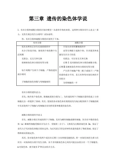
第三章遗传的染色体学说1、有丝分裂和减数分裂的区别在哪里?从遗传学角度来看,这两种分裂各有什么意义?那么,无性生殖会发生分离吗?试加说明。
答:有丝分裂和减数分裂的区别列于下表:有丝分裂的遗传意义:首先:核内每个染色体,准确地复制分裂为二,为形成的两个子细胞在遗传组成上与母细胞完全一样提供了基础。
其次,复制的各对染色体有规则而均匀地分配到两个子细胞的核中从而使两个子细胞与母细胞具有同样质量和数量的染色体。
减数分裂的遗传学意义首先,减数分裂后形成的四个子细胞,发育为雌性细胞或雄性细胞,各具有半数的染色体(n)雌雄性细胞受精结合为合子,受精卵(合子),又恢复为全数的染色体2n。
保证了亲代与子代间染色体数目的恒定性,为后代的正常发育和性状遗传提供了物质基础,保证了物种相对的稳定性。
其次,各对染色体中的两个成员在后期I分向两极是随机的,即一对染色体的分离与任何另一对染体的分离不发生关联,各个非同源染色体之间均可能自由组合在一个子细胞里,n对染色体,就可能有2n种自由组合方式。
例如,水稻n=12,其非同源染色体分离时的可能组合数为212 = 4096。
各个子细胞之间在染色体组成上将可能出现多种多样的组合。
此外,同源染色体的非妹妹染色单体之间还可能出现各种方式的交换,这就更增加了这种差异的复杂性。
为生物的变异提供了重要的物质基础。
2、水稻的正常的孢子体组织,染色体数目是12对,问下列各组织的染色体数目是多少?(1)胚乳;(2)花粉管的管核;(3)胚囊;(4)叶;(5)根端;(6)种子的胚;(7)颖片;答;(1)36;(2)12;(3)12×8=96;(4)24;(5)24;(6)24;(7)24;3、用基因型Aabb的玉米花粉给基因型AaBb的玉米雌花授粉,你预期下一代胚乳的基因型是什么类型,比例如何?答:即下一代胚乳有八种基因型,且比例相等。
4、某生物有两对同源染色体,一对染色体是中间着丝粒,另一对是端部着丝粒,以模式图方式画出:(1)第一次减数分裂的中期图。
刘祖洞(遗传学)课后习题标准答案!全面版
刘祖洞《遗传学》参考答案全面版ﻫ第二章孟德尔定律1、为什么分离现象比显、隐性现象有更重要的意义?(1)分离规律是生物界普遍存在的一种遗传现象,而显性现象的表现是相对的、有条件的;ﻫ(2)答:因为ﻫ只有遗传因子的分离和重组,才能表现出性状的显隐性。
可以说无分离现象的存在,也就无显性现象的发生。
ﻫ2、解:序号杂交基因型表现型(1)RR×rr Rr 红果色ﻫ(2) Rr×rr 1/2Rr,1/2rr 1/2红果色,1/2黄果色(3)Rr×Rr 1/4RR,2/4Rr,1/4rr3/4红果色,1/4黄果色(4)Rr×RR 1/2RR,1/2Rr 红果色(5)rr×rr rr黄果色ﻫ3、下面是紫茉莉的几组杂交,基因型和表型已写明。
问它们产生哪些配子?杂种后代的基因型和表型怎样?(1)Rr× RR(2)rr ×Rr(3)Rr ×Rr粉红红色白色粉红粉红粉红ﻫ解:序号杂交配子类型基因型表现型(1)Rr× RR R,r;R 1/2RR,1/2Rr 1/2红色,1/2粉红(2)rr × Rrr;R,r1/2Rr,1/2rr 1/2粉红,1/2白色4、在南瓜中,果实的白色(W)(3) Rr× Rr R,r1/4RR,2/4Rr,1/4rr1/4红色,2/4粉色,1/4白色ﻫ对黄色(w)是显性,果实盘状(D)对球状(d)是显性,这两对基因是自由组合的。
问下列杂交可以产生哪些基因型,哪些表型,它们的比例如何?(1)WWDD×wwdd(2)XwDd×wwdd(3)Wwdd×wwDd(4)Wwdd×WwDd解:ﻫ序号杂交基因型表现型1WWDD×wwdd WwDd 白色、盘状果实2WwDd×wwdd 1/4WwDd,1/4Wwdd,1/4wwDd,1/4wwdd,1/4白色、盘状,1/4白色、球状,1/2wwDd×wwdd 1/2wwDd,1/2wwdd 1/2黄色、盘状,1/2黄色、4黄色、盘状,1/4黄色、球状ﻫ球状3 Wwdd×wwDd 1/4WwDd,1/4Wwdd,1/4wwDd,1/4wwdd,1/4白色、盘状,1/4白色、球4Wwdd×WwDd1/8WWDd,1/8WWdd,2/8WwDd,2/状,1/4黄色、盘状,1/4黄色、球状ﻫ8Wwdd,1/8wwDd,1/8wwdd 3/8白色、盘状,3/8白色、球状,1/8黄色、盘状,1/8黄色、球状ﻫ5.在豌豆中,蔓茎(T)对矮茎(t)是显性,绿豆荚(G)对黄豆荚(g)是显性,圆种子(R)对皱种子(r)是显性。
遗传学第十三章遗传与进化优秀课件
第十三章:遗传与进化
第二节:进化理论 一、进化学说
进化论一词最早是由法国博物学家拉马克 (marck,1744-1829)在1802年提出的。拉马克的学说被称为 用进废退学说,或获得性状遗传学说。英国博物学家达尔文 (CharlesDarwen,1809-1882)在1859年发表的《物种起源》一书为 进化论奠定了科学基础。现代生命科学的发展大大丰富了进化论 的内容。现代进化理论主要从群体遗传学和分子生物学的角度解 释生物的进化机制。
递给下一代。 (E)新物种是由渐变的方式而形成的
2020/12/10
苏州科技学院生物系 叶亚新
第二节:进化理论 一、进化学说
1、拉马克学说
获得性:是指个体在生活过程中为了适应外界环境而发生 的定向变异。
拉马克认为:累积起来而形成为显著 的变异,最终引起生物类型的改变。
苏州科技学院生物系 叶亚新
第十三章:遗传与进化
第一节:分子进化 三、遗传体系的进化 1、遗传物质在进化中的变化
RNA ---------- DNA 2、蛋白质合成机制在进化中的变化
“三中读二”理论 3、染色体的演化
2020/12/10
苏州科技学院生物系 叶亚新
2020/12/10
苏州科技学院生物系 叶亚新
A
B
加热变性
A、B杂种双链
温育 不同种
同种
2020/12/10
苏州科技学院生物系 叶亚新
二、核酸进化 2、DNA 质的变化
A
A、B杂种双链
加热变性 温育
不同种
B
同种
TmA-A =TmB-B =TmA-B A、B属同一种 TmA-B < TmA-A 或 TmA-B < TmB-B A、B有差异
《遗传学》朱军版习题及答案
《遗传学(第三版)》朱军主编课后习题与答案目录第一章绪论 (1)第二章遗传的细胞学基础 (2)第三章遗传物质的分子基础 (6)第四章孟德尔遗传 (9)第五章连锁遗传和性连锁 (12)第六章染色体变异 (15)第七章细菌和病毒的遗传 (21)第八章基因表达与调控 (27)第九章基因工程和基因组学 (31)第十章基因突变 (34)第十一章细胞质遗传 (35)第十二章遗传与发育 (38)第十三章数量性状的遗传 (39)第十四章群体遗传与进化 (44)第一章绪论1.解释下列名词:遗传学、遗传、变异。
答:遗传学:是研究生物遗传和变异的科学,是生物学中一门十分重要的理论科学,直接探索生命起源和进化的机理。
同时它又是一门紧密联系生产实际的基础科学,是指导植物、动物和微生物育种工作的理论基础;并与医学和人民保健等方面有着密切的关系。
遗传:是指亲代与子代相似的现象。
如种瓜得瓜、种豆得豆。
变异:是指亲代与子代之间、子代个体之间存在着不同程度差异的现象。
如高秆植物品种可能产生矮杆植株:一卵双生的兄弟也不可能完全一模一样。
2.简述遗传学研究的对象和研究的任务。
答:遗传学研究的对象主要是微生物、植物、动物和人类等,是研究它们的遗传和变异。
遗传学研究的任务是阐明生物遗传变异的现象及表现的规律;深入探索遗传和变异的原因及物质基础,揭示其内在规律;从而进一步指导动物、植物和微生物的育种实践,提高医学水平,保障人民身体健康。
3.为什么说遗传、变异和选择是生物进化和新品种选育的三大因素?答:生物的遗传是相对的、保守的,而变异是绝对的、发展的。
没有遗传,不可能保持性状和物种的相对稳定性;没有变异就不会产生新的性状,也不可能有物种的进化和新品种的选育。
遗传和变异这对矛盾不断地运动,经过自然选择,才形成形形色色的物种。
同时经过人工选择,才育成适合人类需要的不同品种。
因此,遗传、变异和选择是生物进化和新品种选育的三大因素。
4. 为什么研究生物的遗传和变异必须联系环境?答:因为任何生物都必须从环境中摄取营养,通过新陈代谢进行生长、发育和繁殖,从而表现出性状的遗传和变异。
(完整word版)刘祖洞遗传学考试库
遗传学试题库(一)一、名词解释:(每小题3分,共18分)1、外显子2、复等位基因3、F因子4、母性影响5、伴性遗传6、杂种优势矚慫润厲钐瘗睞枥庑赖。
二、填空题:(每空0.5分,共20分)1、豌豆中,高茎(T)对矮茎(t)为显性,黄子叶(Y)对绿子叶(y)为显性,假设这两个位点的遗传符合自由组合规律,若把真实遗传的高茎黄子叶个体与矮茎绿子叶个体进行杂交,F2中矮茎黄子叶的概率为。
聞創沟燴鐺險爱氇谴净。
2、人类中,苯丙酮尿症的常染色体隐性纯合体是一种严重的代谢缺馅。
如果正常的双亲生了一个患病的女儿,一个正常表型的儿子.问:儿子是此病基因携带者的概率是。
残骛楼諍锩瀨濟溆塹籟。
3、大麦中,密穗对稀穗为显性,抗条诱对不抗条诱为显性。
一个育种工作者现有一个能真实遗传的密穗染病材料和一个能真实遗传的稀穗抗病材料,他想用这两个材料杂交,以选出稳定的密穗抗病品种,所需要类型有第______代就会出现,所占比例为_______,到第________代才能肯定获得,如果在F3代想得到100个能稳定遗传的目标株系,F2代至少需种植_________株。
酽锕极額閉镇桧猪訣锥。
4、某一植物二倍体细胞有10条同源染色体,在减数分裂前期Ⅰ可观察到个双价体,此时共有条染色单体,到中期Ⅱ每一细胞可观察到条染色单体。
彈贸摄尔霁毙攬砖卤庑.5、人类的性别决定属于型,鸡的性别决定属于型,蝗虫的性别决定属于型.謀荞抟箧飆鐸怼类蒋薔。
6、有一杂交:CCDD ×ccdd,假设两位点是连锁的,而且相距20个图距单位。
F2中基因型(ccdd)所占比率为。
厦礴恳蹒骈時盡继價骚。
7、遗传力是指_____________________________;广义遗传力是_________方差占________方差的比值。
遗传力越_____,说明性状传递给子代的能力就越_____,选择效果越________.茕桢广鳓鯡选块网羈泪。
8、萝卜甘蓝是萝卜和甘蓝的杂种,若杂种体细胞染色体数为36,甘蓝亲本产生的配子染色体数为9条,萝卜单倍体数应为______条,可育的杂种是________倍体。