GPNMB促进周围神经损伤修复的作用及其机制
周围神经损伤修复后再生是神经与末梢效应器交互作用的系统重塑过程
周围神经损伤修复后再生是神经与末梢效应器交互作用的系统重塑过程来自中国北京大学人民医院的张培训所在团队主持的中国2014年“国家重点基础研究发展计划”(973)所在项目组北京大学人民医院创伤骨科,率先提出了“周围神经损伤修复后的再生是神经与末梢效应器交互作用的系统重塑过程”的整体思路,以神经修复过程的3个重要环节,即再生机制、调控机制、重塑机制作为本项目的关键科学问题,设置了以下6个课题作为主要研究内容。
(1)周围神经再生的组织诱导机制。
(2)神经元轴突生长与髓鞘形成的机制。
(3)周围神经损伤后失神经支配肌肉萎缩调控机制。
(4)周围神经损伤后疼痛、感觉异常的机制与干预策略。
(5)周围神经损伤后中枢结构重塑与功能修复。
(6)周围神经创新性修复模式下末梢效应器诱导的系统功能重塑。
本项目将从整体上诠释周围神经损伤再生机制的重大理论问题,而更重要的是将为周围神经损伤的治疗提供新的科学方法。
文章发表在《中国神经再生研究(英文版)》杂志2015年1月第1期。
Article: "Neural regeneration after peripheral nerve injury repair is a system remodelling process of interaction between nerves and terminal effector," by Pei-xun Zhang, Xiao-feng Yin, Yu-hui Kou, Feng Xue, Na Han, Bao-guo Jiang (Department of Trauma & Orthopedics, Peking University People’s Hospital, Beijing, China)Zhang PX, Yin XF, Kou YH, Xue F, Han N, Jiang BG (2015) Neural regeneration after peripheral nerve injury repair is a system remodelling process of interaction between nerves and terminal effector. Neural Regen Res 10(1):52.欲获更多资讯:文章全文请见:Neural Regen ResNeural regeneration after peripheral nerve injury repair is a system remodelling processPei-xun Zhang, Peking University People’s Hospital, China and his colleagues, i.e., the 973 program team, was the first to propose that “neural regeneration after peripheral nerve injury repair is a system remodelling process of interaction between nerves and terminal effector” and discussed the following six topics regarding the regeneration, regulatory and remodelling mechanisms of nerve repair: (1) mechanisms underlying tissue inducing peripheral nerve injury; (2) regulatory mechanisms underlying neuronal axon growth and myelination; (3) regulatory mechanisms underlying denervated muscular atrophy after peripheral nerve injury; (4) mechanisms underlying pain and paraesthesia after peripheral nerve injury and interventional strategies; (5) central structure remodelling and function repair after peripheral nerve injury; (6) terminal effector-induced system function remodelling during innovative repair of injured peripheral nerve. This program will, on the whole, elucidate the major mechanisms underlying regeneration after peripheral nerve injury and more importantly provide new solutions for the treatment of peripheral nerve injury. The relevant paper was published in the Neural Regeneration Research (Vol. 10, No. 1, 2015).Article: "Neural regeneration after peripheral nerve injury repair is a system remodelling process of interaction between nerves and terminal effector," by Pei-xun Zhang, Xiao-feng Yin, Yu-hui Kou, Feng Xue, Na Han, Bao-guo Jiang (Department of Trauma & Orthopedics, Peking University People’s Hospital, Beijing, China)Zhang PX, Yin XF, Kou YH, Xue F, Han N, Jiang BG (2015) Neural regeneration after peripheral nerve injury repair is a system remodelling process of interaction between nerves and terminal effector. Neural Regen Res 10(1):52.。
神经延长:促进损伤周围神经的再生
神经延长:促进损伤周围神经的再生周围神经损伤后局部微环境对损伤神经的再生起重要作用。
最新研究表明,周围神经损伤后适度的张力可以促进轴突再生。
美国加州大学圣地亚哥分校矫形外科与生物工程系Sameer B. Shah博士假设,近端未受损的神经可以向损伤远侧神经伸长,并且忠实地再现体内和体外创建的机械环境,这种神经延长可以跨越神经缺损,确保解剖和功能匹配到远端残端并可能加速功能恢复。
作者研究团队据此研制出新型的固定器,能线性拉长近端残端向远端残端并加速与远端重新连接,促进损伤周围神经的修复与再生。
此观点发表在《中国神经再生研究(英文版)》2014年8月第16期杂志上。
Article: "Peripheral nerve lengthening as a regenerative strategy" Kenneth M. Vaz1, Justin M. Brown2, Sameer B. Shah3 (1Department of Orthopaedic Surgery, University of California, San Diego, La Jolla, CA, USA; 2Department of Neurosurgery, University of California, San Diego, La Jolla, CA, USA; 3Departments of Orthopaedic Surgery and Bioengineering, University of California, San Diego, La Jolla, CA, USA)Vaz KM, Brown JM, Shah SB. Peripheral nerve lengthening as a regenerative strategy. Neural Regen Res. 2014;9(16):1498-1501.欲获更多资讯:文章全文请见:Neural Regen ResPeripheral nerve lengthening as a regenerative strategyExcessive mechanical loading has long been implicated in nerve injury, and thus tension is actively avoided during nerve repair. However, recent evidence suggests that moderate levels of mechanical loading may, in fact, promote neuronal growth. Sameer B. Shah and her colleagues have hypothesized that in vivo elongation of intact proximal stumps towards the injured distal stumps of severed peripheral nerves can faithfully reproduce the mechanical environment created during the above-mentioned in vivo and in vitro procedures. Such lengthening across a nerve gap may accelerate functional recovery and ensures an anatomic and functional match to the distal stump. Nerve transfers require rerouting of redundant branches or fascicles from functional nerves to functionally paralyzed recipient nerves. The transfer restores neuromuscular function in the recipient nerve, enabling individuals to execute critical tasks such as elbow extension or key pinch. However, the choice of donor nerves is often limited byanatomical proximity to the recipient, resulting in the use of an intervening graft or reconnection more proximally than desired. It is possible that sufficiently lengthening a donor nerve, even while intact, could create additional candidate donors for nerve transfers than those currently available. The years to come should offer an exciting expansion of our understanding and application of principles of nerve lengthening towards nerve repair. The perspective article is published on Neural Regeneration Research (VOl. 9, No. 16)Article: "Peripheral nerve lengthening as a regenerative strategy" Kenneth M. Vaz1, Justin M. Brown2, Sameer B. Shah3 (1 Department of Orthopaedic Surgery, University of California, San Diego, La Jolla, CA, USA; 2 Department of Neurosurgery, University of California, San Diego, La Jolla, CA, USA; 3 Departments of Orthopaedic Surgery and Bioengineering, University of California, San Diego, La Jolla, CA, USA)Vaz KM, Brown JM, Shah SB. Peripheral nerve lengthening as a regenerative strategy. Neural Regen Res. 2014;9(16):1498-1501.。
细胞外基质成分可影响周围神经损伤的修复与再生(精)
细胞外基质成分可影响周围神经损伤的修复与再生瑞士洛桑联邦理工学院生物材料 -材料 -组织接口联合实验室 di Summa教授所带团队研究人员正在合作开发一种新型多功能促进周围神经损伤修复的再生导管,这种导管整合仿生材料,微细加工技术和细胞治疗技术。
在《中国神经再生研究(英文版》最新一期杂志中,作者介绍了他们在细胞外基质分子在周围神经损伤修复在的重要作用及试验应用结果,表明其可以成为人工导管的最佳选择。
周围神经损伤严重影响患者的生活。
人工导管是周围神经损伤修复的有效替代方法,能够为周围神经修复创造适宜微环境,并指引轴突生长方向。
好的生物材料需要具备良好的生物相容性,能够减轻炎症和瘢痕组织形成。
充填细胞外基质的神经组织能延长细胞的存活,促进移植细胞在损伤部位生长,减少所需的内源雪旺氏细胞的滞后时间,在修复神经缺损方面表现出较好的应用潜能。
“我们的最新研究成果显示出促进周围神经再生是有希望”,作者如此说。
“未来我们团队的研究将集中于导管内腔的完善,使其可以更好的与涂覆有的细胞外基质成分结合,以增强细胞表面的相互作用,从而更好的促进损伤神经的再生”。
Article: " Extracellular matrix components in peripheral nerve repair: how to affect neural cellular response and nerve regeneration? " by Alba C. de Luca 1, Stephanie P. Lacour1, Wassim Raffoul2, Pietro G. di Summa2 (1 EPFL, Centre for Neuroprosthetics, Laboratory for Soft Bioelectronic Interfaces, Station 17, 1015 Lausanne, Switzerland; 2 Department of Plastic, Reconstructive and Hand Surgery, University Hospital of Lausanne (CHUV, Lausanne, Switzerlandde Luca AC, Lacour SP, Raffoul W, di Summa PG. Extracellular matrix components in peripheral nerve repair: how to affect neural cellular response and nerve regeneration? Neural Regen Res. 2014;9(22: 1943-1948.欲获更多资讯:N eural Regen ResImpact and effect of the ECM molecules in peripheral nerve repair and regeneration Peripheral nerve injury is a serious problem affecting significantly patients’ life. New advancedstrategies have been developed to improve the regeneration of the injured nerve, including artificial conduits. Biomimetic materials aim at simulating the native neural tissue, creating a friendly environment for cells and tissue to growth. This allows the regeneration of longer gaps and extending cell survival.Researchers at the EPFL and at the CHUV in Lausanne, Switzerland, are working together to develop novel multifunctional regenerative conduits for peripheral nerve repair, integrating biomimetic materials, microfabrication techniques and cell therapy. In a recent review accepted for publication in Neural Regeneration Research, they report recent progresses in the fabrication of biomimetic materials for peripheral nerve peripheral. As suggested by the tile, the impact and the effect of extracellular matrix (ECM molecules in nerve regeneration is therefore presented and critically discussed, including future perspectives in the field.ECM molecules can either be used for filling artificial nerve guidance conduits or for coating the inner lumens, resulting in the ability of repairing long injury gaps. Bio-fillers can provide a suitable and natural environment to support cell survival and proliferation inside the tube, shortening the delay that triggers and activates the nerve regeneration. In addition, it has been demonstrated that ECM molecules can provide binding sites for specific growth factors and neurotrophins, making them suitable to develop a drug delivery system localized at the injury site.“The latest results achieved in the field of peripheral nerve regeneration are promising”, state the authors. As reported, future research will focus on more advanced modifica tions of the inner lumen, which can be then further “coated with ECM components, in order to enhance cell-surface interactions, hence promoting higherregeneration of the injured tissue”. The perspective article is published in Neural Regeneration Research (Vol. 9, No. 22, 2014.Article: " Extracellular matrix components in peripheral nerve repair: how to affect neural cellular response and nerve regeneration? " by Alba C. de Luca 1, Stephanie P. Lacour1, Wassim Raffoul2, Pietro G. di Summa2 (1 EPFL, Centre for Neuroprosthetics, Laboratory for Soft Bioelectronic Interfaces, Station 17, 1015 Lausanne, Switzerland; 2 Department of Plastic, Reconstructive and Hand Surgery, University Hospital of Lausanne (CHUV, Lausanne, Switzerlandde Luca AC, Lacour SP, Raffoul W, di Summa PG. Extracellular matrix components in peripheral nerve repair: how to affect neural cellular response and nerve regeneration? Neural Regen Res. 2014;9(22: 1943-1948.。
神经元修复药物,nmn重大研究修复受损神经元的新策略
神经元修复药物,nmn重大研究修复受损神经元的新策略神经元修复药物,nmn重大研究修复受损神经元的新策略!神经细胞是终末分化的细胞,再生能力非常有限,如果神经细胞受到损害,死亡之后不可以生。
通常所说的神经修复,指让存活的神经细胞的纤维,或者突触构建新的神经网络,替代死亡的神经细胞的功能。
目前没有药物能够让死亡的神经细胞复活,所谓修复神经或者营养神经,只能改善存活的受损神经细胞,促使其结构恢复和功能改善。
这些药物根据作用部位分为两类,一类是营养修复中枢神经的药物,另一类是营养修复周围神经的药物。
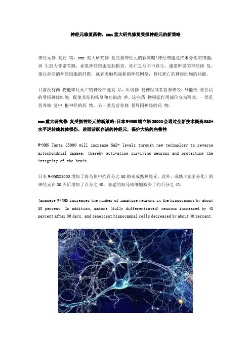
nmn重大研究修复受损神经元的新策略:日本W+NMN端立塔25000会通过全新技术提高NAD+水平逆转线粒体损伤,进而活跃存活的神经元,保护大脑的完整性W+NMN Tanta 25000 will increase NAD+ levels through new technology to reverse mitochondrial damage, thereby activating surviving neurons and protecting the integrity of the brain日本W+NMN25000增加了海马体中约百分之50的未成熟神经元。
此外,成熟(完全分化)的神经元在30天后增加了百分之48,衰老的海马体细胞减少了约百分之40。
Japanese W+NMN increases the number of immature neurons in the hippocampus by about 50 percent. In addition, mature (fully differentiated) neurons increased by 48 percent after 30 days, and senescent hippocampal cells decreased by about 40 percent.此外,日本W+NMN25000诱导了神经发生的增加,5天增加了2.6倍,表现为未成熟神经元数量的显著增加。
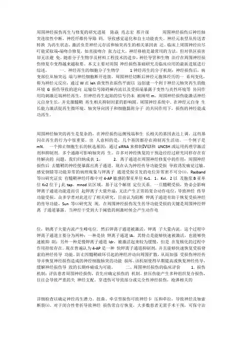
间充质干细胞在周围神经损伤修复中的应用现状及前景分析
写作提纲: (1)间充质干细胞在周围神经损伤修复中的应用; (2)间充质干细胞外泌体在周围神经损伤修复中的应
用; (3)神经导管负载间充质干细胞在周围神经损伤修复
中的应用; (4)间充质干细胞基因工程在周围神经损伤中的应用。
曹丽芝,女,1992 年生, 山东省潍坊市人,汉族, 吉林大学在读硕士,主要 从事神经内科研究。
Cao Lizhi, Master candidate, The First Hospital of Jilin University, Changchun 130000, Jilin Province, China
通讯作者:陈嘉峰,教授, 吉林大学第一医院,吉林 省长春市 130000
文献标识码:A 稿件接受:2019-06-12
文题释义: 外泌体:是指包含了复杂 RNA 和蛋白质的小膜泡(30-150 nm)。多种细胞在正常及病理状态下均可分泌外泌 体,主要来源于细胞内溶酶体微粒内陷形成的多囊泡体,经多囊泡体外膜与细胞膜融合后释放到胞外基质中。 外泌体的功能取决于其所来源的细胞类型,可参与到机体免疫应答、抗原提呈、细胞迁移、细胞分化、肿瘤 侵袭等多个方面。 髓鞘化:也称鞘化,是指髓鞘发展的过程,它使神经兴奋在沿神经纤维传导时速度加快,并保证其定向传导。 在周围神经修复时,再生神经纤维髓鞘化可以改善损伤神经功能,更利于神经修复。
Mesenchymal stem cells: present status and prospect for its application in peripheral nerve injury
Cao Lizhi1, Feng Naibo2, Wang Juan1, Chen Jiafeng1 (1The First Hospital of Jilin University, Changchun 130000, Jilin Province, China; 2The Second Hospital of Jilin University, Changchun 130000, Jilin Province, China)
周围神经损伤再生与修复的研究进展
周围神经损伤再生与修复的研究进展陈焱肖志宏邢廾谋周围神经损伤后神经轴突连续性中断,神经纤维传导障码.导致感觉退化和自主功能丧失。
神经元表型从传送者转换为再生状态,激活负责神经元存活和轴突再生的相关基因表达。
临床上周围神经应尽叮能采取端-端吻合修复,如直接吻合张力过大,神经移植是最常用的方法,但对供区损害却无法避免。
随着分子生物学及材料工程技术的进步,神经导管和生物治疗在周围神经损伤修复巾变得越来越取要。
本文主要对周围神经损伤基础研究及临床应用的最新进展进行综述。
一.神经再生的细胞分子生物学1神经再生的分子机制:神经损伤后,病变部位从轴突远端与神经细胞断开连接。
周围神经切断后神经元胞体经历的一系列变化,称为神经元反应,通过W豇leh曲变性在损伤平面以远创建一个利于神经元轴突再生的傲环境G损伤导致的逆向运输信号障碍癣内流以及受损端暴露于变性与炎性环境等协同作用均刺激近端神经再生,但神经再生起始的信号仍未被阐明m。
周围神经损伤能激话神经元自身生长,并克服髓鞘再生相关抑制因素的影响圆。
周围神经系统中.在神经元自身生长能力激活捉再生微环境、轴突导向因子和细胞黏附分子的共同作用下,损伤的神经能成功再生。
周围神经轴突的再生是复杂的,在神经损伤远侧残端和生长相关的基因表达上调,这些基闪在再生的行为中很重要。
出人意料的是,几个基因都存在抑制再生活动。
一个例子是mN.一个抑止细胞生长的候选基因:通过siRNA来抑制Ⅳ瞓和UNC5H或运用药理学激活剂和抑制剂.多个通路可影响轴突再生。
许多对神经恢复的干预途径经过研究同样存在有待解决的问题,我们归纳成表1。
2.离子通道在周围神经修复中的作用:周围神经损伤后去髓鞘的神经便暴露出离子通道。
现在认为神经传导功能受损导致诱发痛觉过敏、感觉倒错等功能异常的病理现象与钾离子通道受损引发的电位异常密不可分㈤。
Rasband 等㈤研究证实有髓鞘神经纤维中对4-AP敏感的掣亚单位Kv1.1、KvI.2以及胞浆B亚单位Kv2位于j此tap。
细胞外基质成分可影响周围神经损伤的修复与再生(精)
细胞外基质成分可影响周围神经损伤的修复与再生瑞士洛桑联邦理工学院生物材料 -材料 -组织接口联合实验室 di Summa教授所带团队研究人员正在合作开发一种新型多功能促进周围神经损伤修复的再生导管,这种导管整合仿生材料,微细加工技术和细胞治疗技术。
在《中国神经再生研究(英文版》最新一期杂志中,作者介绍了他们在细胞外基质分子在周围神经损伤修复在的重要作用及试验应用结果,表明其可以成为人工导管的最佳选择。
周围神经损伤严重影响患者的生活。
人工导管是周围神经损伤修复的有效替代方法,能够为周围神经修复创造适宜微环境,并指引轴突生长方向。
好的生物材料需要具备良好的生物相容性,能够减轻炎症和瘢痕组织形成。
充填细胞外基质的神经组织能延长细胞的存活,促进移植细胞在损伤部位生长,减少所需的内源雪旺氏细胞的滞后时间,在修复神经缺损方面表现出较好的应用潜能。
“我们的最新研究成果显示出促进周围神经再生是有希望”,作者如此说。
“未来我们团队的研究将集中于导管内腔的完善,使其可以更好的与涂覆有的细胞外基质成分结合,以增强细胞表面的相互作用,从而更好的促进损伤神经的再生”。
Article: " Extracellular matrix components in peripheral nerve repair: how to affect neural cellular response and nerve regeneration? " by Alba C. de Luca 1, Stephanie P. Lacour1, Wassim Raffoul2, Pietro G. di Summa2 (1 EPFL, Centre for Neuroprosthetics, Laboratory for Soft Bioelectronic Interfaces, Station 17, 1015 Lausanne, Switzerland; 2 Department of Plastic, Reconstructive and Hand Surgery, University Hospital of Lausanne (CHUV, Lausanne, Switzerlandde Luca AC, Lacour SP, Raffoul W, di Summa PG. Extracellular matrix components in peripheral nerve repair: how to affect neural cellular response and nerve regeneration? Neural Regen Res. 2014;9(22: 1943-1948.欲获更多资讯:N eural Regen ResImpact and effect of the ECM molecules in peripheral nerve repair and regeneration Peripheral nerve injury is a serious problem affecting significantly patients’ life. New advancedstrategies have been developed to improve the regeneration of the injured nerve, including artificial conduits. Biomimetic materials aim at simulating the native neural tissue, creating a friendly environment for cells and tissue to growth. This allows the regeneration of longer gaps and extending cell survival.Researchers at the EPFL and at the CHUV in Lausanne, Switzerland, are working together to develop novel multifunctional regenerative conduits for peripheral nerve repair, integrating biomimetic materials, microfabrication techniques and cell therapy. In a recent review accepted for publication in Neural Regeneration Research, they report recent progresses in the fabrication of biomimetic materials for peripheral nerve peripheral. As suggested by the tile, the impact and the effect of extracellular matrix (ECM molecules in nerve regeneration is therefore presented and critically discussed, including future perspectives in the field.ECM molecules can either be used for filling artificial nerve guidance conduits or for coating the inner lumens, resulting in the ability of repairing long injury gaps. Bio-fillers can provide a suitable and natural environment to support cell survival and proliferation inside the tube, shortening the delay that triggers and activates the nerve regeneration. In addition, it has been demonstrated that ECM molecules can provide binding sites for specific growth factors and neurotrophins, making them suitable to develop a drug delivery system localized at the injury site.“The latest results achieved in the field of peripheral nerve regeneration are promising”, state the authors. As reported, future research will focus on more advanced modifica tions of the inner lumen, which can be then further “coated with ECM components, in order to enhance cell-surface interactions, hence promoting higherregeneration of the injured tissue”. The perspective article is published in Neural Regeneration Research (Vol. 9, No. 22, 2014.Article: " Extracellular matrix components in peripheral nerve repair: how to affect neural cellular response and nerve regeneration? " by Alba C. de Luca 1, Stephanie P. Lacour1, Wassim Raffoul2, Pietro G. di Summa2 (1 EPFL, Centre for Neuroprosthetics, Laboratory for Soft Bioelectronic Interfaces, Station 17, 1015 Lausanne, Switzerland; 2 Department of Plastic, Reconstructive and Hand Surgery, University Hospital of Lausanne (CHUV, Lausanne, Switzerlandde Luca AC, Lacour SP, Raffoul W, di Summa PG. Extracellular matrix components in peripheral nerve repair: how to affect neural cellular response and nerve regeneration? Neural Regen Res. 2014;9(22: 1943-1948.。
周围神经损伤修复及其功能恢复分析
周围神经损伤修复及其功能恢复分析发表时间:2018-08-20T14:46:51.280Z 来源:《医药界》2018年1月下作者:王挺厅[导读] 对周围神经损伤修复及其功能恢复进行分析,了解生物型人工材料的性能以及应用、功能恢复评定方法。
方法:使用网络数据库对周围神经、损伤修复、功能恢复等关键词进行搜索,查找近几年的相关文章,排除主题重复的文章,以筛选出来的文章为主对周围神经损伤修复及其功能恢复进行分析。
嘉善县第一人民医院浙江省嘉兴市 314100摘要:目的:对周围神经损伤修复及其功能恢复进行分析,了解生物型人工材料的性能以及应用、功能恢复评定方法。
方法:使用网络数据库对周围神经、损伤修复、功能恢复等关键词进行搜索,查找近几年的相关文章,排除主题重复的文章,以筛选出来的文章为主对周围神经损伤修复及其功能恢复进行分析。
结果:在周围神经损失修复中能够使用的比较理想的支架材料为脱细胞神经基质和人工合成材料构成的复合型材料,这种材料基本不会发生排斥反应,并能够促进周围组织神经细胞的再生。
结论:虽然我国周围神经损伤修复的人工材料取得了较大的发展,但是并没有超出自体神经移植支架材料,脱细胞神经基质和人工合成材料构成的复合型材料能够作为良好的周围神经支架使用,但是仍然需要和种子细胞进行联合构建。
关键词:周围神经;损伤修复;功能恢复中图分类号:R745 文献标识码:A引言周围神经损伤是我国医疗当中常见病症之一,如果受损伤的周围神经没有得到及时ID修复,就会对损伤神经支配的肌肉产生影响,促使神经肌肉功能障碍病症的出现,影响患者后期康复。
近几年生物学技术取得了较大的发展,进行神经修复时可以使用组织工程神经移植物,解决了传统治疗中移植物材料单一的问题,但是周围神经损伤修复是一项复杂、难度较大的治疗工作,还需要对修复材料进行优选。
一、资料与方法(一)一般资料标准资料纳入标准:首先与组织工程神经移植物这个主题相关的文献;其次与生物型人工修复神经材料相关的文献;最后组织工程神经中新技术应用与进展的相关文献。
gpnmb基因结构
gpnmb基因结构GPNMB基因结构分析一、引言GPNMB基因(Glycoprotein non-metastatic melanoma protein B)是人类基因组中的一个重要基因,属于GPNMB/DC-HIL家族。
该基因编码的蛋白质是一种糖蛋白,在多种生理和病理过程中发挥着重要作用。
本文将对GPNMB基因的结构进行详细分析,以期增进对该基因的理解。
二、基因组定位GPNMB基因位于人类染色体7q11.22区域。
该区域是一个基因密集区,与多种疾病的发生和发展密切相关。
GPNMB基因在基因组中的位置对其功能具有重要影响。
三、基因结构GPNMB基因由多个外显子和内含子组成。
根据已有研究,GPNMB基因共有10个外显子,分布在基因组上的连续区域。
这些外显子的长度不等,从几百到几千个碱基不等。
外显子与内含子的交替排列形成了GPNMB基因的结构。
四、启动子区域GPNMB基因的启动子区域是基因的调控中心,其中包含多个转录因子结合位点。
这些转录因子可以与启动子区域结合,调控GPNMB基因的转录水平。
通过研究GPNMB基因的启动子区域,可以深入了解GPNMB基因的调控机制。
五、转录过程GPNMB基因在转录过程中需要依赖转录因子和RNA聚合酶等分子的参与。
在启动子区域的调控下,转录因子结合到GPNMB基因的启动子区域,引导RNA聚合酶在GPNMB基因的DNA模板上进行转录,合成GPNMB基因的RNA分子。
六、剪接异构体GPNMB基因的外显子和内含子的排列方式不同,可以产生多个剪接异构体。
这些剪接异构体可能具有不同的功能和调控模式。
通过研究GPNMB基因的剪接异构体,可以揭示其在不同生理和病理过程中的作用机制。
七、翻译过程GPNMB基因的转录产物是一种RNA分子,需要经过翻译过程才能生成蛋白质。
翻译过程包括核糖体结合到RNA分子上,根据RNA分子的密码子序列合成相应的氨基酸,最终形成GPNMB基因所编码的蛋白质。
GPNMB促进周围神经损伤修复的作用及其机制
GPNMB促进周围神经损伤修复的作用及其机制周围神经损伤是临床常见损伤,损伤后的再生修复涉及很多因素,其中施万细胞(Schwann cells,SCs)在周围神经损伤修复中发挥着重要的作用。
周围神经损伤后,SCs通过上调多种神经营养因子(neurotrophic factors,NTFs)和粘附分子的表达,并在损伤局部募集巨噬细胞,与巨噬细胞共同吞噬裂解的轴突和髓鞘,改善再生微环境;在基膜内形成Büngner’s带,引导轴突再生;轴突再生后,SCs 包绕其形成髓鞘,促进神经功能恢复。
损伤远侧端SCs长期失神经,则导致周围神经再生能力下降,影响神经修复。
因此,激活失神经SCs,促进SCs增殖、分化、迁移、表达和分泌NTFs等,或者通过补充外源性营养因子、小分子物质等改善再生微环境,是治疗周围神经损伤的重要策略。
非转移性糖蛋白黑色素瘤蛋白B(Glycoprotein Non-Metastatic Melanoma Protein B,GPNMB),也叫骨活化素(osteoactivin,OA)、树突状细胞肝素整合素配体(dendritic cell-heparin integrin ligand,DC-HIL)、造血生长因子诱导的神经激肽-Ⅰ型(hematopoietic growth factor inducible neurokinin-Ⅰtype,HGFIN),是一种Ⅰ型跨膜蛋白,最早发现于黑色素瘤细胞中。
GPNMB广泛分布于中枢神经系统,具有非常重要的作用:如改善记忆、抗炎、减少神经元死亡、保护神经元。
GPNMB在周围神经系统特别是SCs也有表达,然而,它对SCs和周围神经系统是否有激活或者保护作用,以及在周围神经再生修复中的作用仍不清楚。
基于以上考虑,本研究通过对坐骨神经损伤后远侧端进行基因芯片分析,了解GPNMB在周围神经损伤后的表达变化,并探讨GPNMB对周围神经损伤再生修复的作用和对SCs的作用及其机制,为GPNMB的临床应用提供理论和实验基础。
- 1、下载文档前请自行甄别文档内容的完整性,平台不提供额外的编辑、内容补充、找答案等附加服务。
- 2、"仅部分预览"的文档,不可在线预览部分如存在完整性等问题,可反馈申请退款(可完整预览的文档不适用该条件!)。
- 3、如文档侵犯您的权益,请联系客服反馈,我们会尽快为您处理(人工客服工作时间:9:00-18:30)。
GPNMB促进周围神经损伤修复的作用及其机制周围神经损伤是临床常见损伤,损伤后的再生修复涉及很多因素,其中施万细胞(Schwann cells,SCs)在周围神经损伤修复中发挥着重要的作用。
周围神经损伤后,SCs通过上调多种神经营养因子(neurotrophic factors,NTFs)和粘附分子的表达,并在损伤局部募集巨噬细胞,与巨噬细胞共同吞噬裂解的轴突和髓鞘,改善再生微环境;在基膜内形成Büngner’s带,引导轴突再生;轴突再生后,SCs 包绕其形成髓鞘,促进神经功能恢复。
损伤远侧端SCs长期失神经,则导致周围神经再生能力下降,影响神经修复。
因此,激活失神经SCs,促进SCs增殖、分化、迁移、表达和分泌NTFs等,或者通过补充外源性营养因子、小分子物质等改善再生微环境,是治疗周围神经损伤的重要策略。
非转移性糖蛋白黑色素瘤蛋白B(Glycoprotein Non-Metastatic Melanoma Protein B,GPNMB),也叫骨活化素(osteoactivin,OA)、树突状细胞肝素整合素配体(dendritic cell-heparin integrin ligand,DC-HIL)、造血生长因子诱导的神经激肽-Ⅰ型(hematopoietic growth factor inducible neurokinin-Ⅰtype,HGFIN),是一种Ⅰ型跨膜蛋白,最早发现于黑色素瘤细胞中。
GPNMB广泛分布于中枢神经系统,具有非常重要的作用:如改善记忆、抗炎、减少神经元死亡、保护神经元。
GPNMB在周围神经系统特别是SCs也有表达,然而,它对SCs和周围神经系统是否有激活或者保护作用,以及在周围神经再生修复中的作用仍不清楚。
基于以上考虑,本研究通过对坐骨神经损伤后远侧端进行基因芯片分析,了解GPNMB在周围神经损伤后的表达变化,并探讨GPNMB对周围神经损伤再生修复的作用和对SCs的作用及其机制,为GPNMB的临床应用提供理论和实验基础。
第一部分坐骨神经损伤后GPNMB的表达变化目的:通过对坐骨神经损伤后远侧端进行基因芯片分析,以了解损伤神经远侧端基因表达变化,尤其是GPNMB的表达变化。
方法:对坐骨神经横断伤后0、1、3、7、14、21、28 d,远侧端进行基因芯片分析,STEM分析筛选表达变化趋势显著性的基因集,并对其进行生物信息学分析和聚类分析。
筛选出其中变化最明显的基因,并使用qRT-PCR、免疫印迹(Western bloting,WB)、免疫组织化学(immunohistochemistry,IHC)对其在转录和蛋白水平进行验证。
结果:在坐骨神经横断损伤后0、1、3、7、14、21、28 d,远侧端基因表达趋势显著性的基因集共有12个,其中基因集41的表达趋势在坐骨神经损伤后先上升、至峰值、继而下降,而这种变化趋势与远侧端急性失神经SCs的增殖趋势相一致。
GO、KEGG分析预测基因集41中的基因可能参与细胞增殖过程。
聚类分析发现GPNMB是基因集41中表达变化最明显的基因,其表达从1 d开始上升、从3 d 开始迅速上升,至7 d时上升至峰值,约为0 d的48倍、从14 d开始下降,直至28 d下降至0 d的15倍左右。
qRT-PCR、WB、IHC结果显示GPNMB的表达趋势和芯片结果基本一致。
结论:在坐骨神经损伤后,损伤远侧端GPNMB表达变化最明显,且呈先上升、至峰值、继而下降的趋势,GPNMB可能参与坐骨神经损伤后的自我再生修复过程,尤其是细胞增殖的过程。
第二部分GPNMB促进坐骨神经损伤再生修复目的:探讨GPNMB对坐骨神经损伤再生修复的作用方法:坐骨神经重度压榨伤模型建模后3周,于损伤处由远及近、多点注射重组人GPNMB(rhGPNMB)蛋白或PBS,实验大鼠共分为3组:PBS组、100ng rhGPNMB组、500ng rhGPNMB组,并于4周后进行检测。
免疫荧光检测SCs 增殖及轴突、髓鞘的再生;透射电镜观察髓鞘的超微结构并计算G-Ratio值;体外检测损伤神经兴奋传导速度和动作电位振幅;注射rhGPNMB后的每周观察大鼠行走足印、计算坐骨神经功能指数,检测神经功能恢复情况;观察腓肠肌大体形态、对腓肠肌称重、HE染色检测其萎缩情况。
结果:rhGPNMB组SCs数目、再生轴突数、再生髓鞘数以及增殖细胞核抗原的表达均明显多于PBS组,差异具有统计学意义。
100ng rhGPNMB组和500ng rhGPNMB组的SCs数量分别为PBS组的1.55±0.21和2.75±0.28倍,且形状和排列上更加规则;再生轴突数为PBS组的1.38±0.19和1.78±0.18倍,且分布更加均匀、直径更大、延续性更完整;再生髓鞘数分别为PBS组的1.18±0.18、1.67±0.08倍,且完整性更好、分布更加均匀。
透射电镜结果显示:rhGPNMB组再生髓鞘数多于PBS组,形状更加完整、髓鞘板层较厚且更加致密,微管、微丝等超微结构排列更规则。
计算G-Ratio值,500ng rhGPNMB组G-Ratio值最小,表明其再生髓鞘厚度最厚,差异具有统计学意义。
体外检测发现,100ng rhGPNMB和500ng rhGPNMB两组的动作电位振幅分别为PBS组的1.10±0.22和1.44±0.30倍;神经兴奋传导速度分别为PBS组的1.32±0.08、2.15±0.17倍,差异具有统计学意义。
按照3组大鼠行走足印,计算坐骨神经功能指数,PBS、100ng rhGPNMB、500ng rhGPNMB 3组坐骨神经功能指数均有降低,至第4周3组的坐骨神经功能指数值分别为-72.83±5.85、-60.06±4.77、-40.60±5.00,差异具有统计学意义,表明坐骨神经在损伤再生修复过程中,3组神经功能均有一定的恢复,而rhGPNMB可促进坐骨神经损伤后功能的进一步恢复。
大体形态观察发现rhGPNMB组腓肠肌萎缩较PBS组轻,尤其是500ng rhGPNMB 组。
PBS、100ng rhGPNMB、500ng rhGPNMB组腓肠肌质量分别为正常腓肠肌质量的(44.68±4.08)%、(54.55±3.05)%、(66.85±4.10)%,差异具有统计学意义。
横切面HE结果显示rhGPNMB组肌纤维直径和截面积显著大于PBS组,尤其是500ng rhGPNMB组,差异具有统计学意义。
结论:GPNMB促进损伤坐骨神经SCs增殖、轴突再生以及损伤神经再髓鞘化;改善其电生理特性;促进损伤坐骨神经的功能恢复,延缓腓肠肌萎缩。
总之,GPNMB促进损伤坐骨神经再生修复。
第三部分GPNMB对SCs的作用及其机制目的:探讨GPNMB对SCs的作用及其机制。
方法:在施万细胞系RSC96培养基中分别加入12.5ng/ml或25ng/ml rhGPNMB,CCK8法和WB检测RSC96的增殖;TUNEL实验检测RSC96的凋亡;细胞划痕法检测RSC96的迁移;qRT-PCR、WB、ELISA检测RSC96 NTFs和粘附分子的表达和分泌;WB检测Erk1/2、Akt磷酸化;免疫共沉淀、免疫荧光双标检测rhGPNMB 与RSC96胞膜上哪种蛋白结合;使用该蛋白拮抗剂或干扰RNA(small interfering RNA,siRNA)慢病毒与rhGPNMB共同处理RSC96,CCK8法、细胞划痕法、WB、ELISA分别检测RSC96的增殖、迁移、以及NTFs和粘附分子表达、分泌;WB检测Erk1/2、Akt磷酸化。
结果:RSC96在加入rhGPNMB培养1 d时,增殖无明显变化;从2 d开始,增殖明显高于对照组;培养3 d时,rhGPNMB组RSC96增殖细胞核抗原表达明显高于对照组,尤其是25ng/ml rhGPNMB组。
25ng/ml rhGPNMB组增殖明显快于12.5ng/ml rhGPNMB和对照组,差异具有统计学意义。
rhGPNMB对RSC96凋亡无明显作用。
25ng/ml rhGPNMB组划痕修复速度较快,在48 h时,修复达到(70.35±0.20)%,高于12.5ng/ml rhGPNMB组的(59.74±1.76)%和对照组的(41.23±0.82)%,差异具有统计学意义。
rhGPNMB促进NGF、BDNF、NT-3和N-cadherin在转录、蛋白水平的表达和分泌。
同时25ng/ml rhGPNMB组p-Erk/total-Erk、p-Akt/total-Akt分别为对照组的4.20±0.21、2.58±0.52倍,亦高于12.5ng/ml rhGPNMB组,差异具有统计学意义。
免疫共沉淀结果显示rhGPNMB和NKAα1可共沉淀,免疫荧光双标结果显示rhGPNMB和NKAα1共定位于RSC96胞膜上,这说明rhGPNMB结合RSC96胞膜上的NKAα1。
NKA拮抗剂Ouabain和rhGPNMB共同处理RSC96时,RSC96增殖速度、划痕修复速度、NTFs和N-cadherin的表达和分泌、p-Erk/total-Erk、p-Akt/total-Akt 与单用rhGPNMB相比明显降低,差异具有统计学意义,而与对照组和单用Ouabain 无明显差异。
NKAα1 siRNA和rhGPNMB共同处理RSC96时,NKAα1 siRNA+rhGPNMB 组RSC96增殖速度、划痕修复速度、NTFs和N-cadherin的表达和分泌、p-Erk/total-Erk、p-Akt/total-Akt低于rhGPNMB、NC siRNA+rhGPNMB组,但是高于其他3个非rhGPNMB处理组,差异具有统计学意义。
结论:GPNMB可能通过结合SCs胞膜上NKAα1,通过促进Erk1/2、Akt磷酸化,进而促进SCs的增殖、迁移、表达和分泌神经营养因子和粘附分子。