A Fluorescent Receptor Assay for Benzodiazepines
联用多步骤虚拟筛选方法发现具有新母核的GABAA受体正性变构调节剂

物 理 化 学 学 报Acta Phys. -Chim. Sin. 2024, 40 (1), 2302044 (1 of 13)Received: February 24, 2023; Revised: April 18, 2023; Accepted: April 19, 2023; Published online: April 25, 2023.†These authors contributed equally to this work.*Correspondingauthors.Emails:**************(Z.H.);*****************.cn(S.Z.);*********************(W.M.)The project was supported by the National Natural Science Foundation of China (81903539, 32000674, 82271498), Science Foundation of Peking University Cancer Hospital (JC202304), Chinese National Programs for Brain Science and Brain-like Intelligence Technology (2021ZD0202102), Youth Innovation team of Shandong Province (2022KJ145), Ningxia Hui Autonomous Region Key Research and Development Project (2022BEG02042).国家自然科学基金(81903539, 32000674, 82271498), 北京大学肿瘤医院科学基金(JC202304), 中国脑科学与类脑智能技术国家计划(2021ZD0202102), 山东省高等学校青创团队计划(2022KJ145), 宁夏回族自治区重点研发计划(2022BEG02042)资助© Editorial office of Acta Physico-Chimica Sinica[Article] doi: 10.3866/PKU.WHXB202302044 Identification of Novel GABA A Receptor Positive Allosteric Modulators with Novel Scaffolds via Multistep Virtual ScreeningWeikaixin Kong 1,2,3,4,†, Jingjing Lian 1,†, Chao Peng 1,†, Jie Zhu 5,†, Yulin Zheng 1, Weiran Huang 1, Bowen Zhang 1,6, Guifang Duan 1, Lin Ma 2, Xiaodong Peng 2, Weining Ma 7,*, Sujie Zhu 5,*,Zhuo Huang 1,8,*1 Department of Molecular and Cellular Pharmacology, School of Pharmaceutical Sciences, Peking University Health Science Center, Beijing 100191, China.2 School of Basic Medical Science, Ningxia Medical University, Yinchuan 750004, China.3 Institute for Molecular Medicine Finland (FIMM), University of Helsinki, Helsinki 00250, Finland.4 Institute Sanqu Technology (Hangzhou) Co., Ltd., Hangzhou 310012, China.5 Institute of Translational Medicine, The Affiliated Hospital of Qingdao University, Qingdao 266023, Shandong Province, China.6 ComMedX (Computational Medicine Beijing Co., Ltd.), Beijing 100094, China.7 Department of Neurosurgery, Shengjing Hospital affiliated to China Medical University, Shenyang 110022, China.8 State Key Laboratory of Natural and Biomimetic Drugs, Peking University Health Science Center, Beijing 100191, China.Abstract: The GABA A receptor mainly mediates inhibitory signaltransmission in mammalian central nervous systems and is the keytarget of sedative-hypnotics. However, the long-term use of sedative-hypnotics often leads to drug resistance, necessitating thedevelopment of novel sedative-hypnotics. This development can beachieved with novel scaffolds designed via the computer-aided drugdesign methods to obtain significant advantages. In this study, robustvirtual screening models were established by identifying effectivepositive allosteric modulators of the GABA A receptor from ChEMBLand BindingDB databases. These compounds combined withrandomly extracted negative compounds were firstly applied for a 10-fold cross validation and grid search to establish machine learning models which were subsequently evaluated in an independent test set. In this step, 4 machine learning methods and 6 fingerprints were used to establish 24 models. In the test set, the CDK_LR model performed the best (MCC = 0.751) and was used for subsequent virtual screening. Two effective molecular docking models were also established based on conformation 6D6T and 6D6U, wherein the root mean square deviation (RMSD) values of redocking experiments were 1.141 and 1.505 Å (1 Å = 0.1 nm), respectively. During the virtual screening, 41112 compounds from a commercial database were scanned by machine learning, molecular docking, and molecular mechanics-generalized Born surface area models. After the screening, 16 hits were obtained, 4 of which were structurally novel positive hits verified by whole-cell patch-clamp electrophysiology experiments. The compound GPR120 was verified experimentally at both the cell and animal levels. In cortical neurons recombinantly expressing α1β2γ2-type receptors, at 10 and 50 µmol∙L −1, GPR120 could potentiate GABA EC 3-10 current by 71.5% and 163.8%, respectively. Total decomposition contribution analysis and point mutation experiment showed that the key binding site between GPR120 and the GABA Areceptor is H102, similar to that of the positive drug Diazepam. To further verify物理化学学报 Acta Phys. -Chim. Sin.2024,40 (1), 2302044 (2 of 13)GPR120 function at the animal level, locomotor activity and loss of righting reflex (LORR) tests were performed. GPR120 inhibited the locomotor activity of mice, which recovered after 6 h, indicating that GPR120 is a moderate sedative. In the pentobarbital sodium-induced righting reflex hour test, GPR120 (20 mg∙kg−1) significantly shortened the time to start LORR and prolonged its duration compared with the saline control group. In summary, using integrated virtual screening methods, GPR120 was identified as a moderate sedative with a novel scaffold.Key Words: Machine learning; Ion channel; Molecular docking; Virtual screening; GABA A receptor联用多步骤虚拟筛选方法发现具有新母核的GABA A受体正性变构调节剂孔维恺忻1,2,3,4,†,廉靖靖1,†,彭超1,†,朱杰5,†,郑钰琳1,黄巍然1,张博文1,6,段桂芳1,马琳2,彭晓东2,马维宁7,*,朱素杰5,*,黄卓1,8,*1北京大学医学部药学院分子与细胞药理学系,北京1001912宁夏医科大学基础医学院,宁夏7500043赫尔辛基大学分子医学研究所,赫尔辛基00250,芬兰4三驱科技(杭州)有限公司,杭州3100125青岛大学附属医院转化医学研究所,山东青岛2660236康迈迪森(北京)医药科技有限公司,北京1000947中国医科大学附属盛京医院神经外科,沈阳1100228北京大学医学部天然药物与仿生药物国家重点实验室,北京100191摘要:GABA A受体主要介导哺乳动物中枢神经系统的抑制性信号传递,是镇静催眠药的关键靶点。
氟雷拉纳标准

氟雷拉纳标准的相关标准和规范1. 引言氟雷拉纳(Flurazepam)是一种属于苯二氮䓬类药物的安眠药,通常用于治疗失眠症状。
为了确保氟雷拉纳的质量和疗效,以及保障患者的安全性,相关标准和规范得到了广泛制定和执行。
本文将对氟雷拉纳标准的相关标准和规范进行详细描述,包括标准的制定、执行和效果等。
2. 氟雷拉纳标准的制定2.1 标准委员会的设立制定氟雷拉纳标准的第一步是成立一个标准委员会。
该委员会应该由相关领域的专家组成,包括药学、化学、药品监管等方面的专家。
标准委员会的主要任务是收集和评估相关信息,并根据国际标准制定相应的氟雷拉纳标准。
2.2 数据收集和分析标准制定过程中的关键步骤是数据收集和分析。
标准委员会应该收集关于氟雷拉纳的各种信息,包括化学性质、制造工艺、质量控制方法等。
这些数据将用于制定标准和规范的依据。
2.3 标准的制定根据收集到的数据,并参考国际标准,标准委员会制定和修订相应的氟雷拉纳标准。
标准应该包括以下内容:•药物的命名和分类;•化学性质的要求,如纯度、溶解度等;•制造工艺和生产设备的要求;•药品的质量控制和检测方法;•包装和标签的要求;•药品储存和运输的规定;•不良反应和副作用的监测和报告;•药品的有效期和保存条件等。
2.4 标准的论证和审查制定好的标准需要进行论证和审查。
标准委员会应该邀请相关的专家和组织对标准进行审查,并根据反馈意见进行修改和完善。
论证和审查的结果将决定标准是否可以正式实施。
3. 氟雷拉纳标准的执行3.1 药品生产者的责任药品生产者是执行氟雷拉纳标准的主体,他们应该按照标准要求进行生产和质量控制。
制造过程中需要确保原料药的质量、生产设备的合规性、工艺的规范性等。
同时,药品生产者还应建立相关记录和文件,以便监管部门的审查和评估。
3.2 监管部门的职责监管部门对氟雷拉纳的生产企业进行监督和管理,确保其生产行为符合相关标准和规范。
监管部门应该定期进行检查和抽样检测,检验药品是否符合标准要求。
作用于烟碱乙酰胆碱受体的新型杀虫剂flupyrimin及其开发

第43卷第1期 世 界 农 药2021年1月 World Pesticides ·39·作者简介:谭海军(1985-),男,湖南常德人,工程师,研究方向:绿色农药的开发、应用和推广。
E-mail:*********************。
收稿日期:2020-08-10。
作用于烟碱乙酰胆碱受体的新型杀虫剂flupyrimin 及其开发谭海军(苏州艾科尔化工科技有限公司,江苏昆山 215300)摘要:Flupyrimin 是由日本明治制果药业株式会社新开发的杀虫剂,对靶标害虫的神经元烟碱乙酰胆碱受体起拮抗作用。
该品种对哺乳动物低毒,对传粉昆虫等非靶标生物具有较好的安全性,可有效控制对吡虫啉等传统杀虫剂产生抗药性的害虫,其研究和开发有望为烟碱乙酰胆碱受体杀虫剂的应用和发展注入新的活力。
对flupyrimin 的理化性质、毒性、作用机制、生物活性、代谢残留、产品合成与应用等方面进行总结,以期进一步促进flupyrimin 的研究应用以及同类新品种的创制开发。
关键词:flupyrimin ;烟碱乙酰胆碱受体;杀虫活性;毒性;抗性管理;合成;制剂应用 中图分类号:TQ453 文献标志码:A 文章编号:1009-6485(2021)01-0039-07 DOI :10.16201/10-1660/tq.2021.01.06Flupyrimin, a new insecticide targeting nAChR, and its developmentTAN Haijun(Suzhou ACE Chemical Technology Co., Ltd., Kunshan 215300, Jiangsu, China)Abstract : Flupyrimin is an insecticide newly developed by Japan Meiji Seika Pharma Co., Ltd., which antagonizes neuronal nicotinic acetylcholine receptor (nAChR) of target pests. This insecticide presents low-toxic to mammals and good safety to non-target organisms like pollinators. It can also effectively control the pests that are resistant to imidacloprid and other traditional insecticides, and its research and development are expected to invigorate new vitality into application and development of the insecticides targeting nAChR. Physicochemical properties, toxicity, mechanism of action, biological activity, metabolic residue, product synthesis and application of flupyrimin were reviewed in this chapter, with the aim to further promote research and application of flupyrimin as well as innovation and development of the similar new products.Keywords : flupyrimin; nicotinic acetylcholine receptor (nAChR); insecticidal activity; toxicity; resistance management; synthesis, formulation application在昆虫中枢神经系统中,烟碱乙酰胆碱受体(nAChR)对介导突触传递起重要作用,一直是杀虫剂领域研究的重要作用靶标。
线粒体膜电位检测
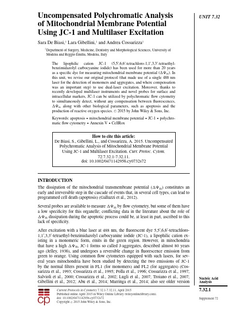
UNIT7.32 Uncompensated Polychromatic Analysisof Mitochondrial Membrane PotentialUsing JC-1and Multilaser ExcitationSara De Biasi,1Lara Gibellini,1and Andrea Cossarizza11Department of Surgery,Medicine,Dentistry and Morphological Sciences,University ofModena and Reggio Emilia,Modena,ItalyThe lipophilic cation JC-1(5,5ʹ,6,6ʹ-tetrachloro-1,1ʹ,3,3ʹ-tetraethyl-benzimidazolyl carbocyanine iodide)has been used for more than20yearsas a specific dye for measuring mitochondrial membrane potential( m).Inthis unit,we revise our original protocol(that made use of a single488nmlaser for the detection of monomers and aggregates,and where compensationwas an important step)to use dual-laser excitation.Moreover,thanks torecently developed multilaser instruments and novel probes for surface andintracellular markers,JC-1can be utilized by polychromaticflow cytometryto simultaneously detect,without any compensation betweenfluorescences,m along with other biological parameters,such as apoptosis and theproduction of reactive oxygen species.C 2015by John Wiley&Sons,Inc.Keywords:apoptosis r mitochondrial membrane potential r JC-1r polychro-maticflow cytometry r Annexin V r CellRoxHow to cite this article:De Biasi,S.,Gibellini,L.,and Cossarizza,A.2015.UncompensatedPolychromatic Analysis of Mitochondrial Membrane PotentialUsing JC-1and Multilaser Excitation.Curr.Protoc.Cytom.72:7.32.1-7.32.11.doi:10.1002/0471142956.cy0732s72INTRODUCTIONThe dissipation of the mitochondrial transmembrane potential( m)constitutes anearly and irreversible step in the cascade of events that,in several cell types,can lead toprogrammed cell death(apoptosis)(Galluzzi et al.,2012).Several probes are available to measure m byflow cytometry,but some of them havea low specificity for this organelle;conflicting data in the literature about the role ofm dissipation during the apoptotic process could be,at least in part,ascribed to thislack of specificity.After excitation with a blue laser at488nm,thefluorescent dye5,5ʹ,6,6ʹ-tetrachloro-1,1ʹ,3,3ʹ-tetraethyl-benzimidazolyl carbocyanine iodide(JC-1),a lipophilic cation ex-isting in a monomeric form,emits in the green region.However,in mitochondriathat have a high m,JC-1forms so called J-aggregates,described almost80yearsago(Jelley,1936),and undergoes a reversible change influorescence emission fromgreen to ing commonflow cytometers equipped with such lasers,for sev-eral years mitochondria have been studied by detecting the two emissions of JC-1by the normalfilters present in FL1(for monomers)and FL2(for aggregates)(Cos-sarizza et al.,1993;Cossarizza et al.,1995;Polla et al.,1996;Cossarizza et al.,1997;Salvioli et al.,2000;Cossarizza et al.,2002;Lugli et al.,2007;Troiano et al.,2007; Gibellini et al.,2012;Abu et al.,2014;Marringa et al.,2014;also see older version Current Protocols in Cytometry7.32.1-7.32.11,April2015Published online April2015in Wiley Online Library().doi:10.1002/0471142956.cy0732s72Copyright C 2015John Wiley&Sons,Inc.Nucleic Acid Analysis7.32.1 Supplement72of this unit at /doi/10.1002/0471142956.cy0732s41/full).Measurements using this dye provide information on changes in m(typically,adecrease in m causes a relevant shift from orange to greenfluorescence emis-sion),as well as on total mitochondrial content(based on the intensity of the greenfluorescence emission).A number of studies have since shown the superiority ofJC-1over other dyes—e.g.,rhodamine123(R123)or3,3ʹ-dihexyloxadicarbocyanineiodide[DiOC6(3)]—that were used for the same purpose,and demonstrated thatJC-1is also unaffected by changes in plasma membrane potential(Salvioli et al.,1997;Lugli et al.,2007;Troiano et al.,2007;also see older version of this unit at/doi/10.1002/0471142956.cy0732s41/full).This unit discusses a new method to detect JC-1(see Basic Protocol1),based upon theuse of two lasers,one to excite JC-1monomers(by the canonical488-nm laser line),and the other to excite JC-1aggregates(by a yellow laser emitting at561nm).Thetypical excitation by the blue laser excites JC-1with high efficiency,but sometimesrequires significant compensation between FL1and FL2.In contrast,yellow laser allowsa better resolution,and thus a clearer visualization of monomers and aggregates withoutcompensation(Perelman et al.,2012).For this reason,we have revised our basic JC-1protocol using the two different lasers quoted above.Furthermore,we have recently developed another polychromaticflow cytometric assay(see Basic Protocol2)utilizing JC-1and other probes for the simultaneous detection of m,reactive oxygen species(ROS,by CellRox DeepRed),and apoptosis(by Annexin V,detecting the exposure of phosphatidylserine on the plasma membrane).This protocolcan be applied when the simultaneous analysis of multiple parameters during apoptosisis required,e.g.,in investigating the role of certain proteins on cell phenotype or whentesting the cytotoxicity of compounds of pharmacological interest.CAUTION:For the protection of laboratory personnel from potential infectious agents (e.g.,hepatitis and HIV),handle human samples using disposable gloves in a biological safety cabinet.CAUTION:All probes described in this unit are potentially hazardous(see manufacturers’MSDSs),and users should wear gloves during the staining procedures.BASIC PROTOCOL1BASIC DETERMINATION OF MITOCHONDRIAL MEMBRANE POTENTIAL USING JC-1:DUAL-LASER EXCITATION OF THE DYEA VOIDS COMPENSATION ISSUESThis protocol is intended for cells such as peripheral blood mononuclear cells(PBMCs)or cell lines such as RKO,HL60,MCF7,and U937.Other cell types may also be stainedusing minor adjustments to the steps described below.Typically,by using a488-nm blue laser,it can be observed that cells with high m(that form JC-1aggregates)emit orangefluorescence(atß590nm);those with low m(containing JC-1in its monomeric form)emit greenfluorescence(atß520nm) (Cossarizza et al.,1993).Recently it has been demonstrated that alternative excitationwavelengths can facilitate the detection of m,and,most importantly,use of twowavelengths avoids the need for compensation.Indeed,the excitation wavelength561nm(i.e.,yellow laser)is above the emission spectra of JC-1monomers,and selectivelyexcites J-aggregates;hence there is no need to compensate green and orangefluorescence(Perelman et al.,2012).Thus,we have adapted our original protocol(that made use ofa single488-nm laser,and where compensation was an important step)to an instrumentequipped with a blue and a yellow laser(like the Attune NxT,from Life Technologies).Analysis ofMitochondrialMembranePotential UsingJC-1andMultilaserExcitation7.32.2Supplement72Current Protocols in CytometryMaterialsExperimental samples:human peripheral blood lymphocytes or monocytes,orhuman tumor cell lines(e.g.,RKO,HL60,U937,MCF7);here we use RKOcells,which derive from a colon carcinoma and grow adherent to the plasticflask Complete RPMI culture medium,1ml per sample1M valinomycin[dissolve valinomycin(mol.wt.1111.32;Sigma-Aldrich)indimethylformamide(DMF)and store in a glass container up to6months at4°C]or1mM carbonyl cyanide p-(trifluoromethoxy)phenylhydrazone(FCCP;Sigma Aldrich)2.5mg/ml JC-1(5,5ʹ,6,6ʹ-tetrachloro-1,1ʹ,3,3ʹ-tetraethylbenzimidazolyl-carbocyanine iodide):prepare by dissolving JC-1(Life Technologies,ThermoFisher Scientific)in dimethylformamide(DMF);store in a glass container up to2years at–20°C,protected from lightPhosphate-buffered saline(PBS)3.5-ml,55×12–mm plastic tubes(Sarstedt,or equivalent)Centrifuge(Minifuge RF;Heraeus),or equivalentFlow cytometer equipped with a488-nm blue laser and with a561-nm yellow laser,e.g.,Attune NxT(Life Technologies)Additional reagents and equipment for counting(APPENDIX3A)and culturing(APPENDIX3B)mammalian cellsPrepare cells1.Count a sample of the experimental cells of interest(APPENDIX3A).This protocol can be used to stain either cells growing in suspension or adherent cellsafter they have been released from the plate by trypsinization(APPENDIX3B)and counted(APPENDIX3A).2.Collect at least2×105cells from the experimental samples in55×12–mm tubesby centrifuging5min at300×g,room temperature.Collect the same number ofcells to use for a positive control.3.Decant and discard the medium and resuspend the cell pellet in1ml fresh completeRPMI culture medium.4.For obtaining a so-called“positive control,”i.e.,a sample where all cells have de-polarized mitochondria,prepare one sample of cells treated with valinomycin(finalconcentration0.1μM)or with carbonyl cyanide p-(trifluoromethoxy)phenylhydra-zone(FCCP,final concentration250nM).Incubate10min or45min,respectively,at37°C.Drugs such as the K+ionophore valinomycin or the proton translocator FCCP are ableto collapse theΔΨm.Note that to avoid problems related to intracellular drug metabolism,in some instancesvalinomycin is preferred over FCCP or ClCCP(and is also less expensive).Stain with JC-15.Add1μl of2.5mg/ml JC-1fluorescent probe(2.5μg/mlfinal concentration)to theexperimental and positive control cells and shake the cell suspension until the dyeis well dispersed and gives a uniform red-violet color.JC-1tends to form aggregates when added to normal aqueous medium.To avoid this,add the probe while gently vortexing.6.Incubate the samples10min in the dark,37°C.Nucleic AcidAnalysis7.32.3 Current Protocols in Cytometry Supplement72Figure 7.32.1Changes in JC-1fluorescence after mitochondrial membrane depolarization in RKO cells treated with valinomycin,as described in Basic Protocol 1.Samples were acquired using 488-nm laser only (A ),or with dual-laser excitation (B ).Control cells (CTR)were stained with 2.5μg/ml JC-1.Note the shift to the bottom and to the right of cells with mitochondria depolarized by treatment with 100nM valinomycin.Right panel shows the merging of the left and center panels.Green-orange compensation was ß4%and orange-green compensation was ß10%;compensation was required to better visualize monomers and aggregates.All reagents must be at room temperature and carefully checked for pH (7.4)when used,because ΔΨm is very sensitive to alterations of these conditions.The staining procedure must be carried out away from direct intense light,and incubation must be in the dark because of the light sensitivity of JC-1.7.Wash the cells by centrifuging 5min at 300×g ,room temperature,discarding the supernatant,and resuspending the cells in 1ml PBS for analysis on cytometer.Set up flow cytometer 8.Detect JC-1fluorescence of the experimental and positive control samples using a classical green band-pass filter centered at 525/50nm for monomers detection (channel of blue laser)and a classical greenish orange band-pass filter centered at 585/42nm (usually those for a channel collecting fluorescence signals deriving from the excitation with the blue or the yellow laser).The most common flow cytometers are typically equipped with only a 488-nm argon or solid-state laser;no special requirements are needed to analyze ΔΨm .The gain of photomultipliers (PMTs)obviously depends on the cytometer used,but generally JC-1does not require any substantial increase in PMT amplification;green-orange compensation can be ß4%and orange-green compensation ß10%.However,note that no compensation is needed if a blue and a yellow laser are used to detect monomers and aggregates,respectively.See Figure 7.32.1for a typical example of JC-1staining of control (CTR)RKO cells,and of RKO cells treated with valinomycin.Detection was performed by using a single blue laser (A)or using blue and yellow lasers (B).This treatment results in a relevant change in the fluorescence distribution:cells with depolarized mitochondria can be easily identified as those going from the center of the plot to the lower right quadrant.Analysis ofMitochondrial Membrane Potential UsingJC-1andMultilaser Excitation7.32.4Supplement 72Current Protocols in Cytometry9.On the basis of the laser used,adjust the voltage of the respective PMTs to obtainthe bivariate green versus orange distributions similar to those shown in Figure7.32.1A and B,and then record the control e the same PMT settings forthe subsequent samples.Analyze JC-1stained experimental samples10.Acquire fluorescence data for experimental samples in listmode,using a log scalefor the fluorescence channels.Cells with high ΔΨm are those forming J-aggregates;thus,they show high orangefluorescence.On the other hand,cells with low ΔΨm are those in which JC-1maintains (orre-acquires)its monomeric form,and thus show green fluorescence.Once mitochondriaare depolarized,JC-1monomers redistribute in other membranous compartments withlower ΔΨ.As a consequence,the green fluorescence intensity of depolarized cells is alittle bit higher than that of polarized ones simply because of the presence of a higheramount of JC-1monomers inside the cell.11.Recommended for samples with heterogeneous cell populations:Set a gate on thepopulation of interest,then proceed with adjustment of PMTs,as well as compen-sation if a 488-nm laser is used.Dual-laser excitation of the dye does not requirecompensation.When the sample contains a heterogeneous cell population,it is possible to see differentfluorescence patterns due to different autofluorescences and the variable content in termsof membranes and mitochondria of cell subpopulations.This is the case for peripheralblood mononuclear cells (PBMCs),lymphocytes,and monocytes,the first being smallerand having fewer mitochondria than the latter.Accordingly,the fluorescence pattern ofJC-1for such a sample shows at least two distinct peaks,one corresponding to lympho-cytes,and the second,brighter in both green and orange,corresponding to monocytes.It is thus recommended to first set a gate on the population of interest,then proceed withadjustment of PMTs and compensation.BASIC PROTOCOL 2ANALYSIS OFM ,APOPTOSIS,AND REACTIVE OXYGEN SPECIESCONTENT BY 4-LASER POLYCHROMATIC FLOW CYTOMETRYThis protocol allows the analysis of m along with the detection of early apoptotic cells,and the quantification of the amount of reactive oxygen species in the cells of interest.It has been developed taking into account the possibility of simultaneously using fourlasers (by using an Attune NxT from Life Technologies)and avoiding any compensationamong dyes.Fine analysis of the apoptotic process requires the detection of multiple cell functions atthe same time,and it could be highly informative to reveal whether cells with differentm also differ with respect to other parameters.This assay is recommended whenstudying compounds that can have differential effects on the cell populations of interest.This protocol uses three different probes:JC-1(for m ),annexin V conjugated withPacific Blue (for detecting the exposure of phosphatidylserine on the plasma membrane,a well known phenomenon which identifies early apoptotic cells),and CellRox DeepRed (for measuring ROS production).CellRox is a cytoplasmic cell-permeable non-fluorescent (or very weakly fluorescent)reagent which,in a reduced state and uponoxidation,exhibits a strong fluorogenic signal.CellRox Deep Red can be excited by a638-nm laser,and emits at ß665nm.For complete information regarding the probesdescribed here,see Internet Resources at the end of this unit.Annexins are a family of soluble proteins (13different isoforms)with four to eightrepeats of a 75–amino acid consensus sequence relevant for Ca 2+binding.They areinvolved in membrane transport,regulation of protein kinase C,formation of ion channels,Nucleic Acid Analysis 7.32.5Current Protocols in Cytometry Supplement 72endocytosis,exocytosis,and membrane-cytoskeleton interactions.Annexin V binds with peculiar specificity to phosphatidylserine residues,which are precociously exposed on the external leaflet of the plasma membrane during apoptosis (Lizarbe et al.,2013).Thus,when cells are annexin V positive,they have entered into an early phase of apoptosis.The annexin V–Pacific Blue conjugate is violet excitable,making it ideal for instruments with a laser at 405nm,and for multicolor experiments that include green-or red-fluorescent dyes.The Pacific Blue-conjugated annexin V emits at ß455nm after excitation by a violet light source.Before starting with sample analysis,running samples stained with single fluorochromes (see steps below)is suggested to properly set up fluorescence levels.Note that also in this case there are no compensation requirements.Materials Cells in culture (ATCC):in suspension or adherent in 24-well tissue culture plate (as in Basic Protocol 1,we use RKO cells derived from human colon carcinoma Complete RPMI culture medium Phosphate-buffered saline (PBS)CellRox Deep Red Reagent (Life Technologies)2.5mg/ml JC-1(5,5ʹ,6,6ʹ-tetrachloro-1,1ʹ,3,3ʹ-tetraethylbenzimidazolylcarbocyanine iodide);prepare by dissolving JC-1(Life Technologies,Thermo Fisher Scientific)in dimethylformamide (DMF);store in a glass container up to 2years at –20°C,protected from light Annexin V binding buffer (see recipe)Pacific Blue-conjugated annexin V (Life Technologies,Thermo Fisher Scientific):store at 4°C,protected from light 3.5ml,55×12–mm plastic tubes (Sarstedt,or equivalent)Centrifuge (Minifuge RF;Heraeus),or equivalent.Attune NxT cytometer or equivalent cytometer equipped with four light sources for excitation at 405nm (violet laser,for Annexin V),488and 561nm (blue and yellow lasers,for JC-1),and 638nm (red laser,for CellRox)and filters for collecting fluorescence emissions at 455/40(for annexin V),520/20(for JC-1monomers),585/42(JC-1aggregates),and 660/40(CellRox)Additional reagents and equipment for counting (APPENDIX 3A )and culturing (APPENDIX 3B )mammalian cells and detaching adherent cells using trypsin (see APPENDIX 3B )Prepare cells 1.Count a sample of the cells in culture (see APPENDIX 3A ).For cells in suspension 2a.Collect at least 3×10⁵cells from experimental samples by centrifuging 5min at 300×g ,room temperature.Collect the same number of cells to use for a positive control.3a.Decant and discard the medium and bring the total volume up to 1ml with prewarmed RPMI culture medium.For adherent cells 2b.Decant and discard the growth medium.3b.Add 1ml prewarmed culture medium (RPMI or similar)to the cells in the plate.Analysis ofMitochondrial Membrane Potential UsingJC-1andMultilaser Excitation7.32.6Supplement 72Current Protocols in CytometryThis protocol has been set up using blood cells and has been shown to work with differentcell lines.However,particular attention should be given to adherent cell lines,detachmentof which from the culture plate by trypsin-EDTA is required before cytofluorimetricanalysis.The detachment procedure could be particularly harmful to those cells thathave been damaged during the in vitro treatment,i.e.,by the presence of an apoptogenicsubstance.In this case,the multistaining procedure described here could be performedon still-adherent cells by adding the probes directly to the culture plate.Stain cells4.Add the CellROX Reagent at afinal concentration of5μM to the cells and incubatefor30min at37°C.For cells in suspension5a.To wash the staining solution from the cells,add1ml PBS,mix by shaking gently,and centrifuge5min at300×g,room temperature.Decant and discard the supernatant.Because this protocol requires many centrifugations for the cells in suspension,theauthors suggest setting the centrifugation speed as low as possible in order to avoidcellular damages due to stress.Adding10%fetal bovine serum to PBS can decrease cellloss during washing steps.6a.Resuspend the cells in1ml complete culture medium.Proceed to step7.For adherent cells5b.Decant the staining solution from the cells and wash by adding1ml PBS,swirling, and decanting.6b.Detach the adherent cells as follows.i.Trypsinize cells as described in APPENDIX3B.The minimal amount of trypsin should be used in order to avoid both cellular damage andthe presence of aggregates in the cell suspension.In fact,cell aggregates could augmentbackground or J-aggregatefluorescence.In this case,aggregates can be eliminated fromanalysis by gating on singlets,which can be identified by plotting FS-area versus FS-height.In any case,when adherent cell lines are treated with apoptogenic substances,remember that apoptotic cells spontaneously detach andfloat in the supernatant;theyshould not be discarded but collected and analyzed separately or together with attachedcells.ii.Add1ml complete culture medium to neutralize trypsin activity.iii.Centrifuge5min at300×g,room temperature,and discard the supernatant.Proceed to step7.7.Add1μl of2.5mg/ml JC-1(2.5μg/mlfinal concentration)to the pellet from step6a or6b and mix until the dye is well dispersed and gives a uniform red-violet color.Incubate the samples10min in the dark,room temperature.JC-1tends to form aggregates when added to normal aqueous medium.To avoid this,add the probe while gently vortexing.8.Wash with1ml PBS as in step5a or5b.9.Resuspend the cells in195μl annexin V binding buffer.10.Add5μl of Pacific Blue–conjugated annexin V(at concentration provided by themanufacturer)and incubate15min at room temperature.Staining with annexin V is the last step of the protocol because annexin V binding tophosphatidylserine is affected by the presence of its incubation buffer.In the authors’Nucleic AcidAnalysis7.32.7 Current Protocols in Cytometry Supplement72experience,washing or resuspending cells with PBS causes annexin V detachment from phosphatidylserine.11.Resuspend the cells in1ml annexin V binding buffer.Acquire samples on cytometer12.First acquire blank samples and cells without CellRox,to set the level of backgroundfluorescence for the Alexa647channel.This type of analysis requires aflow cytometer equipped with three light sources and appropriate collectionfilters for all the dyes(see Materials list).13.Acquire at least30,000total events.Analyze data14.Identify cell populations on the basis of annexin V,i.e.,live(Annexin V–),apoptotic(Annexin V+).Analyze m and ROS content in these subsets.Since multiple parameters are simultaneously analyzed,different techniques for data interpretation can be adopted depending on the user’s interests.In this case,should the researcher be interested in detectingΔΨm and ROS production in early apoptotic or healthy cells,a gate can be designed on annexin V positive or negative cells,where the other parameters are thus analyzed(see Fig.7.32.2).REAGENTS AND SOLUTIONSUse deionized,distilled water in all recipes and protocol steps.Annexin V binding buffer0.477g HEPES(10mM)1.636g NaCl(140mM)0.073g CaCl2(2.5mM)H2O to200mlAdjust pH to7.4and store up to1year at4°CMilli-Q-purified(double purified)water may also be used in this recipe. COMMENTARYBackground InformationMitochondria play an active role in theregulation of programmed cell death,and in-deed the collapse in m can occur duringthe apoptotic process(Green et al.,2011).The opening of the mitochondrial permeabilitytransition pore—a mitochondrial protein com-plex formed by the adenine nucleotide translo-cator(ANT),the voltage-dependent anionchannel(VDAC),and the peripheral benzo-diazepine receptor(PBR)—can induce loss of m,release of apoptogenic factors,and loss of oxidative phosphorylation(Martel et al.,2014).However,whether loss of m is acause or a consequence of the triggering ofapoptosis still remains a matter of debate.De-pending on the apoptotic model used,loss of m may be a late(Cossarizza et al.,1994)or an early(Zamzami et al.,1995)event.More-over,loss of m is responsible for the release of apoptosis-inducing factor(AIF),which con-sequently translocates to the nucleus and pro-motes chromatin condensation and fragmen-tation(Kroemer et al.,2007).Other mecha-nisms initiating apoptosis(e.g.,cytochromec release or activation of executioner cas-pases)are independent of the disruption of m(Kluck et al.,1997;Bossy-Wetzel et al., 1998).Several techniques are used to investigatethe role of this organelle,including classicalbiochemical or molecular biology methods;flow cytometry clearly represents the mostrapid and powerful tool for investigating m at the single-cell level.Many probes are available for this purpose,but some of them, e.g.,R123and DiOC6(3),are not fully adequate(Salvioli et al.,1997).As a consequence,discrepancies in the data regarding the role of m in the regulation of the apoptotic process may be also attributed to the use of inappropriate probes.A detailed analysis of other dyes is reported in UNIT9.14 (Cossarizza and Salvioli,2000).Analysis ofMitochondrialMembranePotential UsingJC-1andMultilaserExcitation7.32.8Supplement72Current Protocols in CytometryFigure7.32.2Multilaser,uncompensated analysis of apoptosis,mitochondrial membrane potential,and production of reactive oxygen species.RKO cells were cultured in the absence(A)or presence(B)of H2O2(1hr)and(C)5μM CDDO (24hr).Cell were stained as described in Basic Protocol2.Viable and apoptotic cells were identified by positivity for annexin V; m was analyzed by JC-1,ROS production by CellRox Deep Red.We have demonstrated that JC-1is an excel-lent potentiometric probe,having the peculiar ability to change color reversibly depending on the m.This property is due to the reversible formation of JC-1aggregates upon polariza-tion of mitochondrial membrane,which causes a shift in emitted light fromß530nm(emis-sion of monomers)toß590nm(emission of J-aggregates).In living cells,the color of the dye changes reversibly from green to orange as the mitochondrial membrane becomes more polarized(Reers et al.,1991).Aggregate for-mation begins at potential values on the order of80to100mV,and reaches the zenith at ß200mV.When488nm was the sole available laser line,researchers had to cope with compen-sation,which had to be set up considering the spillover of the twofluorescences,and re-quired not only the preparation of“biologi-cal negative controls”(i.e.,samples of cells treated with a depolarizing agent to see the area where cells with a low m tended to go),but also a certain experience on the part of the operator.In any case,excitation with 488-nm laser was quite efficient and allowed,and is currently allowing,a significant num-ber of studies.Modernflow cytometers havemore excitation sources than in the past.Themain advantage of a second excitation sourcefor JC-1aggregates is well evidenced by factthat compensation is no longer needed,sinceyellow laser does not excite JC-1monomers(Perelman et al.,2012).JC-1staining can be combined with multi-ple probes in a polychromaticflow cytometricassay to detect changes in m together withother parameters during apoptosis;Basic Pro-tocol2can be useful and informative,becauseseveral cell functional subsets with different characteristics can be simultaneously identi-fied in a given population.This makes it pos-sible not only to discriminate cell death,butalso to investigate whether similar compoundsexert differential effects in the same cell type.This type of analysis,combined with high-throughput technologies,could be adopted forthe screening of the toxicity of a variety of compounds,in order to obtain multiple infor-mation about the investigated molecules.Nucleic AcidAnalysis7.32.9Current Protocols in Cytometry Supplement72。
大鼠星形胶质细胞组织因子活性的表达及其调控
生理学报,1999年6月,51(3),291~296 291 Acta Physiologica Sinica大鼠星形胶质细胞组织因子活性的表达及其调控3朱发明33 文志斌 何晓凡 李俊成 贺石林(湖南医科大学生理教研室,长沙410078)摘 要 本实验观察了基础培养条件和凝血酶刺激条件下,大鼠星形胶质细胞组织因子活性的表达及其信号传递途径。
结果显示,基础条件下,A23187(42brom o calcium ionophore)和佛波醇脂(phorbol122myristate132acetate,P M A)能明显提高星形胶质细胞组织因子活性的表达,而三氟吡啦嗪(trifluoperazine,TFP)和12(52异喹啉磺胺)232甲基哌嗪[12(52is oquinolinyl sulfonyl)232methyl2piperazine,H7]则降低星形胶质细胞组织因子活性的表达。
凝血酶能明显增加星形胶质细胞表达组织因子活性;当凝血酶与TFP或H7联合应用时,凝血酶的刺激作用受到明显抑制。
实验表明,星形胶质细胞在基础培养条件下能表达组织因子,凝血酶能刺激它表达组织因子活性。
Ca2+/钙调素和PK C途径参与了在基础条件以及凝血酶刺激条件下星形胶质细胞组织因子活性的表达。
关键词:星形胶质细胞;组织因子;蛋白激酶C;钙调素;凝血酶学科分类号:Q461;R33111 近几年的研究表明,组织因子途径是生理性止血中凝血过程与病理条件下血栓形成的启动环节[1,2]。
组织因子(tissue factor,TF)作为因子Ⅶa的受体在组织因子途径中起关键性作用。
它广泛分布于脑、肺等组织。
血管内皮下的成纤维细胞、平滑肌细胞以及血管外的各种组织细胞都程度不同地表达组织因子,尤其在血管周围组织因子分布比较丰富,这对血液循环系统的完整性起着保护套的作用。
早已知道,脑组织富含组织因子,但长期来对它的来源并未确定。
新近发现星形胶质细胞(astrocyte,AS)是脑组织中TF的主要来源,并已达成广泛的共识[3,4]。
Fluo-4, AM 产品说明书
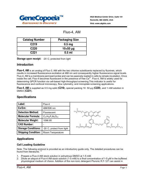
Fluo-4, AMStorage upon receipt: -20°C, protected from lightIntroductionFluo-4, AM is an analog of Fluo-3, AM with the two chlorine substituents replaced by fluorines, which results in increased fluorescence excitation at 488 nm and consequently higher fluorescence signal levels. Fluo-4, AM is a membrane permeant probe and can be passively loaded in cells by simple incubation. Once inside the cell, Fluo-4 becomes fluorescent in the presence of free Ca2+. Fluo-4, AM is widely used for determining GPCR function via cell-based high-throughput screening.This indicator is useful for fluorescence and confocal microscopy, flow cytometry, and microplate screening applications.Fluo-4, AM is supplied as 0.5 mg solid (C219), special packing 10×50 µg (C220), and 1 mM solution in DMSO (C221).SpecificationsApplicationsCell Loading GuidelineNote: The following protocol is provided as an introductory guide only. The detailed procedures can be found from literatures.1,21. Prepare a Fluo-4 AM stock solution in anhydrous DMSO at 1-5 mM.2. Dilute an aliquot of Fluo-4 AM stock solution (1-5 mM) to a final concentration of 1-5 µM in the bufferedphysiological medium of choice. Addition of the non-ionic detergent Pluronic R F-127 can assist inPage 1dispersion of the nonpolar Fluo-4 AM ester in aqueous media. This can be conveniently accomplished by mixing the aliquot of Fluo-4 AM ester stock solution in DMSO with an equal volume of 20% (w/v) Pluronic in DMSO (Cat No. C021) before dilution into the loading medium, making the final Pluronic concentration about 0.02%.3. The organic anion-transport inhibitors probenecid (1-2.5 mM) may be added to the cell medium toreduce leakage of the de-esterified indicator.4. Cells are normally incubated with the Fluo-4 AM ester for 15–60minutes at 20–37°C. Exact loadingconcentration, time, and temperature will need to be determined empirically; in general it is desirable to use the minimum dye concentration required to yield fluorescence signals with adequate signal to noise.Subcellular compartmentalization, an inherent problem with the AM ester loading technique, is usually lessened by lowering the incubation temperature.5. Before fluorescence measurements are commenced, cells should be washed in indicator-free medium(containing an anion transport inhibitor, if applicable) to remove any dye that is nonspecificallyassociated with the cell surface, and then incubated for a further 30 minutes to allow completede-esterification of intracellular Fluo-4 AM ester.High-Throughput ScreeningIntracellular Ca2+ measurements in 96-well and 384-well microplates are an essential tool for high- throughput pharmacological screening. Cell samples in microplate wells are loaded with the AM ester form of the indicator using protocol basically similar to those described in Cell Loading Guideline. References:1. Methods Cell Biol 40, 155 (1994);2. Cell Biology: A Laboratory Handbook, 2nd Edition, J.E. Celis, Ed., Volume 3, pp 363–374,Academic Press (1998);Page 2。
Capto Core 700 AN 29000334AA
GE Healthcare Life SciencesApplication note 29-0003-34 AAVaccinesPurification of influenza A/H1N1using Capto ™Core 700Capto Core 700 is a chromatography medium (resin) optimized for purification of viruses and other large biomolecules. The medium is designed to be used in flowthrough mode for large targets (> M r 700 000) while scavenging smaller contaminants. Capto Core 700 is built on the core bead technology which allows for dual functionality combining size separation with binding chromatography. The size separating mode of operation could be compared to gel filtration (size exclusion chromatography), which is a commonapproach for polishing steps in vaccine processes. Here, the performance of Capto Core 700 in three different processes for the purification of influenza H1N1 virus from infected mammalian cells was evaluated andcompared. This study shows that while offering the same purity as gel filtration, Capto Core 700 enables significant improvements in productivity and process economy.IntroductionInfluenza vaccine has historically and is today primarily produced in embryonated chicken eggs. However, to meet the needs for pandemic preparedness and scalability of vaccine productions, cell-based processes are being developed and implemented to a greater extent in the industry. The methods for purification processes have typically involved a combination of sucrose density gradient ultracentrifugation, ultrafiltration/diafiltration (UF/ D F) with hollow-fiber membranes, and chromatography using affinity-, ion exchange-, and/or gel filtration (GF) media. In these processes, both sucrose density gradient ultracentrifugation and GF have limitations in, for example, scalability and productivity. In GF, the low productivity relates to low flow rates and limited sample loads.The Capto Core 700 medium has a core bead design and consists of an inactive shell and a ligand-activated core. Small contaminant molecules enter into the beads where they are captured. Viruses and other large entities with a molecular mass (M r ) greater than approximately 700 000 (700 kDa) are excluded and are collected in the chromatography flowthrough. The octylamine ligands in the core of the bead are multimodal, being bothhydrophobic and positively charged. These internalized ligands bind various contaminants strongly over a wide range of pH and salt concentrations.Using Capto Core 700 allows higher flow rates (1) and significantly higher sample loads than traditional GF (typically several column volumes compared with 0.1 to 0.3 column volumes in GF).The aim of this work was to evaluate Capto Core 700 chromatography medium as an alternative to current chromatography technologies used in vaccine processes. An experiment to determine the binding capacity and window of operation with Capto Core 700 for Madin-Darby canine kidney (MDCK) cell protein was performed using PreDictor™ 96-well plates for high-throughputprocess development. The approach taken for designing a purification process for flu vaccine is also described (Fig 1).Finally, the process economy impact of Capto Core 700compared with Sepharose™ 4 Fast Flow, a GF medium often used in the vaccine manufacturing industry today, was also investigated.Fig 1. Three alternative process streams for the purification of influenza A/H1N1 were evaluated in this study.Materials and methodsCell culture and infectionMDCK cells (inoculation concentration of 500 000 cells/mL) were grown on Cytodex™ 3 microcarriers for 48 h in an Applikon™ Bioreactor (Applikon Technology). The finalcell density was approximately 2 500 000 cells/mL at which point cells were infected with influenza A/Solomon Islands/3/2006 (H1N1) and harvested at 72 h post infection. Preliminary studiesInvestigation of operating window (binding study) for Capto Core 700The binding capacity of Capto Core 700 for host cell protein (HCP) from MDCK cell lysate was evaluated in buffers containing sodium phosphate and Tris, 150–1000 mM NaCl, pH 6.5–8.0. PreDictor 96-well plates were filled with 10 µL of Capto Core 700 for the binding study. Clarified MDCK cell lysate (200 µL) was applied to the wells of the plate and incubated in the various equilibration buffers for 60 min. After incubation, unbound sample was removed by centrifugation and the medium was washed with buffer. Collected fractions were analyzed for total protein using the Bradford protein assay.Benchmarking of purification performance:Capto Core 700 vs Sepharose 4 Fast FlowThe performance of Capto Core 700 was compared to that of Sepharose 4 Fast Flow. Capto Core 700 was packedin Tricorn™ 5/50 column (column volume [CV], 1 mL) and10 CV of clarified and concentrated virus material was loaded. Sepharose 4 Fast Flow was packed in Tricorn10/600 (CV, 47 mL) and 0.1 CV of virus feed was loaded. Recovery of virus as measured by the quantitation of hemagglutinin (HA) and reduction of HCP was compared between the two media.Process developmentClarificationClarification of harvested cells was achieved by microfiltration (MF) using ULTA Prime GF normal-flowfilter capsules. A 2.0 µm (4“ membrane, 0.10 m2 effective filtration area) rating was used initially followed by a0.6 µm (4” membrane, 0.11 m2 effective filtration area) rating. The ULTA Prime GF filters were washed with buffer (20 mM sodium phosphate, 150 mM NaCl, 0.05% sodium azide, pH 7.2) before use.Degradation of DNABenzonase endonuclease treatment was applied for the removal of DNA in process stream 2 (Fig 1). Degradationof DNA using Benzonase results in small oligonucleotide fragments that enter through the inactive layer and into the core where they are captured by the internalized ligands. Benzonase was applied after MF and before column purification with Capto Core 700 in process stream 2 (Fig 1).Capto DeVirS: binding/elution of influenza virusCapto DeVirS is a chromatography medium designed for the capture and intermediate purification of viruses. The Capto DeVirS ligand is dextran sulfate, which allows for pseudo affinity for several virus types including influenza and was therefore selected for virus capture in process stream 3. Chromatography runs were performed in a HiScale™50/20 column packed with 202 mL of Capto DeVirS.ÄKTAexplorer™ 100 chromatography system was used for chromatography runs on Capto DeVirS.Capto Core 700: polishing stepIn process stream 1–3, chromatography runs were performed in an XK 16/20 column packed with 25 mL of Capto Core 700. ÄKTAexplorer 10 chromatography system was used for chromatography runs on Capto Core 700. Analytical methodsVirus quantitationIn this study, Biacore™ T200 system and Sensor Chip CM5 were used to measure HA content according to a previously described method (2). The potency of influenza vaccines are mainly determined by quantitation of HA using the single radial immunodiffusion (SRID) assay. This method, although approved by both FDA and EMEA, is labor-intensiveand suffers from low precision and sensitivity. Biacore biosensor assays offer greater precision in the quantitation of influenza HA and faster analysis than SRID in vaccine development and manufacturing.HCP quantitationHCP is usually quantitated as total protein with, for example, the Bradford protein assay. This method is not sensitive or specific enough to detect levels below the regulatory critical limits. Therefore, Biacore biosensor assay was used for the quantitation of HCP using the same instrumentation as mentioned above. In house produced polyclonal anti-MDCK HCP antibodies were immobilized on the Biacore chip for MDCK HCP protein binding. The HCP standard was set using Bradford to estimate protein content.As reference, the total protein content (including HA)was also measured with the Bradford protein assay. The assay was performed according to the manufacturer’s recommended methods (3).Determination of infectious particlesThe median Tissue Culture Infectious Dose (TCID50)assay measures dilution that generates cytopathic effect in 50% of the cell culture and is an infectivity method that is oneof the most commonly used methods for detection of the infective virus. The TCID50assay is simple to perform and requires no specific instrumentation for result interpretation. The outcome is either presented as a log10 titer (10x.x TCID50 units/ m L) or a dilution (10-x.x/mL).2 29-0003-34 AASodium phosphate Tris20 mM sodium phosphate + 150 mM NaCl 20 mM sodium phosphate + 1000 mM NaCl 02468101214H C P c a p a c i t y (m g p r o t e i n /m L m e d i u m)10002000300040005000Volume (mL)A 280 (m A U )Conductivity (mS/cm)29-0003-34 AA 3Results and discussionInvestigation of operating window (binding study) for Capto Core 700The performance of Capto Core 700 was robust in both 20 mM sodium phosphate and Tris buffer containing up to 1 M NaCl and in pH from 6.5 to 7.5 for sodium phosphate and pH 7.5 to 8.0 for Tris, respectively (Fig 2). In general, the low NaCl concentration of 150 mM resulted in higher binding capacity while lower pH yielded higher binding capacity for the MDCK cell lysate proteins.The robust performance of the multimodal octylamine ligand in a relatively wide range of NaCl concentration and pH gives Capto Core 700 a wide window of operation. This reduces the need for optimization such as buffer exchange or dilution between steps, even with different feed materials when working with Capto Core 700.Benchmarking of purification performance: Capto Core 700 vs Sepharose 4 Fast FlowThe purification performance of Capto Core 700 was compared to that of Sepharose 4 Fast Flow, which is a chromatography medium typically used for GFpurification of a range of viruses in vaccine processes. Both chromatography methods provided a similar yield of virus HA and reduction of HCP (Table 1).Amount of medium: 10 µL of Capto Core 700 in PreDictor 96-well filter plate Sample: Clarified MDCK cell lysate in different equilibration buffers Sample load: 200 µL of cell lysate (60 min incubation)Equilibration buffers: 20 mM sodium phosphate, 150–1000 mM NaCl, pH 6.5–7.520 mM Tris, 150–1000 mM NaCl, pH 7.5–8.0Equilibration: 3 × 200 µL of equilibration buffer Wash: 200 µL of equilibration bufferFig 2. Protein binding capacity of Capto Core 700 in sodium phosphate and Tris buffers with different NaCl concentrations and a range of pH. The performance of Capto Core 700 is robust in terms of buffer component and NaCl concentration. At higher pH, the ligand gradually loses charge, which consequently decreases binding capacity.Table 1. Recovery of HA and reduction of HCP on Capto Core 700 comparedwith Sepharose 4 Fast Flow*, a standard medium used for GF in vaccine processes MediaRecovery HA (%)Recovery normalized (%)Reduction HCP (%)Reduction normalized (%)Capto Core 700†859932104Sepharose 4 Fast Flow †8610031100*Chromatography conditions:Columns:T ricorn 5/50 (CV, 1 mL) packed with Capto Core 700; Tricorn 10/600 (CV, 47 mL) packed with Sepharose 4 Fast FlowSample: Clarified virus materialSample load: Capto Core 700, 10 CV; Sepharose 4 Fast Flow, 0.1 CV Buffer: 20 mM Tris, 150 mM NaCl, pH 7.5†Capto Core 700 runs were performed in triplicate, Sepharose 4 Fast Flow runs in duplicateFig 3. Purification of influenza A/H1N1 on Capto Core 700 following MF. Labels on the chromatogram indicate gDNA and virus in the flowthrough fraction and the peak of impurities obtained during CIP.Column: XK 16/20, packed with 25 mL of Capto Core 700Sample and load: Clarified virus material, 20 CVEquilibration/wash: 20 mM sodium phosphate, 500 mM NaCl, 0.05% sodium azide,pH 7.2Flow velocity during loading: 250 cm/h (3 min residence time) CIP: 1 M NaOH, 27% 1-propanol (total contact time 60 min)System: ÄKTAexplorer 10Results from the combination of MF with ULTA Prime GF and single-step purification with Capto Core 700 are shown in Table 2. Virus yield (measured in HA content) in fractions collected in the flowthrough was excellent and protein removal significant, while it was clear that a greater reduction of DNA would be required. Full-length genomic DNA (gDNA) will not enter the beads and hence not bind to the ligands. An expected result would therefore be low reduction of DNA in the column flowthrough.Process stream 1: MF and single-step purification using Capto Core 700Figure 3 shows the broad flowthrough peak containing the virus and residual DNA/HCP obtained on Capto Core 700.Volume (mL)A 280 (m A U )Conductivity (mS/cm)pHFlowthrough (DNA and HCP)CIPVirus Volume (mL)A 280 (m A U )Conductivity (mS/cm)50010001500200020406080100120140pHFlowthrough (virus)CIP4 29-0003-34 AATable 2. Virus HA yield, TCID 50, DNA, total protein, and HCP/HA quotient in a purification scheme incorporating MF and single-step chromatography using Capto Core 700StepHA yield (%)Titer (TCID 50/mL)DNA/HA (ng/µg)Total protein/HA (µg/µg)HCP/HA (µg/µg)Microfiltration: ULTA Prime GF649.7267219.831.6Chromatography: Capto Core 7001059.314594.211.2Column: HiScale 50/20 column packed with 202 mL Capto DeVirS Sample and sample load: Clarified virus material, 5 CVBinding and wash buffer: 20 mM sodium phosphate, 150 mM NaCl, 0.05% sodium azide, pH 7.2Elution buffer:20 mM sodium phosphate, 750 mM NaCl, 0.05% sodium azide, pH 7.2 (giving ~500 mM NaCl in elution pool)Flow velocityduring loading: 60 cm/h CIP: 1 M NaOH System: ÄKTAexplorer 100Process stream 2: MF, DNA removal step, andsingle-step purification using Capto Core 700In an attempt to decrease DNA concentration obtained in process 1 and potentially also decrease DNA associated HCPs, a DNA degrading step using Benzonase treatment was added to the workflow.Results from this experiment are shown in Table 3. As expected, virus yield in flowthrough fractions in the Capto Core 700 chromatography step was excellent. DNA/HA content was effectively reduced by Benzonase treatment. Protein levels remained similar as process stream 1, that is a 3–5 fold reduction. Benzonase was also removed in the Capto Core 700 step as it entered the core and was bound. The TCID 50 titer remained high meaning that the infectivity of the virus was not affected either by filtration, Benzonase treatment, or chromatography.Table 3. Virus HA yield, TCID 50, DNA, total protein, and HCP/HA quotient in a purification scheme incorporating MF, DNA reduction step using Benzonase endonuclease, and final chromatography step using Capto Core 700StepHA yield (%)Titer (TCID 50/mL)DNA/ HA (ng/µg)Total protein/HA (µg/µg)HCP/HA (µg/µg)Microfiltration: ULTA Prime GF 649.7267222.032.3Benzonase endonuclease treatment7.022.930.3Chromatography:Capto Core 7001059.37.03.813.1Column:XK 16/20 packed with 25 mL of Capto Core 700Sample:Eluted fractions from Capto DeVirS stepSample load: 8 CV of Capto DeVirS eluate, 250 cm/h (3 min residence time)Equilibration/ wash:20 mM sodium phosphate, 500 mM NaCl, 0.05% sodium azide, pH 7.2Flow velocityduring loading: 250 cm/h CIP: 1 M NaOH, 27% 1-propanol (total contact time 60 min)System: ÄKTAexplorer 10SFig 4. Two-step purification of influenza A/H1N1 virus after MF. Capture of the virus was achieved using A) Capto DeVirs and final purification using B) Capto Core 700. Labels on the chromatograms indicate the elution of virus, DNA, and HCP.A)B)Process stream 3: MF and two-stepchromatography using Capto DeVirS and Capto Core 700Processes 1 and 2 both gave high yield of HA whileinsufficient DNA and HCP reduction was observed. Thus, it was considered desirable to further reduce both DNA and HCP content. For comparison to Process 1 and 2, a two-step chromatography process using Capto DeVirS for capture and Capto Core 700 for final purification of the virus was evaluated.Chromatograms showing both chromatography steps after MF are shown in Figure 4. Capto DeVirS was effective for the capture of the virus (Fig 4A) and eluted fractions from this step were applied to the XK column packed with Capto Core 700 for final purification (Fig 4B). On account of the robust binding performance of Capto Core 700, equilibration of Capto Core 700 was achieved using the buffer usedfor elution in the Capto DeVirS step. The need for buffer exchange or dilution between steps was thereby eliminated, contributing to speeding up the chromatography process. This demonstrates the advantages from the wide window of operation that is enabled by Capto Core 700.Table 4 shows the results in terms of HA recovery, TCID50,DNA- and protein removal at each step of the process.In this case, good yield of virus HA as well as significant removal of HCP and DNA were observed. DNA was reduced 2.8 log and proteins 5–7 fold by Capto DeVirS. CaptoCore 700 further reduced protein levels by 3–5 fold. The infectivity of the virus was retained throughout the process, as shown by the titer measured with TCID50.Table 4. Virus HA yield, TCID50, DNA, total protein, and HCP/HA quotient ina purification scheme incorporating MF and two-step purification using Capto DeVirS and Capto Core 700Step HAyield(%)Titer(TCID50/mL)DNA/HA(ng/µg)Totalprotein/HA(µg/µg)HCP/HA(µg/µg)Microfiltration:ULTA Prime GF649.7267222.032.3 Chromatographyfirst-step:Capto DeVirS94 4.0 3.1 6.1 Chromatographysecond step:Capto Core 700949.3 5.0 1.1 1.1Cost analysis – comparison of Capto Core 700 approach vs GFAn economic evaluation of Capto Core 700 as an alternative to GF for polishing step was performed based on the example above. The comparison was made under the following conditions:1. Calculation was based on a 1000 L fermentation volume2. Same recoveries and purities of virus were assumedfor Capto Core 700 and GF, as shown by previousexperiments3. Process conditions for Capto Core 700 step was thesame as process 3 above, while the conditions for GFwere chosen based on experience and presented as two different examples4. Costs were calculated per batch and costs expected tobe same for both techniques were excluded from theanalysis5. Labor costs included both salary and overheadcosts relating to facility buildings etc. This was set to800 USD/h but will vary from case to caseThe comparison of Capto Core 700 and GF is shown in Table 5. The higher load volumes achieved with Capto Core 700 compared to GF resulted in smaller volumesof chromatography medium needed to process thesame amount of material within a shorter timeframe(GF alternative 1). As a consequence, the labor costs, buffer costs, and costs for Capto Core 700 medium would be significantly lower than for traditional GF. The resulting total cost per batch for this step could be lowered by almost two-fold. Other benefits, not included in this simplified example, are that smaller buffer tanks and smaller columns could be used leading to reduced footprint, and the need for larger hardware investments is reduced.Another alternative could be to use a smaller columnfor GF and compensate the low load volume by a larger number of cycles per batch (GF alternative 2, Table 5). The medium cost per batch would still be the same for GF as this calculation takes into account the complete lifetime of the medium. However, labor cost would increase further as a consequence of the longer process time, giving a three-fold higher total cost than for Capto Core 700. On the other hand, the benefits with reduced footprint and minimized hardware investments would in this case be the same as for the Capto Core 700 alternative.29-0003-34 AA 56 29-0003-34 AAConclusionsCapto Core 700 allows a wide window of operation for pH and NaCl concentration, securing robust purification and often simplified process design in vaccine manufacturing. The combination of MF, capture of virus with CaptoDeVirS, and final purification on Capto Core 700 showed excellent results in terms of virus purity and reduction of DNA and HCP. Using Capto Core 700 in this approach had the advantage of allowing direct transfer of elutedfractions containing target virus without the need for buffer exchange or dilution, which enables faster and simpler processing. Capto Core 700 combines scalability and high productivity with improved process economy, all common drawbacks of GF. Capto Core 700 enables chromatography processes with characteristics that the vaccine industry is looking for—scalability, high productivity, good process economy, and short start-up times.References1. Data file: Capto Core 700. GE Healthcare, 28-9983-07, Edition AA (2012).2. Estmer Nilsson, C. et al. A novel assay for influenza virus quantification using surface plasmon resonance . Vaccine 28(3), 759–756 (2010).3.Bradford, M. M. A rapid and sensitive method for the quantitation of microgram quantities of protein utilizing the principle of protein-dye binding. Anal. Biochem. 72, 248-254 (1976).Table 5. Key process parameters and cost comparison between an approach using Capto Core 700 and traditional GFCapto Core 700GF alternative 1GF alternative 2Column volume (L)2520050Load volume (CV)80.20.2Number of cycles per batch 1520Medium cost (USD)185060006000Buffer cost (USD)100020002000Labor cost (USD)4165510012 750Total cost (USD)701513 10020 750Ordering informationProduct Code number Cytodex 3, 2.5 kg 17-0485-25ULTA Prime GF, 2.0 µm 28-9084-21ULTA Prime GF, 0.6 µm 28-9083-33Capto DeVirS, 1 L 17-5466-03Capto Core 700, 1 L 17-5481-04Tricorn 5/50 column 28-4064-09Tricorn 10/600 column 28-4064-19XK 16/20 column 28-9889-37HiScale 50/20 column 28-9644-44Sepharose 4 Fast Flow17-0149-0129-0003-34 AA 7imagination at work GE, imagination at work and GE monogram are trademarks of General Electric Company.ÄKTAexplorer, Biacore, Capto, Cytodex, HiScale, PreDictor, Sepharose, Tricorn, and ULTA are trademarks of GE Healthcare companies. Applikon is a trademark of Applikon Biotechnology B.V.Benzonase is a trademark of Merck KGaA.© 2012 General Electric Company — All rights reserved.First published Mar. 2012All goods and services are sold subject to the terms and conditions of sale of the company within GE Healthcare which supplies them. A copy of these terms and conditions is available on request. Contact your local GE Healthcare representative for the most current information. GE Healthcare UK LimitedAmersham PlaceLittle ChalfontBuckinghamshire, HP7 9NAUKGE Healthcare Europe, GmbHMunzinger Strasse 5D-79111 FreiburgGermanyGE Healthcare Bio-Sciences Corp.800 Centennial Avenue, P.O. Box 1327Piscataway, NJ 08855-1327USAGE Healthcare Japan CorporationSanken Bldg., 3-25-1, HyakuninchoShinjuku-ku, Tokyo 169-0073Japan29-0003-34 AA 03/2012For local office contact information, visit /contact /captocore GE Healthcare Bio-Sciences ABBjörkgatan 30751 84 UppsalaSweden。
罗氟司特片说明书(美国,FDA,英文)
5 10 15 20 25 30 35 40 1 HIGHLIGHTS OF PRESCRIBING INFORMATION2 These highlights do not include all the information needed3 to use DALIRESP safely and effectively. See full4 prescribing information for DALIRESP. 6 DALIRESP ®(roflumilast) tablets 7 Initial U.S. Approval: 20XX 8 9 ------------------------INDICATIONS AND USAGE----------------------DALIRESP is indicated as a treatment to reduce the risk of11 COPD exacerbations in patients with severe COPD associated 12 with chronic bronchitis and a history of exacerbations. (1, 14) 13 14 Limitations of Use: DALIRESP is not a bronchodilator and is notindicated for the relief of acute bronchospasm. (1, 14) 16 17 ----------------DOSAGE AND ADMINISTRATION--------------------18 The recommended dosage for patients with COPD is one 50019 mcg tablet per day, with or without food. (2)21 ------------------DOSAGE FORMS AND STRENGTHS--------------- 22 Tablets: 500 mcg (3) 23 24 ----------------------CONTRAINDICATIONS---------------------------- • Moderate to severe liver impairment (Child-Pugh B or C) 26 (4)27 28 ----------------WARNINGS AND PRECAUTIONS---------------------- 29 • Acute bronchospasm: Do not use for the relief of acutebronchospasm. (5.1) 31 • Psychiatric Events including Suicidality: Advise patients ,32 their caregivers, and families to be alert for the emergence33 or worsening of insomnia, anxiety, depression, suicidal34 thoughts or other mood changes, and if such changesoccur to contact their healthcare provider. Carefully weigh36 the risks and benefits of treatment with DALIRESP in 37 patients with a history of depression and/or suicidal 38 thoughts or behavior. (5.2) 39 •Weight Decrease: Monitor weight regularly. If unexplainedor clinically significant weight loss occurs, evaluate weight41loss and consider discontinuation of DALIRESP. (5.3)7542•Drug Interactions: Use with strong cytochrome P45043 enzyme inducers (e.g. rifampicin, phenobarbital,44 carbamazepine, phenytoin) is not recommended. (5.4)45 46 -----------------------------ADVERSE REACTIONS----------------------47 Most common adverse reactions (≥ 2%) are diarrhea, weight48 decrease, nausea, headache, back pain, influenza, insomnia, 49 dizziness and decreased appetite. (6.1) 50 51 To report SUSPECTED ADVERSE REACTIONS, Contact52 Forest Laboratories, Inc. at 1-800-678-1605 or FDA at 1-80053 FDA-1088 or /medwatch. 54 55 ----------------------DRUG INTERACTIONS------------------------------ 56 •Use with inhibitors of CYP3A4 or dual inhibitors of 57 CYP3A4 and CYP1A2 (e.g, erythromycin, ketoconazole,58 fluvoxamine, enoxacin, cimetidine) will increase roflumilast59 systemic exposure and may result in increased adverse60 reactions. The risk of such concurrent use should be61 weighed carefully against benefit. (7.2) 62 63 ---------------------USE IN SPECIFIC POPULATIONS----------------64 •Nursing Mothers: DALIRESP should not be used by 65 women who are nursing as excretion of roflumilast and/or 66 its metabolites into human milk is probable and there are67 no human studies that have investigated effects of68 DALIRESP on breast-fed infants. (8.3)69 70 See 17 for PATIENT COUNSELING INFORMATON AND71 MEDICATION GUIDE 72 73 74REVISED XX/20XX76FULL PRESCRIBING INFORMATION: CONTENTS*1. INDICATIONS AND USAGE 11. DESCRIPTION2. DOSAGE AND ADMINISTRATION 12. CLINICAL PHARMACOLOGY3. DOSAGE FORMS AND STRENGTHS 12.1 Mechanism of Action4. CONTRAINDICATIONS 12.2 Pharmacodynamics5. WARNINGS AND PRECAUTIONS 12.3 Pharmacokinetics5.1 Treatment of Acute Bronchospasm 13. NONCLINICAL TOXICOLOGY5.2 Psychiatric Events including Suicidality 13.1 Carcinogenesis, Mutagenesis, Impairment ofFertility5.3 Weight Decrease 14. CLINICAL STUDIES5.4 Drug Interactions 14.1 Chronic Obstructive Pulmonary Disease6. ADVERSE REACTIONS 16. HOW SUPPLIED/STORAGE AND HANDLING6.1 Adverse Reactions in Clinical Trials 16.1 How Supplied7. DRUG INTERACTIONS 16.2 Storage and Handling7.1 Drugs that induce Cytochrome P450 (CYP) 17. PATIENT COUNSELING INFORMATIONEnzymesDrugs7.2 that inhibit Cytochrome P450 (CYP) 17.1 BronchospasmEnzymes7.3 Oral combination contraceptives containing 17.2 Psychiatric Events including Suicidalitygestodene and ethinyl estradiol8. USE IN SPECIFIC PATIENT POPULATIONS 17.3 Weight Decrease8.1 Pregnancy 17.4 Drug Interactions8.2 Labor and Delivery8.3 Nursing Mothers8.4 Pediatric Use8.5 Geriatric Use8.6 Hepatic Impairment8.7 Renal Impairment10. OVERDOSAGE10.1 Human Experience10.2 Management of Overdose*Sections or subsections omitted from the full prescribinginformation are not listed77 FULL PRESCRIBING INFORMATION7879 1 INDICATIONSUSAGEAND80 DALIRESP® is indicated as a treatment to reduce the risk of COPD exacerbations in patients with severe COPD associated with81 chronic bronchitis and a history of exacerbations.8283 Limitations of Use84 DALIRESP is not a bronchodilator and is not indicated for the relief of acute bronchospasm.8586 2 DOSAGEADMINISTRATIONAND87 The recommended dose of DALIRESP is one 500 microgram (mcg) tablet per day, with or without food.8889 3 DOSAGE FORMS AND STRENGTHS90 DALIRESP is supplied as white to off-white, round tablets, embossed with “D” on one side and “500” on the other side. Each tablet91 contains 500 mcg of roflumilast.9293 4 CONTRAINDICATIONS94 The use of DALIRESP is contraindicated in the following conditions:95 •Moderate to severe liver impairment (Child-Pugh B or C) [see Clinical Pharmacology (12.3) and Use in Special Populations (8.6)].9697 5 WARNINGS AND PRECAUTIONS9899 5.1 Treatment of Acute Bronchospasm100 DALIRESP is not a bronchodilator and should not be used for the relief of acute bronchospasm.101102 5.2 Psychiatric Events including Suicidality103 Treatment with DALIRESP is associated with an increase in psychiatric adverse reactions. In 8 controlled clinical trials 5.9% (263) of 104 patients treated with DALIRESP 500 mcg daily reported psychiatric adverse reactions compared to 3.3% (137) treated with placebo. 105 The most commonly reported psychiatric adverse reactions were insomnia, anxiety, and depression which were reported at higher 106 rates in those treated with DALIRESP 500 mcg daily (2.4%, 1.4%, and 1.2% for DALIRESP versus 1.0%, 0.9%, and 0.9% for placebo, 107 respectively) [see Adverse Reactions (6.1)]. Instances of suicidal ideation and behavior, including completed suicide, have been 108 observed in clinical trials. Three patients experienced suicide-related adverse reactions (one completed suicide and two suicide 109 attempts) while receiving DALIRESP compared to one patient (suicidal ideation) who received placebo.110111 Before using DALIRESP in patients with a history of depression and/or suicidal thoughts or behavior, prescribers should carefully 112 weigh the risks and benefits of treatment with DALIRESP in such patients. Patients, their caregivers, and families should be advised of 113 the need to be alert for the emergence or worsening of insomnia, anxiety, depression, suicidal thoughts or other mood changes, and if 114 such changes occur to contact their healthcare provider. Prescribers should carefully evaluate the risks and benefits of continuing 115 treatment with DALIRESP if such events occur.116117 5.3 WeightDecrease118 Weight loss was a common adverse reaction in DALIRESP clinical trials and was reported in 7.5% (331) of patients treated with 119 DALIRESP 500 mcg once daily compared to 2.1% (89) treated with placebo [see Adverse Reactions (6.1)]. In addition to being 120 reported as adverse reactions, weight was prospectively assessed in two placebo-controlled clinical trials of one year duration. In 121 these studies, 20% of patients receiving roflumilast experienced moderate weight loss (defined as between 5-10% of body weight) 122 compared to 7% of patients who received placebo. In addition, 7% of patients who received roflumilast compared to 2% of patients 123 receiving placebo experienced severe (>10% body weight) weight loss. During follow-up after treatment discontinuation, the majority of 124 patients with weight loss regained some of the weight they had lost while receiving DALIRESP. Patients treated with DALIRESP 125 should have their weight monitored regularly. If unexplained or clinically significant weight loss occurs, weight loss should be 126 evaluated, and discontinuation of DALIRESP should be considered.127128 5.4 Drug Interactions129 A major step in roflumilast metabolism is the N-oxidation of roflumilast to roflumilast N-oxide by CYP3A4 and CYP1A2. The 130 administration of the cytochrome P450 enzyme inducer rifampicin resulted in a reduction in exposure, which may result in a decrease 131 in the therapeutic effectiveness of DALIRESP. Therefore, the use of strong cytochrome P450 enzyme inducers (e.g. rifampicin, 132 phenobarbital, carbamazepine, phenytoin) with DALIRESP is not recommended. [see Drugs That Induce Cytochrome P450 (CYP) 133 Enzymes (7.1) and Clinical Pharmacology (12.3)].134135 6 ADVERSE REACTIONS136 The following adverse reactions are described in greater detail in other sections:137 • Psychiatric Events Including Suicidality [see Warnings and Precautions (5.2)]138 • Weight Decrease [see Warnings and Precautions (5.3)]139140 6.1 Adverse Reactions in Clinical Studies141 Because clinical trials are conducted under widely varying conditions, adverse reaction rates observed in the clinical trials of a drug 142 cannot be directly compared to rates in the clinical trials of another drug and may not reflect the rates observed in practice.143144 The safety data described below reflect exposure of 4438 patients to DALIRESP 500 mcg once daily in four 1-year placebo-controlled 145 trials, two 6-month placebo-controlled trials, and two 6-month drug add-on trials [see Clinical Studies (14.1)]. In these trials, 3136 and 146 1232 COPD patients were exposed to DALIRESP 500 mcg once daily for 6 months and 1-year, respectively.147148 The population had a median age of 64 years (range 40-91), 73% were male, 92.9% were Caucasian, and had COPD with a mean 149 pre-bronchodilator forced expiratory volume in one second (FEV1) of 8.9 to 89.1% predicted. In these trials, 68.5% of the patients 150 treated with DALIRESP reported an adverse reaction compared with 65.3% treated with placebo.151152 The proportion of patients who discontinued treatment due to adverse reaction was 14.8% for DALIRESP-treated patients and 9.9% 153 for placebo-treated patients. The most common adverse reactions that led to discontinuation of DALIRESP were diarrhea (2.4%) and 154 nausea (1.6%).155156 Serious adverse reactions, whether considered drug-related or not by the investigators, which occurred more frequently in DALIRESP157 treated patients include diarrhea, atrial fibrillation, lung cancer, prostate cancer, acute pancreatitis, and acute renal failure.158159 Table 1 summarizes the adverse reactions reported by ≥ 2% of patients in the DALIRESP group 8 controlled COPD clinical trials. 160161 Table 1: Adverse Reactions Reported by ≥ 2% of Patients162 Treated with DALIRESP 500 mcg daily and Greater Than PlaceboTreatmentAdverse Reactions (Preferred Term) DALIRESP Placebo (N=4438) (N=4192) n (%) n (%)Diarrhea 420 (9.5) 113 (2.7)Weight decreased 331 (7.5) 89 (2.1)Nausea 209 (4.7) 60 (1.4)Headache 195 (4.4) 87 (2.1)Back pain 142 (3.2) 92 (2.2)Influenza 124 (2.8) 112 (2.7)Insomnia 105 (2.4) 41 (1.0)Dizziness 92 (2.1) 45 (1.1)Decreased appetite 91 (2.1) 15 (0.4)163164 Adverse reactions that occurred in the DALIRESP group at a frequency of 1 to 2% where rates exceeded that in the placebo group 165 include:166167 Gastrointestinal disorders - abdominal pain, dyspepsia, gastritis, vomiting168 Infections and infestations - rhinitis, sinusitis, urinary tract infection,169 Musculoskeletal and connective tissue disorders - muscle spasms170 Nervous system disorders - tremor171 Psychiatric disorders - anxiety, depression172173 7 DRUGINTERACTIONS174 A major step in roflumilast metabolism is the N-oxidation of roflumilast to roflumilast N-oxide by CYP3A4 and CYP1A2 [see Clinical 175 Pharmacology (12.3)].176177 7.1 Drugs That Induce Cytochrome P450 (CYP) Enzymes178 Strong cytochrome P450 enzyme inducers decrease systemic exposure to roflumilast and may reduce the therapeutic effectiveness of 179 DALIRESP. Therefore the use of strong cytochrome P450 inducers (e.g., rifampicin, phenobarbital, carbamazepine, and phenytoin) 180 with DALIRESP is not recommended [see Drug Interactions (5.4) and Clinical Pharmacology (12.3)].181182 7.2 Drugs That Inhibit Cytochrome P450 (CYP) Enzymes183 The co-administration of DALIRESP (500 mcg) with CYP3A4 inhibitors or dual inhibitors that inhibit both CYP3A4 and CYP1A2 184 simultaneously (e.g., erythromycin, ketoconazole, fluvoxamine, enoxacin, cimetidine) may increase roflumilast systemic exposure and 185 may result in increased adverse reactions. The risk of such concurrent use should be weighed carefully against benefit. [see Clinical 186 Pharmacology (12.3)].187188 7.3 Oral Contraceptives Containing Gestodene and Ethinyl Estradiol189 The co-administration of DALIRESP (500 mcg) with oral contraceptives containing gestodene and ethinyl estradiol may increase190 roflumilast systemic exposure and may result in increased side effects. The risk of such concurrent use should be weighed carefully 191 against benefit [see Clinical Pharmacology (12.3)].192193 8 USE IN SPECIFIC POPULATIONS194195 8.1 Pregnancy196 Teratogenic effects: Pregnancy Category C: There are no adequate and well controlled studies of DALIRESP in pregnant women. 197 DALIRESP was not teratogenic in mice, rats, or rabbits. DALIRESP should be used during pregnancy only if the potential benefit 198 justifies the potential risk to the fetus.199200 DALIRESP induced stillbirth and decreased pup viability in mice at doses corresponding to approximately 16 and 49 times, 201 respectively, the maximum recommended human dose (MRHD) (on a mg/m2 basis at maternal doses > 2 mg/kg/day and 6 mg/kg/day, 202 respectively). DALIRESP induced post-implantation loss in rats at doses greater than or equal to approximately 10 times the MRHD 203 (on a mg/m2 basis at maternal doses ≥ 0.6 mg/kg/day). No treatment-related effects on embryo-fetal development were observed in204 mice, rats, and rabbits at approximately 12, 3, and 26 times the MRHD, respectively (on a mg/m2 basis at maternal doses of 1.5, 0.2, 205 and 0.8 mg/kg/day, respectively).206207 Nonteratogenic effects: DALIRESP has been shown to adversely affect pup post-natal development when dams were treated with the 208 drug during pregnancy and lactation periods in mice. These studies found that DALIRESP decreased pup rearing frequencies at 209 approximately 49 times the MRHD (on a mg/mg2 basis at a maternal dose of 6 mg/kg/day) during pregnancy and lactation. DALIRESP 210 also decreased survival and forelimb grip reflex and delayed pinna detachment in mouse pups at approximately 97 times the MRHD 211 (on a mg/m2 basis at a maternal dose of 12 mg/kg/day) during pregnancy and lactation.212213 8.2 Labor and Delivery214 DALIRESP should not be used during labor and delivery. There are no human studies that have investigated effects of DALIRESP on 215 preterm labor or labor at term; however, animal studies showed that DALIRESP disrupted the labor and delivery process in mice. 216 DALIRESP induced delivery retardation in pregnant mice at doses greater than or equal to approximately 16 times the MRHD (on a 217 mg/m2 basis at a maternal dose of > 2 mg/kg/day).218219 8.3 NursingMothers220 Roflumilast and/or its metabolites are excreted into the milk of lactating rats. Excretion of roflumilast and/or its metabolites into human 221 milk is probable. There are no human studies that have investigated effects of DALIRESP on breast-fed infants. DALIRESP should not 222 be used by women who are nursing.223224 8.4 PediatricUse225 COPD does not normally occur in children. The safety and effectiveness of DALIRESP in pediatric patients have not been established. 226227 8.5 GeriatricUse228 Of the 4438 COPD subjects exposed to DALIRESP for up to 12 months in 8 controlled clinical trials, 2022 were > 65 years of age and 229 471 were > 75 years of age. No overall differences in safety or effectiveness were observed between these subjects and younger 230 subjects and other reported clinical experience has not identified differences in responses between the elderly and younger patients, 231 but greater sensitivity of some older individuals cannot be ruled out. Based on available data for roflumilast, no adjustment of dosage 232 in geriatric patients is warranted [see Clinical Pharmacology (12.3)].233234 8.6 Hepatic Impairment235 Roflumilast 250 mcg once daily for 14 days was studied in subjects with mild-to-moderate hepatic impairment classified as Child-Pugh 236 A and B (8 subjects in each group). The AUCs of roflumilast and roflumilast N-oxide were increased by 51% and 24%, respectively in 237 Child-Pugh A subjects and by 92% and 41%, respectively in Child-Pugh B subjects, as compared to age-, weight- and gender238 matched healthy subjects. The C max of roflumilast and roflumilast N-oxide were increased by 3% and 26%, respectively in Child-Pugh A 239 subjects and by 26% and 40%, respectively in Child-Pugh B subjects, as compared to healthy subjects. DALIRESP 500 mcg has not 240 been studied in hepatically impaired patients. Clinicians should consider the risk-benefit of administering DALIRESP to patients who 241 have mild liver impairment (Child-Pugh A). DALIRESP is not recommended for use in patients with moderate or severe liver 242 impairment (Child-Pugh B or C) [see Contraindications (4) and Clinical Pharmacology (12.3)].243244 8.7 RenalImpairment245 In twelve subjects with severe renal impairment administered a single dose of 500 mcg roflumilast, the AUCs of roflumilast and 246 roflumilast N-oxide were decreased by 21% and 7%, respectively and C max were reduced by 16% and 12%, respectively. No dosage 247 adjustment is necessary for patients with renal impairment [see Clinical Pharmacology (12.3)].248249 10 OVERDOSAGE250251 10.1 Human Experience252 No case of overdose has been reported in clinical studies with DALIRESP. During the Phase I studies of DALIRESP, the 253 following symptoms were observed at an increased rate after a single oral dose of 2500 mcg and a single dose of 5000 mcg: 254 headache, gastrointestinal disorders, dizziness, palpitations, lightheadedness, clamminess and arterial hypotension.255256 10.2 Management of Overdose257 In case of overdose, patients should seek immediate medical help. Appropriate supportive medical care should be provided. Since 258 roflumilast is highly protein bound, hemodialysis is not likely to be an efficient method of drug removal. It is not known whether 259 roflumilast is dialyzable by peritoneal dialysis.260261 11 DESCRIPTION262 The active ingredient in DALIRESP tablets is roflumilast. Roflumilast and its active metabolite (roflumilast N-oxide) are selective 263 phosphodiesterase 4 (PDE4) inhibitors. The chemical name of roflumilast is N-(3,5-dichloropyridin-4-yl)-3-cyclopropylmethoxy-4264 difluoromethoxy-benzamide. Its empirical formula is C17H14Cl2F2N2O3 and the molecular weight is 403.22.265266 The chemical structure is:267268269270271 The drug substance is a white to off-white non-hygroscopic powder with a melting point of 160°C. It is practically insoluble in water and 272 hexane, sparingly soluble in ethanol and freely soluble in acetone.273274 DALIRESP is supplied as white to off-white, round tablets, embossed with “D” on one side and “500” on the other side. Each tablet 275 contains 500 mcg of roflumilast.276277 Each tablet of DALIRESP for oral administration contains the following inactive ingredients: lactose monohydrate, corn starch, 278 povidone and magnesium stearate.279280 12 CLINICALPHARMACOLOGY281282 12.1 Mechanism of Action283 Roflumilast and its active metabolite (roflumilast N-oxide) are selective inhibitors of phosphodiesterase 4 (PDE4). Roflumilast and 284 roflumilast N-oxide inhibition of PDE4 (a major cyclic-3′,5′-adenosine monophosphate (cyclic AMP)-metabolizing enzyme in lung 285 tissue) activity leads to accumulation of intracellular cyclic AMP. While the specific mechanism(s) by which DALIRESP exerts its 286 therapeutic action in COPD patients is not well defined, it is thought to be related to the effects of increased intracellular cyclic AMP in 287 lung cells.288289 12.2 Pharmacodynamics290 In COPD patients, 4 week treatment with DALIRESP 500 mcg oral once daily reduced sputum neutrophils and eosinophils by 31%, 291 and 42%, respectively. In a pharmacodynamic study in healthy volunteers, DALIRESP 500 mcg once daily reduced the number of total 292 cells, neutrophils and eosinophils found in bronchoalveolar lavage fluid following segmental pulmonary lipopolysaccharide (LPS) 293 challenge by 35%, 38% and 73%, respectively. The clinical significance of these findings is unknown.294295 12.3 Pharmacokinetics296 Absorption297 The absolute bioavailability of roflumilast following a 500 mcg oral dose is approximately 80%. Maximum plasma concentrations (C max) 298 of roflumilast typically occur approximately one hour after dosing (ranging from 0.5 to 2 hours) in the fasted state while plateau-like 299 maximum concentrations of the N-oxide metabolite are reached in approximately eight hours (ranging from 4 to 13 hours). Food has 300 no affect on total drug absorption, but delays time to maximum concentration (T max) of roflumilast by one hour and reduces C max by 301 approximately 40%, however, C max and T max of roflumilast N-oxide are unaffected. An in vitro study showed that roflumilast and 302 roflumilast N-oxide did not inhibit P-gp transporter.303304 Distribution305 Plasma protein binding of roflumilast and its N-oxide metabolite is approximately 99% and 97%, respectively. Volume of distribution for 306 single dose 500 mcg roflumilast is about 2.9 L/kg. Studies in rats with radiolabeled roflumilast indicate low penetration across the 307 blood-brain barrier.308309 Metabolism310 Roflumilast is extensively metabolized via Phase I (cytochrome P450) and Phase II (conjugation) reactions. The N-oxide metabolite is 311 the only major metabolite observed in the plasma of humans. Together, roflumilast and roflumilast N-oxide account for the majority 312 (87.5%) of total dose administered in plasma. In urine, roflumilast was not detectable while roflumilast N-oxide was only a trace 313 metabolite (less than 1%). Other conjugated metabolites such as roflumilast N-oxide glucuronide and 4-amino-3,5-dichloropyridine N314 oxide were detected in urine.315316 While roflumilast is three times more potent than roflumilast N-oxide at inhibition of the PDE4 enzyme in vitro, the plasma AUC of 317 roflumilast N-oxide on average is about 10-fold greater than the plasma AUC of roflumilast.318319 In vitro studies and clinical drug-drug interaction studies suggest that the biotransformation of roflumilast to its N-oxide metabolite is 320 mediated by CYP 1A2 and 3A4. Based on further in vitro results in human liver microsomes, therapeutic plasma concentrations of 321 roflumilast and roflumilast N-oxide do not inhibit CYP 1A2, 2A6, 2B6, 2C8, 2C9, 2C19, 2D6, 2E1, 3A4/5, or 4A9/11. Therefore, there is 322 a low probability of relevant interactions with substances metabolized by these P450 enzymes. In addition, in vitro studies 323 demonstrated no induction of the CYP 1A2, 2A6, 2C9, 2C19, or 3A4/5 and only a weak induction of CYP 2B6 by roflumilast.324325 Elimination326 The plasma clearance after short-term intravenous infusion of roflumilast is on average about 9.6 L/h. Following an oral dose, the 327 median plasma effective half-life of roflumilast and its N-oxide metabolite are approximately 17 and 30 hours, respectively. Steady 328 state plasma concentrations of roflumilast and its N-oxide metabolite are reached after approximately 4 days for roflumilast and 6 days 329 for roflumilast N-oxide following once daily dosing. Following intravenous or oral administration of radiolabeled roflumilast, about 70%330 of the radioactivity was recovered in the urine.331332 Special Populations333334 Hepatic Impairment335 Roflumilast 250 mcg once daily for 14 days was studied in subjects with mild-to-moderate hepatic impairment classified as Child-Pugh 336 A and B (8 subjects in each group). The AUC of roflumilast and roflumilast N-oxide were increased by 51% and 24%, respectively in 337 Child-Pugh A subjects and by 92% and 41%, respectively in Child-Pugh B subjects, as compared to age-, weight- and gender338 matched healthy subjects. The C max of roflumilast and roflumilast N-oxide were increased by 3% and 26%, respectively in Child-Pugh A 339 subjects and by 26% and 40%, respectively in Child-Pugh B subjects, as compared to healthy subjects. DALIRESP 500 mcg has not 340 been studied in hepatically impaired patients. Clinicians should consider the risk-benefit of administering DALIRESP to patients who 341 have mild liver impairment (Child-Pugh A). DALIRESP is not recommended for use in patients with moderate or severe liver 342 impairment (Child-Pugh B or C) [see Contraindications (4) and Use in Specific Populations (8.6)].343344 Renal Impairment345 In twelve subjects with severe renal impairment administered a single dose of 500 mcg roflumilast, roflumilast and roflumilast N-oxide 346 AUCs were decreased by 21% and 7%, respectively and C max were reduced by 16% and 12%, respectively. No dosage adjustment is 347 necessary for patients with renal impairment [see Use in Specific Populations (8.7)].348349 Age350 Roflumilast 500 mcg once daily for 15 days was studied in young, middle aged, and elderly healthy subjects. The exposure in elderly 351 (> 65 years of age) were 27% higher in AUC and 16% higher in C max for roflumilast and 19% higher in AUC and 13% higher in C max for 352 roflumilast-N-oxide than that in young volunteers (18-45 years old). No dosage adjustment is necessary for elderly patients [see Use in 353 Specific Populations (8.5)].354355 Gender356 In a Phase I study evaluating the effect of age and gender on the pharmacokinetics of roflumilast and roflumilast N-oxide, a 39% and 357 33% increase in roflumilast and roflumilast N-oxide AUC were noted in healthy female subjects as compared to healthy male subjects. 358 No dosage adjustment is necessary based on gender.359360 Smoking361 The pharmacokinetics of roflumilast and roflumilast N-oxide were comparable in smokers as compared to non-smokers. There was no 362 difference in C max between smokers and non-smokers when roflumilast 500 mcg was administered as a single dose to 12 smokers and 363 12 non-smokers. The AUC of roflumilast in smokers was 13% less than that in non-smokers while the AUC of roflumilast N-oxide in 364 smokers was 17% more than that in non-smokers.365366 Race367 As compared to Caucasians, African Americans, Hispanics, and Japanese showed 16%, 41%, and 15% higher AUC, respectively, for 368 roflumilast and 43%, 27%, and 16% higher AUC, respectively, for roflumilast N-oxide. As compared to Caucasians, African Americans, 369 Hispanics, and Japanese showed 8%, 21%, and 5% higher C max, respectively, for roflumilast and 43%, 27%, and 17% higher C max, 370 respectively, for roflumilast N-oxide. No dosage adjustment is necessary for race.371372 Drug Interactions373 Drug interaction studies were performed with roflumilast and other drugs likely to be coadministered or drugs commonly used as 374 probes for pharmacokinetic interaction [see Drug Interactions]. No significant drug interactions were observed when 500 mcg oral 375 roflumilast was administered with inhaled salbutamol, formoterol, budesonide and oral montelukast, digoxin, theophylline, warfarin, 376 sildenafil, midazolam, or antacids.377378 The effect of concomitant drugs on the exposure of roflumilast and roflumilast N-oxide is shown in the Figure 1 below.379380。
- 1、下载文档前请自行甄别文档内容的完整性,平台不提供额外的编辑、内容补充、找答案等附加服务。
- 2、"仅部分预览"的文档,不可在线预览部分如存在完整性等问题,可反馈申请退款(可完整预览的文档不适用该条件!)。
- 3、如文档侵犯您的权益,请联系客服反馈,我们会尽快为您处理(人工客服工作时间:9:00-18:30)。
AFluorescentReceptorAssayforBenzodiazepinesUsingCoumarin-LabeledDesethylflumazenilasLigand
M.J.Janssen,K.Ensing,*andR.A.deZeeuwUniversityCentreforPharmacy,DepartmentofAnalyticalChemistryandToxicology,AntoniusDeusinglaan1,9713AVGroningen,TheNetherlands
Thisarticledescribesanovelnonisotopicreceptorassayforbenzodiazepineswithfluorescencedetection.Asla-beledligand(coumarin-labeleddesethylflumazenil,CLDEF),ametaboliteofthebenzodiazepineantagonistflumazenil(desetheylflumazenil,Ro15-3890)hasbeencoupledtoacoumarinfluorophore,viaaspacer.CLDEFhadaKiof
6.5nM.Toavoidtheinterferenceofthebackgroundfluorescenceofthereceptorsinthemeasurementstep,theboundCLDEFwasdissociatedfromthereceptorsafterthefiltrationstep.ThisdissociationwasachievedbyincubatingtheCLDEFsboundtothereceptorsonthefiltersswithaweaklyacetatebuffer.ThesecondfiltratesthencontainedthepreviouslyboundCLDEF,whichwasthenquantitatedwithaRP-HPLCsystemwithafluores-cencedetector.Theresultswithafluorescentreceptorassaywereverysimilartothosewitharadioreceptorassay,inthattheIC50valuesoflorazepamwere7.2(
0.5and6.6(0.7nM,respectively.
Receptorassayscanbeusedasananalyticaltooltorapidlydetectandquantitatedrugsandrelatedsubstancesinbiologicalmatrixes.Theprincipleofthemethodisbasedonthecompetitionofalabeledligandandtheanalyteforbindingtoacertainreceptor.Upuntilnow,receptorassaysaremostlyperformedwithradio-activeligands.Radioactiveligandscanbeselectivelymeasuredatverylowlevelsbecauseoftheirhighspecificactivity.Besides,sincemostoftheradioisotopesareincorporatedinthemolecule,thistypeoflabelinghasnoinfluenceonthebindingaffinityoftheligandtowardthereceptor.However,sincetheuseofradioactivityalsohasseveraldisadvantages,nonradioactiveligands,suchasfluorescent-labeledligands,havebeensynthesizedfordifferentreceptortypes.Forthebenzodiazepinereceptor,thesefluorescent-labeledligandshavebeenusedforthecharacterizationofthereceptoritself1-3aswellasforaquantitativebenzodiazepinereceptor
assay.4-6Yet,theaboveligandsandtheassaysbasedthereupon
haveanumberofdrawbacksintermsofsensitivityandinterfer-
encebymatrixcompoundsorreceptorproteins.Therefore,wesynthesizedandevaluatedanumberofnovelfluorescentligands.7Regardingthebindingaffinitiesandfluorescencecharacteristicsofthedifferentfluorescent-labeledbenzodiazepines,weconcludedthatcoumarin-labeleddesethylflumazenil,Mmc-O-CO-(CH2)3-Ro15-
3890(CLDEF),isthefluorescent-labeledbenzodiazepineofchoiceforuseaslabeledligandinthefluorescentreceptorassayforbenzodiazepines.8Inthisarticle,wedescribeafluorescentreceptorassayforbenzodiazepineswithCLDEFasthelabeledligandandinwhichtheboundandunboundfractionsofCLDEFareseparatedbyfiltration.Sincemembrane-boundreceptorsshowedsubstantialbackgroundfluorescenceduetoautofluorescenceoftheproteins,itwasnotpossibletomeasureCLDEFinthepresenceofreceptormaterial.Althoughsolubilizedbenzodiazepinereceptorsshowedlowerbackgroundfluorescence,9thelatterwasstilltoohigh.
Therefore,weusedmembrane-boundreceptorsanddissociatedCLDEFbeforequantitatingtheboundCLDEF.ThiswasachievedbyincubatingtheCLDEFboundtothereceptorsonthefilterwithaweakacetatebuffer(pH4)afterthefirstfiltration.ThesecondfiltratesthencontainedthepreviouslyboundCLDEF.ForquantitationoftheboundfractionsofCLDEF,weusedaRP-HPLCsystemwithafluorescencedetector.Thiswasdonebecausethefiltratescontainedsomeimpuritiesfromthereceptormaterialandalsobecausefluorescencedetectorsforchromatographicpurposesappearedtobemoresensitivethanconventional,staticfluores-cencedetectors.ThebindingofCLDEFtowardthebenzodiazepinereceptorwasestablishedbyperformingsaturationexperiments.Further,calibrationcurvesofthreebenzodiazepines(diazepam,lorazepam,andflumazenil,respectively)weredeterminedwithCLDEFaslabel.Thecalibrationcurveoflorazepamwasalsocomparedwithacalibrationcurveobtainedwiththeradioactive-labeledligand[3H]flunitrazepam.
(1)Havunjian,R.J.;deCosta,B.R.;Rice,K.C.;Skolnick,P.J.Biol.Chem.1990,265,22181-22186.(2)McCabe,R.T.;deCosta,B.R.;Miller,R.I.;Havunjian,R.H.;Rice,K.C.;Skolnick,P.FASEBJ.1990,4,2934-2940.(3)Velazquez,J.L.;Thompson,C.L.;Barnes,E.M.;Angelides,K.J.J.Neurosci.1989,9,2163-2169.(4)Takeuchi,T.;Rechnitz,G.A.Anal.Biochem.1991,194,250-255.
(5)Takeuchi,T.;Nishikawa,T.;Matsukawa,R.;Matsui,J.Anal.Chem.1995,67,2655-2658.(6)Takeuchi,T.;Yoshida,M.;Kabasawa,Y.;Matsukawa,R.;Tamiya,E.;Karube,I.Anal.Lett.1993,26,1535-1545.(7)Janssen,M.J.;Hulst,R.;Kellogg,R.M.;Hendriks,M.M.W.B.;Ensing,K.;deZeeuw,R.A.,Pharmazie2000,55,42-48.(8)Janssen,M.J.;Ensing,K.;deZeeuw,R.A.,Pharmazie2000,55,102-106.(9)Janssen,M.J.;Stegeman,M.;Ensing,K.;deZeeuw,R.A.,J.Pharm.Biomed.Anal.1996,14,989-996.